Abstract
Accumulating evidence suggests a role for microRNAs in human carcinogenesis as novel types of tumor suppressors or oncogenes. However, their precise biological role remains largely elusive. In the present study, we aimed to identify microRNA species involved in the regulation of cell proliferation. Using quantitative RT-PCR analysis, we demonstrated that miR-34a was highly up-regulated in a human colon cancer cell line, HCT 116, treated with a DNA-damaging agent, adriamycin. Transient introduction of miR-34a into two human colon cancer cell lines, HCT 116 and RKO, caused complete suppression of cell proliferation and induced senescence-like phenotypes. Moreover, miR-34a also suppressed in vivo growth of HCT 116 and RKO cells in tumors in mice when complexed and administered with atelocollagen for drug delivery. Gene-expression microarray and immunoblot analyses revealed down-regulation of the E2F pathway by miR-34a introduction. Up-regulation of the p53 pathway was also observed. Furthermore, 9 of 25 human colon cancers (36%) showed decreased expression of miR-34a compared with counterpart normal tissues. Our results provide evidence that miR-34a functions as a potent suppressor of cell proliferation through modulation of the E2F signaling pathway. Abrogation of miR-34a function could contribute to aberrant cell proliferation, leading to colon cancer development.
Keywords: microRNA, p53, adriamycin, atelocollagen
Development of human tumors is associated with genetic and/or epigenetic alterations, which result in abnormal gene-expression profiles (1–3). Genetic alterations, such as gene amplifications, deletions, chromosomal translocations, and point mutations, induced in cells result in activation or inactivation of oncogenes and tumor suppressor genes (1), whereas epigenetic changes are defined by hyper- or hypomethylation of CpG sites in promoter regions and modifications of histones (2, 3). In addition, another class of perturbation has recently attracted attention in relation to cancer development; namely, posttranscriptional regulation of gene expression by noncoding RNA, including microRNA (miRNA), short interference RNA, and repeat-associated short interference RNA (4).
miRNA consists of ≈22 nucleotides and regulates gene expression in a posttranscriptional manner by pairing with complementary nucleotide sequences in 3′ untranslated regions (UTRs) of target mRNAs (5). Precise chronological and topological regulation of posttranscriptional gene silencing by miRNA is essential for animal development and tissue differentiation (6), and abnormal expression is suggested to be associated with various human disorders, including cancer (7–9). Recently, mutations of miR-16-1 and miR-15a genes have been reported in chronic lymphocytic leukemia patients (10), and the available results suggest a crucial involvement of aberrant miRNA expression in human carcinogenesis. However, the precise, critical roles of individual miRNAs largely remain to be elucidated.
We recently identified SND1/Tudor-SN as a C-rich DNA/RNA-binding protein (11), and Caudy et al. (12) reported it to be a component of RNA-induced silencing complex (RISC). We also demonstrated its frequent up-regulation in human colon cancers (13). Furthermore, it was also overexpressed in precancerous lesions induced by chemical carcinogens in rats (13). Although the detailed molecular mechanisms underlying the induction of SND1 in colon epithelial cells are not yet clear, alteration of its expression could be accompanied by the changes in the expression of miRNA species caused by some environmental insults. Therefore, we hypothesize that expression of a subset of miRNA species and components of miRNA effector complexes, including SND1, is affected by cytotoxic stresses and could play an important role in the onset and progression of colon carcinogenesis.
Recently, aberrant up- and down-regulation of miRNA species in human colon cancers has been reported (7–9). However, which miRNA species are actually implicated in human colon cancer development remains to be elucidated. Therefore, we have attempted to isolate miRNA species associated with cell proliferation control in colon epithelial cells. Aiming to this goal, we here used human colon cancer HCT 116 cells harboring wild-type p53 to identify miRNA species induced after cell proliferation arrest when treated with a low concentration of ADR. By comparing the miRNA responses after ADR treatment between HCT 116 and HCT 116 p53 knockout (HCT 116 p53−/−) cell lines (14), we identified the miR-34 family as an ADR-responsive miRNA group in a p53-dependent manner. Focusing on miR-34a that showed relatively high-expression levels among the miR-34 family members in HCT 116 cells, we further investigated the biological effects of miR-34a on cell proliferation both in vitro and in vivo settings. Expression levels of miR-34a in human colon cancers were also determined, and their possible role in human colon cancer development is discussed below.
Results
Induction of the miR-34 Family in Response to ADR in Human Colon Cancer HCT 116 Cells.
Among 157 miRNAs assembled in the list of the TaqMan MicroRNA Assays Human Panel, seven miRNAs (miR-16, -34a, -34b, -34c, -146, -147, and -205) were increased 2-fold or greater in ADR-treated (100 ng/ml, 16 h) HCT 116 cells as compared with nontreated cells in the first experiment [supporting information (SI) Table 1]. In two other independent experiments, we analyzed the expression of these seven miRNAs as described above along with 17 other miRNAs that had shown no change after ADR treatment in the first experiment. The miR-34 family, miR-34a, -34b, and -34c, were reproducibly induced >2-fold after ADR treatment, whereas four other miRNAs (miR-16, -146, -147, and -205) were not induced consistently (SI Table 1). The expression of 17 miRNAs, representing the miRNA not being responsive to ADR in HCT 116 cells, did not change compared with the nontreated cells. For further analysis, we focused on miR-34a because substantial expression of miR-34a was observed, although, in contrast, expression levels of the other miR-34 family members, miR-34b and -34c, were very low.
miR-34a Induction Depends on p53 Activation.
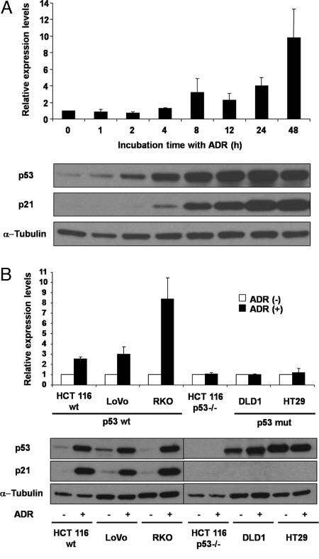
miR-34a expression was increased in a time-dependent manner after ADR treatment, rising 3.2-fold at 8 h and >10-fold at 48 h (Fig. 1A). We also observed that p53 and p21 started to accumulate at 2 and 4 h, respectively, and the accumulation continued until 48 h after treatment (Fig. 1A). HCT 116 p53−/− cells showed no change in expression of miR-34a after ADR treatment (Fig. 1B). To confirm that the induction of miR-34a depends on p53, other human colon cancer cell lines, either with wild-type p53 genes (LoVo and RKO) or mutated p53 genes (DLD1 and HT29), were analyzed (Fig. 1B). As expected, LoVo and RKO cells exhibited increased expression of miR-34a similar to HCT 116 cells, but DLD1 and HT29 cells showed no change, like the HCT 116 p53−/− cells. Accumulation of p53 and p21 was observed in HCT 116, LoVo, and RKO cells, whereas HCT 116 p53−/− cells showed no accumulation, and DLD1 and HT29 cells expressing mutant p53 showed consistent levels of p53 and no accumulation of p21. These results indicate that miR-34a is induced in a p53-dependent manner after ADR treatment.
Fig. 1.
Induction of miR-34a expression after treatment with ADR in colon cancer cell lines with wild-type p53. (A) HCT 116 cells were incubated in the presence of ADR at a concentration of 100 ng/ml, and miR-34a expression was analyzed at the indicated time points by using quantitative real-time RT-PCR. The value for miR-34a at time 0 was set at 1, and the relative amounts of miR-34a at the other time points were plotted as fold induction. Immunoblots under the graph indicate the accumulation of p53 and p21 at each time point. α-Tubulin was used as a loading control. (B) Induction of miR-34a in p53 wild-type and p53-knockout or -mutated colon cancer cell lines after 16 h incubation in the culture medium with or without ADR. The relative amounts of miR-34a in ADR-treated cells were calculated as described above. Open and filled bars represent nontreated and ADR-treated cells, respectively. Immunoblots under the graph indicate the accumulation of p53 and p21 detected in the six colon cancer cell lines.
miR-34a Inhibits Cell Proliferation of HCT 116 and RKO Cells.
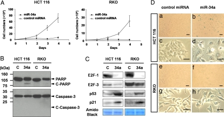
The marked induction of miR-34a after p53 activation, prompted us to investigate whether miR-34a functions as a tumor suppressor. The introduction of miR-34a caused a remarkable inhibition of cell proliferation in both HCT 116 and RKO cells compared with that of control miRNA (Fig. 2A). Immunoblot analysis for apoptosis-specific markers, PARP and caspase-3, revealed no significant induction of apoptosis-related cellular responses in either cell line by miR-34a (Fig. 2B), indicating that the inhibitory effect of miR-34a on cell proliferation is not mainly caused by apoptotic response.
Fig. 2.
miR-34a inhibits cell proliferation and induces senescence-like phenotypes through down-regulation of E2F and up-regulation of p53/p21 in HCT 116 and RKO cells. (A) HCT 116 and RKO cells, seeded at 1.0 × 10⁴ cells in 24-well plates, were transfected with control miRNA (open circles) or miR-34a (filled circles) on day 0, and cells were counted on the days indicated. The data are expressed as mean values with standard deviations. Cultures were performed in triplicate. (B) Immunoblot analysis of apoptotic cell markers. Cell extracts from HCT 116 and RKO cells were subjected to immunoblot analysis by using anti-PARP and anti-caspase-3 antibodies. Lanes C and 34a indicate the lysates of cells transfected with control miRNA and miR-34a, respectively. The molecular sizes of the intact and cleaved forms of PARP (Upper) and those of caspase-3 (Lower) are indicated on the right. (C) Cells transfected with control miRNA (C) and miR-34a (34a) were incubated for 3 days, and cell lysates were then subjected to immunoblot analysis. Amido black staining for an 80-kDa protein in the immunoblot is shown as a protein loading control. Marked down-regulation of E2F-1 and -3, and up-regulation of p53 and p21 were observed after miR34a transfection in both HCT 116 and RKO cells. (D) Induction of senescence-like appearance in cells by miR-34a. Cells transfected with control miRNA (a, c, e, and g) or miR-34a (b, d, f, and h) were subjected to SA-β-gal staining. a, b, e, and f are low-magnification images for visualizing SA-β-gal-positive cells, and c, d, g, and h are high-magnification images for morphological observation. (Scale bars, 10 μm.)
miR-34a Down-Regulates the E2F Signaling Pathway and Up-Regulates the p53 Signaling Pathway.
Comprehensive gene-expression analysis using an Agilent (Agilent Technologies, Santa Clara, CA) microarray platform revealed 287 genes to be down-regulated and 326 genes to be up-regulated 2-fold or greater in both HCT 116 and RKO cells transfected with miR-34a, compared with those transfected with control miRNA (SI Tables 2–4). As for the down-regulated genes, E2F1, E2F2, and some E2F-target genes, including DHFR, MCM3, and MCM10, were observed among the list. Genes associated with cell-cycle progression, CDK4 and CDC25C, were also down-regulated. Among the up-regulated genes, a subset of p53-target genes, including CDKN1A (p21), TP53INP1, ATF3, IKIP, ICAM1, and PTPRE, was apparent. Based on these observations, we hypothesized that E2F-family proteins could be candidate targets for miR-34a.
We then examined the protein levels of E2F-family proteins, E2F-1, -2, and -3, in HCT 116 and RKO cells. E2F-3 is a predicted target for miR-34a in the databases (PicTar web site, http://pictar.bio.nyu.edu), but no further information on E2F-3 with regard to its biological relationship with miR-34a is available. As shown in Fig. 2C, introduction of miR-34a decreased the accumulation of E2F-1 and -3. E2F-2 was not detectable in either case (data not shown). Accumulation of the p53 and p21 proteins was also observed by introduction of miR-34a in both cell lines, reflecting the results of global gene-expression analysis by microarray system. These observations indicate that introduction of miR-34a causes the down-regulation of the E2F pathway, leading to the up-regulation of the p53/p21 pathway. A possible mechanism for the up-regulation of p53 and its downstream target is discussed below.
miR-34a Induces Senescence-Like Phenotypes.
We observed that introduction of miR-34a in HCT 116 cells caused senescence-like phenotypes with positive staining for senescence-associated β-galactosidase (SA-β-gal) and enlarged cellular size (Fig. 2D). RKO cells also showed similar morphological changes with enlarged cellular size by miR-34a introduction, although few SA-β-gal-positive cells were observed (Fig. 2D). We also observed that p53-mutated human colon cancer cells exhibited morphological changes and SA-β-gal positive staining characteristic of cellular senescence (SI Fig. 5). These results suggest that suppression of cell proliferation by miR-34a is mainly associated with the induction of senescence-like phenotypes.
Administration of miR-34a with Atelocollagen Suppresses Tumor Growth in Vivo.
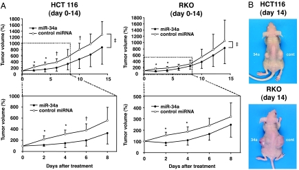
Atelocollagen has been recently shown to be a very useful system to efficiently deliver small interfering RNA molecules into tumors in vivo (15–17). Subcutaneous administration of miR-34a/atelocollagen complexes caused significant suppression of growth of both HCT 116 and RKO cells by day 4 compared with the administration of the control miRNA/atelocollagen complex (Fig. 3). Significant reduction of tumor volume was also observed for HCT 116 cells until day 6 after the miR-34a administration. The averaged tumor volumes were decreased significantly with administration of miR-34a compared with those that were administered control miRNA throughout the entire experimental period of 14 days. Tumor tissues treated with miR-34a showed a considerable amount of necrotic tissue but showed no significant differences in Ki67 and p21 immunostaining compared with those treated with control miRNA (SI Fig. 6). These results indicate that introduction of miR-34a suppresses the growth of human colon cancer cells to tumors in an in vivo setting as well.
Fig. 3.
Administration of miR-34a with atelocollagen suppresses tumor cell growth in vivo. (A) Upper graphs are growth curves of HCT 116 and RKO tumors after transplantation into nude mice with miR-34a/atelocollagen or control miRNA/atelocollagen complexes. The volume of the tumors derived from both cell lines at day 0, which was when control miRNA (open circles) and miR-34a (filled circles) treatment was performed, were set as 100%, and relative tumor volume was evaluated at 2-day intervals for 14 days and is plotted as the percentage relative to day 0. Lower graphs are enlarged images of the growth curves of tumors during the first 8 days after miR-34a administration. The data at each time point are expressed as mean values of 8 tumors in each experimental group with standard deviations. Note the significant suppression of tumor growth on days 2, 4, 6, and 10 for HCT 116 tumors, and on days 2 and 4 for RKO tumors (*, P < 0.005; †, P < 0.05). Significant differences (‡, P < 0.05) in the averaged tumor volumes were also observed in HCT 116 and RKO tumors between mir-34a- and control miRNA-administered tumors throughout the entire experimental period by adopting the linear mixed effect model (34). (B) Photographs illustrating representative features of mouse tumors derived from HCT 116 and RKO cells 14 days after treatment with control miRNA (cont) or miR-34a (34a).
A Subset of Human Colon Cancers Shows Decreased miR-34a Expression.
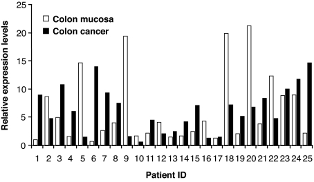
To gain insight into the biological role of miR-34a in human colon carcinogenesis, we analyzed the expression of miR-34a in human colon cancer and paired counterpart normal tissue (Fig. 4). Nine of 25 colon cancer tissues (36%) (patient IDs 2, 5, 9, 10, 12, 16, 18, 20, and 22) showed decreased expression of miR-34a, suggesting that down-regulation of miR-34a may be, at least in part, involved in human colon cancer development.
Fig. 4.
Down-regulation of miR-34a expression in human colon cancer specimens. Quantitative real-time RT-PCR for miR-34a was carried out by using 25 surgical specimens of human colon cancer (filled bars) and paired noncancerous counterpart (open bars). The relative expression levels of miR-34a were calculated as described in Fig. 1.
Discussion
Altered expression of microRNA genes has recently been reported to impact human carcinogenesis (7–9). In the present study, miR-34a was identified as a DNA damage-responsive gene in the HCT 116 colon cancer cell line after treatment with a low concentration of ADR, at which cell proliferation is completely arrested without substantial induction of apoptosis (18). The induction of miR-34a by ADR treatment led us to hypothesize that miR-34a is a responsive gene for chemically induced cellular stress. This hypothesis is supported by recent reports that miR-34a is up-regulated by treatment with sodium arsenite in human immortalized lymphoblasts (19) and with tamoxifen in rat hepatocytes (20).
The introduction of miR-34a into two colon cancer cell lines, HCT 116 and RKO, showed a profound inhibition of cell proliferation accompanying the down-regulation of the E2F family. The E2F family is a well characterized group of transcription factors comprising eight subclasses, E2F1–E2F8 (21). E2F1–E2F3 are positive regulators for the cell cycle, whereas E2F4 and -5 negatively regulate, and the functions of E2F6–8 are unknown. Embryonic fibroblasts derived from knockout mice lacking E2f1, -2, or -3 demonstrate slow cell-cycle progression, especially at entry to the S phase (22, 23), but there is not a complete block of the cell cycle and of proliferation. In contrast, fibroblasts with triple knockout of E2f1–E2f3 demonstrate strong inhibition of cell proliferation throughout the cell cycle. Interestingly, induction of p53 and its downstream target p21 was observed in the triple knockout cells (23–25). This phenotype resembles the results obtained in our current study with miR-34a introduction in colon cancer cells.
Among the E2F family, E2F-3 seems to be a strong candidate for the miR-34a targets. Because E2F-3 is reported to be a regulator for E2F-1 gene expression at the transcriptional level (26), repression of other E2F family proteins might thus occur as a subsequent event by miR-34a overexpression. Activation of the p53 pathway could be caused as a consequence of the repression of the E2F family members as in the case of fibroblast with triple knockout of E2f1–E2f3 (23–25) as described above. A positive feedback loop through the p53 pathway for miR-34a could be another important molecular basis for its transcriptional control, explaining the time-dependent increment in miR-34a induction in p53 wild-type colon cancer cells upon continuous exposure to ADR (Fig. 1A).
It is also important to note that introduction of miR-34a represses cell proliferation of p53-knockout HCT 116 and other colon cancer cell lines with mutant p53 (SI Fig. 5). This observation indicates that enforced induction of exogenous miR-34a causes cell growth arrest in a p53-independent manner. The down-regulation of E2F family members, possibly through down-regulation of E2F-3 by overexpression of miR-34a, appears to be a key event for the induction of cellular senescence. Maehara et al. (27), indeed, recently revealed that reduction of E2F/DP activity induces senescence-like phenotypes in human cancer cells. Introduction of miR-34a also caused the up-regulation of the HBP1 gene (SI Table 4), which is associated with oncogenic RAS-induced premature senescence (28). Thus, miR-34a-mediated modulation of several signaling pathways, including E2F- and RAS-related pathways, would collectively and effectively contribute to the induction of senescence-like phenotypes in human colon cancer cells.
Tumor cell growth in nude mice was significantly inhibited for 4–6 days after administration of miR-34a/atelocollagen complexes, but the suppressive effect was less prominent at later time points. Because complexed miRNA with atelocollagen is expected to stay stable for ≈1 week in this system (17), repeated injections of miR-34a/atelocollagen complexes may be required to exert tumor suppressive effects more efficiently and continuously.
Finally, it is of great interest to note that approximately one-third of human colon cancer specimens revealed down-regulation of the miR-34a expression. Similarly, quite recently, miR-34a was reported to be down-regulated in human primary neuroblastomas with heterozygous deletion of chromosome 1p36, in which miR-34a resides (29). Because the same genomic region is also frequently deleted in colon cancers (30), down-regulation of miR-34a could be associated with the deletion of chromosome 1p36. Furthermore, deletions or mutations of the p53 gene are also suggested to be causative genetic events underlying the down-regulation of miR-34a because its induction is tightly associated with p53 status (Fig. 1B). We indeed observed p53 mutations and chromosomal losses of 17p, including the p53 locus, in some of the colon cancer specimens that showed down-regulation of miR-34a (unpublished observations). Epigenetic inactivation of miR-34a should also be considered as an underlying mechanism. Unexpectedly, we also observed that one-third of colon cancer cases showed up-regulation of miR-34a. There may be alternative mechanisms that remain to be identified in the regulation of miR-34a.
To conclude, the present demonstration of previously uncharacterized biological functions of miR-34a, with the ability to control cell proliferation and induce senescence-like changes in cancer cells, points to its significance as a unique type of tumor suppressor in colon cancer development.
Materials and Methods
Human Colon Cancer Cell Lines and ADR Treatment.
The human colon cancer cell line, HCT 116, and the isogenic HCT 116 p53−/− cells (14) were kindly provided by Bert Vogelstein (The Johns Hopkins University, Baltimore, MD). Other human colon cancer cell lines, such as LoVo, RKO, DLD1, and HT29, were obtained from the American Type Culture Collection (Manassas, VA). DLD1 and HT29 cells are reported to harbor missense mutations in the p53 gene at exon 7 (Ser241Phe) and exon 8 (Arg273His), respectively (31). HCT 116, HCT 116 p53−/−, and HT29 cells were maintained in McCoy's 5A medium; and LoVo, RKO, and DLD1 cells were maintained in Ham's F12K medium, Dulbecco's modified Eagle's medium, and RPMI medium 1640, respectively. All media were supplemented with 10% FBS, 50 units/ml penicillin and 50 μg/ml streptomycin. The cells were routinely incubated at 37°C in a humidified atmosphere with 5% CO2.
ADR was purchased from Sigma–Aldrich (St. Louis, MO). For isolation of total RNA and protein samples from various colon cancer cell lines, cells were seeded at 1.5 × 10⁶ cells per 100-mm dish and treated with ADR at a concentration of 100 ng/ml for 16 h.
For the time course experiment of ADR treatment, HCT 116 cells were seeded at 6 × 10⁵ cells per 60-mm dish and then incubated afterward with or without ADR in the culture medium. Cells were collected and subjected to extraction of total RNA and protein at 0, 1, 2, 4, 8, 12, 24, and 48 h thereafter.
Isolation of miRNA and Quantitative Real-Time RT-PCR Analysis.
Total RNA, including miRNA, was extracted from the cells by using a mirVana miRNA Isolation Kit (Ambion, Austin, TX) according to the manufacturer's instructions and quantified with a NanoDrop spectrophotometer (NanoDrop Technologies, Wilmington, DE). cDNA was synthesized from 10 ng of total RNA by using a High Capacity cDNA Archive kit (Applied Biosystems, Foster City, CA), and the expression levels of 157 mature miRNA species were quantified by using a TaqMan MicroRNA Assays Human Panel (Applied Biosystems). Real-time RT-PCR was performed by using the Applied Biosystems 7300 Sequence Detection system (Applied Biosystems), the expression of each miRNA was defined from the threshold cycle (Ct), and relative expression levels were calculated by using the 2−ΔΔCt method (32) after normalization with reference to expression of U6 small nuclear RNA.
Transient Transfection of Cells with miR-34a and Cell Proliferation Assay.
HCT 116 and RKO cells were seeded at a density of 1.0 × 10⁴ cells in 24-well tissue culture plates 24 h before transfection with 5 nM of either miR-34a (Ambion) or nontargeting control miRNA (control miRNA; Ambion) by using HiPerfect transfection reagents (Qiagen, Valencia, CA). The transfection efficiency under the conditions we adopted in this study was estimated to be close to 100% according to our observations made by using a fluorescence-labeled double-stranded short interference RNA. After transfection, cells were harvested and counted every day for 4 days. The average number of cells was determined at each time point in triplicate.
Microarray Analysis.
Comprehensive gene-expression analysis of HCT 116 and RKO cells transfected with miR-34a or control miRNA was carried out by using the Whole Human Genome (4 × 44K) Oligo Microarray system (Agilent Technologies). Cells, seeded at 5 × 10⁴ cells per ml in six-well tissue culture plates, were transfected with miR-34a or control miRNA as described above and propagated for 3 days. Total RNAs were isolated with an RNeasy column (Qiagen), and Cy3-labeled cRNA was prepared with an Agilent One Color Spike Mix Kit (Agilent Technologies) and then hybridized by using a Gene Expression Hybridization Kit (Agilent Technologies) at 65°C for 17 h. Signal intensity was calculated from digitized images captured by Laser Scanner (Agilent Technologies), and data analysis was performed by using GeneSpring GX software (Agilent Technologies).
Immunoblot Analysis.
At day 3 after transfection with miR-34a or control miRNA, whole cell lysate was prepared in a lysis buffer (50 mM Tris·HCl, pH 7.4/150 mM NaCl/1% Triton X-100) with a protease inhibitor mixture (Complete Mini; Roche, Indianapolis, IN). Proteins were electrophoresed on a 5–20% linear gradient Tris·HCl-ready gels (Bio-Rad, Hercules, CA) and transferred to polyvinylidene fluoride membranes (Immobiron-P; Millipore, Billerica, MA). Blots were blocked with 3% nonfat dry milk in TBS-T (Tris-buffered saline/0.1% Tween-20, pH 7.4) at room temperature for 30 min. For primary antibodies, we used mouse anti-p53 mAb (1:200, DO-1; Santa Cruz Biotechnology, Santa Cruz, CA), mouse anti-p21 mAb (1:2,000, DCS60; Cell Signaling Technology, Beverly, MA), mouse anti-α-tubulin mAb (1:5,000, DM1A, ICN Biomedicals, Irvine, CA), mouse PARP mAb (1:1,000, C-2–10, Oncogene Research Products, San Diego, CA), mouse anti-caspase-3 mAb (1:1,000, 3G2; Cell Signaling Technology), rabbit anti-E2F-1 polyclonal antibody (pAb) (1:500, C-20; Santa Cruz Biotechnology), rabbit anti-E2F-2 pAb (1:1,000, C-20; Santa Cruz Biotechnology), and rabbit anti-E2F-3 pAb (1:1,000, C-18; Santa Cruz Biotechnology). Horseradish peroxidase-conjugated antibodies against mouse IgG or rabbit IgG (1:5,000–10,000, NA9310V; Amersham Biosciences, Piscataway, NJ) were used as secondary antibodies. Immunoreactive bands on the blots were visualized with enhanced chemiluminescence substrates (Immobilon Western; Millipore).
SA-β-Gal Staining.
At day 4 after transfection with miR-34a or control miRNA, HCT 116 and RKO cells were fixed and stained by using an SA-β-gal kit (Cell Signaling Technology) according to the manufacturer's instructions.
Human Tumor Xenograft Model and Administration of miR-34a/Atelocollagen Complexes.
Animal experimental protocols were approved by our institute's Committee for Ethics in Animal Experimentation, and the experiments were conducted in accordance with the guidelines for Animal Experiments of the National Cancer Center. HCT 116 or RKO cells were inoculated with 5 × 10⁶ cells per site bilaterally on the backs of female athymic nude mice aged 6 weeks (Charles River Laboratories, Wilmington, MA). Tumor size was monitored by measuring the length and width with calipers, and volumes were calculated with the formula: (L × W2) × 0.5, where L is length and W is width of each tumor.
Atelocollagen was pretreated with pepsin to remove the telopeptides, which confer most of the collagen's antigenicity (33), and complexes of miR-34a (Ambion) and control miRNA (Ambion) with atelocollagen were prepared as described in refs. 15–17. The miR-34a and control miRNA (15 μg) with 0.5% atelocollagen in a 200-μl volume were administered into the s.c. spaces around the tumors when they had reached a volume of 50–100 mm3 at day 6 after inoculation. The volume of each tumor at the point of miR-34a or control miRNA administration was set at 100%, and the relative tumor volumes at each time point was evaluated every other day from days 2 to 14.
Immunohistochemistry.
Tumors were fixed in 10% neutralized formalin and embedded in paraffin blocks. Sections (4 μm) were prepared for hematoxylin/eosin staining and also for immunohistochemical examination. After deparaffinization and rehydration, antigen retrieval was performed by microwave irradiation at 600 W for 10 min in 10 mM citrate buffer (pH 6.0). Tissue sections were incubated with rabbit anti-Ki67 polyclonal antibody (pAb) (1:1,000; Novocastra Laboratories, Newcastle, U.K.), mouse anti-p21 mAb (1:100) and rabbit anti-cleaved caspase-3 mAb (1:200, 5A1; Cell Signaling Technology). The sections were then incubated with goat biotinylated anti-rabbit IgG antibody and goat biotinylated anti-mouse IgG antibody (1:200; Vector Laboratories, Burlingame, CA). Immunoreactive signals were visualized by using 3,3′-diaminobenzidine tetrahydrochloride solution, and the nuclei were counterstained with hematoxylin.
Human Colon Cancer Samples.
Primary human colon cancers and paired noncancerous normal colon samples were obtained from 25 patients treated at the National Cancer Center Hospital, Tokyo, with documented informed consent in each case. Samples were frozen in liquid nitrogen and stored at −80°C until use. Total RNA was isolated from frozen samples by using TRIzol reagent (Invitrogen), and then quantitative real-time RT-PCR for miR-34a and U6 small nuclear RNA was performed as described above. The relative expression level of miR-34a in each sample was calculated and quantified by using the 2−ΔΔCt method after normalization for expression of U6 small nuclear RNA as described above.
Statistical Analysis.
The Mann–Whitney U test was used to test for statistical significance of differences in tumor volumes at each time point after administration of miR-34a and control miRNA. We also used linear mixed effect models (34) to evaluate the differences in averaged volumes of tumors throughout the entire experimental period between the two experimental groups, namely miR-34a- and control miRNA-administered groups. P < 0.05 was considered significant.
Supplementary Material
Acknowledgments
We thank Dr. Bert Vogelstein for providing the HCT 116 and HCT 116 p53−/− cells; Dr. Takahiro Ochiya (National Cancer Center Research Institute) and Dr. Fumitaka Takeshita (National Cancer Center Research Institute) for providing us with atelocollagen and for helpful discussion; and Dr. Kenichi Yoshimura for his kind help in statistical analysis. This work was supported in part by a Grant-in-Aid for Cancer Research for the Third-Term Comprehensive 10-Year Strategy for Cancer Control from the Ministry of Health, Labour and Welfare of Japan and by Research Grant 06-016 of the Sankyo Foundation of Life Science. M.I. is a recipient of the Research Resident Fellowship from the Foundation for Promotion of Cancer Research in Japan.
Abbreviations
- miRNA
microRNA
- ADR
adriamycin
- SA-β-gal
senescence-associated β-galactosidase.
Note.
During the process of submitting our manuscript, three related papers by Raver-Shapira et al. (35), Chang et al. (36), and He et al. (37) were published.
Footnotes
The authors declare no conflict of interest.
This article contains supporting information online at www.pnas.org/cgi/content/full/0707351104/DC1.
References
- 1.Lengauer C, Kinzler KW, Vogelstein B. Nature. 1998;396:643–649. doi: 10.1038/25292. [DOI] [PubMed] [Google Scholar]
- 2.Baylin SB, Ohm JE. Nat Rev Cancer. 2006;6:107–116. doi: 10.1038/nrc1799. [DOI] [PubMed] [Google Scholar]
- 3.Esteller M. Nat Rev Genet. 2007;8:286–298. doi: 10.1038/nrg2005. [DOI] [PubMed] [Google Scholar]
- 4.Tomaru Y, Hayashizaki Y. Cancer Sci. 2006;97:1285–1290. doi: 10.1111/j.1349-7006.2006.00337.x. [DOI] [PMC free article] [PubMed] [Google Scholar]
- 5.Bartel DP. Cell. 2004;116:281–297. doi: 10.1016/s0092-8674(04)00045-5. [DOI] [PubMed] [Google Scholar]
- 6.Alvarez-Garcia I, Miska EA. Development (Cambridge, UK) 2005;132:4653–4662. doi: 10.1242/dev.02073. [DOI] [PubMed] [Google Scholar]
- 7.Calin GA, Croce CM. Nat Rev Cancer. 2006;6:857–866. doi: 10.1038/nrc1997. [DOI] [PubMed] [Google Scholar]
- 8.Lu J, Getz G, Miska EA, Alvarez-Saavedra E, Lamb J, Peck D, Sweet-Cordero A, Ebert BL, Mak RH, Ferrando AA, et al. Nature. 2005;435:834–838. doi: 10.1038/nature03702. [DOI] [PubMed] [Google Scholar]
- 9.Volinia S, Calin GA, Liu CG, Ambs S, Cimmino A, Petrocca F, Visone R, Iorio M, Roldo C, Ferracin M, et al. Proc Natl Acad Sci USA. 2006;103:2257–2261. doi: 10.1073/pnas.0510565103. [DOI] [PMC free article] [PubMed] [Google Scholar]
- 10.Calin GA, Ferracin M, Cimmino A, Di Leva G, Shimizu M, Wojcik SE, Iorio MV, Visone R, Sever NI, Fabbri M, et al. N Engl J Med. 2005;353:1793–1801. doi: 10.1056/NEJMoa050995. [DOI] [PubMed] [Google Scholar]
- 11.Fukuda H, Sugimura T, Nagao M, Nakagama H. Biochim Biophys Acta. 2001;1528:152–158. doi: 10.1016/s0304-4165(01)00186-6. [DOI] [PubMed] [Google Scholar]
- 12.Caudy AA, Ketting RF, Hammond SM, Denli AM, Bathoorn AM, Tops BB, Silva JM, Myers MM, Hannon GJ, Plasterk RH. Nature. 2003;425:411–414. doi: 10.1038/nature01956. [DOI] [PubMed] [Google Scholar]
- 13.Tsuchiya N, Ochiai M, Nakashima K, Ubagai T, Sugimura T, Nakagama H. Cancer Res. 2007 doi: 10.1158/0008-5472.CAN-06-2707. in press. [DOI] [PubMed] [Google Scholar]
- 14.Bunz F, Dutriaux A, Lengauer C, Waldman T, Zhou S, Brown JP, Sedivy JM, Kinzler KW, Vogelstein B. Science. 1998;282:1497–1501. doi: 10.1126/science.282.5393.1497. [DOI] [PubMed] [Google Scholar]
- 15.Minakuchi Y, Takeshita F, Kosaka N, Sasaki H, Yamamoto Y, Kouno M, Honma K, Nagahara S, Hanai K, Sano A, et al. Nucleic Acids Res. 2004;32:e109. doi: 10.1093/nar/gnh093. [DOI] [PMC free article] [PubMed] [Google Scholar]
- 16.Takeshita F, Minakuchi Y, Nagahara S, Honma K, Sasaki H, Hirai K, Teratani T, Namatame N, Yamamoto Y, Hanai K, et al. Proc Natl Acad Sci USA. 2005;102:12177–12182. doi: 10.1073/pnas.0501753102. [DOI] [PMC free article] [PubMed] [Google Scholar]
- 17.Takei Y, Kadomatsu K, Yuzawa Y, Matsuo S, Muramatsu T. Cancer Res. 2004;64:3365–3370. doi: 10.1158/0008-5472.CAN-03-2682. [DOI] [PubMed] [Google Scholar]
- 18.Bunz F, Hwang PM, Torrance C, Waldman T, Zhang Y, Dillehay L, Williams J, Lengauer C, Kinzler KW, Vogelstein B. J Clin Invest. 1999;104:263–269. doi: 10.1172/JCI6863. [DOI] [PMC free article] [PubMed] [Google Scholar]
- 19.Marsit CJ, Eddy K, Kelsey KT. Cancer Res. 2006;66:10843–10848. doi: 10.1158/0008-5472.CAN-06-1894. [DOI] [PubMed] [Google Scholar]
- 20.Pogribny IP, Tryndyak VP, Boyko A, Rodriguez-Juarez R, Beland FA, Kovalchuk O. Mutat Res. 2007;619:30–37. doi: 10.1016/j.mrfmmm.2006.12.006. [DOI] [PubMed] [Google Scholar]
- 21.Tsantoulis PK, Gorgoulis VG. Eur J Cancer. 2005;41:2403–2414. doi: 10.1016/j.ejca.2005.08.005. [DOI] [PubMed] [Google Scholar]
- 22.Leone G, Sears R, Huang E, Rempel R, Nuckolls F, Park CH, Giangrande P, Wu L, Saavedra HI, Field SJ, et al. Mol Cell. 2001;8:105–113. doi: 10.1016/s1097-2765(01)00275-1. [DOI] [PubMed] [Google Scholar]
- 23.Wu L, Timmers C, Malti B, Saavedra HI, Sang L, Chong GT, Nuckolls F, Giangrande P, Wright FA, Field SJ, et al. Nature. 2001;414:457–462. doi: 10.1038/35106593. [DOI] [PubMed] [Google Scholar]
- 24.Sharma N, Timmers C, Trikha P, Saavedra HI, Obery A, Leone G. J Biol Chem. 2006;281:36124–36131. doi: 10.1074/jbc.M604152200. [DOI] [PubMed] [Google Scholar]
- 25.Timmers C, Sharma N, Opavsky R, Maiti B, Wu L, Wu J, Orringer D, Trikha P, Saavedra HI, Leone G. Mol Cell Biol. 2007;27:65–78. doi: 10.1128/MCB.02147-05. [DOI] [PMC free article] [PubMed] [Google Scholar]
- 26.Johnson DG, Ohtani K, Nevins JR. Genes Dev. 1994;8:1514–1525. doi: 10.1101/gad.8.13.1514. [DOI] [PubMed] [Google Scholar]
- 27.Maehara K, Yamakoshi K, Ohtani N, Kubo Y, Takahashi A, Arase S, Jones N, Hara E. J Cell Biol. 2005;168:553–560. doi: 10.1083/jcb.200411093. [DOI] [PMC free article] [PubMed] [Google Scholar]
- 28.Zhang X, Kim J, Ruthazer R, McDevitt MA, Wazer DE, Paulson KE, Yee AS. Mol Cell Biol. 2006;26:8252–8266. doi: 10.1128/MCB.00604-06. [DOI] [PMC free article] [PubMed] [Google Scholar]
- 29.Welch C, Chen Y, Stallings RL. Oncogene. 2007;26:5017–5022. doi: 10.1038/sj.onc.1210293. [DOI] [PubMed] [Google Scholar]
- 30.Martens F, Johansson B, Hoglund M, Mitelman F. Cancer Res. 1997;57:2765–2780. [PubMed] [Google Scholar]
- 31.Rodrigues NR, Rowan A, Smith ME, Kerr IB, Bodmer WF, Gannon JV, Lane DP. Proc Natl Acad Sci USA. 1990;87:7555–7559. doi: 10.1073/pnas.87.19.7555. [DOI] [PMC free article] [PubMed] [Google Scholar]
- 32.Livak KJ, Schmittgen TD. Methods. 2001;25:402–408. doi: 10.1006/meth.2001.1262. [DOI] [PubMed] [Google Scholar]
- 33.Ochiya T, Takahama Y, Nagahara S, Sumita Y, Hisada A, Itoh H, Nagai Y, Terada M. Nat Med. 1999;5:707–710. doi: 10.1038/9560. [DOI] [PubMed] [Google Scholar]
- 34.Laird NM, Ware JH. Biometrics. 1982;38:963–974. [PubMed] [Google Scholar]
- 35.Raver-Shapira N, Marciano E, Meiri E, Spector Y, Rosenfeld N, Moskovits N, Bentwich Z, Oren M. Mol Cell. 2007;26:731–743. doi: 10.1016/j.molcel.2007.05.017. [DOI] [PubMed] [Google Scholar]
- 36.Chang TC, Wentzel EA, Kent OA, Ramachandran K, Mullendore M, Lee KH, Feldmann G, Yamakuchi M, Ferlito M, Lowenstein CJ, et al. Mol Cell. 2007;26:745–752. doi: 10.1016/j.molcel.2007.05.010. [DOI] [PMC free article] [PubMed] [Google Scholar]
- 37.He L, He X, Lim LP, de Stanchina E, Xuan Z, Liang Y, Xue W, Zender L, Magnus J, Ridzon D, et al. Nature. 2007;447:1130–1134. doi: 10.1038/nature05939. [DOI] [PMC free article] [PubMed] [Google Scholar]
Associated Data
This section collects any data citations, data availability statements, or supplementary materials included in this article.


