Abstract
Inflammation can play a regulatory role in cancer progression and metastasis. Previously, we have shown that metastatic tumor cells entering the liver trigger a proinflammatory response involving Kupffer cell-mediated release of tumor necrosis factor-α and the up-regulation of vascular endothelial cell adhesion receptors, such as E-selectin. Here, we analyzed spatio-temporal aspects of the ensuing tumor-endothelial cell interaction using human colorectal carcinoma CX-1 and murine carcinoma H-59 cells and a combination of immunohistochemistry, confocal microscopy, and three-dimensional reconstruction. E-selectin expression was evident mainly on sinusoidal vessels by 6 and 10 hours, respectively, following H-59 and CX-1 inoculation, and this corresponded to a stabilization of the number of tumor cells within the sinuses. Tumor cells arrested in E-selectin+ vessels and appeared to flatten and traverse the vessel lining, away from sites of intense E-selectin staining. This process was evident by 8 (H-59) and 12 (CX-1) hours after inoculation, coincided with increased endothelial vascular cell adhesion molecule-1 expression, and involved tumor cell attachment in areas of intense vascular cell adhesion molecule-1 and platelet endothelial cell adhesion molecule-1 expression. Nonmetastatic (human) MIP-101 and (murine) M-27 cells induced a weaker response and could not be seen to extravasate. The results show that metastatic tumor cells can alter the hepatic microvasculature and use newly expressed endothelial cell receptors to arrest and extravasate.
The host microenvironment plays an important role in the regulation of tumor progression at the primary site. It can also facilitate tumor dissemination by promoting neovascularization and providing growth-enhancing factors to the metastatic cells at the secondary sites of growth.1,2 Resident and tumor-infiltrating host inflammatory cells constitute an integral component of the tumor microenvironment and can participate in regulating tumor growth and dissemination through the release of proinflammatory cytokines and chemokines, proangiogenic factors, and extracellular matrix-degrading proteinases.3,4
The liver is a major site of metastasis for some of the most common human malignancies, carcinomas of the gastrointestinal tract in particular. Liver metastases are frequently inoperable and are associated with poor prognosis.5,6 A better understanding of the molecular interactions that underlie liver metastasis formation, particularly during the early stages of the process, may provide novel approaches for prevention and treatment.
Among the rate-limiting steps in the process of hematogenous metastasis are tumor cell arrest into and extravasation out of the blood vessels. Tumor necrosis factor-α (TNF-α)-inducible cell adhesion molecules (CAMs) on the luminal surface of the microvascular endothelium are thought to mediate tumor-endothelial cell adhesion and thereby facilitate tumor cell arrest and transmigration into the extravascular space.7,8,9 Among the vascular endothelial cell receptors that have been implicated in cell-cell adhesion and transendothelial migration are P-, E-, and L-selectin,10,11,12,13 as well as vascular adhesion receptors of the immunoglobulin superfamily such as intercellular adhesion molecules ICAM-1, -2, and -3, vascular cell adhesion molecule (VCAM)-1, and mucosal addressin cell adhesion molecule-1.14,15 However, direct evidence for their involvement in metastasis, in vivo, is still lacking. Under normal physiological conditions, E-selectin and VCAM-1 expression on vascular endothelial cells is low. In response to cytokines such as interleukin (IL)-1α and TNF-α, E-selectin expression is induced through activation of the nuclear factor-κB and Raf/MEK/MAPK pathways, and this, in turn, can lead to up-regulation of VCAM-1 and ICAM-1 expression.16,17 Adhesion to E-selectin is generally mediated by sialylated, glycosylated, or sulfated glycans such as the tetrasaccharides sialyl-Lewisx (sLewx) and sialyl-Lewisa (sLewa) that are present on cell surface glycoproteins, glycolipids, or proteoglycans.10,11,18 These molecules have been identified as markers of progression in carcinomas of the gastrointestinal tract, such as colorectal carcinoma,19,20,21 and have been implicated in tumor cell adhesion to the hepatic microvascular endothelial cells during liver metastasis.7,22,23 Recently, a splice variant of CD44 was also identified as an E-selectin ligand.24 Adhesion to VCAM-1 can be mediated by the α4 integrins α4β1 (VLA-4) and α4β7,15,25 whereas ICAM-1 adhesion is mediated by the counter-receptor αLβ2 (LFA-1).15,26
In a series of studies, we have previously shown that tumor cell entry into the hepatic microvasculature can trigger a rapid, proinflammatory cascade that begins with increased local TNF-α and IL-1β production by activated Kupffer cells and leads to up-regulated expression of E-selectin and other vascular adhesion receptors such as ICAM-1 and VCAM-1 on the hepatic sinusoidal vessels.27,28 We have shown that a blockade of TNF-α-mediated E-selectin induction by mouse-specific C-raf antisense oligonucleotides, or an inhibition of E-selectin function by specific neutralizing antibodies, markedly reduced the number of experimental liver metastases formed by human colorectal carcinoma CX-1 and mouse Lewis lung carcinoma-derived H-59 cells, respectively.12,17 The objective of the present study was to analyze more directly the role of TNF-α-induced vascular endothelial cell adhesion receptors in liver metastasis. Using a combination of immunostaining and confocal microscopy, we were able to visualize tumor cell adhesion to hepatic microvascular endothelial cells in vivo and analyze spatio-temporal aspects of the tumor-endothelial cell interaction during the very early stages of liver metastasis.
Materials and Methods
Cells
The tumor cells used in this study were the murine Lewis lung carcinoma sublines H-59 (highly metastatic to liver) and M-27 (poorly metastatic) and the human colorectal carcinoma lines CX-1 (highly metastatic) and MIP-101 (nonmetastatic) (a kind gift from Dr. Peter Thomas, Boston University School of Medicine, Boston, MA).17,18,28 All of the cells were maintained in RPMI 1640 medium containing 10% fetal bovine serum, 100 μg/ml penicillin, 100 μg/ml streptomycin, and 300 μg/ml glutamine. Routine testing confirmed that the cells were free of mycoplasma and viral contaminants during the study period. M-27GFP and MIP-101GFP cells were generated by transfection with 1 to 5 μg of the pLEGFP-N1 plasmid (Clontech, Mountain View, CA) using Lipofectamine Plus, as per the manufacturer’s instructions (Invitrogen Canada, Burlington, ON, Canada). Transfectants were selected using 100 μg/ml G-418 (Invitrogen) and maintained in complete RPMI 1640 medium containing the same concentration of G-418. They were used in this study without further cloning. The H-59 and CX-1 cells were transduced with the vLTR-GFP retrovirus at multiplicities of infection of 15 and 10, respectively, as we previously described.28,29 H-59GFP cells were used without further selection. Highly fluorescent CX-1GFP cells were obtained by sorting the top 30% of fluorescent cells using a fluorescence-activated cell sorter (FACS-Vantage Flow Cytometer/Cell Sorter; Becton, Dickinson and Company, Franklin Lakes, NJ).
Antibodies
Rat monoclonal antibodies to mouse E-selectin (clone 10E9.6), VCAM-1 (clone 429-MVCAM-A), CD31 (clone MEC13.3), and ICAM-1 were from Pharmingen (San Diego, CA). The Alexa Fluor 568 goat anti-rat antibody was from Molecular Probes, Inc. (Eugene, OR).
Tumor Cell Tracking
Green fluorescent protein (GFP)-tagged tumor cells (2 × 10⁶ per mouse) were injected by the intrasplenic/portal route into C57Bl/6 (H-59 and M-27, CX-1 and MIP-101) or nu/nu (CX-1 and MIP-101) mice. The animals were euthanized at different time intervals ranging from 20 minutes to 72 hours thereafter, and the livers were perfused with 50 ml of a 4% paraformaldehyde solution, fixed for 48 hours in the same fixative, transferred into a 30% sucrose solution for 4 days, and placed in OCT medium (Somagen Diagnostics, Edmonton, AB, Canada) before freezing at −80°C. For each liver (four per interval per experiment), multiple, 7-μm cryostat sections were prepared from each of the lobes and the sections mounted in Prolong Gold Antifade reagent (Molecular Probes). GFP+ cells were enumerated using a Zeiss imaging station (Carl Zeiss Inc., Thornwood, NY) equipped with the Northern Eclipse imaging software (Empix Imaging, Inc., Mississauga, ON, Canada).
Immunohistochemistry and Confocal Microscopy
Following the injection of 2 × 10⁶ tumor cells, the livers were perfused with 50 ml of a 4% paraformaldehyde solution and processed as described above. Immunohistochemistry was performed on 7-μm cryostat sections. The sections were incubated first in a blocking solution (5% normal goat serum and 0.1% Triton X-100 in phosphate-buffered saline) for 30 minutes and then overnight at 4°C with the primary antibodies diluted 1:50 (E-selectin), or 1:100 (VCAM-1, ICAM-1, and CD31) in the blocking buffer. After several washes with phosphate-buffered saline, the sections were incubated with Alexa Fluor 568 goat anti-rat IgG antibody (diluted 1:200 in the blocking buffer) for 1 hour at 4°C and mounted in Prolong Gold Antifade reagent (Molecular Probes). For confocal analysis and three-dimensional (3D) reconstruction, the same protocol was used but with 100-μm sections, and the antibody dilutions were 1:50 for each primary antibody and 1:100 for the secondary antibody. Incubation with the secondary antibody was for 4 hours at 4°C. Confocal images were captured with a Zeiss LSM 510 equipped with an inverted Axiovert 100M microscope (Zeiss). Lasers used were the Argon 488 nm (for visualization of green fluorescence) and the HeNe 543 nm (for visualization or red fluorescence). Image processing was with the Zeiss LSM 510 software. 3D images were reconstructed using the MetaMorph software (Molecular Devices).
Enumeration of E-Selectin- and VCAM-1-Positive Vessels
For each section, 20 random fields were analyzed for the presence of E-selectin or VCAM-1 immunolabeling. A vessel was considered positive if a single fluorescent pixel was present on or around it. Results are expressed as the mean number (±SD) of E-selectin- or VCAM-1-positive vessels counted in 20 microscope fields/time point based on four different livers. A tumor cell was considered to be associated with an E-selectin-positive vessel if it was observed within a vessel that stained positively based on the above criterion.
Statistical Analysis
The univariate t-test was used to analyze differences between the responses to different cell lines, and a P value ≤0.05 was considered significant.
Results
Metastatic Carcinoma Cells Induce Intense E-Selectin Expression within Hours of Entry into the Liver
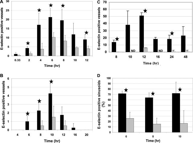
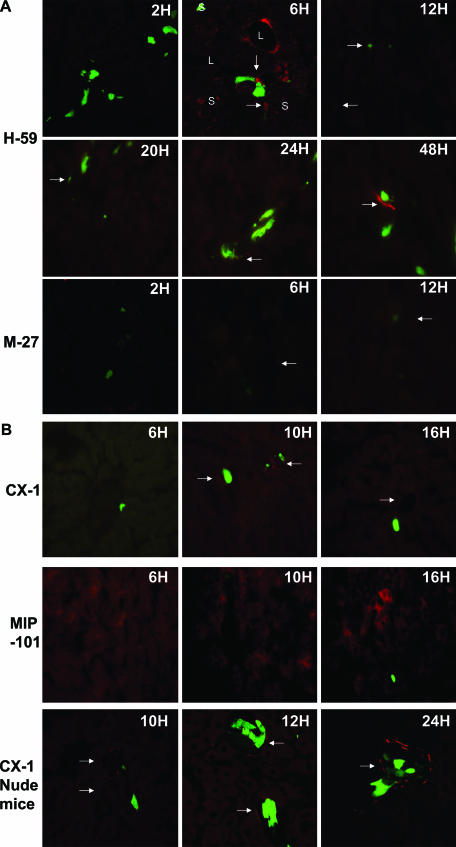
We have shown that tumor cell entry into the liver can trigger a rapid proinflammatory response involving increased macrophage-mediated TNF-α production. We also reported that this is rapidly followed by increased expression of E-selectin mRNA in vascular endothelial cells.17,27,28 To investigate further temporal aspects of tumor-induced E-selectin expression, we inoculated mice with GFP-expressing carcinoma cells, and the relative proportion, number, and type of liver vessels expressing E-selectin were analyzed at different intervals thereafter by a combination of immunohistochemistry and confocal microscopy. Minimal E-selectin expression (<1 positive vessel/20 microscope fields) was detected in livers of saline-injected control mice, and these levels were not significantly altered for up to 1 hour following tumor injection, regardless of the type of tumor cell injected. The proportion of E-selectin-positive vessels began to rise significantly 2 hours following the inoculation of (metastatic) H-59 cells, increased by >10-fold by 6 hours, and remained high for up to 12 hours, with sporadic vessel staining still evident at 48 hours following tumor injection (Figures 1A and 2A). E-selectin staining was preferentially observed on sinusoidal endothelial cells (Figure 1D). In contrast, (poorly metastatic) M-27 cells elicited a much weaker response with some, low-intensity E-selectin staining seen between 6 and 8 hours after tumor injection. Interestingly, E-selectin positivity in these livers was seen preferentially along portal veins and in centrolobular venules (Figures 1, A and D, and 2A).
Figure 1.
Rapid increase in E-selectin expression in response to tumor cell injections. Cryostat sections were prepared from livers removed at different time intervals following the inoculation of H-59 and M-27 (A, H-59: black bars; M-27: gray) or CX-1 and MIP-101 (B and C, CX-1: black; MIP-101: gray) cells into C57Bl/6 mice (A and B) or nude mice (C). Sections were stained with an E-selectin-specific antibody and an Alexa Fluor 568 secondary antibody. E-selectin-positive vessels were counted at ×630 magnification in 20 fields per liver using a total of four livers per time point. Results are expressed as the mean number (±SD) of E-selectin-positive vessels counted in 20 microscope fields/time point. Shown in D are the proportions (%) of E-selectin+ sinusoids as calculated for mice inoculated with H-59 (black) or M-27 (gray) cells. ★P < 0.05 (t-test) for H-59 (A and D) and CX-1 (B and C) cells compared with M-27 and MIP-101 cells, respectively.
Figure 2.
Localization of tumor cells in E-selectin-positive vessels. Shown are images of E-selectin+ vessels (arrows) captured at different time intervals following the inoculation of H-59 and M-27 (A) or CX-1 and MIP-101 (B) cells. Tumor cells are in green. S, sinusoidal vessel; L, centrolobular or portal venules. Original magnification, ×400.
Similar to the mouse carcinoma cells, there was also a marked increase in E-selectin expression following the injection of metastatic human colorectal carcinoma CX-1 cells. However, the time course and duration of the response were distinct. The E-selectin signal began to rise only 6 to 8 hours after tumor injection and peaked at 10 to 12 hours, declining thereafter but still detectable on some vessels 48 hours after tumor inoculation (Figures 1B and 2B). This pattern was consistent with the delayed but more sustained increase in TNF-α production observed in response to CX-1 inoculation.17 The response to human colorectal carcinoma cells was also tumor specific, since significantly fewer E-selectin-positive vessels were observed following the injection of nonmetastatic MIP-101 cells (Figures 1B and 2B). Interestingly, the number of E-selectin-positive vessels was significantly higher in athymic nude mice (where hepatic metastases eventually develop) than in conventional mice injected with CX-1 cells, although the duration of the response was comparable. E-selectin positivity in response to MIP-101 cells was, however, low in both types of animals (Figures 1, B and C, and 2B).
Tracking of Tumor Cells in Vivo Reveals No Difference in Tumor Cell Numbers and Distribution in the Liver Immediately after Tumor Injection
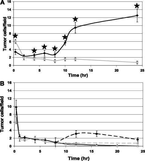
To determine whether the differential ability of the tumor cells to induce vascular endothelial E-selectin expression resulted from differences in tumor cell arrest/survival in the hepatic microvasculature, multiple cryostat sections were prepared from livers of tumor-injected mice at different intervals, ranging from 20 minutes onwards after injection, and fluorescence microscopy was used to track and enumerate the tumor cells. Within the first 2 hours after injection, there was no significant difference in the total number of GFP+ murine tumor cells that could be detected in the sections. However, from 4 hours onwards, a significant difference was noted as the number of M-27 cell slowly declined while the number of H-59 cells stabilized and then began to rise at 10 hours after injection as a consequence of cell extravasation and the onset of cell division (Figure 3A). We did not observe this difference in tumor cell numbers when CX-1 and MIP-101 cells were analyzed (Figure 3B). Interestingly, we noted that the number of CX-1 (but not MIP-101) cells detectable in livers of athymic nu/nu mice was significantly higher than that seen in conventional mice from 12 to 24 hours following tumor inoculation (Figure 3B), the period during which E-selctin positivity was also significantly higher in nude mice.
Figure 3.
Tracking of tumor cells in vivo reveals no differences in tumor cell distribution following injection. Cryostat sections were prepared from livers removed at different intervals following the inoculation of H-59 (black line in A), M-27 (gray line in A), CX-1 (black diamonds and circles in B), or MIP-101 (gray squares or triangles in B) into C57Bl/6 (A and solid lines in B) or nude (dashed lines in B) mice. Fluorescent tumor cells were counted in 20 fields/slide in sections prepared from each of four different livers. Results are expressed as the mean number of cells (±SD) counted per field at a magnification of ×630. There was a significant difference between the numbers of H-59 and M-27 cells counted (A) at all time points >2 hours (★P < 0.05). There was no significant difference between the numbers of CX-1 and MIP-101 cells observed following the injection into C57Bl/6 or nude mice. The numbers of CX-1 cells observed in livers derived from nude mice were significantly higher than those seen in livers from C57Bl/6 mice (P < 0.05) at 12 and 24 hours.
Tumor Cells Arrest in E-Selectin-Positive Vessels
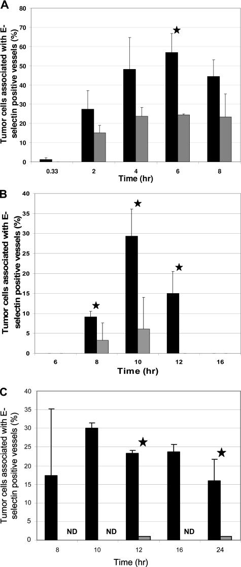
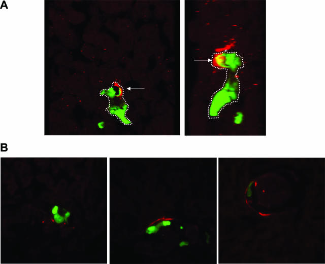
The finding that the initial stabilization and eventual increase in the number of H-59 cells seen in the liver corresponded temporally to the peak in vascular endothelial E-selectin expression suggested that these events may be causally linked. We therefore used immunohistochemistry and fluorescence microscopy to assess whether H-59 cells were preferentially associated with E-selectin-positive vessels. Indeed, we observed that between 4 and 6 hours after tumor injection, more than 50% of all of the detectable GFP+ H-59 cells were localized within E-selectin-positive hepatic vessels (Figure 4A). Confocal microscopy and 3D reconstruction revealed areas of close contact between the green fluorescent tumor cells and (red fluorescent) E-selectin, suggesting that the tumor cells, in fact, used this vascular adhesion molecule for attachment (Figure 5A, arrow). M-27 cells did not seem to associate preferentially with E-selectin-positive vessels. The proportion of cells detected in E-selectin-positive vessels was approximately 20% at all time points analyzed and did not change with time, suggesting a more random distribution of the cells (Figure 4A). Furthermore, we noted that subsequent to the initial attachment, H-59 cells began to spread, aligning their tails along E-selectin-positive endothelial cells with the bulk of the cell moving away from regions of intense positive staining, where extravasation, as revealed by 3D reconstruction, seems to have occurred (Figure 5A).
Figure 4.
Time-dependent increase in tumor cell association with E-selectin+ vessels. Cryostat sections were prepared from livers removed at different time intervals following the inoculation of H-59 and M-27 (A, H-59: black; M-27: gray) or CX-1 and MIP-101 into C57B1/6 (B, CX-1: black; MIP-101: gray) or nude (C, CX-1: black; MIP-101: gray) mice. Sections were stained with E-selectin-specific antibody and an Alexa Fluor 568 secondary antibody. Tumor cells associated (or not) with E-selectin-positive vessels were counted in 20 different fields per liver (n = 4) at a magnification of ×630. Results are expressed as the mean percentage of tumor cells per field that were associated with E-selectin-positive vessels. ★P < 0.05 (t-test) for H-59 (A) and CX-1 (B and C) cells as compared with M-27 and MIP-101 cells, respectively. ND, not determined.
Figure 5.
Confocal microscopy reveals close contact between tumor cells and E-selectin. Cryostat sections were prepared from livers removed at different time intervals following the inoculation of H-59 (A) or CX-1 (B) cells and immunostained with anti E-selectin antibodies. Sections were analyzed with a Zeiss confocal microscope at a magnification of ×630, and 3D reconstruction was performed using the MetaMorph software. Tumor cells are shown in green. Arrows (A) on the 3D reconstructed images of red and green fluorescence indicate co-localized tumor cell/E-selectin signals, demonstrating areas of contact. The transmigrating cell is outlined by the dashed line. The right image in A is an enlarged and rotated (255°) version of the left image. Cell spreading along E-selectin+ vessels is shown in A and B.
The number of CX-1 cells that localized in E-selectin-positive vessels also increased with time, reaching maximal levels (close to 30%) at 10 hours after injection in both conventional and nude mice and declining sharply thereafter in conventional mice (Figure 4B) and more gradually in nude mice (Figure 4C). However, we did not observe a preferential localization (>50%) of these tumor cells in E-selectin+ vessels. Similar to H-59 cells, CX-1 cells could be seen spreading along E-selectin+ endothelial cells (Figure 5B).
The Decline in E-Selectin Expression Coincides with Increased VCAM-1 Expression Levels
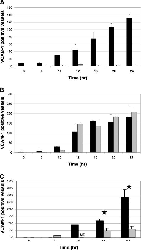
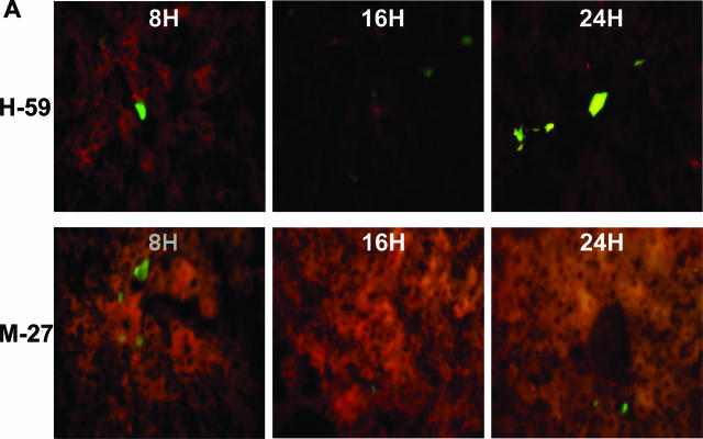
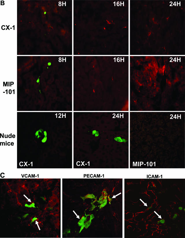
In the course of inflammation-induced transendothelial migration of leukocytes, the decline in vascular E-selectin expression has been shown to coincide with increased expression of vascular adhesion receptors of the immunoglobulin superfamily such as ICAM-1 and VCAM-1.15,30,31,32 We previously reported that VCAM-1 mRNA expression in the liver also rises in response to the influx of metastatic cells.28 Here, we show that a significant increase in cell surface VCAM-1 expression on sinusoidal endothelial cells was evident as early as 10 hours following H-59 and CX-1 injection, when the gradual decline in E-selectin expression began (Figure 6, A–C). The VCAM-1 signal remained high for up to 24 to 48 hours after injection and was frequently seen in close proximity to the tumor cells with areas of contact revealed by confocal imaging, particularly for H-59 cells (Figure 7C, VCAM-1, arrow; and Supplemental Figure 1, see http://ajp.amjpathol.org). No significant VCAM-1 expression was noted following M-27 inoculation at any of the time intervals analyzed (Figures 6A and 7A), in agreement with our reverse transcriptase-polymerase chain reaction data.28 Interestingly, despite the absence of E-selectin expression following MIP-101 injection, up-regulated expression of VCAM-1 was observed and was comparable in magnitude to that seen following CX-1 injection in conventional mice but lower in nude mice (Figure 6, B and C). Platelet endothelial cell adhesion molecule-1 (PECAM-1) (CD31) is constitutively expressed in vascular endothelial cells of the liver. Our previous polymerase chain reaction data suggested that PECAM-1 mRNA levels were not measurably increased in response to tumor cell injections. When cell surface expression of PECAM-1 was analyzed in liver sections obtained 8 hours following the injection of H-59 cells, we observed that most of the sinusoidal vessels were PECAM-1-positive (Figure 7C and data not shown). Interestingly, confocal imaging showed regions of close contact between the tumor cells and PECAM-1, and 3D reconstruction revealed that tumor cell extravasation was preferentially associated with areas of intense PECAM-1 staining (Figure 7C, PECAM-1, and Supplemental Figure 2, see http://ajp.amjpathol.org), suggesting that this adhesion receptor may also participate in the transendothelial migration of the tumor cells, as was also reported for leukocyte transmigration.33 ICAM-1 is also constitutively expressed on vascular endothelial cells of the liver and, indeed, we found high-intensity ICAM-1 labeling on endothelial cells of both large (centrolobular and portal veins) and sinusoidal vessels at all time points analyzed (Figure 7C and not shown). In contrast to PECAM-1, however, tumor H-59 cells that were observed in proximity to vascular ICAM-1 did not appear to be actively transmigrating (Figure 7C, ICAM-1).
Figure 6.
VCAM-1 expression increases in response to tumor cell injection. Cryostat sections prepared following the inoculation of H-59 and M-27 (A, H-59: black; M-27: gray) or CX-1 and MIP-101 cells into C57B1/6 (B, CX-1: black; MIP-101: gray) or nude (C, CX-1: black; MIP-101: gray) mice were used for quantification of VCAM-1+ vessels. Sections were stained with anti-VCAM-1 antibodies and an Alexa Fluor 568 secondary antibody. VCAM-1-positive vessels were enumerated in 20 fields per liver at a magnification of ×630. Results are expressed as the mean number of VCAM-1-positive vessels (±SD, n = 4) in 20 microscope fields per time point. The difference between animals injected with H-59 and M-27 cells (A) was significant (P < 0.05) at all time points from 10 hours onward. ★P < 0.05.
Figure 7.
Confocal microscopy reveals areas of contact between H-59 and vascular endothelial CAM. Shown are representative images of VCAM-1+ vessels observed following injection of H-59 and M-27 (A) or CX-1 and MIP-101 (B) cells. Tumor cells are in green. Magnification, ×400. Shown in C are representative images captured with 100-μm liver sections that were obtained following inoculation of H-59 cells and stained with antibodies to VCAM-1, PECAM-1, or ICAM-1, as indicated. A Zeiss confocal microscope was used for the analysis at magnifications of ×630 (VCAM-1 and PECAM-1) or ×400 (ICAM-1). Cells are in green. Arrows on the merged images of red and green fluorescence indicate co-localized tumor cell/CAM signals.
Discussion
Our results provide direct evidence that tumor cells entering the liver can rapidly alter their microenvironment by triggering a host inflammatory cascade that leads to up-regulation of vascular endothelial cell adhesion molecules and then use this altered microenvironment to arrest, extravasate, and ultimately survive in the liver.
We show here that the ability to activate this response is variable and correlates with the metastatic phenotype of the cells. Thus, whereas H-59 cells rapidly induced intense E-selectin expression followed by VCAM-1 up-regulation, M-27 cells induced a weak E-selectin signal that was selectively observed on larger venules (ie, centrolobular and portal venules), and no VCAM-1 induction was seen. This low level of CAM induction correlated with the low levels of TNF-α and IL-1β induced in response to these tumor cells.27,28 Human colorectal CX-1 cells also induced an inflammatory cascade similar to that observed with H-59 cells, but the induction of endothelial cell CAMs was delayed and more sustained, as was also observed for TNF-α production in response to tumor inoculation.28 This distinct pattern may be related to the observation that CX-1-induced TNF-α production corresponded temporally to the formation of tumor-Kupffer cell conjugates, a process that may both delay and stabilize the response.28 Our findings with CX-1 cells are also consistent with reports by others implicating tumor cell-derived carcinoembryonic antigen in Kupffer cell activation and the induction of endothelial cell adhesion receptors.34,35,36 Interestingly, we observed a greater number of E-selectin-positive vessels in nude mice than in conventional mice injected with CX-1 cells. Although the underlying mechanism is unclear, it may be related to the more rapid clearance of CX-1 cells from the liver in the conventional, immunocompetent mice (Figure 3, B and C). Although T-cell-mediated immune rejection of the xenografted human tumor cells is unlikely during the first 48 hours following injection, it is possible that natural antibody- or cell-mediated innate immunity37,38,39 is differentially expressed in these mice and contribute to a more rapid clearance of the human carcinoma cells. Nonmetastatic MIP-101 cells, although failing to induce TNF-α, IL-1, and E-selectin expression, did trigger an increase in VCAM-1 expression that was detectable at 10 to 12 hours and persisted for up to 24 hours following tumor inoculation. This suggests that VCAM-1 up-regulation can occur independently of increased TNF-α or IL-1 levels, possibly as a response to other liver or tumor-derived cytokines such as IL-1840 or oxidative stress triggered by tumor cell entry into the blood vessels.41 The finding that in the athymic mice MIP-101 induced a weaker VCAM-1 signal suggests that T cells may also be involved in this response, and this is in agreement with data based on in vitro studies that implicated resting and activated T cells in VCAM-1 induction on endothelial cells.42 The increase in VCAM-1 expression did not, however, result in MIP-101 adhesion or transmigration, consistent with the reported failure of these cells to adhere to VCAM-1 on cultured endothelial cells.35
The tumor-specific patterns of cytokine and CAM induction that we observed most likely reflects the distinct repertoires of cytokines/chemokines produced by the tumor cells or, for the human colorectal carcinoma lines, a differential ability to produce carcinoembryonic antigen.43 Indeed, we have recently shown that M-27, but not H-59 cells express high levels of the secretory leukocyte protease inhibitor SLPI, an 11.7-kd secreted molecule that can block nuclear factor-κB-mediated TNF-α production in macrophages and thereby attenuate the host inflammatory response.44,45,46 We further demonstrated that SLPI production diminished the proinflammatory response triggered by the tumor cells and, as a consequence, reduced liver metastasis.44
Our findings are in line with other studies that implicated the proinflammatory host response and vascular endothelial CAM in the process of liver metastasis. Increased hepatic metastasis of B16F1 melanoma cells could be observed following exogenous IL-1α administration, and this was attributed to increased vascular adhesion receptor expression and tumor cell arrest in terminal portal venules.7,47 Likewise, pretreatment with TNF-α was shown to increase expression of vascular adhesion molecules ICAM-1, VCAM-1, and E-selectin, and this resulted in increased adhesion, extravasation, and metastasis of syngeneic colon adenocarcinoma C-26 cells.48 In another study, the same carcinoma cells were also shown to trigger an endogenous inflammatory cascade upon entry into the liver, and this cascade was absent in TNFR1-deficient mice, leading to reduced liver metastases formation.49 In addition to TNF-α and IL-1α, endogenous, endothelial cell-mediated IL-18 production in response to invading tumor cells was also shown to play a role in increased vascular CAM expression and hepatic metastasis of B16 melanoma cells.40
In our studies, H-59 cells were found to preferentially associate with E-selectin-positive sinusoidal vessels (Figure 4A), suggesting that E-selectin could mediate tumor-endothelial cell adhesion, as was also suggested by confocal imaging (Figure 5A). These findings are in agreement with results of our in vitro adhesion assays12 and with our finding that animals treated with anti-E-selectin antibodies had markedly reduced numbers of hepatic metastases.12 The E-selectin ligands on H-59 cells remain to be identified, since neither sLewx nor sLewa could be detected on these cells.12 The possible role of PSGL-1 and of CD43 and CD44, E-selectin ligands identified on T cells and colon cancer cells, respectively,24,50 is unknown and presently under investigation. Our data also suggest that although E-selectin could mediate tumor cell attachment to the sinusoidal vessels, transendothelial migration actually occurred away from areas of intense E-selectin expression (Figure 5A), as is also the case for leukocyte transmigration.32 Moreover, confocal analysis revealed areas of close contact between the tumor cells and endothelial VCAM-1 or PECAM-1 (Figure 7C), suggesting that tumor cells can co-opt molecular mediators of leukocyte adhesion and transmigration to attach to the hepatic endothelial cells.51 CX-1 adhesion in close proximity to vascular E-selectin was also observed (Figure 5B), consistent with the high expression of sialylated glycoproteins found on these cells.52 In athymic nude mice, this coincided with a transient increase in the total number of cells seen in the liver, but in both nude and conventional mice, the numbers of tumor cells eventually declined and remained low at 24 hours after injection. This is consistent with the lower number of metastases produced by these cells and their slower rate of growth compared with H-59 cells.17 The rapid decline in the number of CX-1 and MIP-101 cells in conventional mice may also reflect a higher rate of tumor cell death immediately following tumor injection.
Cytokine-induced endothelial cell adhesion receptors have previously been implicated in hepatic metastasis of human colorectal carcinoma and other malignancies.7,9,53,54 Studies in animal models have shown that the inhibition of cytokine-mediated E-selectin or VCAM-1 expression by reagents such as antibodies12 and anti-inflammatory drugs,55 by the selective depletion of Kupffer cells,48 or by an IL-18-binding protein40 effectively blocked liver metastases formation, even when administered at a time when micrometastases were already present.40 These findings suggest that the proinflammatory response may be critical not only for the initiation but also for the maintenance and growth of hepatic metastases. The present study is in line with these reports and adds several novel elements to this body of evidence. Namely, it provides a spatio-temporal context for the host inflammatory cascade that is triggered by liver-infiltrating tumor cells and shows that the kinetics of the host response are tumor type-specific and may depend, among other factors, on the tumor-Kupffer cell interaction. It also provides the first direct evidence that tumor cells can attach to sinusoidal endothelial E-selectin in vivo and that this attachment precedes the expression of and attachment to VCAM-1 or PECAM-1 and subsequent extravasation. Taken together, these studies suggest that drugs that target proinflammatory cytokines such as TNF-α, their receptors, or downstream effectors may be beneficial in prevention and/or treatment of hepatic micrometastases.56,57,58
Supplementary Material
Acknowledgments
We are grateful to Ms. Jacynthe Laliberte for her generous help with the confocal microscope.
Footnotes
Address reprint requests to Pnina Brodt, Surgical Labs, Royal Victoria Hospital, Room H6.25, 687 Pine Ave., W. Montreal, QC, H3A 1A1, Canada. E-mail: pnina.brodt@muhc.mcgill.ca.
Supported by Canadian Institute for Health Research grant MOP-13646 (P.B.) and a Fonds de la Recherche en Santé du Québec/INSERM Research Collaboration Travel award (P.B. and A.B.). N.W. was supported by a Valorization Recherche Québec grant and a Cedar Cancer Institute Post-Doctoral Fellowship, and J.B. is supported by a McGill University Health Center Research Institute Fellowship.
P.A. and L.F. contributed equally to this article.
P.A. was on sabbatical leave at the Department of Surgery, McGill University Health Center, during the course of this study.
Supplemental material for this article can be found on http://ajp.amjpathol.org.
References
- Bissell MJ, Kenny PA, Radisky DC. Microenvironmental regulators of tissue structure and function also regulate tumor induction and progression: the role of extracellular matrix and its degrading enzymes. Cold Spring Harb Symp Quant Biol. 2005;70:343–356. doi: 10.1101/sqb.2005.70.013. [DOI] [PMC free article] [PubMed] [Google Scholar]
- Witz IP, Levy-Nissenbaum O. The tumor microenvironment in the post-PAGET era. Cancer Lett. 2006;242:1–10. doi: 10.1016/j.canlet.2005.12.005. [DOI] [PubMed] [Google Scholar]
- Coussens LM, Werb Z. Inflammation and cancer. Nature. 2002;420:860–867. doi: 10.1038/nature01322. [DOI] [PMC free article] [PubMed] [Google Scholar]
- Robinson SC, Coussens LM. Soluble mediators of inflammation during tumor development. Adv Cancer Res. 2005;93:159–187. doi: 10.1016/S0065-230X(05)93005-4. [DOI] [PubMed] [Google Scholar]
- Fahy B, Bold RJ. Epidemiology and molecular genetics of colorectal cancer. Surg Oncol. 1998;7:115–123. doi: 10.1016/s0960-7404(99)00021-3. [DOI] [PubMed] [Google Scholar]
- Boring CC, Squires TS, Tong T, Montgomery S. Cancer statistics, 1994. CA Cancer J Clin. 1994;44:7–26. doi: 10.3322/canjclin.44.1.7. [DOI] [PubMed] [Google Scholar]
- Wang HH, Qiu H, Qi K, Orr FW. Current views concerning the influences of murine hepatic endothelial adhesive and cytotoxic properties on interactions between metastatic tumor cells and the liver. Comp Hepatol. 2005;4:8. doi: 10.1186/1476-5926-4-8. [DOI] [PMC free article] [PubMed] [Google Scholar]
- Brodt P, editor. Austin, TX, Landes: Heidelberg, Springer-Verlag; Adhesion receptors and proteolytic mechanisms in cancer invasion and metastasis. Cell Adhesion and Invasion in Cancer Metastasis. 1996:pp 167–242. [Google Scholar]
- Rudmik LR, Magliocco AM. Molecular mechanisms of hepatic metastasis in colorectal cancer. J Surg Oncol. 2005;92:347–359. doi: 10.1002/jso.20393. [DOI] [PubMed] [Google Scholar]
- Bresalier RS, Byrd JC, Brodt P, Ogata S, Itzkowitz SH, Yunker CK. Liver metastasis and adhesion to the sinusoidal endothelium by human colon cancer cells is related to mucin carbohydrate chain length. Int J Cancer. 1998;76:556–562. doi: 10.1002/(sici)1097-0215(19980518)76:4<556::aid-ijc19>3.0.co;2-5. [DOI] [PubMed] [Google Scholar]
- Bresalier RS, Ho SB, Schoeppner HL, Kim YS, Sleisenger MH, Brodt P, Byrd JC. Enhanced sialylation of mucin-associated carbohydrate structures in human colon cancer metastasis. Gastroenterology. 1996;110:1354–1367. doi: 10.1053/gast.1996.v110.pm8613039. [DOI] [PubMed] [Google Scholar]
- Brodt P, Fallavollita L, Bresalier RS, Meterissian S, Norton CR, Wolitzky BA. Liver endothelial E-selectin mediates carcinoma cell adhesion and promotes liver metastasis. Int J Cancer. 1997;71:612–619. doi: 10.1002/(sici)1097-0215(19970516)71:4<612::aid-ijc17>3.0.co;2-d. [DOI] [PubMed] [Google Scholar]
- Witz IP. The involvement of selectins and their ligands in tumor-progression. Immunol Lett. 2006;104:89–93. doi: 10.1016/j.imlet.2005.11.008. [DOI] [PubMed] [Google Scholar]
- Wang J, Springer TA. Structural specializations of immunoglobulin superfamily members for adhesion to integrins and viruses. Immunol Rev. 1998;163:197–215. doi: 10.1111/j.1600-065x.1998.tb01198.x. [DOI] [PubMed] [Google Scholar]
- Carlos TM, Harlan JM. Leukocyte-endothelial adhesion molecules. Blood. 1994;84:2068–2101. [PubMed] [Google Scholar]
- Kim I, Moon SO, Kim SH, Kim HJ, Koh YS, Koh GY. Vascular endothelial growth factor expression of intercellular adhesion molecule 1 (ICAM-1), vascular cell adhesion molecule 1 (VCAM-1), and E-selectin through nuclear factor-kappa B activation in endothelial cells. J Biol Chem. 2001;276:7614–7620. doi: 10.1074/jbc.M009705200. [DOI] [PubMed] [Google Scholar]
- Khatib AM, Fallavollita L, Wancewicz EV, Monia BP, Brodt P. Inhibition of hepatic endothelial E-selectin expression by C-raf antisense oligonucleotides blocks colorectal carcinoma liver metastasis. Cancer Res. 2002;62:5393–5398. [PubMed] [Google Scholar]
- Harvey BE, Toth CA, Wagner HE, Steele GD, Jr, Thomas P. Sialyltransferase activity and hepatic tumor growth in a nude mouse model of colorectal cancer metastases. Cancer Res. 1992;52:1775–1779. [PubMed] [Google Scholar]
- Nakamori S, Kameyama M, Imaoka S, Furukawa H, Ishikawa O, Sasaki Y, Kabuto T, Iwanaga T, Matsushita Y, Irimura T. Increased expression of sialyl Lewisx antigen correlates with poor survival in patients with colorectal carcinoma: clinicopathological and immunohistochemical study. Cancer Res. 1993;53:3632–3637. [PubMed] [Google Scholar]
- Sato M, Narita T, Kimura N, Zenita K, Hashimoto T, Manabe T, Kannagi R. The association of sialyl Lewis(a) antigen with the metastatic potential of human colon cancer cells. Anticancer Res. 1997;17:3505–3511. [PubMed] [Google Scholar]
- Yamada N, Chung YS, Takatsuka S, Arimoto Y, Sawada T, Dohi T, Sowa M. Increased sialyl Lewis A expression and fucosyltransferase activity with acquisition of a high metastatic capacity in a colon cancer cell line. Br J Cancer. 1997;76:582–587. doi: 10.1038/bjc.1997.429. [DOI] [PMC free article] [PubMed] [Google Scholar]
- Laferriere J, Houle F, Huot J. Regulation of the metastatic process by E-selectin and stress-activated protein kinase-2/p38. Ann NY Acad Sci. 2002;973:562–572. doi: 10.1111/j.1749-6632.2002.tb04702.x. [DOI] [PubMed] [Google Scholar]
- Matsushita Y, Kitajima S, Goto M, Tezuka Y, Sagara M, Imamura H, Tanabe G, Tanaka S, Aikou T, Sato E. Selectins induced by interleukin-1beta on the human liver endothelial cells act as ligands for sialyl Lewis X-expressing human colon cancer cell metastasis. Cancer Lett. 1998;133:151–160. doi: 10.1016/s0304-3835(98)00220-1. [DOI] [PubMed] [Google Scholar]
- Hanley WD, Burdick MM, Konstantopoulos K, Sackstein R. CD44 on LS174T colon carcinoma cells possesses E-selectin ligand activity. Cancer Res. 2005;65:5812–5817. doi: 10.1158/0008-5472.CAN-04-4557. [DOI] [PubMed] [Google Scholar]
- Berlin C, Berg EL, Briskin MJ, Andrew DP, Kilshaw PJ, Holzmann B, Weissman IL, Hamann A, Butcher EC. Alpha 4 beta 7 integrin mediates lymphocyte binding to the mucosal vascular addressin MAdCAM-1. Cell. 1993;74:185–195. doi: 10.1016/0092-8674(93)90305-a. [DOI] [PubMed] [Google Scholar]
- Salas A, Shimaoka M, Phan U, Kim M, Springer TA. Transition from rolling to firm adhesion can be mimicked by extension of integrin alpha 1beta 2 in an intermediate-affinity state. J Biol Chem. 2006;128:10876–10882. doi: 10.1074/jbc.M512472200. [DOI] [PMC free article] [PubMed] [Google Scholar]
- Khatib AM, Kontogiannea M, Fallavollita L, Jamison B, Meterissian S, Brodt P. Rapid induction of cytokine and E-selectin expression in the liver in response to metastatic tumor cells. Cancer Res. 1999;59:1356–1361. [PubMed] [Google Scholar]
- Khatib AM, Auguste P, Fallavollita L, Wang N, Samani A, Kontogiannea M, Meterissian S, Brodt P. Characterization of the host proinflammatory response to tumor cells during the initial stages of liver metastasis. Am J Pathol. 2005;167:749–759. doi: 10.1016/S0002-9440(10)62048-2. [DOI] [PMC free article] [PubMed] [Google Scholar]
- Samani AA, Chevet E, Fallavollita L, Galipeau J, Brodt P. Loss of tumorigenicity and metastatic potential in carcinoma cells expressing the extracellular domain of the type 1 insulin-like growth factor receptor. Cancer Res. 2004;64:3380–3385. doi: 10.1158/0008-5472.CAN-03-3780. [DOI] [PubMed] [Google Scholar]
- Carman CV, Springer TA. A transmigratory cup in leukocyte diapedesis both through individual vascular endothelial cells and between them. J Cell Biol. 2004;167:377–388. doi: 10.1083/jcb.200404129. [DOI] [PMC free article] [PubMed] [Google Scholar]
- Weber C, Springer TA. Interaction of very late antigen-4 with VCAM-1 supports transendothelial chemotaxis of monocytes by facilitating lateral migration. J Immunol. 1998;161:6825–6834. [PubMed] [Google Scholar]
- Kakkar AK, Lefer DJ. Leukocyte and endothelial adhesion molecule studies in knockout mice. Curr Opin Pharmacol. 2004;4:154–158. doi: 10.1016/j.coph.2004.01.003. [DOI] [PubMed] [Google Scholar]
- Wang Y, Sheibani N. PECAM-1 isoform-specific activation of MAPK/ERKs and small GTPases: implications in inflammation and angiogenesis. J Cell Biochem. 2006;98:451–468. doi: 10.1002/jcb.20827. [DOI] [PubMed] [Google Scholar]
- Gangopadhyay A, Bajenova O, Kelly TM, Thomas P. Carcinoembryonic antigen induces cytokine expression in Kuppfer cells: implications for hepatic metastasis from colorectal cancer. Cancer Res. 1996;56:4805–4810. [PubMed] [Google Scholar]
- Gangopadhyay A, Lazure DA, Thomas P. Adhesion of colorectal carcinoma cells to the endothelium is mediated by cytokines from CEA stimulated Kupffer cells. Clin Exp Metastasis. 1998;16:703–712. doi: 10.1023/a:1006576627429. [DOI] [PubMed] [Google Scholar]
- Meterissian SH, Toth CA, Steele G, Jr, Thomas P. Kupffer cell/tumor cell interactions and hepatic metastasis in colorectal cancer. Cancer Lett. 1994;81:5–12. doi: 10.1016/0304-3835(94)90157-0. [DOI] [PubMed] [Google Scholar]
- Vollmers HP, Brandlein S. Death by stress: natural IgM-induced apoptosis. Methods Find Exp Clin Pharmacol. 2005;27:185–191. doi: 10.1358/mf.2005.27.3.890876. [DOI] [PubMed] [Google Scholar]
- Brändlein S, Pohle T, Ruoff N, Wozniak E, Muller-Hermelink HK, Vollmers HP. Natural IgM antibodies and immunosurveillance mechanisms against epithelial cancer cells in humans. Cancer Res. 2003;63:7995–8005. [PubMed] [Google Scholar]
- Diefenbach A, Raulet DH. The innate immune response to tumors and its role in the induction of T-cell immunity. Immunol Rev. 2002;188:9–21. doi: 10.1034/j.1600-065x.2002.18802.x. [DOI] [PubMed] [Google Scholar]
- Carrascal MT, Mendoza L, Valcarcel M, Salado C, Egilegor E, Telleria N, Vidal-Vanaclocha F, Dinarello CA. Interleukin-18 binding protein reduces b16 melanoma hepatic metastasis by neutralizing adhesiveness and growth factors of sinusoidal endothelium. Cancer Res. 2003;63:491–497. [PubMed] [Google Scholar]
- Mendoza L, Carrascal T, De Luca M, Fuentes AM, Salado C, Blanco J, Vidal-Vanaclocha F. Hydrogen peroxide mediates vascular cell adhesion molecule-1 expression from interleukin-18-activated hepatic sinusoidal endothelium: implications for circulating cancer cell arrest in the murine liver. Hepatology. 2001;34:298–310. doi: 10.1053/jhep.2001.26629. [DOI] [PubMed] [Google Scholar]
- Yarwood H, Mason JC, Mahiouz D, Sugars K, Haskard DO. Resting and activated T cells induce expression of E-selectin and VCAM-1 by vascular endothelial cells through a contact-dependent but CD40 ligand-independent mechanism. J Leukoc Biol. 2000;68:233–242. [PubMed] [Google Scholar]
- Gangopadhyay A, Lazure DA, Thomas P. Carcinoembryonic antigen induces signal transduction in Kupffer cells. Cancer Lett. 1997;118:1–6. doi: 10.1016/s0304-3835(97)00216-4. [DOI] [PubMed] [Google Scholar]
- Wang N, Thuraisingam T, Fallavollita L, Ding A, Radzioch D, Brodt P. The secretory leukocyte protease inhibitor is a type 1 insulin-like growth factor receptor-regulated protein that protects against liver metastasis by attenuating the host proinflammatory response. Cancer Res. 2006;66:3062–3070. doi: 10.1158/0008-5472.CAN-05-2638. [DOI] [PubMed] [Google Scholar]
- Lentsch AB, Jordan JA, Czermak BJ, Diehl KM, Younkin EM, Sarma V, Ward PA. Inhibition of NF-kappaB activation and augmentation of IkappaBbeta by secretory leukocyte protease inhibitor during lung inflammation. Am J Pathol. 1999;154:239–247. doi: 10.1016/s0002-9440(10)65270-4. [DOI] [PMC free article] [PubMed] [Google Scholar]
- Jin FY, Nathan C, Radzioch D, Ding A. Secretory leukocyte protease inhibitor: a macrophage product induced by and antagonistic to bacterial lipopolysaccharide. Cell. 1997;88:417–426. doi: 10.1016/s0092-8674(00)81880-2. [DOI] [PubMed] [Google Scholar]
- Orr FW, Wang HH, Lafrenie RM, Scherbarth S, Nance DM. Interactions between cancer cells and the endothelium in metastasis. J Pathol. 2000;190:310–329. doi: 10.1002/(SICI)1096-9896(200002)190:3<310::AID-PATH525>3.0.CO;2-P. [DOI] [PubMed] [Google Scholar]
- Sturm JW, Magdeburg R, Berger K, Petruch B, Samel S, Bonninghoff R, Keese M, Hafner M, Post S. Influence of TNFA on the formation of liver metastases in a syngenic mouse model. Int J Cancer. 2003;107:11–21. doi: 10.1002/ijc.11320. [DOI] [PubMed] [Google Scholar]
- Kitakata H, Nemoto-Sasaki Y, Takahashi Y, Kondo T, Mai M, Mukaida N. Essential roles of tumor necrosis factor receptor p55 in liver metastasis of intrasplenic administration of colon 26 cells. Cancer Res. 2002;62:6682–6687. [PubMed] [Google Scholar]
- Matsumoto M, Atarashi K, Umemoto E, Furukawa Y, Shigeta A, Miyasaka M, Hirata T. CD43 functions as a ligand for E-selectin on activated T cells. J Immunol. 2005;175:8042–8050. doi: 10.4049/jimmunol.175.12.8042. [DOI] [PubMed] [Google Scholar]
- DeLeve LD, Wang X, Hu L, McCuskey MK, McCuskey RS. Rat liver sinusoidal endothelial cell phenotype is maintained by paracrine and autocrine regulation. Am J Physiol. 2004;287:G757–G763. doi: 10.1152/ajpgi.00017.2004. [DOI] [PubMed] [Google Scholar]
- Petrick AT, Meterissian S, Steele G, Jr, Thomas P. Desialylation of metastatic human colorectal carcinoma cells facilitates binding to Kupffer cells. Clin Exp Metastasis. 1994;12:108–116. doi: 10.1007/BF01753977. [DOI] [PubMed] [Google Scholar]
- Laferrière J, Houle F, Huot J. Adhesion of HT-29 colon carcinoma cells to endothelial cells requires sequential events involving E-selectin and integrin beta4. Clin Exp Metastasis. 2004;21:257–264. doi: 10.1023/b:clin.0000037708.09420.9a. [DOI] [PubMed] [Google Scholar]
- Gulubova MV. Expression of cell adhesion molecules, their ligands and tumour necrosis factor alpha in the liver of patients with metastatic gastrointestinal carcinomas. Histochem J. 2002;34:67–77. doi: 10.1023/a:1021304227369. [DOI] [PubMed] [Google Scholar]
- Kobayashi K, Matsumoto S, Morishima T, Kawabe T, Okamoto T. Cimetidine inhibits cancer cell adhesion to endothelial cells and prevents metastasis by blocking E-selectin expression. Cancer Res. 2000;60:3978–3984. [PubMed] [Google Scholar]
- Karin M. Inflammation-activated protein kinases as targets for drug development. Proc Am Thorac Soc. 2005;2:386–390; discussion 394–385. doi: 10.1513/pats.200504-034SR. [DOI] [PMC free article] [PubMed] [Google Scholar]
- Luo JL, Kamata H, Karin M. IKK/NF-kappaB signaling: balancing life and death—a new approach to cancer therapy. J Clin Invest. 2005;115:2625–2632. doi: 10.1172/JCI26322. [DOI] [PMC free article] [PubMed] [Google Scholar]
- Karin M, Greten FR. NF-kappaB: linking inflammation and immunity to cancer development and progression. Nat Rev Immunol. 2005;5:749–759. doi: 10.1038/nri1703. [DOI] [PubMed] [Google Scholar]
Associated Data
This section collects any data citations, data availability statements, or supplementary materials included in this article.


