Abstract
IL-15 is a common γ-chain cytokine that has been shown to be more active than IL-2 in several murine cancer immunotherapy models. Although T lymphocytes do not produce IL-15, murine lymphocytes carrying an IL-15 transgene demonstrated superior antitumor activity in the immunotherapy of B16 melanoma. Thus, we sought to investigate the biological impact of constitutive IL-15 expression by human lymphocytes. In this report we describe the generation of a retroviral vector encoding a codon-optimized IL-15 gene. Alternate codon usage significantly enhanced the translational efficiency of this tightly regulated gene in retroviral vector-transduced cells. Activated human CD4+ and CD8+ human lymphocytes expressed IL-15Rα and produced high levels of cytokine upon retroviral transduction with the IL-15 vector. IL-15-transduced lymphocytes remained viable for up to 180 days in the absence of exogenous cytokine. IL-15 vector-transduced T cells showed continued proliferation after cytokine withdrawal and resistance to apoptosis while retaining specific Ag recognition. In the setting of adoptive cell transfer, IL-15-transduced lymphocytes may prolong lymphocyte survival in vivo and could potentially enhance antitumor activity.
Interleukins-2 and -15 are common γ-chain cytokines that share the ability to stimulate activated T cells in vitro (1, 2), but have other contrasting properties. IL-2, the first of the γ-chain cytokines to be described, has been used to grow T lymphocytes in vitro and to support lymphocyte survival after adoptive transfer into mice and humans (3). However, IL-2 has several properties that limit its effectiveness as a T cell growth factor. In vivo, IL-2 plays a central role in the activation-induced cell death of T cells and mediates T cell tolerance via promotion of CD4+CD25+ regulatory T cell production, growth, survival, and suppressor function (4, 5). In contrast, IL-15 inhibits IL-2-induced activation-induced cell death (6) and plays a role in the generation and/or maintenance of memory CD8+ T cells (7–10). Thus, it has been suggested that IL-15 may be superior to IL-2 as a component of therapeutic regimens for the immunotherapy of cancer (11, 12).
In a recent model of the immunotherapy of a murine melanoma, IL-15 was shown to be superior to IL-2 in the generation of antitumor T cells; furthermore, the in vivo antitumor activity of Ag-specific CD8+ lymphocytes was enhanced in cells bearing an IL-15 transgene (13). This was an intriguing finding, given that T lymphocytes do not produce IL-15 (14). Implicitly, the effector function or survival of T lymphocytes may be enhanced by constitutive IL-15 expression. To evaluate the findings reported in this murine model in the context of human immunobiology, we developed a retroviral vector capable of efficiently transducing human lymphocytes with the IL-15 gene.
One of the challenges in engineering lymphocytes to express IL-15 is that IL-15 protein production is obstructed by several mechanisms. Although IL-15 mRNA is widely expressed by a variety of tissues (placenta, skeletal muscle, heart, lung, liver, kidney, fibroblasts, epithelial cells, and activated monocytes), unexpectedly low levels of cytokine are actually produced by cells expressing abundant mRNA (14). It is now understood that IL-15 gene expression is highly regulated at multiple levels. In addition to control at the level of transcription, IL-15 is post-transcriptionally regulated by elements upstream from the mature protein (14–16). Furthermore, analysis of the coding sequence for the mature protein revealed numerous infrequently used codons and free energy levels predisposing mRNA transcripts to the formation of inhibitory secondary structures.
In this report we describe the development of a retroviral vector encoding a codon-optimized (CO)3 IL-15 gene that efficiently transduced human lymphocytes to express high levels of IL-15. We demonstrate that human lymphocytes transduced with the CO IL-15 gene persisted in vitro for up to 180 days in the absence of exogenous cytokine support. IL-15-transduced lymphocytes resisted apoptosis resulting from cytokine withdrawal and retained specific Ag recognition. The potential use of genetically modified lymphocytes carrying the IL-15 gene in adoptive immunotherapy is discussed.
Materials and Methods
Cell lines
The cell lines used include the rhabdomyosarcoma line TE 671 (American Type Culture Collection (ATCC) HTB-139), the highly transfectable human renal epithelial line 293T (ATCC CRL-11268), the mouse fibroblast line NIH-3T3 (ATCC CRL-1658), the human lymphoid cell line Sup T1 (ATCC CRL-1942), the TAP-deficient lymphoblastoid cell line T2 (17), the PG13 gibbon ape leukemia virus-packaging cell line (ATCC CRL-10686), and the human ecotropic packaging cell line Phoenix Eco (provided by G. Nolan, Stanford University, Stanford, CA). Cell culture medium consisted of RPMI 1640 (Invitrogen Life Technologies) supplemented with 10% FCS, 100 U/ml penicillin, 100 μg/ml streptomycin, 2 mM l-glutamine, and 25 mM HEPES buffer solution (all from Invitrogen Life Technologies). Cells lines were cultured at 37°C in a 5% CO2 humidified incubator.
Construction of retroviral vector plasmids
The pMSGV1 retroviral vector contains a murine stem cell virus long-terminal repeat and RNA processing signals similar to the MFG class of retroviral vectors. The detailed construction of this vector was recently described (18).
Two retroviral vector plasmids coding for human IL-15 were constructed. pMSGV1 preprolactin leader (PPL) IL-15, which carries the native IL-15 sequence linked to the bovine PPL, was assembled by insertion of an NcoI/BamHI fragment of an IL-15 expression construct (provided by Y. Tagaya, National Institutes of Health, Bethesda, MD) (6) into the NcoI/SnaBI sites of pMSGV1. A CO IL-15 gene was synthesized in which 63 codon substitutions were made in the mature protein region of the PPL IL-15 sequence (Blue Heron Biotechnology). These changes in the coding sequence minimized the use of rare codons while maintaining a low free energy as calculated by the Vienna RNA package (19). This synthetic gene was then subcloned and inserted into the NcoI/SnaBI sites of pMSGV1 to yield pMSGV1 PPL CO IL-15.
The anti-MART-1 TCR retroviral vector, designated AIB, was used for control transductions. This vector also uses the pMSGV1 retroviral vector backbone (18).
Preparation of retrovirus for comparison of pMSGV1 PPL IL-15 and pMSGV1 PPL CO IL-15
Phoenix Eco packaging cells (5 × 10⁵) were plated into each well of a six-well tissue culture plate. On the following day, the cells were transfected with 2 μg of plasmid DNA from either pMSGV1 PPL IL-15 or pMSGV1 PPL CO IL-15 using the GenePorter2 reagent (Gene Therapy Systems). Twenty-four hours later, the medium was aspirated and replaced with 2 ml of fresh medium/well. Forty-eight hours post-transfection, the cell culture supernatant containing retrovirus was collected. The transfections were performed in triplicate for each plasmid; thus, three separate preparations of retrovirus were produced from each plasmid.
Retroviral transduction
Nontissue culture-treated 24-well plates (BD Biosciences) were coated with 25 μg/ml recombinant fibronectin fragment (RetroNectin; Takara). Retroviral vector supernatants were added, and the plates were incubated at 32°C for 2–4 h, followed by storage at 4°C overnight. Plates were warmed to room temperature, supernatant was removed, and 0.1–1.0 × 10⁶ cells were added to each well with 1 ml of tissue culture medium/well. The plates were then incubated overnight in a 5% CO2 humidified incubator at 37°C.
Transduction of NIH-3T3 cells: cytokine production and PCR amplification to detect vector integration
Retroviral supernatants were used to transduce NIH-3T3 cells to assess target cell IL-15 production. Four hundred microliters of undiluted and serially diluted retroviral supernatants were applied to recombinant fibronectin-coated, nontissue culture, 24-well plates as described previously. NIH-3T3 cells (1 × 10⁵) were then transduced in each well of the retrovirus-coated plates. Twenty-four hours after transduction, the wells were washed three times with 2 ml of PBS (Biofluids) and then filled with 2 ml of fresh medium. The cell culture medium was collected 72 h after transduction, and the IL-15 content was analyzed by ELISA (R&D Systems).
PCR amplification used genomic DNA extracted from cell lysates using QuickExtract solution (Epicentre). Oligonucleotide primers flanking the multiple cloning site of the pMSGV1 retroviral vector backbone were synthesized: ggggtggaccatcctctaga and accgtcgactgcagaattcg. Oligonucleotide primers for the amplification of β-actin were also used. PCR was performed for 30 cycles at 96°C for 30 s, 60°C for 30 s, and 72°C for 90 s in a PTC-200 Thermal Cycler (Global Medical Instrumentation). The PCR products were run on a 1% agarose gel and subsequently imaged and quantitated with an LAS-1000 luminescent image analyzer system (Fujifilm Medical Systems USA). The ratio of vector insert band intensity compared with the corresponding β-actin band was calculated for each of the cell lysate samples.
Generation of high titer packaging cell clones
Phoenix Eco packaging cells were transfected with pMSGV1 PPL IL-15 or pMSGV1 PPL CO IL-15 using the GenePorter2 reagent. The retroviral supernatant was harvested 48 h after transfection and transferred to non-tissue culture plates coated with recombinant fibronectin (Takara). PG13 cells were then transduced on these plates. Clones were isolated by limiting dilution culture and screened for IL-15 production by ELISA (R&D Systems). Retroviral supernatants produced by these clones were then applied to RetroNectin (Takara)-coated plates and used to transduce Sup T1 cells. A high titer clone produced from pMSGV1 PPL CO IL-15 was selected on the basis of IL-15 production by transduced Sup T1 cells. Genomic DNA was then extracted from the selected PG13 clone, and a Southern blot was performed to verify vector integrity and evaluate the number of integrations. This clone was used to produce retrovirus for all subsequent experiments in which human lymphocytes were transduced.
Transduction of PBLs and cytokine withdrawal studies
PBLs were obtained by leukapheresis from patients with a history of melanoma who were treated on an adjuvant peptide vaccine protocol at the National Cancer Institute. Lymphocytes were purified by centrifugation on a Ficoll/Hypaque cushion, washed in HBSS, and cultured in medium consisting of AIM-V medium (Invitrogen Life Technologies) supplemented with 300 IU/ml IL-2, 5% human AB serum (Valley Biomedical), 100 U/ml penicillin, 100 μg/ml streptomycin, 2 mM l-glutamine (all from Invitrogen Life Technologies), 55 μM 2-ME, and 25 mM HEPES buffer solution (Invitrogen Life Technologies). Polyclonally activated lymphocytes were generated by adding 50 ng/ml OKT3 on culture day 0. Peptide-reactive lymphocytes were generated from PBLs obtained from patients previously vaccinated with the anchor residue-modified gp100:209–217(210M) peptide (20); these PBLs were activated by adding 1 μg/ml gp100:209–217(210M) to the lymphocyte culture medium, in the absence of IL-2, on day 0. On day 1, 300 IU/ml IL-2 was added to the cells. Lymphocytes were cultured at 37°C in a 5% CO2 humidified incubator. OKT3-activated cells were transduced on culture days 2 and 3, whereas peptide-stimulated cells were transduced between culture days 5 and 7. Each culture was exposed to retrovirus a total of two times, on successive days, by transferring 0.5–1.0 × 10⁶ cells in a volume of 1 ml of medium to each well of a 24-well, nontissue culture plate that had been coated sequentially with RetroNectin (Takara) and then retrovirus.
Studies requiring withdrawal of the cells from exogenous IL-2 were undertaken by harvesting and washing lymphocytes three times in lymphocyte culture medium without IL-2. After the final wash, the medium was tested by IL-2 and/or IL-15 ELISA (Endogen and R&D Systems, respectively) to verify that no residual cytokine was present. The cells were then resuspended in lymphocyte culture medium without IL-2 at a concentration of 1 × 10⁶ cells/ml, returned to tissue culture vessels, and enumerated and assessed for viability by trypan exclusion every 3–7 days. The medium was refreshed every 3–7 days by removing half the spent medium and replacing the volume with lymphocyte culture medium without IL-2.
Lymphocyte IL-15 production assay
Control or IL-15-transduced lymphocytes were washed three times in culture medium. Lymphocytes (2 × 10⁵) were then resuspended in cell culture medium without IL-2 and plated in round-bottom, 96-well plates. Half the wells were precoated with OKT3 Ab (200 μg/well). After 3 days in culture, the cell culture supernatants were harvested for analysis by ELISA (R&D Systems).
Lymphocyte proliferation assay
Lymphocytes were washed three times with culture medium and plated at 1 × 10⁵ cells/well in a 96-well, round-bottom microplate in the presence or the absence of 300 IU/ml IL-2. The cells were cultured for a total of 4 days, and in the final 16 h of culture, 1 μCi/well [methyl-3H]thymidine (PerkinElmer Life Sciences) was added to each well. Cellular DNA was harvested and counted by liquid scintillation counting.
Flow cytometry
Cell surface expression of CD3, CD4, CD8, CD27, CD28, CD45RA, CD45RO, CD62L, and CCR7 was measured using FITC-, PE-, or allophycocyanin-conjugated Abs and the corresponding isotype controls (BD Pharmingen). IL-15Rα was detected with a polyclonal biotinylated Ab and a biotinylated isotype-matched control Ab; cell surface-bound Ab was then labeled with streptavidin-PE (R&D Systems). Intracellular proteins Bcl-2 and Bcl-xL were detected using FITC- and PE-conjugated Abs and their respective isotype controls (BD Pharmingen and Southern Biotechnology Associates). Cells were washed twice in staining buffer composed of PBS with 0.5% BSA and stained with cell surface Abs or matched isotype controls, followed by incubation in the dark for 20 min at 4°C. Cells were then washed twice more with staining buffer before analysis. Intracellular staining was performed by fixing and permeablizing the cells with Cytofix/Cytoperm solution (BD Pharmingen), two washes in Perm/Wash buffer (BD Pharmingen), staining with intracellular Abs or isotype controls, and two additional washes before analysis. Annexin V and 7-aminoactinomycin (7-AAD) staining for the detection of apoptotic cells was performed using the Annexin V-PE Apoptosis Kit I (BD Pharmingen). Immunofluorescence was measured using a FACScan flow cytometer and was analyzed using CellQuest Pro software (BD Biosciences).
Lymphocyte Ag reactivity assay
Target cells were prepared by pulsing T2 cells with 10–1000 ng/ml peptide in cell culture medium for 2 h at 37°C. The following HLA-A2-restricted peptides were used: influenza peptide (GILGFVFTL), MART-1:27–35 peptide, or native gp100:209–217. Effector cells (PBL; 1 × 10⁵) were cocultured with 1 × 10⁵ target cells (peptide-pulsed T2) in a final volume of 0.2 ml in each well of a round-bottom, 96-well plate. Twenty-four hours later, cell culture supernatants were harvested and assayed for IFN-γ production by ELISA (Endogen).
Results
Generation of wild-type and CO IL-15 vectors
The redundancy of the genetic code is not reflected in a homogeneous distribution of tRNAs for certain amino acids, leading to the potential for rare codons in any given cistron. Analysis of the DNA sequence of the mature IL-15 protein revealed that 29 of the 114 codons (25%) were used with a frequency of <20% in human genes. In comparison, the IL-2 gene contains 20 rare codons in a coding sequence totaling 133 codons (15%). We designed an alternate IL-15-coding sequence taking into consideration: 1) codon frequency and 2) minimalization of nucleic acid folding, as predicted by free energy calculations. The final nucleic acid sequence of the synthetic IL-15 gene includes a total of 63 codon substitutions. Nineteen of these alternate codons replace native codons with utilization frequencies <20%. The amino acid sequence of the optimized IL-15 gene remains identical with that of the wild-type sequence (Fig. 1). The wild-type RNA transcript has a predicted free energy of 76 kcal/mol; the CO molecule has a predicted free energy of 38 kcal/mol. This synthetic gene was subcloned into the wild-type IL-15 retroviral vector, replacing the wild-type mature protein sequence while leaving the bovine PPL sequence intact.
FIGURE 1.
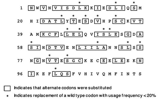
Amino acid sequence of the mature IL-15 protein, illustrating codon optimization. The mature protein contains 114 aa. Sixty-three codons were replaced with alternate sequences coding for the same amino acid. Nineteen of the substitutions resulted in a shift from a rarely used codon (<20%) to a more frequently used codon.
Wild-type and CO IL-15 retroviral vectors have comparable expression in transfected cell lines
We tested the activity of the vectors pMSGV1 PPL IL-15 and pMSGV1 PPL CO IL-15 by transfecting NIH-3T3, TE671, and 293T cell lines in triplicate with serial dilutions of retroviral vector plasmid DNA. The cytokine production of each cell line was assessed by ELISA. Under all conditions tested and in every cell line, IL-15 was produced at similar levels by cells receiving either the native or CO vector DNA (Fig. 2).
FIGURE 2.
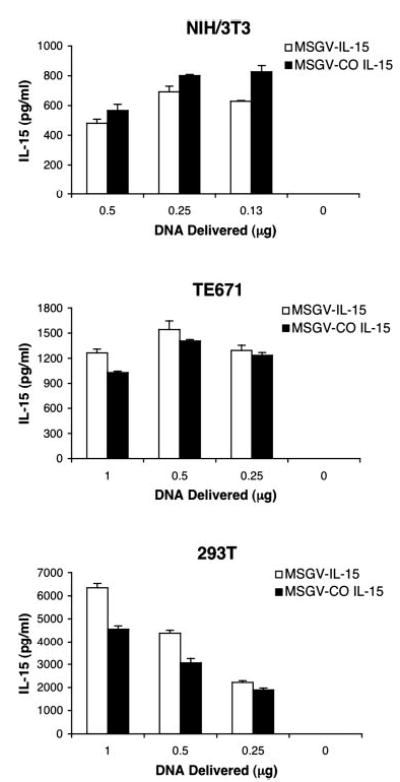
IL-15 expression by transfected cell lines. NIH-3T3, TE671, and 293T cells were transfected by lipofection with wild-type or CO IL-15 retroviral vector plasmids. The tissue culture plates were subsequently washed and refreshed with complete medium. After 24 h, the cell culture medium was tested for IL-15. Data are representative of two experiments.
To assess whether codon optimization influences the production of retrovirus by packaging cell lines, Phoenix Eco packaging cells were transfected with pMSGV1 PPL IL-15 and pMSGV1 PPL CO IL-15. The resultant retrovirus was titrated by Northern dot blot analysis (21) using a probe common to both vectors. Retroviral supernatants from Phoenix Eco cells transduced with either native or CO vector contained comparable amounts of RNA (data not shown).
Enhanced IL-15 expression in cells transduced with the CO retroviral vector
Retroviral vector preparations described in the previous section were used to transduce NIH-3T3 cells. Twenty-four hours after transduction, the plates were washed to eliminate any trace of IL-15 that could be carried over from the retroviral supernatant. Forty-eight hours later, the cell culture medium was collected and assayed for IL-15 (Fig. 3A). Cells transduced with a retrovirus coding for GFP did not produce detectable levels of IL-15 (data not shown). The cell culture medium from NIH-3T3 transduced with 2-fold serial dilutions of pMSGV1 PPL IL-15 retrovirus contained 436, 205, 98, and 44 pg/ml IL-15. In contrast, cell culture medium from NIH-3T3 transduced with similar dilutions of pMSGV1 PPL CO IL-15 retrovirus contained 1051, 498, 248, and 135 pg/ml IL-15, an ~2.5-fold increase in IL-15 production.
FIGURE 3.

Cytokine production and vector integration by IL-15-transduced cells. NIH-3T3 cells were transduced in triplicate with three separate preparations of serially diluted retroviral supernatants derived from either wild-type or CO IL-15 retroviral vectors. A, After 48 h in culture with fresh medium, the cell culture supernatants were harvested and assayed for IL-15. B, Genomic DNA was isolated from cell lysates. PCR amplification was conducted using primers flanking the insert as well as primers for β-actin. The PCR products were run on an agarose gel; the individual bands were then quantitated, and the ratio of retroviral insert to β-actin was calculated. Data are representative of two independent experiments.
To rule out differences in transduction efficiencies of the wild-type and CO IL-15 retroviral vectors, genomic DNA was isolated and analyzed for vector sequences. Using identical oligonucleotide primers, PCR was performed, and vector-specific amplification was normalized to coamplified β-actin. These studies demonstrated that transduction with either the wild-type or CO retrovirus led to comparable efficiencies of gene transfer (Fig. 3B).
IL-15 vector transduction of human PBLs results in stimulation-dependent cytokine production and persistence of the cells in the absence of exogenous cytokine support
PG13 retroviral packaging cell clones carrying the IL-15 gene were generated using pMSGV1 PPL CO IL-15. A high titer producer cell clone was selected on the basis of its ability to transduce Sup T1 cells to express IL-15. A Southern blot verified vector integrity and demonstrated four sites of vector integration (data not shown). This CO IL-15 retroviral vector-packaging cell was used to produce the retrovirus used in all of the subsequent studies involving IL-15-transduced human lymphocytes. Transduction efficiencies were 50–70% after two sequential transductions, as assessed by semiquantitative PCR and real-time PCR (data not shown).
OKT3-stimulated PBLs from five patients were transduced with the IL-15 vector. Cytokine production from these cells ranged from 251 to 2095 pg/ml (untransduced cells did not produce detectable quantities of IL-15). Stimulation of the cells with plate-bound OKT3 resulted in a 2- to 5-fold increase in cytokine production; between 1186 and 3957 pg/ml IL-15 was produced after stimulation (Fig. 4A). Stimulation-dependent activation of the retroviral long-terminal repeat is well described (22–24) and was previously reported in lymphocytes retrovirally transduced with IL-2 (25).
FIGURE 4.
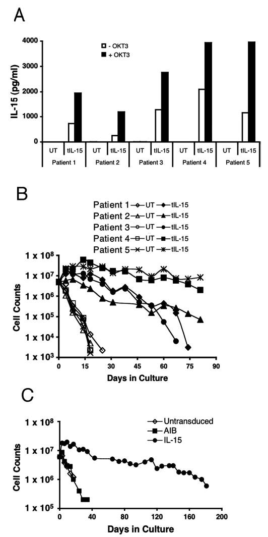
Cytokine production and growth of IL-15-transduced PBLs after withdrawal from exogenous cytokine support. OKT3-activated PBLs from five patients were transduced with IL-15. Control cells were not transduced. A, On culture day 7, cytokine production was measured in the presence or the absence of plate-bound OKT3. B, The PBLs were washed extensively, and 5 × 10⁶ cells from each culture were plated in fresh medium in the absence of exogenous cytokine. Viable cells were enumerated every 4–7 days by trypan blue exclusion; concurrently, the cell culture medium was refreshed by replacing half the conditioned medium with fresh medium. C, A similar experiment was performed in which the culture was conducted for >180 days. AIB, lymphocytes transduced with a control vector.
After 7 days in culture and 4 days after the final transduction, lymphocytes were thoroughly washed to remove all traces of soluble cytokines and were returned to culture in the absence of exogenous cytokine. Untransduced lymphocytes rapidly declined in both viability and cell count. By day 30 after cytokine withdrawal, no untransduced cells could be detected. In contrast, IL-15-transduced lymphocytes uniformly persisted in vitro for >60 days (Fig. 4B). After 60 days, two of five IL-15 vector-transduced cultures significantly declined in viability, whereas the remaining three cultures persisted beyond 80 days in the absence of added cytokine. In a similar experiment conducted over a longer time course, IL-15-transduced cells persisted in vitro for 181 days, whereas untransduced lymphocytes and lymphocytes transduced with a control gene were undetectable after culture for 30 days in the absence of exogenous cytokine (Fig. 4C).
During the course of these studies, lymphocytes from 17 patients were transduced with the IL-15 vector. Consistently, IL-15-transduced lymphocyte cultures demonstrated prolonged in vitro persistence after IL-2 withdrawal compared with control cultures. In 16 of 17 cultures, viable IL-15-transduced lymphocytes were detected from 40–181 days after cytokine withdrawal. However, one of the 17 IL-15-transduced cultures exhibited logarithmic, clonal expansion for >365 days (data not shown); this cell line is under active investigation.
Activated human T cells express IL-15Rα
IL-15- and IL-15Rα-deficient mice manifest similar phenotypes, exhibiting decreased numbers of CD8+ cells and nearly a total lack of memory CD8 cells, suggesting that the high affinity IL-15Rα is critical to the function of IL-15 in vivo (26). In vitro, supraphysiologic levels of cytokine can engage intermediate and low affinity IL-15 receptors on lymphocytes lacking IL-15Rα. To determine whether IL-15Rα was expressed in OKT3-activated T cells, we evaluated IL-15-transduced lymphocytes as well as untransduced control cells grown in medium containing either IL-2 or IL-15 (Fig. 5A). Untransduced lymphocytes, stimulated with OKT3 and IL-2 on day 0 of culture, expressed IL-15Rα in both CD4+ and CD8+ subsets (86 and 69% positive compared with isotype control, respectively). Untransduced lymphocytes stimulated with OKT3 and grown in medium containing 100 ng/ml IL-15 did not manifest IL-15Rα staining, nor did IL-15 vector-transduced lymphocytes. Thus, exogenous or endogenous IL-15 appeared to down-regulate the expression of IL-15Rα.
FIGURE 5.
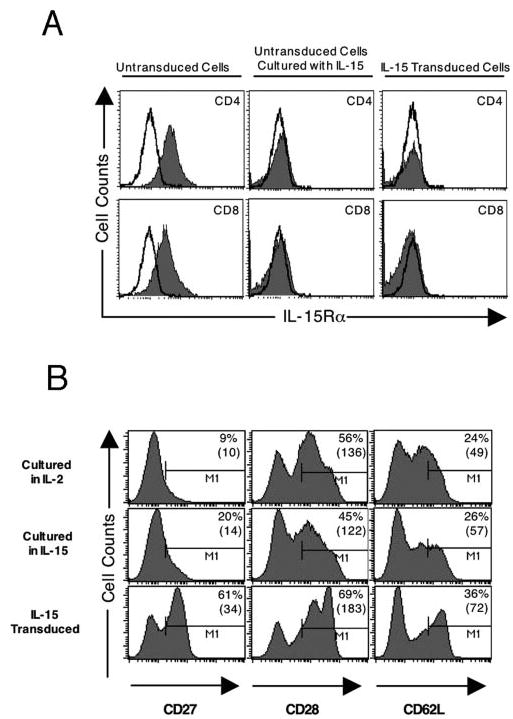
Cell surface molecule profile of OKT3-activated lymphocytes. A, IL-15Rα expression in OKT3-activated lymphocytes. Day 10 untransduced lymphocytes were grown in the presence of IL-2 (300 IU/ml) or IL-15 (100 ng/ml). IL-15-transduced lymphocytes from the same patient were grown in the absence of exogenous cytokine. After 3 days in culture, flow cytometric analysis was performed using FITC-coupled CD4 or CD8 and biotinylated anti-IL-15Rα secondarily stained with streptavidin-PE. The unshaded histograms represent isotype control staining, and the shaded histograms represent IL-15Rα staining for the indicated condition. B, CD27, CD28, and CD62L expression in lymphocytes after prolonged culture. After activation with OKT3, untransduced lymphocytes were grown in either IL-2 (300 IU/ml) or IL-15 (10 ng/ml), whereas IL-15-transduced lymphocytes were withdrawn from IL-2 after transduction. After 3 wk in culture, FACS analysis was performed on CD3-gated cells. The numbers in each histogram represent the percentages of cells exhibiting positive staining, followed by the mean fluorescence intensity in parentheses.
Phenotype of IL-15-transduced lymphocytes
T lymphocyte subsets are often defined by cell surface expression of costimulatory molecules, adhesion molecules, and receptors; these cell surface proteins, in turn, are subject to influences such as TCR stimulation and cytokine engagement. To further define the influence of IL-15 transduction on T lymphocytes, we evaluated long-term cultures of IL-15-transduced lymphocytes grown in the absence of exogenous cytokine as well as untransduced lymphocytes grown in medium containing either IL-2 or IL-15. IL-15-transduced lymphocytes demonstrated modest increases in staining for CD27, CD28, and CD62L compared with untransduced lymphocytes cultured in IL-2 or IL-15 (Fig. 5B). This was reflected in both the percentage of cells exhibiting positive staining compared with isotype controls and the mean fluorescence intensity. No differences were seen in the expression of CD45RA, CD45RO, or CCR7 (data not shown).
OKT3-stimulated PBLs transduced with IL-15 continue to proliferate in the absence of exogenous cytokine support and resist cytokine withdrawal-induced apoptosis
When OKT3-stimulated PBLs were transduced with the IL-15 gene, they remained viable for prolonged periods in culture, but ceased to increase in cell number. We sought to dissect the mechanisms for this persistence by first evaluating thymidine incorporation. On culture day 7, untransduced, control vector-transduced, and IL-15-transduced lymphocytes were washed free of IL-2, then cultured in the presence or the absence of IL-2 for 4 days. [3H]thymidine was added during the final 16 h of culture. Assessment of thymidine incorporation revealed that the IL-15-transduced cells continued to proliferate in the absence of exogenous cytokine, whereas the untransduced or control transduced cells did not (Fig. 6).
FIGURE 6.
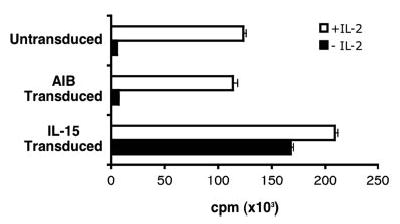
Thymidine incorporation assay. PBLs were untransduced (UT), control vector-transduced (AIB), or transduced with IL-15. On culture day 7, the cells were washed extensively and plated in triplicate in fresh medium with or without exogenous IL-2 (300 IU/ml). The cells were cultured for 4 days and were pulsed with [3H]thymidine during the final 16 h of culture. Data represent the mean ± SEM.
We next evaluated whether the IL-15-transduced cells were protected from apoptotic death upon IL-2 withdrawal. Fractions of cultures of untransduced, control vector-transduced, and IL-15-transduced PBLs were sequentially withdrawn from IL-2 for 3 consecutive days, starting on culture day 7. Four days later, cells were stained for 7-AAD/annexin V and analyzed by flow cytometry (Fig. 7). Untransduced and control gene-transduced cell populations demonstrated an increase in apoptotic cells within 24 h after cytokine withdrawal; the percentage of cells undergoing apoptosis (positivity for annexin V, but not 7-AAD) peaked at ~14–17%, 48 h after cytokine withdrawal. Necrotic cells, defined by 7-AAD positivity, accumulated continuously in these cultures after IL-2 was withdrawn, and ~35% of the cells in the populations lacking cytokine support were necrotic or undergoing apoptosis by 72 h. In contrast, IL-15-transduced PBLs resisted apoptosis after withdrawal from IL-2. Over 72 h, the percentage of apoptotic cells increased slightly from 4.3 to 5.7%, whereas necrotic cells increased from 3.5 to 6.4%.
FIGURE 7.
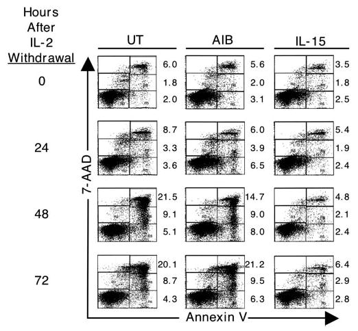
IL-2 withdrawal-induced apoptosis. OKT3-stimulated PBLs were untransduced, transduced with control vector (AIB), or transduced with IL-15. For 3 consecutive days, starting on culture day 7, a fraction of cells from each transduction condition was washed extensively and replated in the absence of exogenous IL-2. On day 10, the cells were stained with 7-AAD/annexin V and analyzed by flow cytometry. For each plot, the upper right sextant contains necrotic cells, the middle right contains late apoptotic cells, and the lower right contains early apoptotic cells. The numbers to the right of each plot represent the percentage of cells in the corresponding sextant.
IL-15-transduced PBLs maintain Bcl-2 and Bcl-xL expression after withdrawal from IL-2
CD8 lymphocytes from HIV patients demonstrate decreased levels of the antiapoptotic proteins Bcl-2 and Bcl-xL, which can be reversed by exposure to IL-15 (27). Because IL-15-transduced lymphocytes resisted apoptosis after cytokine withdrawal, we evaluated CD4 and CD8 lymphocytes for Bcl-2 and Bcl-xL expression (Fig. 8). Levels of Bcl-2 and Bcl-xL were down-regulated in untransduced and control-transduced cells 2 days after cytokine withdrawal. In contrast, the expression of these proteins was maintained in IL-15-transduced CD4 and CD8 lymphocytes after withdrawal from IL-2; Bcl-xL demonstrated no decrease in expression, whereas Bcl-2 displayed a modest decrease in expression (18 and 16% decreases in IL-15-transduced CD4 and CD8 cells vs 47–48 and 53–54% decreases in control CD4 and CD8 cultures).
FIGURE 8.
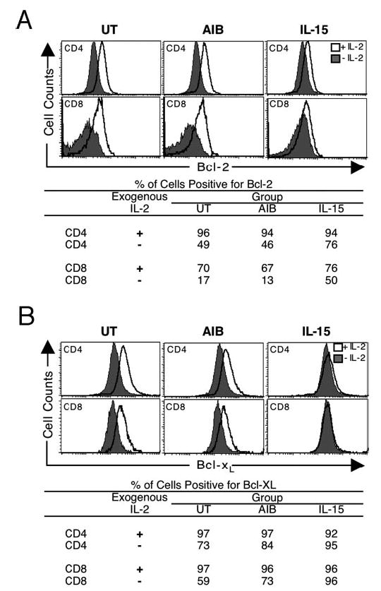
Bcl-2 and Bcl-xL expression in activated lymphocytes. OKT3-stimulated PBLs were untransduced, transduced with a control vector (AIB), or transduced with IL-15. On culture day 7, a fraction of the cells was washed extensively and plated in fresh medium without IL-2. Forty-eight hours later, the cells were stained with anti-CD3 and anti-CD8 Abs. Subsequently, intracellular staining was performed with either anti-Bcl-2 (A) or anti-Bcl-xL (B). Lymphocytes were forward and side scatter gated to exclude dead cells. Subsequently, staining in CD3 and CD8 populations was used to delineate CD4+ and CD8+ cells. The unshaded histogram depicts cells maintained in medium containing IL-2, and the shaded histogram represents cells after IL-2 withdrawal. Tables below each set of histograms show the percentages of cells exhibiting positive staining for Bcl-2 or Bcl-xL, respectively.
Specific peptide recognition by IL-15-transduced lymphocytes is maintained after withdrawal from IL-2
To further evaluate the function of IL-15-transduced lymphocytes, peptide-stimulated lymphocyte cultures with reactivity to the melanoma Ag gp100 were generated and subsequently transduced with the CO IL-15 vector. Untransduced and IL-15-transduced lymphocytes exhibited identical patterns of staining with gp100 tetramer, with 50–80% of CD8 cells staining positively after one round of peptide stimulation (data not shown). Subsequently, coculture experiments were performed using peptide-pulsed T2 cells as stimulators. Comparable quantities of IFN-γ were released by control and IL-15-transduced lymphocytes upon exposure to T2 cells pulsed with serial dilutions of the gp100 peptide (Fig. 9A). Peptide-specific reactivity was demonstrated by secretion of IFN-γ upon culture of control and IL-15-transduced lymphocytes with T2 cells pulsed with gp100, but not the HLA-A2-restricted influenza peptide (Flu). MART peptide reactivity was seen only in the culture transduced with the MART TCR (Fig. 9B). When the lymphocyte cultures were withdrawn from IL-2, control cultures (untransduced and MART TCR transduced) declined in viability and number, whereas IL-15-transduced lymphocytes remained viable; this was similar to the pattern previously demonstrated in OKT3-activated lymphocytes (data not shown). Five days after IL-2 withdrawal, the cells were again tested for peptide reactivity against T2-pulsed target cells; control cultures demonstrated diminished IFN-γ secretion upon encounter with gp100-pulsed T2, whereas IL-15-transduced cells maintained a high level of specific IFN-γ secretion (Fig. 9C).
FIGURE 9.
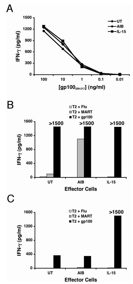
Peptide-specific reactivity is maintained in IL-15-transduced lymphocytes after withdrawal from IL-2. The gp100-reactive PBL cultures were generated and subsequently untransduced (UT), transduced with the MART TCR vector (AIB), or transduced with IL-15. These lymphocytes were tested for peptide reactivity by coculturing them with peptide-pulsed T2 cells at a 1:1 ratio. Cell culture supernatants were harvested and assayed for IFN-γ content 24 h after cocultures were initiated. A, On culture day 7, a peptide titration curve was established by coculturing lymphocytes with T2 cells that were pulsed with serially diluted gp100 peptide. B, To evaluate specific reactivity, culture day 7 lymphocytes were cocultured with T2 cells pulsed with 1 μg of peptide derived from influenza virus (Flu), MART, or gp100. C, In a similar fashion, to evaluate peptide reactivity after cytokine withdrawal, day 7 cultured cells were withdrawn from IL-2 and subsequently tested for reactivity to peptide-pulsed T2 cells on culture day 12.
Discussion
In this study we have described a novel approach in the retroviral transduction of activated human lymphocytes with a CO IL-15 gene. The codon optimization process improved IL-15 expression in transduced cells. Transduced human lymphocytes produced IL-15 in quantities with a measurable biological impact, persisting in vitro for up to 180 days in the absence of exogenous cytokine and resisting cytokine withdrawal-induced apoptosis. Transduction with IL-15 did not perturb lymphocyte Ag recognition or specificity. Furthermore, IL-15-transduced lymphocytes retained the ability to recognize Ag and secrete IFN-γ after withdrawal from exogenous cytokine support.
Codon optimization is a term that has been applied to a variety of approaches in which codons are systematically altered to enhance gene expression. Highly expressed genes evolve to use codons that are highly represented in the genome (28). In some instances, gene expression can be augmented by replacing rare codons with codons favored by highly expressed genes. This strategy has been applied to enhancing murine or human expression of parasitic and viral genes, which often contain codons infrequently used in mammalian genes (29–31). Conversely, altered codon usage has been applied to systems in which production of mammalian proteins by bacterial systems is desired (32, 33). In addition to codon bias considerations, RNA secondary structure formation and stability clearly impact protein expression and can be influenced by alternate codon usage (34). This approach continues to be refined, and its use is expanding. To our knowledge, codon optimization of cytokine expression by human cells has not been previously demonstrated.
Inefficient expression of IL-15 has been well established. The 5′-untranslated region, which contains multiple AUGs, has been implicated in the obstruction of mRNA translation (15, 16). Furthermore, an inhibitory effect of the secreted isoform of the IL-15 signal peptide and mature protein C terminus was demonstrated (16, 35). The expression of IL-15 was markedly increased when the native leader sequence was replaced with either the mouse IL-2 signal peptide or the bovine PPL sequence (6). We generated a construct containing the PPL sequence and further modified the coding sequence through alternate codon usage, thereby minimizing the usage of rare codons and minimizing the free energy (and thus potential folding) of the mRNA transcript.
Interestingly, cells transfected with either the wild-type or CO IL-15 constructs produced similar amounts of protein (Fig. 2). This may be explained by the fact that transfected cells receive hundreds of copies of the plasmid DNA, possibly saturating the protein production machinery. However, retrovirally transduced NIH-3T3 cells receiving the CO gene demonstrated an ~2.5-fold increase in protein expression compared with cells receiving the wild-type gene (Fig. 3A). The NIH-3T3 cells were transduced with retrovirus of comparable titer, and assessment of retroviral integration demonstrated similar efficiency of transduction (Fig. 3B). Thus, we conclude that the codon optimization improved gene expression at the level of mRNA stability and/or efficiency of translation.
Having established a means to engineer human lymphocytes to efficiently express IL-15, we were able to make several observations. We detected IL-15Rα on the surface of activated CD4+ and CD8+ T cells, contrary to a previous report in which activated human CD8+, but not CD4+, T cells, expressed this receptor (36). The IL-15Rα Ab used in our studies did not bind to lymphocytes cultured in exogenous IL-15 or transduced with the IL-15 gene, implying an interaction between IL-15 and the high affinity α receptor (Fig. 5A). In these OKT3-activated T cells, we speculate that the Ab-binding site on the receptor is altered or blocked upon binding IL-15. Alternatively, IL-15Rα may be internalized after capturing IL-15. Based on the observed expression of IL-15Rα on activated T cells, it is possible that IL-15 gene expression in individual cells led to their persistence via an autocrine mechanism or that paracrine effects from a fraction of lymphocytes excreting IL-15 were sufficient to sustain the entire lymphocyte culture.
Common γ-chain cytokines promote the survival of activated T lymphocytes through common signaling pathways that induce the expression of antiapoptotic Bcl-2 family proteins (37–39). We have demonstrated that IL-15-transduced lymphocytes, withdrawn from exogenous cytokine support, exhibited continued proliferation as well as resistance to apoptosis; this coincided with the maintenance of Bcl-2 and Bcl-xL (Figs. 6–8). In vitro, this resulted in prolonged survival in IL-15-transduced lymphocytes (Fig. 4). Furthermore, peptide-stimulated, IL-15-transduced lymphocytes retained Ag recognition and specificity even after withdrawal from exogenous IL-2 (Fig. 9). In most respects, IL-15-transduced lymphocytes behaved similarly to control cell populations maintained in the presence of IL-2. This is not entirely unexpected given that IL-2Rs and IL-15Rs share β and γ subunits as well as signaling through common Jak/Stat pathways (26). Our studies corroborate previous reports in which IL-2 and IL-15 shared the ability to stimulate activated T cells in vitro (1, 2).
In several preclinical models, the addition of IL-15 to cell transfer therapy regimens significantly improved antitumor activity (13, 40, 41). Briefly, these studies have demonstrated that culturing antitumor lymphocytes ex vivo in the presence of IL-15, delivering lymphocytes bearing an IL-15 transgene, or administering adjuvant IL-15 during the course of adoptive cell transfer resulted in superior antitumor activity. These models also demonstrated superior treatment effects of IL-15 compared with IL-2 when both cytokines were used in a similar fashion. The fact that IL-2 and IL-15 seem to have similar functions in vitro, but distinct and often opposing actions in vivo, is at least partially explained by murine models demonstrating that IL-15 is captured by APCs and stromal cells expressing high levels of the high affinity IL-15Rα and subsequently presented to lymphocytes and NK cells (42–45). It seems likely that the complex interactions between IL-15-presenting cells and responding lymphocytes powerfully influence the differentiation of effector T cells. This is strongly supported by the finding that IL-15Rα-deficient memory CD8+ T cells could be sustained by other cells expressing IL-15Rα (46). Furthermore, IL-15 antitumor activity in vivo may also be enhanced by the effects of IL-15 on cells other than the transferred lymphocyte, such as APCs or NK cells. To determine whether results reported in murine models, demonstrating superiority of IL-15 in adoptive cell transfer cancer models, would be recapitulated in humans would require clinical trials in which patients receive treatment with IL-15.
The use of IL-15-expressing T cells in adoptive transfer studies involving human patients has potential drawbacks. IL-15-transgenic mice develop autoimmunity and can ultimately succumb to lymphocytic leukemia (47). In humans, abnormal expression of IL-15 has been associated with rheumatoid arthritis, inflammatory bowel disease, adult T cell leukemia, and tropical spastic paresis (48, 49). In our experience with IL-15-transduced human lymphocytes, one of 17 cultures evolved into a clonal population that exhibited growth for >1 year in the absence of exogenous cytokine (data not shown); it remains uncertain whether this is a consequence of IL-15 overexpression or vector integration at a site critical to cell cycle regulation. With these considerations, the clinical application of gene-engineered T cells constitutively expressing IL-15 would probably require a reliable way to terminate the response should adverse effects of the treatment arise. HSV thymidine kinase-engineered lymphocytes have proven effective in controlling graft-vs-host disease in human bone marrow transplant recipients (50, 51). Thus, the development of an IL-15 retroviral vector carrying HSV thymidine kinase might permit the safe administration of IL-15-transduced, tumor-reactive lymphocytes to patients with metastatic cancer.
Acknowledgments
We express gratitude to Arnold Mixon and Shawn Farid for FACS analyses, to Yong Li for DNA sequencing, to Paul Robbins for insightful discussions, and to Yutaka Tagaya for providing the IL-15 expression plasmid.
Footnotes
This work was supported by the Intramural Research Program of the National Institutes of Health, National Cancer Institute, Center for Cancer Research.
Abbreviations used in this paper: CO, codon optimized; 7-AAD, 7-aminoactinomycin; PPL, preprolactin leader.
Disclosures
The authors have no financial conflict of interest.
References
- 1.Burton JD, Bamford RN, Peters C, Grant AJ, Kurys G, Goldman CK, Brennan J, Roessler E, Waldmann TA. A lymphokine, provisionally designated interleukin T and produced by a human adult T-cell leukemia line, stimulates T-cell proliferation and the induction of lymphokine-activated killer cells. Proc Natl Acad Sci USA. 1994;91:4935–4939. doi: 10.1073/pnas.91.11.4935. [DOI] [PMC free article] [PubMed] [Google Scholar]
- 2.Grabstein KH, Eisenman J, Shanebeck K, Rauch C, Srinivasan S, Fung V, Beers C, Richardson J, Schoenborn MA, Ahdieh M, et al. Cloning of a T cell growth factor that interacts with the β chain of the interleukin-2 receptor. Science. 1994;264:965–968. doi: 10.1126/science.8178155. [DOI] [PubMed] [Google Scholar]
- 3.Dudley ME, Rosenberg SA. Adoptive-cell-transfer therapy for the treatment of patients with cancer. Nat Rev Cancer. 2003;3:666–675. doi: 10.1038/nrc1167. [DOI] [PMC free article] [PubMed] [Google Scholar]
- 4.Waldmann TA. IL-15 in the life and death of lymphocytes: immuno-therapeutic implications. Trends Mol Med. 2003;9:517–521. doi: 10.1016/j.molmed.2003.10.005. [DOI] [PubMed] [Google Scholar]
- 5.Malek TR, Bayer AL. Tolerance, not immunity, crucially depends on IL-2. Nat Rev Immunol. 2004;4:665–674. doi: 10.1038/nri1435. [DOI] [PubMed] [Google Scholar]
- 6.Marks-Konczalik J, Dubois S, Losi JM, Sabzevari H, Yamada N, Feigenbaum L, Waldmann TA, Tagaya Y. IL-2-induced activation-induced cell death is inhibited in IL-15 transgenic mice. Proc Natl Acad Sci USA. 2000;97:11445–11450. doi: 10.1073/pnas.200363097. [DOI] [PMC free article] [PubMed] [Google Scholar]
- 7.Schluns KS, Williams K, Ma A, Zheng XX, Lefrancois L. Cutting edge: requirement for IL-15 in the generation of primary and memory antigen-specific CD8 T cells. J Immunol. 2002;168:4827–4831. doi: 10.4049/jimmunol.168.10.4827. [DOI] [PubMed] [Google Scholar]
- 8.Oh S, Berzofsky JA, Burke DS, Waldmann TA, Perera LP. Coadministration of HIV vaccine vectors with vaccinia viruses expressing IL-15 but not IL-2 induces long-lasting cellular immunity. Proc Natl Acad Sci USA. 2003;100:3392–3397. doi: 10.1073/pnas.0630592100. [DOI] [PMC free article] [PubMed] [Google Scholar]
- 9.Ku CC, Murakami M, Sakamoto A, Kappler J, Marrack P. Control of homeostasis of CD8+ memory T cells by opposing cytokines. Science. 2000;288:675–678. doi: 10.1126/science.288.5466.675. [DOI] [PubMed] [Google Scholar]
- 10.Berard M, Brandt K, Bulfone-Paus S, Tough DF. IL-15 promotes the survival of naive and memory phenotype CD8+ T cells. J Immunol. 2003;170:5018–5026. doi: 10.4049/jimmunol.170.10.5018. [DOI] [PubMed] [Google Scholar]
- 11.Fehniger TA, Cooper MA, Caligiuri MA. Interleukin-2 and interleukin-15: immunotherapy for cancer. Cytokine Growth Factor Rev. 2002;13:169–183. doi: 10.1016/s1359-6101(01)00021-1. [DOI] [PubMed] [Google Scholar]
- 12.Waldmann TA, Dubois S, Tagaya Y. Contrasting roles of IL-2 and IL-15 in the life and death of lymphocytes: implications for immunotherapy. Immunity. 2001;14:105–110. [PubMed] [Google Scholar]
- 13.Klebanoff CA, Finkelstein SE, Surman DR, Lichtman MK, Gattinoni L, Theoret MR, Grewal N, Spiess PJ, Antony PA, Palmer DC, et al. IL-15 enhances the in vivo antitumor activity of tumor-reactive CD8+ T cells. Proc Natl Acad Sci USA. 2004;101:1969–1974. doi: 10.1073/pnas.0307298101. [DOI] [PMC free article] [PubMed] [Google Scholar]
- 14.Tagaya Y, Bamford RN, DeFilippis AP, Waldmann TA. IL-15: a pleiotropic cytokine with diverse receptor/signaling pathways whose expression is controlled at multiple levels. Immunity. 1996;4:329–336. doi: 10.1016/s1074-7613(00)80246-0. [DOI] [PubMed] [Google Scholar]
- 15.Bamford RN, Battiata AP, Burton JD, Sharma H, Waldmann TA. Interleukin (IL) 15/IL-T production by the adult T-cell leukemia cell line HuT-102 is associated with a human T-cell lymphotrophic virus type I region/IL-15 fusion message that lacks many upstream AUGs that normally attenuates IL-15 mRNA translation. Proc Natl Acad Sci USA. 1996;93:2897–2902. doi: 10.1073/pnas.93.7.2897. [DOI] [PMC free article] [PubMed] [Google Scholar]
- 16.Bamford RN, DeFilippis AP, Azimi N, Kurys G, Waldmann TA. The 5′ untranslated region, signal peptide, and the coding sequence of the carboxyl terminus of IL-15 participate in its multifaceted translational control. J Immunol. 1998;160:4418–4426. [PubMed] [Google Scholar]
- 17.Salter RD, Howell DN, Cresswell P. Genes regulating HLA class I antigen expression in T-B lymphoblast hybrids. Immunogenetics. 1985;21:235–246. doi: 10.1007/BF00375376. [DOI] [PubMed] [Google Scholar]
- 18.Hughes MS, Yu YY, Dudley ME, Zheng Z, Robbins PF, Li Y, Wunderlich J, Hawley RG, Moayeri M, Rosenberg SA, et al. Transfer of a TCR gene derived from a patient with a marked antitumor response conveys highly active T-cell effector functions. Hum Gene Ther. 2005;16:457–472. doi: 10.1089/hum.2005.16.457. [DOI] [PMC free article] [PubMed] [Google Scholar]
- 19.Hofacker IL. Vienna RNA secondary structure server. Nucleic Acids Res. 2003;31:3429–3431. doi: 10.1093/nar/gkg599. [DOI] [PMC free article] [PubMed] [Google Scholar]
- 20.Rosenberg SA, Yang JC, Schwartzentruber DJ, Hwu P, Marincola FM, Topalian SL, Restifo NP, Dudley ME, Schwarz SL, Spiess PJ, et al. Immunologic and therapeutic evaluation of a synthetic peptide vaccine for the treatment of patients with metastatic melanoma. Nat Med. 1998;4:321–327. doi: 10.1038/nm0398-321. [DOI] [PMC free article] [PubMed] [Google Scholar]
- 21.Onodera M, Yachie A, Nelson DM, Welchlin H, Morgan RA, Blaese RM. A simple and reliable method for screening retroviral producer clones without selectable markers. Hum Gene Ther. 1997;8:1189–1194. doi: 10.1089/hum.1997.8.10-1189. [DOI] [PubMed] [Google Scholar]
- 22.Plavec I, Agarwal M, Ho KE, Pineda M, Auten J, Baker J, Matsuzaki H, Escaich S, Bonyhadi M, Bohnlein E. High transdominant RevM10 protein levels are required to inhibit HIV-1 replication in cell lines and primary T cells: implication for gene therapy of AIDS. Gene Ther. 1997;4:128–139. doi: 10.1038/sj.gt.3300369. [DOI] [PubMed] [Google Scholar]
- 23.Auten J, Agarwal M, Chen J, Sutton R, Plavec I. Effect of scaffold attachment region on transgene expression in retrovirus vector-transduced primary T cells and macrophages. Hum Gene Ther. 1999;10:1389–1399. doi: 10.1089/10430349950018058. [DOI] [PubMed] [Google Scholar]
- 24.Parkman R, Weinberg K, Crooks G, Nolta J, Kapoor N, Kohn D. Gene therapy for adenosine deaminase deficiency. Annu Rev Med. 2000;51:33–47. doi: 10.1146/annurev.med.51.1.33. [DOI] [PubMed] [Google Scholar]
- 25.Liu K, Rosenberg SA. Interleukin-2-independent proliferation of human melanoma-reactive T lymphocytes transduced with an exogenous IL-2 gene is stimulation dependent. J Immunother. 2003;26:190–201. doi: 10.1097/00002371-200305000-00003. [DOI] [PMC free article] [PubMed] [Google Scholar]
- 26.Kovanen PE, Leonard WJ. Cytokines and immunodeficiency diseases: critical roles of the γc-dependent cytokines interleukins 2, 4, 7, 9, 15, and 21, and their signaling pathways. Immunol Rev. 2004;202:67–83. doi: 10.1111/j.0105-2896.2004.00203.x. [DOI] [PubMed] [Google Scholar]
- 27.Petrovas C, Mueller YM, Dimitriou ID, Bojczuk PM, Mounzer KC, Witek J, Altman JD, Katsikis PD. HIV-specific CD8+ T cells exhibit markedly reduced levels of Bcl-2 and Bcl-xL. J Immunol. 2004;172:4444–4453. doi: 10.4049/jimmunol.172.7.4444. [DOI] [PubMed] [Google Scholar]
- 28.Rocha EP. Codon usage bias from tRNA’s point of view: redundancy, specialization, and efficient decoding for translation optimization. Genome Res. 2004;14:2279–2286. doi: 10.1101/gr.2896904. [DOI] [PMC free article] [PubMed] [Google Scholar]
- 29.Narum DL, Kumar S, Rogers WO, Fuhrmann SR, Liang H, Oakley M, Taye A, Sim BK, Hoffman SL. Codon optimization of gene fragments encoding Plasmodium falciparum merzoite proteins enhances DNA vaccine protein expression and immunogenicity in mice. Infect Immun. 2001;69:7250–7253. doi: 10.1128/IAI.69.12.7250-7253.2001. [DOI] [PMC free article] [PubMed] [Google Scholar]
- 30.Bradel-Tretheway BG, Zhen Z, Dewhurst S. Effects of codon-optimization on protein expression by the human herpesvirus 6 and 7 U51 open reading frame. J Virol Methods. 2003;111:145–156. doi: 10.1016/s0166-0934(03)00173-3. [DOI] [PubMed] [Google Scholar]
- 31.Cid-Arregui A, Juarez V, zur Hausen H. A synthetic E7 gene of human papillomavirus type 16 that yields enhanced expression of the protein in mammalian cells and is useful for DNA immunization studies. J Virol. 2003;77:4928–4937. doi: 10.1128/JVI.77.8.4928-4937.2003. [DOI] [PMC free article] [PubMed] [Google Scholar]
- 32.Wang H, O’Mahony DJ, McConnell DJ, Qi SZ. Optimization of the synthesis of porcine somatotropin in Escherichia coli. Appl Microbiol Biotechnol. 1993;39:324–328. doi: 10.1007/BF00192086. [DOI] [PubMed] [Google Scholar]
- 33.Kirkpatrick RB, McDevitt PJ, Matico RE, Nwagwu S, Trulli SH, Mao J, Moore DD, Yorke AF, McLaughlin MM, Knecht KA, et al. A bicistronic expression system for bacterial production of authentic human interleukin-18. Protein Expr Purif. 2003;27:279–292. doi: 10.1016/s1046-5928(02)00606-x. [DOI] [PubMed] [Google Scholar]
- 34.Wu X, Jornvall H, Berndt KD, Oppermann U. Codon optimization reveals critical factors for high level expression of two rare codon genes in Esch-erichia coli: RNA stability and secondary structure but not tRNA abundance. Biochem Biophys Res Commun. 2004;313:89–96. doi: 10.1016/j.bbrc.2003.11.091. [DOI] [PubMed] [Google Scholar]
- 35.Tagaya Y, Kurys G, Thies TA, Losi JM, Azimi N, Hanover JA, Bamford RN, Waldmann TA. Generation of secretable and non-secretable interleukin 15 isoforms through alternate usage of signal peptides. Proc Natl Acad Sci USA. 1997;94:14444–14449. doi: 10.1073/pnas.94.26.14444. [DOI] [PMC free article] [PubMed] [Google Scholar]
- 36.Lu J, Giuntoli RL, Jr, Omiya R, Kobayashi H, Kennedy R, Celis E. Interleukin 15 promotes antigen-independent in vitro expansion and long-term survival of antitumor cytotoxic T lymphocytes. Clin Cancer Res. 2002;8:3877–3884. [PubMed] [Google Scholar]
- 37.Vella AT, Dow S, Potter TA, Kappler J, Marrack P. Cytokine-induced survival of activated T cells in vitro and in vivo. Proc Natl Acad Sci USA. 1998;95:3810–3815. doi: 10.1073/pnas.95.7.3810. [DOI] [PMC free article] [PubMed] [Google Scholar]
- 38.Akbar AN, Borthwick NJ, Wickremasinghe RG, Panayoitidis P, Pilling D, Bofill M, Krajewski S, Reed JC, Salmon M. Interleukin-2 receptor common γ-chain signaling cytokines regulate activated T cell apoptosis in response to growth factor withdrawal: selective induction of anti-apoptotic (bcl-2, bcl-xL) but not pro-apoptotic (bax, bcl-xS) gene expression. Eur J Immunol. 1996;26:294–299. doi: 10.1002/eji.1830260204. [DOI] [PubMed] [Google Scholar]
- 39.Plas DR, Rathmell JC, Thompson CB. Homeostatic control of lymphocyte survival: potential origins and implications. Nat Immunol. 2002;3:515–521. doi: 10.1038/ni0602-515. [DOI] [PubMed] [Google Scholar]
- 40.Brentjens RJ, Latouche JB, Santos E, Marti F, Gong MC, Lyddane C, King PD, Larson S, Weiss M, Riviere I, et al. Eradication of systemic B-cell tumors by genetically targeted human T lymphocytes co-stimulated by CD80 and interleukin-15. Nat Med. 2003;9:279–286. doi: 10.1038/nm827. [DOI] [PubMed] [Google Scholar]
- 41.Roychowdhury S, May KF, Jr, Tzou KS, Lin T, Bhatt D, Freud AG, Guimond M, Ferketich AK, Liu Y, Caligiuri MA. Failed adoptive immunotherapy with tumor-specific T cells: reversal with low-dose interleukin 15 but not low-dose interleukin 2. Cancer Res. 2004;64:8062–8067. doi: 10.1158/0008-5472.CAN-04-1860. [DOI] [PubMed] [Google Scholar]
- 42.Kobayashi H, Dubois S, Sato N, Sabzevari H, Sakai Y, Waldmann TA, Tagaya Y. Role of trans-cellular IL-15 presentation in the activation of NK cell-mediated killing, which leads to enhanced tumor immunosurveillance. Blood. 2005;105:721–727. doi: 10.1182/blood-2003-12-4187. [DOI] [PubMed] [Google Scholar]
- 43.Lodolce JP, Burkett PR, Boone DL, Chien M, Ma A. T cell-independent interleukin 15Rα signals are required for bystander proliferation. J Exp Med. 2001;194:1187–1194. doi: 10.1084/jem.194.8.1187. [DOI] [PMC free article] [PubMed] [Google Scholar]
- 44.Koka R, Burkett P, Chien M, Chai S, Boone DL, Ma A. Cutting edge: murine dendritic cells require IL-15Rα to prime NK cells. J Immunol. 2004;173:3594–3598. doi: 10.4049/jimmunol.173.6.3594. [DOI] [PubMed] [Google Scholar]
- 45.Burkett PR, Koka R, Chien M, Chai S, Boone DL, Ma A. Coordinate expression and trans presentation of interleukin (IL)-15Rα and IL-15 supports natural killer cell and memory CD8+ T cell homeostasis. J Exp Med. 2004;200:825–834. doi: 10.1084/jem.20041389. [DOI] [PMC free article] [PubMed] [Google Scholar]
- 46.Burkett PR, Koka R, Chien M, Chai S, Chan F, Ma A, Boone DL. IL-15Rα expression on CD8+ T cells is dispensable for T cell memory. Proc Natl Acad Sci USA. 2003;100:4724–4729. doi: 10.1073/pnas.0737048100. [DOI] [PMC free article] [PubMed] [Google Scholar]
- 47.Fehniger TA, Suzuki K, Ponnappan A, VanDeusen JB, Cooper MA, Florea SM, Freud AG, Robinson ML, Durbin J, Caligiuri MA. Fatal leukemia in interleukin 15 transgenic mice follows early expansions in natural killer and memory phenotype CD8+ T cells. J Exp Med. 2001;193:219–231. doi: 10.1084/jem.193.2.219. [DOI] [PMC free article] [PubMed] [Google Scholar]
- 48.McInnes IB, Leung BP, Sturrock RD, Field M, Liew FY. Interleukin-15 mediates T cell-dependent regulation of tumor necrosis factor-α production in rheumatoid arthritis. Nat Med. 1997;3:189–195. doi: 10.1038/nm0297-189. [DOI] [PubMed] [Google Scholar]
- 49.Waldmann TA, Tagaya Y. The multifaceted regulation of interleukin-15 expression and the role of this cytokine in NK cell differentiation and host response to intracellular pathogens. Annu Rev Immunol. 1999;17:19–49. doi: 10.1146/annurev.immunol.17.1.19. [DOI] [PubMed] [Google Scholar]
- 50.Bonini C, Ferrari G, Verzeletti S, Servida P, Zappone E, Ruggieri L, Ponzoni M, Rossini S, Mavilio F, Traversari C, et al. HSV-TK gene transfer into donor lymphocytes for control of allogeneic graft-versus-leukemia. Science. 1997;276:1719–1724. doi: 10.1126/science.276.5319.1719. [DOI] [PubMed] [Google Scholar]
- 51.Tiberghien P, Ferrand C, Lioure B, Milpied N, Angonin R, Deconinck E, Certoux JM, Robinet E, Saas P, Petracca B, et al. Administration of herpes simplex-thymidine kinase-expressing donor T cells with a T-cell-depleted allogeneic marrow graft. Blood. 2001;97:63–72. doi: 10.1182/blood.v97.1.63. [DOI] [PubMed] [Google Scholar]


