Abstract
The induction of apoptotic cell death is a hallmark of influenza virus infection. Although a variety of cellular and viral proteins have been implicated in this process, to date no conserved cellular pathway has been identified. In this study, we report that the tumor suppressor protein p53 is essential for the induction of cell death in influenza virus-infected cells. In primary human lung cells, influenza virus increased p53 protein levels. This was also noted in the human lung cell line A549, along with the up-regulation of p53-dependent gene transcription. Reduction of p53 activity in A549 cells inhibited influenza virus-induced cell death as measured by trypan blue exclusion and caspase activity. These findings were not cell type specific. Influenza virus-induced cell death was absent in mouse embryo fibroblasts isolated from p53 knockout mice, which was not the case in wild-type mouse embryo fibroblasts, suggesting that p53 is a common cellular pathway leading to influenza virus-induced cell death. Surprisingly, inhibiting p53 activity led to elevated virus replication. Mechanistically, this may be due to the decrease in interferon signaling in p53-deficient cells, suggesting that functional p53 is involved in the interferon response to influenza infection. To our knowledge, these are the first studies demonstrating that p53 is involved in influenza virus-induced cell death and that inhibiting p53 leads to increased viral titers, potentially through modulation of the interferon response.
Apoptosis is essential in many physiological processes, including tissue atrophy, development of the immune system, and tumor biology (20, 28, 59). It also plays an important role in the pathogenesis of many infectious diseases, including those caused by viruses (4, 5, 7, 15, 26, 40, 42). Although there are many cellular proteins involved in the induction of apoptosis, a central player is the tumor suppressor protein p53. In general, p53 is an essential component of an emergency stress response that prevents the growth and survival of damaged or abnormal cells (12). Various genotoxic stresses, including viral infection, increase p53 transcriptional activity, which induces the expression of genes involved in cell cycle arrest and apoptosis (39). p53 activity is regulated primarily through posttranslational mechanisms, including stabilization of the protein by phosphorylation, increased nuclear localization, and changes in conformation leading to enhanced DNA binding (11, 53, 56). Recent studies demonstrated that the type 1 interferons (IFN-α/β) enhance the p53-mediated response to genotoxic stress by increasing p53 transcription (47). Intriguingly, p53 also regulates key components in the IFN pathway through the induction of the IFN-regulated genes (34). However, nothing is known about the role of p53 in influenza virus-induced apoptosis.
Influenza viruses induce apoptosis in numerous cell types in vivo (18, 31, 32, 49) and in vitro (19, 24, 26, 27, 44, 48). Studies have identified roles for both viral proteins and cell-signaling molecules in the induction of cell death during influenza virus infection (19, 24, 26, 27, 44, 48). Fas and Fas ligand (13), tumor necrosis factor-α (45), double-stranded RNA-dependent protein kinase R (PKR) (50), nitric oxide (29, 36), transforming growth factor-β (23, 33, 43), and mitogen-activated protein kinase signaling (25, 38) have been associated with influenza-induced cell death. Despite the identification of many proteins involved with influenza virus-induced apoptosis, studies have not identified common cellular factors or a unifying theory to explain the involvement of all these factors. Additionally, the role of apoptosis in virus replication or host-cell defense needs to be defined. Recent studies suggest that apoptosis may promote influenza virus replication: specifically, inhibiting caspase 3 activity impaired influenza virus replication (64). Mechanistically, the block in virus propagation appeared to be due to the retention of the viral RNP complexes in the nucleus, preventing the formation of progeny virus particles (64). Further studies identifying the effect of cellular proteins involved in apoptosis on viral replication are needed.
Recent studies demonstrated that p53 levels increased in the lungs of influenza-infected mice. These studies suggested that the inflammatory cells associated with pneumonia may activate p53 indirectly, leading to apoptosis (52). Given this information and the importance of p53 in the regulation of apoptosis, we hypothesized that p53 is directly involved in influenza virus-induced cell death and is a common pathway in diverse cell types. To test this hypothesis, we examined p53 levels and activity during influenza infection in several cell types, including primary human lung bronchial epithelial cells, the human A549 lung cell line, and primary mouse embryo fibroblasts from wild-type and p53-deficient mice. Our studies demonstrate that p53 levels and activity are up-regulated during influenza infection. Using dominant-negative inhibitors and primary mouse embryo fibroblasts lacking p53, we show that p53 is required for influenza-induced cell death and that the inhibition of p53 led to increased viral titers. Additionally, decreasing p53 activity prevents interferon activation in A549 cells, suggesting that the increased virus titers may be a direct result of a decreased interferon response. These studies are the first, to our knowledge, to demonstrate that p53 is involved in influenza virus-induced cell death, limitations of viral titers, and the induction of the IFN response.
MATERIALS AND METHODS
Viruses and cells.
A/Puerto Rico/8/34 and A/WSN/33 (PR8 and WSN; both H1N1) viruses were propagated in Madin-Darby canine kidney (MDCK) cells in the presence of 1 μg/ml of l-1-tosylamide-2-phenylethyl chloromethyl ketone-treated trypsin (Sigma Immunochemicals, St. Louis, MO). The 50% tissue-culture infectious dose (TCID50) was determined by serial titration of virus in MDCK cells. Primary human bronchial epithelial cells were kindly provided by James Gern (University of Wisconsin) and cultured in bronchial epithelial medium (Cambrex Corporation, East Rutherford, NJ). Human pulmonary type II epithelial cells (A549) and MDCK cells were purchased from the American Type Culture Collection (ATCC, Manassas, VA) and cultured in modified Eagle's medium (MEM) (Fisher Scientific, Norcross, GA) supplemented with 4.5 g/liter of glucose, 2 mM glutamine, and 10% fetal bovine serum (FBS) (HyClone, Logan, UT) at 37°C and 5% CO2. A549 cells are susceptible to influenza infection (35) and express wild-type p53 (30). Wild-type p53 was confirmed by immunoprecipitation with a p53-specific antibody (clone PAb1620; Calbiochem, San Diego, CA) followed by Western blot analysis as described below. Primary mouse embryo fibroblasts (MEF) from wild-type mice (MEF p53+/+) and p53-knockout mice (MEF p53−/−) (generous gift of Tyler Jacks, Massachusetts Institute of Technology, Cambridge, MA) were cultured in Dulbecco's modified Eagle's medium (DMEM) containing 4.5 g/liter glucose, 2 mM glutamine, 1 mM HEPES, and 10% heat-inactivated FBS (1).
Generation of A549 dominant-negative mutant p53 clones.
A549 cells were transfected with pCMV-p53mt135 (BD Biosciences, Palo Alto, CA) or control vector or were mock transfected using Lipofectamine Plus (Invitrogen, Carlsbad, CA) following the manufacturer's instructions. The p53mt135 gene differs from the wild-type p53 gene by a G-to-A conversion at nucleotide 1017. It acts in a dominant-negative fashion by binding to wild-type p53 and inhibiting its activity. Clones were selected in the presence of G418 (Fisher Scientific) for 3 weeks and screened for decreased p53 activity.
Viral infections.
Cells were washed with phosphate-buffered saline (PBS) and infected with influenza virus for 1 h at 37°C. Except for viral growth curves, all A549 studies were performed with PR8 at a multiplicity of infection of two (MOI 2). After a 1-h adsorption, the inocula were removed, and cells were incubated with MEM containing 1% bovine serum albumin (BSA) (for A549 cells) or DMEM containing 1% FBS (for MEF) for the indicated times. Noninfectious PR8 was generated by incubating the virus at 70°C for 20 min, and inactivation was confirmed by the lack of cytopathic effect or replication on MDCK cells. The studies with primary human lung cells were performed with WSN at MOI 2. WSN was used due to the limited replication of PR8 in primary human lung cells (unpublished observation). After a 1-h adsorption, the inocula were removed, and cells were incubated with bronchial epithelial medium containing 1% FBS for the indicated times.
Viral titers.
To measure viral titers, A549 cells (parental and dominant-negative mutant p53 clones) and MEF p53+/+ and p53−/− cells were infected with PR8 (MOI 0.05 or MOI 2 for one-step growth curves) for 1 h at 37°C. After a 1-h adsorption, the inocula were removed, and cells were incubated with MEM containing 1% BSA (A549) or DMEM containing 1% FBS (MEF) for the indicated times. Viral titers were determined by TCID50 analysis for MDCK cells. Titers were evaluated by the method of Reed and Muench (41).
Trypan blue exclusion.
Cell viability was monitored by trypan blue exclusion. Briefly, at various times after infection, supernatants were removed from infected and mock-infected (treated with media alone) monolayers. The monolayers were trypsinized and combined with the supernatant. After centrifugation, the supernatant was discarded, the cell pellet was resuspended and incubated with 0.1% trypan blue (Sigma) for 5 min at room temperature, and four fields per well were counted on a hemacytometer. All experiments were performed in triplicate.
Transient transfection.
MEF p53−/− cells were transiently transfected with 0.1 μg pCMV-p53 (wild-type p53) or pp53-EGFP (wild-type p53 fused to enhanced green fluorescent protein [GFP]) (BD Biosciences) or mock transfected using Lipofectamine Plus (Invitrogen). Four hours posttransfection, cells were infected at an MOI of 2 and incubated for an additional 16 h. Cell viability was determined using the trypan blue exclusion method. Transfection efficiency was determined by counting the number of GFP-expressing cells per randomly chosen field of 100 cells at 12 h postinfection (hpi) (multiple fields), and the mean number was determined. The transfection efficiency of the MEF cells was determined to be 40%.
Reporter activity assays.
To measure p53 activity, a p53-luciferase reporter plasmid (PG13-Luc), which contains 13 copies of the p53 consensus binding sequence, and a control reporter plasmid (MG15-Luc, containing 15 copies of a similar sequence with mutations in critical residues) were used (kindly provided by Bert Vogelstein, Johns Hopkins University) (10). The IFN-luciferase reporter plasmid containing five copies of the interferon-stimulated response element binding sequence (pISRE-Luc) and the negative control plasmid lacking cis-acting DNA elements (pCIS-Luc) (Stratagene, San Diego, CA) were used to measure endogenous IFN activity. A plasmid encoding green fluorescent protein (pEGFP; Clontech, Palo Alto, CA) was used to determine transfection efficiency. Briefly, cells were transfected with the reporter construct of interest (0.1 μg) using Lipofectamine Plus. Twelve hours posttransfection, cells were infected with influenza virus at an MOI of 2 and incubated as indicated above. Cell lysates were collected in 30 μl of the Promega cell lysis buffer, and luciferase activity was assayed using a Luciferase assay system (Promega, Madison, WI) and a TD-20/20 luminometer (Turner Biosystems, Sunnyvale, CA). Relative light units were normalized for protein concentration and are reported as luciferase activity. The n-fold increase in activity was determined by dividing the levels of luciferase in infected cells by those in mock-infected cells.
RT-PCR.
A549 cells were treated with media alone or infected with PR8 (MOI 2), and total RNA was extracted at the indicated times using Trizol according to the manufacturer's instructions (Invitrogen). After treatment with DNase I, cDNA was synthesized by incubating 1 μg of total RNA with 10 pmol oligo(dT)18 (Invitrogen) in a 20-μl reaction mixture containing 200 units of SuperScript II reverse transcriptase at 42°C for 50 min. Reverse transcriptase (RT) activity was inactivated at 70°C for 15 min. Human p53 and β-actin sequences were aligned using Lasergene software (DNASTAR Inc., Madison, WI) and used to design primers specific to conserved regions (GenBank accession numbers, AF307851 and NM001101). The human p53-specific forward and reverse primers (5′ACCCAGGTCCAGATGAAGC3′ and 5′ATGTAGTTGTAGTGGATGGTGGTA3′, respectively) amplified a 541-bp product. β-actin forward and reverse primers (5′GCTGTGCTATCCCTGTA3′ and 5′GCCTCAGGGCAGCAGCGG3′, respectively), which amplify a 340-bp fragment, were selected for use in these assays. PCR products were purified by use of a Qiaquick PCR purification system (QIAGEN, Valencia, CA) and sequenced to confirm the amplification of the intended gene (data not shown).
The PCR contained an aliquot (2 μl) of the first strand product used as the template for amplification in a 25-μl reaction mixture containing 10 pmol of primers, 20 pmol of dNTPs, 1.5 mM MgCl2, and 1.5 units of Taq DNA polymerase (Invitrogen). Amplification involved an initial denaturation step at 94°C for 5 min followed by 30 cycles of 94°C for 45 s, 59°C for 30 s, and 72°C for 30 s and finished with a final extension set at 72°C for 2 min. PCR products were separated by electrophoresis at 100 V for 1 h in a 1.5% SeaKem LE agarose gel (FMC, Rockland, ME) in Tris-borate-EDTA electrophoresis buffer and visualized by ethidium bromide staining and UV transillumination.
Immunoprecipitation and Western blot analysis.
Cells were mock infected (treated with media alone) or infected (MOI 2) and cell monolayers lysed in Triton lysis buffer (10 mM Tris-HCl, pH 7.5, 10 mM EDTA, 0.2% Triton X-100) at various times postinfection. Protein concentrations were determined by Lowery assay (DC protein assay; Bio-Rad, Hercules, CA), and 30 μg of protein was analyzed. Cell lysates were separated by sodium dodecyl sulfate-12% polyacrylamide gel electrophoresis in the presence of β-mercaptoethanol. Following transfer to nitrocellulose membrane, the membranes were probed for p53 (DO-1; Oncogene), followed by horseradish peroxidase-conjugated anti-mouse immunoglobulin G secondary antibody (Molecular Probes, Eugene, OR). Immune complexes were detected by enhanced chemiluminescence (Amersham, Piscataway, NJ). To analyze p53 phosphorylation during infection, 50-μg portions of control or infected A549 lysates were incubated with a rabbit anti-p53 Ser15-specific antibody (Cell Signaling Technologies, Beverly, MA) overnight at 4°C. Immune complexes were precipitated with protein G beads (Sigma), washed, and separated by sodium dodecyl sulfate-12% polyacrylamide gel electrophoresis under reducing conditions. p53 levels were detected as described above.
Fluorescent microscopy.
A549 cells were grown on coverslips and mock infected or infected with PR8 (MOI 2). At various times postinfection, cells were fixed with 1:1 cold methanol/acetone and stained for 1 h at room temperature with wild-type p53 (rabbit polyclonal at a 1:500 dilution; Lab Vision, Fremont, CA), influenza virus nonstructural protein (NS) (mouse monoclonal at a 1:500 dilution; generous gift of Robert Webster, St. Jude's, Memphis, TN), and nuclei (DAPI [4′,6′-diamidino-2-phenylindole]; Sigma) for 1 h at room temperature. Secondary antibodies included anti-rabbit Alexa 488 (p53) or anti-mouse Alexa 594 (NS; Molecular Probes) diluted 1:200 in PBS containing 0.1% Tween-20 and 1% BSA for 1 h at room temperature. Coverslips were mounted in ProLong Gold (Molecular Probes) and examined by fluorescence on a motorized Zeiss Axioplan IIi instrument equipped with a rear-mounted excitation filter wheel, a triple-pass (DAPI/fluorescein isothiocyanate/Texas Red) emission cube, and a Zeiss AxioCam black-and-white charge-coupled-device camera. Fluorescence images were pseudocolored using OpenLabs 3.0 software (Improvision Inc., Lexington, MA).
Caspase activity.
Caspase 3/7 activity was measured using the Caspase-glo 3/7 assay according to the manufacturer's instructions (Promega). Briefly, cells were mock or PR8 (MOI 2) infected for 1 h at 37°C, washed twice with PBS, and incubated in MEM containing 1% BSA (for A549) or DMEM containing 1% FBS (for MEF) for 8 or 24 h. At the indicated time points, the cells were incubated with Caspase-glo reagent for 1 h in the dark, and the luminescence was measured with a luminometer. The n-fold increase in protease activity was determined by comparing the levels of luciferase activity of infected cells with those of mock-infected cells. Equal numbers of cells were analyzed by counting a parallel set of cells and determining the total cell number for each sample.
Statistical analysis.
The statistical significance of the data was determined by using analysis of variance (ANOVA; Microsoft Excel).
RESULTS
p53 levels and activity increase during influenza infection.
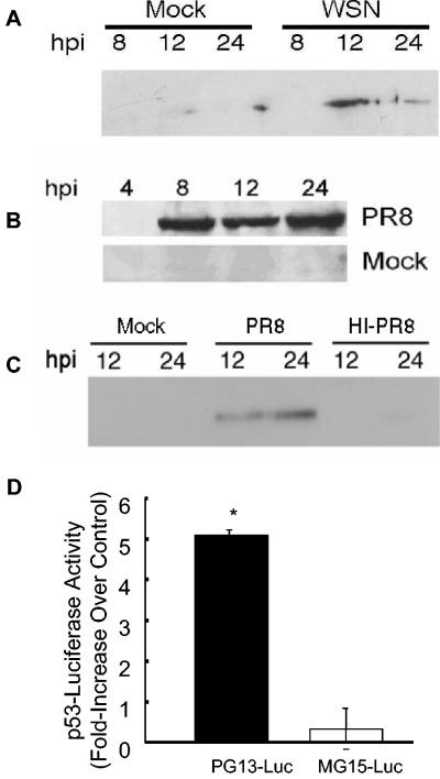
To determine whether influenza infection affects p53 levels or activity, primary human lung cells were infected with A/WSN/33 and p53 protein monitored by Western blot analysis. p53 levels increased in the primary lung cells within 12 hpi compared to mock-infected controls (Fig. 1A). In A549 cells, p53 levels were increased at 8 hpi and remained elevated at 24 hpi (Fig. 1B). Similar results were obtained with MDCK and mink lung epithelial cells (Mv1Lu). To determine if viral replication was required for the observed increase in p53, we examined p53 levels in A549 cells treated with infectious or heat-inactivated PR8 at 12 and 24 hpi. In contrast to what was observed for cells treated with infectious virus, p53 levels did not increase in cells treated with heat-inactivated virus (Fig. 1C). These studies demonstrated that p53 levels increased during influenza virus infection. Human primary lung and A549 cells had similar increases in virus-induced p53 and cell death (data not shown). Thus, the remainder of the studies was performed with A549 cells due to the inherent difficulties of working with the primary cells.
FIG. 1.
p53 activity increases during influenza infection. Equal protein concentrations of mock- (treated with media alone) and WSN-infected (MOI 2) human primary lung cell lysates (A), mock- and PR8-infected (MOI 2) A549 lysates (B), and heat-inactivated (HI) PR8-treated A549 lysates (C) were analyzed for p53 levels by Western blotting. (D) A549 cells transfected with the p53 reporter (PG13-Luc) construct or control (MG15-Luc) construct were mock or PR8 infected (MOI 2), and samples were collected at 24 hpi. The luciferase activity of lysates was standardized for equal protein concentration and expressed as the n-fold increase in luciferase activity in infected cells over that in mock-infected controls. Error bars represent the standard errors of the means, and the asterisk indicates samples that are significant (P < 0.05) as determined by ANOVA.
Increased p53 protein levels do not necessarily correlate with increased transcriptional activity (21). To examine p53 activity, A549 cells were transiently transfected with a p53 reporter construct, PG13-Luc, or the control construct, MG15-Luc, prior to infection with PR8 (MOI 2). The reporter measures the transcriptional activity of p53, driving the expression of luciferase off the p53 consensus response element (10). Activation of p53 was detected in infected cells as early as 4 hpi and remained elevated until 24 hpi (data not shown). At 24 hpi, a fivefold increase in p53 activity was detected in infected cells relative to that in mock-infected controls (Fig. 1D, left). An increase in p53 activity was not detected using the control plasmid (Fig. 1D, right). To confirm these results, we examined the expression of several p53-regulated genes. The activation of p53 by influenza virus led to the increased expression of the specific p53-responsive genes p21/WAF and bax (data not shown). These studies demonstrated that influenza infection up-regulated p53 protein levels and transcriptional activity.
Influenza infection increases p53 nuclear accumulation and phosphorylation.
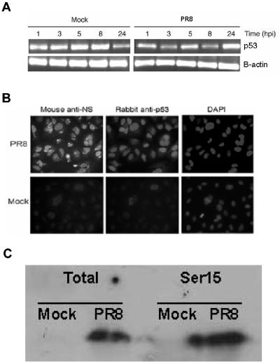
To begin defining p53 activation by influenza virus, we examined p53 transcription, localization, and phosphorylation during infection. No change in p53 mRNA levels was detected in infected A549 cells compared to mock-infected controls, suggesting that the increase in p53 protein was independent of increased transcription (Fig. 2A). Increased p53 activity correlates with localization to the nucleus (16, 36). Thus, we examined the cellular localization of p53 during infection by immunofluorescent microscopy. Within 8 hpi, an increase in the nuclear localization of p53 was detected (as confirmed by DAPI staining) with peak accumulation at 12 hpi (Fig. 2B). In contrast, staining for p53 in control cells was very light and primarily localized to the cytoplasm. Staining for influenza virus NS confirmed that the nuclear accumulation of p53 occurred specifically in infected cells. Nuclear accumulation of p53 often results from posttranslational modifications, such as phosphorylation. Thus, p53 phosphorylation was examined by the immunoprecipitation of infected and mock-infected A549 cell lysates with a phospho-Ser15-specific p53 antibody and p53 detected by Western blotting. Control cells had no detectable Ser15-phosphorylated p53 (Fig. 2C). In contrast, p53 was phosphorylated at Ser15 in infected cells (Fig. 2C). Increased phosphorylation was also observed at Ser6 and Ser20 (data not shown). These studies suggested that influenza virus infection led to nuclear localization and increased phosphorylation of p53.
FIG. 2.
Nuclear accumulation and serine phosphorylation of p53. (A) A549 cells were treated with media alone (mock) or infected with PR8 (MOI 2); total RNA was isolated at 1, 3, 5, 8, and 24 hpi, and RT-PCR was performed for p53 or β-actin. (B) A549 cells, treated with media alone (mock) or PR8 infected (MOI 2), were double-stained for virus antigen (NS) and p53 at 12 hpi and examined by fluorescent microscopy. The nucleus was visualized by DAPI. Magnification, ×42. (C) Cellular lysates from mock- or PR8-infected A549 cells at 24 hpi were immunoprecipitated with a phospho-Ser15-specific p53 antibody followed by binding to protein G Sepharose beads. Equal protein levels of total or immunoprecipitated lysates were analyzed by probing Western blots with a p53 antibody (DO-1).
p53 is involved in influenza virus-induced cell death.
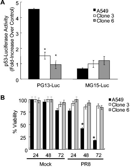
To test our hypothesis that p53 was involved in influenza virus-induced cell death, we examined cell death in A549 cells stably expressing a dominant-negative mutant p53. Thus, A549 cells were transfected with pCMV-p53mt135, a p53 gene that differs from the wild type by a G-to-A conversion at nucleotide 1017. It acts in a dominant-negative fashion by binding to wild-type p53 and inhibiting its activity. To ensure that the dominant-negative mutant inhibited endogenous p53 activity, parental A549 cells or two distinct dominant-negative mutant clones (clones 3 and 6) were tested for p53 transcriptional activity using the p53-luciferase reporter plasmid (PG13-Luc) and the control reporter plasmid (MG15-Luc) (10). Luciferase activity increased fivefold in infected A549 cells transfected with the p53 reporter construct, PG13-Luc, over that in mock-infected controls (Fig. 3A, left). In contrast, there was no increase in p53 activity above the control levels in either of the dominant-negative mutant clones (Fig. 3A, left). Luciferase activity did not increase in cells transfected with the control reporter construct, MG15-Luc (Fig. 3A, right), or with the control vector. Transfection of the A549 parental cells and clones with a plasmid expressing GFP confirmed that the differences in the levels of luciferase activity were not a result of differences in the levels of transfection efficiency (data not shown).
FIG. 3.
Inhibition of p53 decreases influenza virus-induced cell death. (A) Parental A549 cells or A549 cells stably expressing a p53 dominant-negative mutant (clones 3 and 6) were transfected with the p53 reporter (PG13-Luc) or the control (MG15-Luc) construct. Cells were mock or PR8 infected (MOI 2), and the luciferase activities of lysates were measured. Results were standardized for equal protein concentration and are expressed as the n-fold increases in activity in PR8-infected cells over those in mock-infected controls. (B) Cell viabilities of mock- or PR8-infected (MOI 2) parental A549 cells or dominant-negative p53 mutants (clones 3 and 6) were determined by trypan blue exclusion. Error bars represent the standard errors of the means, and asterisks indicate samples that are significant (P < 0.05) as determined by ANOVA.
To examine the role of p53 in influenza virus-induced cell death, cell viability was measured in mock and infected dominant-negative mutant p53 clones and parental A549 cells by trypan blue exclusion (Fig. 3B). There was little cell death in the mock-infected cells at any time after infection. Viability decreased by 25% at 24 hpi, 60% at 48 hpi, and 80% at 72 hpi in infected A549 cells (Fig. 3B). Similarly, viability decreased by 20% at 24 hpi, 70% at 48 hpi, and 90% at 72 hpi in infected primary human lung cells (data not shown). In contrast, the cells expressing the dominant-negative mutant were protected from influenza virus-induced cell death (Fig. 3B), demonstrating that p53 was involved in influenza virus-induced cell death in A549 cells. Additional experiments were performed in MDCK and Mv1Lu cell lines. Using antisense oligonucleotides, p53 activity was decreased, resulting in reduced cell death after infection with influenza virus (data not shown). These studies demonstrated that p53 was involved in influenza virus-induced cell death.
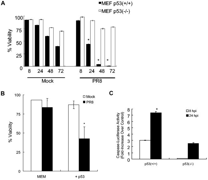
To exclude the possibility that our results were an artifact arising from the use of an immortalized cell line or from the overexpression of an exogenous protein, we repeated the cell death studies using primary MEF isolated from wild-type mice, MEF p53+/+, or p53 knockout mice, MEF p53−/−. When MEF p53+/+ cells were infected with influenza virus, 40% of the infected p53+/+ cells were dead at 24 h (Fig. 4A, right). This increased to 60% by 48 hpi compared to mock-infected controls. In contrast, the MEF p53−/− cells failed to succumb to influenza virus-induced cell death. Viability did not decrease below 70% in either infected or control MEF p53−/− cells at any time point tested.
FIG. 4.
p53 is essential for influenza virus-induced cell death. (A) Cell viabilities of mock and infected MEF p53+/+ and p53−/− cells were determined by trypan blue exclusion. (B) MEF p53−/− cells were transfected with pCMV-p53 (+ p53) or mock transfected (MEM), and treatment with MEM alone (mock) or with infection (PR8) followed. Viabilities were measured by trypan blue exclusion. (C) Caspase 3/7 activity was measured in mock or infected MEF p53+/+ and p53−/− cells. Caspase activity was normalized to equal cell numbers and is graphed as the n-fold increase in infected cells over that in mock-infected cells. All error bars represent the standard errors of the means, and asterisks indicate samples that are significant (P < 0.05) as determined by ANOVA.
To confirm that the lack of cell death was directly related to p53, MEF p53−/− cells were mock or transiently transfected with a plasmid expressing wild-type p53 and infected 4 h after transfection, and cell viability was measured at 16 hpi. Again, mock-transfected MEF p53−/− cells did not die when infected with influenza virus (Fig. 4B). In contrast, there was a 40% increase in cell death in MEF p53−/− cells transfected with a wild-type p53 and infected with influenza virus. This correlated directly with the transfection efficiencies of the cells (data not shown). These studies strongly suggested that p53 was required for influenza virus-induced cell death in A549 and MEF cells.
Caspases are often used to measure apoptosis, as they are early indicators of apoptosis. Thus, to define the role of p53 in influenza virus-induced apoptosis, caspase 3/7 activity was examined in infected and control MEF cells. Caspase activity increased 2.9- and 7.9-fold over that in mock-infected controls in the infected MEF p53+/+ cells at 8 and 24 hpi, respectively (Fig. 4C). While there was no increase in caspase activity in the infected MEF p53−/− cells at 8 hpi, there was an increase detected at 24 hpi (2.6-fold) compared to that in mock-infected controls (Fig. 4C). Caspase 3/7 activity returned to background levels by 48 hpi (data not shown). The decreased caspase activity in the MEF p53−/− cells correlated with the decreased cell death observed in these cells. Caspase activity was not extended past 24 hpi, due to the high levels of cell death in the infected MEF p53+/+ cells at 36 hpi. The cleavage of caspase 3 was confirmed by Western blot analysis (data not shown). These studies demonstrated that caspase 3/7 activation was reduced in infected MEF p53−/− cells compared to that in wild-type MEF p53+/+ cells.
Inhibition of p53 increases viral titers.
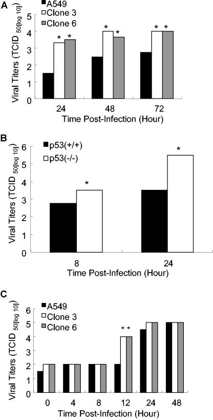
To determine the effect of p53-induced cell death on viral replication, we examined viral titers in A549, MEF p53+/+, and p53−/− cells. A low MOI (0.05) was used to monitor viral spread. Inhibition of p53 in the A549 cells stably expressing dominant-negative p53 resulted in increased viral titers (Fig. 5A). Within 24 hpi, there was approximately a 2-log increase in A549 dominant-negative clones 3 and 6 compared to parental A549 cells. The viral titers from the dominant-negative p53 clones remained elevated over those of the parental A549 cells through 72 hpi (Fig. 5A).
FIG. 5.
p53 inhibition increases viral titers. Confluent cultures of parental A549 cells, i.e., A549 p53 dominant-negative clones 3 and 6 (A) or MEF p53−/− and MEF p53+/+ cells (B) were infected with PR8 at an MOI of 0.05. At various times postinfection, supernatants were collected and viral titers determined by TCID50 on MDCK cells. (C) Confluent cultures of parental A549 cells and p53 dominant-negative clones 3 and 6 were infected with PR8 at an MOI of 2. At various times postinfection, supernatants were collected and viral titers determined by use of the TCID50 for MDCK cells. Asterisks indicate samples that are significant (P < 0.05) as determined by ANOVA.
To determine if the increased virus titers noted in A549 cells were cell type specific, viral titers were measured in MEF cells. Due to the rapid onset of cell death in the infected MEF p53+/+ cells, samples were isolated only at 8 and 24 hpi. Similar to what was observed with the A549 cells, there was a measurable increase in viral titers in MEF p53−/− cells compared to those of MEF p53+/+ cells (Fig. 5B). There was a 1-log increase in virus titers in MEF p53−/− cells by 8 hpi and a 2-log increase by 24 hpi. Similar results were observed for MDCK and mink lung cells with decreased p53 activity (data not shown). Viral titers increased in MDCK (2.5 logs by 24 hpi) and Mv1Lu cells (3 logs by 24 hpi) when p53 was inhibited. These results strongly suggested that a lack of p53 activity during influenza infection increased viral replication.
Based on the increase in viral titers prior to the onset of cell death, we hypothesized that p53 interfered with the viral replication cycle. Thus, one-step growth curves (MOI 2) were performed in parental A549 and p53 dominant-negative clones prior to the appearance of cell death (48 hpi). Only input virus was isolated from 0 to 12 hpi in the A549 and dominant-negative clones (101.5 to 102 TCID50 units). By 24 hpi, viral titers reached 104.5 TCID50 units in the A549 clones and 105 TCID50 units in clones 3 and 6 (Fig. 5C). Titers remained at 105 TCID50 units at 48 hpi. These studies suggest that p53 may not directly interfere with influenza virus replication.
p53 is necessary for activation of ISRE during influenza infection.
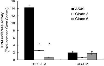
The p53 protein regulates a key component in the IFN pathway, namely, the induction of the interferon genes (34). We hypothesized that the decreased virus replication in parental A549 and MEF cells may be due to p53 regulation of the IFN pathway. To test this hypothesis, endogenous IFN activity was measured in parental A549 and dominant-negative clones using an IFN-stimulated response element (ISRE) binding sequence (pISRE-Luc) driving the expression of luciferase and the negative control plasmid lacking cis-acting DNA elements (pCIS-Luc) (Stratagene). IFN-driven luciferase activity increased 14-fold in infected A549 cells compared to that in mock-infected controls (Fig. 6). In contrast, there was no increase in luciferase activity above control levels in either of the dominant-negative clones, suggesting that IFN activation was inhibited in these cells. Luciferase activity did not increase in cells expressing the control reporter construct (Fig. 6) or the empty vector (data not shown). These studies indicate that p53 may be involved in IFN activation during influenza infection.
FIG. 6.
Inhibition of p53 decreases IFN activity. Parental A549 cells or p53 dominant-negative clones 3 and 6 were transfected with the IFN reporter (ISRE-Luc) or control (CIS-Luc) and then mock or PR8 infected. Luciferase activity was measured in cell lysates at 12 hpi. Results were normalized to equal cell numbers and are expressed as the n-fold increase in activity in infected cells over that in mock-infected controls. Error bars represent the standard errors of the means, and asterisks indicate samples that are significant (P < 0.05) as determined by ANOVA.
DISCUSSION
These studies suggest that p53 plays a critical role in influenza virus-induced cell death. Our studies demonstrated that p53 levels and activity increased during influenza infection prior to the onset of cell death at 24 hpi in primary human lung bronchial epithelial cells and the human lung cell line, A549. Stable expression of a p53 dominant-negative mutant in A549 cells inhibited influenza virus-induced cell death. Moreover, primary MEF from p53 knockout mice failed to succumb to virus-induced cell death unless p53 activity was restored. We also showed an increase in caspase activity in influenza-infected cells, indicating that apoptosis is occurring in these cells. Programmed cell death is proposed to be a host protective mechanism to limit viral spread. In this study, we not only observed elevated viral titers in cells with no or decreased p53 activity but, more surprisingly, noted decreased IFN activation in these cells. We postulate that the lack of IFN activation is partially responsible for the elevated viral titers.
A549 cells were used in these studies because they undergo cell death and support productive viral replication to levels and with kinetics similar to those of human primary lung cells (35) and because they express wild-type p53 (30). p53 was also involved in virus-induced cell death in other cell lines that support viral replication, including MDCK, Mv1Lu, and Vero cells (data not shown). In addition to testing multiple cell lines, numerous human and avian influenza viruses (including highly pathogenic avian isolates) were tested, and all increased p53 activity during infection. These findings suggested that p53 was a commonly activated protein during influenza infection, as it is activated independently of the cell type or viral strain tested.
In contrast to our findings, Technau-Ihling et al. demonstrated that influenza infection failed to increase p53 nuclear localization in C3H10T1/2 cells (52). There are several possible explanations for the differences in the results. First, our studies utilized cells that support productive viral replication. Heat- and UV-inactivated (data not shown) virus failed to increase p53 activity, suggesting viral replication is necessary. There is no evidence in the literature that C3H10T1/2 cells support productive viral replication. Second, our studies utilized a high MOI (MOI 2) and clearly demonstrated viral replication and increased p53 levels and activity from 8 hpi to 24 hpi in all cell types. A much lower MOI (MOI 0.2) was used in the Technau-Ihling study, and only p53 nuclear localization was examined. They demonstrated that 20% of the cells were diffusely stained for viral antigen at 24 hpi and that there was no increase in nuclear localization. Since p53 activity and protein levels were not monitored, it is difficult to predict whether there is increased activity independent of nuclear localization. Finally, it is unclear whether the C3H10T1/2 cells used in these studies contain wild-type or mutant p53. To our knowledge, the cells used in our studies contained only wild-type p53. Studies are under way to determine whether influenza virus also regulates mutant p53 levels and activity.
The activation of p53 involves several posttranslational modifications that stabilize the protein, including phosphorylation. Our data showed an increase in phosphorylation of serine 15 during influenza infection. Phosphorylation of serine 15 by kinases increases the sequence-specific transcriptional activity and stability of p53 (6, 46). This modification results in nuclear translocation and enhanced transcriptional activity (16, 37). Viral infection can induce several cellular p53 activators, including double-stranded RNA-activated PKR or interferon. PKR activates p53 (8, 9), resulting in increased nuclear translocation and enhanced transcriptional activity, and plays a critical role in preventing viral replication (2, 57). Studies are under way to determine the role of PKR in the activation of p53 and the mechanism of increased p53 levels during influenza infection.
IFN enhances the transcription of p53 (47, 55), suggesting that influenza virus-induced IFN could lead to an accumulation of p53 mRNA in surrounding cells. Although we cannot exclude this possibility, in these studies the use of a high MOI and the rapid increase in p53 protein levels suggest that our findings are independent of IFN. Additionally, we observed increased p53 protein levels in influenza virus-infected Vero cells (data not shown), which does not produce type 1 IFN (14), indicating that the induction of p53 by influenza virus occurs independently of IFN. We were surprised to find a decrease in IFN signaling in cells lacking p53. This correlates with studies by Nees et al., where the down-regulation of multiple interferon-responsive genes was reported for cells expressing mutant p53 (34). Specifically, interferon regulatory factor 5 is a downstream target of p53 (3), and p53 was recently shown to physically associate with STAT-1. This interaction is required for STAT-1-induced apoptosis (54).
To date, there have been conflicting results on the impact of apoptosis on influenza virus replication. Previous studies suggested that apoptosis inhibits the replication of influenza virus as an antiviral host response (22). However, the inhibition of apoptosis by the overexpression of Bcl-2 decreased viral titers (17, 26), while inhibition of caspases either had no effect or reduced viral replication (23, 51, 58). This result was thought to be due to caspase activation occurring downstream of virus replication. Recently, the activation of caspase 3 was shown to be required for efficient virus production by coregulating intrinsic viral export (38). In the present studies, the inhibition of p53 led to elevated viral titers in all cell types tested, despite decreased caspase 3/7 activity. Perhaps relatively low concentrations of active caspases are required for viral protein processing, while higher concentrations are required for cell death. The transient increase in caspase 3/7 activity in p53−/− cells suggests that influenza may activate caspase 3/7 through a p53-independent pathway.
Influenza virus-induced cell death is a complex interplay of viral and cellular factors. In this study, we demonstrated that p53 was required for influenza virus-induced cell death, limited viral replication, and was important in the induction of the IFN response. To our knowledge, these are the first studies to demonstrate a role for p53 in the induction of cell death and IFN production during influenza virus infection. By further defining the mechanism, we will potentially identify specific cellular functions essential for influenza virus replication.
Acknowledgments
We thank Laura Knoll, Donna Paulnock, and Jay Bangs (University of Wisconsin) for advice and use of their equipment; James Gern and Rebecca Brockman-Schneider for the primary human bronchial epithelial cells and helpful discussions and advice; Tyler Jacks, (Massachusetts Institute of Technology), Bert Vogelstein (Johns Hopkins Medical Institutions), and Robert Webster (St. Jude's) for reagents; and Erik Johnson for technical assistance. We gratefully acknowledge Curtis Brandt, Matt Koci, Gabrielle Neumann, Chris Olsen, and Lindsey Moser for helpful discussions and critical reviews of the report.
E. Turpin was supported by a postdoctoral training fellowship in tumor virology through the McArdle Cancer Center at the University of Wisconsin (CA09075-28). This work was also supported by Sigma Xi Grants-In-Aid of Research to K. Luke and an American Cancer Society Institutional Research Grant (IRG-58-011-47-11) to S. Schultz-Cherry.
REFERENCES
- 1.Attardi, L. D., S. W. Lowe, J. Brugarolas, and T. Jacks. 1996. Transcriptional activation by p53, but not induction of the p21 gene, is essential for oncogene-mediated apoptosis. EMBO J. 15:3693-3701. [PMC free article] [PubMed] [Google Scholar]
- 2.Barber, G. N. 2001. Host defense, viruses and apoptosis. Cell Death Differ. 8:113-126. [DOI] [PubMed] [Google Scholar]
- 3.Barnes, B. J., M. J. Kellum, K. E. Pinder, J. A. Frisancho, and P. M. Pitha. 2003. Interferon regulatory factor 5, a novel mediator of cell cycle arrest and cell death. Cancer Res. 63:6424-6431. [PubMed] [Google Scholar]
- 4.Blaho, J. A. 2003. Virus infection and apoptosis (issue 1). Dedicated to the memory of Lois K. Miller. Int. Rev. Immunol. 22:321-326. [PubMed] [Google Scholar]
- 5.Blaho, J. A. 2004. Virus infection and apoptosis (issue II) an introduction: cheating death or death as a fact of life? Int. Rev. Immunol. 23:1-6. [DOI] [PubMed] [Google Scholar]
- 6.Bulavin, D. V., S. Saito, M. C. Hollander, K. Sakaguchi, C. W. Anderson, E. Appella, and A. J. Fornace, Jr. 1999. Phosphorylation of human p53 by p38 kinase coordinates N-terminal phosphorylation and apoptosis in response to UV radiation. EMBO J. 18:6845-6854. [DOI] [PMC free article] [PubMed] [Google Scholar]
- 7.Collins, M. 1995. Potential roles of apoptosis in viral pathogenesis. Am. J. Respir. Crit. Care Med. 152:S20-S24. [DOI] [PubMed] [Google Scholar]
- 8.Cuddihy, A., S. Li, N. W. Tam, A. H. Wong, Y. Taya, N. Abraham, J. C. Bell, and A. E. Koromilas. 1999. Double-stranded-RNA-activated protein kinase PKR enhances transcriptional activation by tumor suppressor p53. Mol. Cell. Biol. 19:2475-2484. [DOI] [PMC free article] [PubMed] [Google Scholar]
- 9.Cuddihy, A., A. H. Wong, N. W. Tam, S. Li, and A. E. Koromilas. 1999. The double-stranded RNA activated protein kinase PKR physically associates with the tumor suppressor p53 protein and phosphorylates human p53 on serine 392 in vitro. Oncogene 18:2690-2702. [DOI] [PubMed] [Google Scholar]
- 10.el-Deiry, W. S., S. E. Kern, J. A. Pietenpol, K. W. Kinzler, and B. Vogelstein. 1992. Definition of a consensus binding site for p53. Nat. Genet. 1:45-49. [DOI] [PubMed] [Google Scholar]
- 11.Foster, B., H. A. Coffey, M. J. Morin, and F. Rastinejad. 1999. Pharmacological rescue of mutant p53 conformation and function. Science 286:2507-2510. [DOI] [PubMed] [Google Scholar]
- 12.Fridman, J. S., and S. W. Lowe. 2003. Control of apoptosis by p53. Oncogene 22:9030-9040. [DOI] [PubMed] [Google Scholar]
- 13.Fujimoto, I., T. Takizawa, Y. Ohba, and Y. Nakanishi. 1998. Co-expression of Fas and Fas-ligand on the surface of influenza virus-infected cells. Cell Death Differ. 5:426-431. [DOI] [PubMed] [Google Scholar]
- 14.Garcia-Sastre, A., A. Egorov, D. Matassov, S. Brandt, D. E. Levy, J. E. Durbin, P. Palese, and T. Muster. 1998. Influenza A virus lacking the NS1 gene replicates in interferon-deficient systems. Virology 252:324-330. [DOI] [PubMed] [Google Scholar]
- 15.Goodkin, M. L., E. R. Morton, and J. A. Blaho. 2004. Herpes simplex virus infection and apoptosis. Int. Rev. Immunol. 23:141-172. [DOI] [PubMed] [Google Scholar]
- 16.Haupt, S., I. Louria-Hayon, and Y. Haupt. 2003. P53 licensed to kill? Operating the assassin. J. Cell. Biochem. 88:76-82. [DOI] [PubMed] [Google Scholar]
- 17.Hinshaw, V. S., C. W. Olsen, N. Dybdahl-Sissoko, and D. Evans. 1994. Apoptosis: a mechanism of cell killing by influenza A and B viruses. J. Virol. 68:3667-3673. [DOI] [PMC free article] [PubMed] [Google Scholar]
- 18.Ito, T., Y. Kobayashi, T. Morita, T. Horimoto, and Y. Kawaoka. 2002. Virulent influenza A viruses induce apoptosis in chickens. Virus Res. 84:27-35. [DOI] [PubMed] [Google Scholar]
- 19.Julkunen, I., K. Melen, M. Nyqvist, J. Pirhonen, T. Sareneva, and S. Matikainen. 2000. Inflammatory responses in influenza A virus infection. Vaccine 19(Suppl. 1):S32-S37. [DOI] [PubMed] [Google Scholar]
- 20.Kerr, J. F., C. M. Winterford, and B. V. Harmon. 1994. Apoptosis. Its significance in cancer and cancer therapy. Cancer 73:2013-2026. [DOI] [PubMed] [Google Scholar]
- 21.Kim, E., and W. Deppert. 2003. The complex interactions of p53 with target DNA: we learn as we go. Biochem. Cell Biol. 81:141-150. [DOI] [PubMed] [Google Scholar]
- 22.Kurokawa, M., A. H. Koyama, S. Yasuoka, and A. Adachi. 1999. Influenza virus overcomes apoptosis by rapid multiplication. Int. J. Mol. Med. 3:527-530. [DOI] [PubMed] [Google Scholar]
- 23.Lin, C., S. G. Zimmer, Z. Lu, R. E. Holland, Jr., Q. Dong, and T. M. Chambers. 2001. The involvement of a stress-activated pathway in equine influenza virus-mediated apoptosis. Virology 287:202-213. [DOI] [PubMed] [Google Scholar]
- 24.Lowy, R. J. 2003. Influenza virus induction of apoptosis by intrinsic and extrinsic mechanisms. Int. Rev. Immunol. 22:425-449. [DOI] [PubMed] [Google Scholar]
- 25.Ludwig, S., C. Ehrhardt, E. R. Neumeier, M. Kracht, U. R. Rapp, and S. Pleschka. 2001. Influenza virus-induced AP-1-dependent gene expression requires activation of the JNK signaling pathway. J. Biol. Chem. 276:10990-10998. [PubMed] [Google Scholar]
- 26.Ludwig, S., S. Pleschka, and T. Wolff. 1999. A fatal relationship—influenza virus interactions with the host cell. Viral Immunol. 12:175-196. [DOI] [PubMed] [Google Scholar]
- 27.Lyles, D. S. 2000. Cytopathogenesis and inhibition of host gene expression by RNA viruses. Microbiol. Mol. Biol. Rev. 64:709-724. [DOI] [PMC free article] [PubMed] [Google Scholar]
- 28.Majno, G., and I. Joris. 1995. Apoptosis, oncosis, and necrosis. An overview of cell death. Am. J. Pathol. 146:3-15. [PMC free article] [PubMed] [Google Scholar]
- 29.McKinney, L. C., S. J. Galliger, and R. J. Lowy. 2003. Active and inactive influenza virus induction of tumor necrosis factor-alpha and nitric oxide in J774.1 murine macrophages: modulation by interferon-gamma and failure to induce apoptosis. Virus Res. 97:117-126. [DOI] [PubMed] [Google Scholar]
- 30.Mirzayans, R., S. Pollock, A. Scott, L. Enns, B. Andrais, and D. Murray. 2004. Relationship between the radiosensitizing effect of wortmannin, DNA double-strand break rejoining, and p21WAF1 induction in human normal and tumor-derived cells. Mol. Carcinog. 39:164-172. [DOI] [PubMed] [Google Scholar]
- 31.Mori, I., and Y. Kimura. 2000. Apoptotic neurodegeneration induced by influenza A virus infection in the mouse brain. Microbes Infect. 2:1329-1334. [DOI] [PubMed] [Google Scholar]
- 32.Mori, I., T. Komatsu, K. Takeuchi, K. Nakakuki, M. Sudo, and Y. Kimura. 1995. In vivo induction of apoptosis by influenza virus. J. Gen. Virol. 76:2869-2873. [DOI] [PubMed] [Google Scholar]
- 33.Morris, S. J., G. E. Price, J. M. Barnett, S. A. Hiscox, H. Smith, and C. Sweet. 1999. Role of neuraminidase in influenza virus-induced apoptosis. J. Gen. Virol. 80:137-146. [DOI] [PubMed] [Google Scholar]
- 34.Nees, M., J. M. Geoghegan, T. Hyman, S. Frank, L. Miller, and C. D. Woodworth. 2001. Papillomavirus type 16 oncogenes downregulate expression of interferon-responsive genes and upregulate proliferation-associated and NF-κB-responsive genes in cervical keratinocytes. J. Virol. 75:4283-4296. [DOI] [PMC free article] [PubMed] [Google Scholar]
- 35.Nimmerjahn, F., D. Dudziak, U. Dirmeier, G. Hobom, A. Riedel, M. Schlee, L. M. Staudt, A. Rosenwald, U. Behrends, G. W. Bornkamm, and J. Mautner. 2004. Active NF-{kappa}B signalling is a prerequisite for influenza virus infection. J. Gen. Virol. 85:2347-2356. [DOI] [PubMed] [Google Scholar]
- 36.Peterhans, E. 1997. Oxidants and antioxidants in viral diseases: disease mechanisms and metabolic regulation. J. Nutr. 127:962S-965S. [DOI] [PubMed] [Google Scholar]
- 37.Piette, J., H. Neel, and V. Marechal. 1997. Mdm 2: keeping p53 under control. Oncogene 15:1001-1010. [DOI] [PubMed] [Google Scholar]
- 38.Pleschka, S., T. Wolff, C. Ehrhardt, G. Hobom, O. Planz, U. R. Rapp, and S. Ludwig. 2001. Influenza virus propagation is impaired by inhibition of the Raf/MEK/ERK signalling cascade. Nat. Cell Biol. 3:301-305. [DOI] [PubMed] [Google Scholar]
- 39.Polyak, K., Y. Xia, J. L. Zweier, K. W. Kinzler, and B. Vogelstein. 1997. A model for p53-induced apoptosis. Nature 389:300-305. [DOI] [PubMed] [Google Scholar]
- 40.Razvi, E. S., and R. M. Welsh. 1995. Apoptosis in viral infections. Adv. Virus Res. 45:1-60. [DOI] [PubMed] [Google Scholar]
- 41.Reed, L. J., and H. Muench. 1938. A simple method of estimating fifty percent endpoints. Am. J. Hyg. 27:493-497. [Google Scholar]
- 42.Roulston, A., R. C. Marcellus, and P. E. Branton. 1999. Viruses and apoptosis. Annu. Rev. Microbiol. 53:577-628. [DOI] [PubMed] [Google Scholar]
- 43.Schultz-Cherry, S., and V. S. Hinshaw. 1996. Influenza virus neuraminidase activates latent transforming growth factor β. J. Virol. 70:8624-8629. [DOI] [PMC free article] [PubMed] [Google Scholar]
- 44.Schultz-Cherry, S., R. M. Krug, and V. S. Hinshaw. 1998. Induction of apoptosis by influenza virus. Semin. Virol. 8:491-495. [Google Scholar]
- 45.Sedger, L. M., S. Hou, S. R. Osvath, M. B. Glaccum, J. J. Peschon, N. van Rooijen, and L. Hyland. 2002. Bone marrow B cell apoptosis during in vivo influenza virus infection requires TNF-alpha and lymphotoxin-alpha. J. Immunol. 169:6193-6201. [DOI] [PubMed] [Google Scholar]
- 46.She, Q. B., N. Chen, and Z. Dong. 2000. ERKs and p38 kinase phosphorylate p53 protein at serine 15 in response to UV radiation. J. Biol. Chem. 275:20444-20449. [DOI] [PubMed] [Google Scholar]
- 47.Takaoka, A., S. Hayakawa, H. Yanai, D. Stoiber, H. Negishi, H. Kikuchi, S. Sasaki, K. Imai, T. Shibue, K. Honda, and T. Taniguchi. 2003. Integration of interferon-alpha/beta signalling to p53 responses in tumour suppression and antiviral defence. Nature 424:516-523. [DOI] [PubMed] [Google Scholar]
- 48.Takizawa, T. 1996. Mechanism of the induction of apoptosis by influenza virus infection. Nippon Rinsho 54:1836-1841. (In Japanese with English summary.) [PubMed] [Google Scholar]
- 49.Takizawa, T., S. Matsukawa, Y. Higuchi, S. Nakamura, Y. Nakanishi, and R. Fukuda. 1993. Induction of programmed cell death (apoptosis) by influenza virus infection in tissue culture cells. J. Gen. Virol. 74:2347-2355. [DOI] [PubMed] [Google Scholar]
- 50.Takizawa, T., K. Ohashi, and Y. Nakanishi. 1996. Possible involvement of double-stranded RNA-activated protein kinase in cell death by influenza virus infection. J. Virol. 70:8128-8132. [DOI] [PMC free article] [PubMed] [Google Scholar]
- 51.Takizawa, T., C. Tatematsu, K. Ohashi, and Y. Nakanishi. 1999. Recruitment of apoptotic cysteine proteases (caspases) in influenza virus-induced cell death. Microbiol. Immunol. 43:245-252. [DOI] [PubMed] [Google Scholar]
- 52.Technau-Ihling, K., C. Ihling, J. Kromeier, and G. Brandner. 2001. Influenza A virus infection of mice induces nuclear accumulation of the tumorsuppressor protein p53 in the lung. Arch. Virol. 146:1655-1666. [DOI] [PubMed] [Google Scholar]
- 53.Thut, C. J., J. A. Goodrich, and R. Tjian. 1997. Repression of p53-mediated transcription by MDM2: a dual mechanism. Genes Dev. 11:1974-1986. [DOI] [PMC free article] [PubMed] [Google Scholar]
- 54.Townsend, P. A., T. M. Scarabelli, S. M. Davidson, R. A. Knight, D. S. Latchman, and A. Stephanou. 2004. STAT-1 interacts with p53 to enhance DNA damage-induced apoptosis. J. Biol. Chem. 279:5811-5820. [DOI] [PubMed] [Google Scholar]
- 55.Vilcek, J. 2003. Boosting p53 with interferon and viruses. Nat. Immunol. 4:825-826. [DOI] [PubMed] [Google Scholar]
- 56.Vousden, K. H. 2002. Activation of the p53 tumor suppressor protein. Biochim. Biophys. Acta 1602:47-59. [DOI] [PubMed] [Google Scholar]
- 57.Williams, B. R. G. 1999. PKR: a sentinel kinase for cellular stress. Oncogene 18:6112-6120. [DOI] [PubMed] [Google Scholar]
- 58.Wurzer, W. J., O. Planz, C. Ehrhardt, M. Giner, T. Silberzahn, S. Pleschka, and S. Ludwig. 2003. Caspase 3 activation is essential for efficient influenza virus propagation. EMBO J. 22:2717-2728. [DOI] [PMC free article] [PubMed] [Google Scholar]
- 59.Young, L. S., C. W. Dawson, and A. G. Eliopoulos. 1997. Viruses and apoptosis. Br. Med. Bull. 53:509-521. [DOI] [PubMed] [Google Scholar]


