Abstract
Background
Patients with peritoneal metastasis from colorectal cancer (PM-CRC) have inferior prognosis and respond particularly poorly to chemotherapy. This study aims to identify the molecular explanation for the observed clinical behavior and suggest novel treatment strategies in PM-CRC.
Methods
Tumor samples (230) from a Norwegian national cohort undergoing surgery and hyperthermic intraperitoneal chemotherapy (HIPEC) with mitomycin C (MMC) for PM-CRC were subjected to targeted DNA sequencing, and associations with clinical data were analyzed. mRNA sequencing was conducted on a subset of 30 samples to compare gene expression in tumors harboring BRAF or KRAS mutations and wild-type tumors.
Results
BRAF mutations were detected in 27% of the patients, and the BRAF-mutated subgroup had inferior overall survival compared to wild-type cases (median 16 vs 36 months, respectively, p < 0.001). BRAF mutations were associated with RNF43/RSPO aberrations and low expression of negative Wnt regulators (ligand-dependent Wnt activation). Furthermore, BRAF mutations were associated with gene expression changes in transport solute carrier proteins (specifically SLC7A6) and drug metabolism enzymes (CES1 and CYP3A4) that could influence the efficacy of MMC and irinotecan, respectively. BRAF-mutated tumors additionally exhibited increased expression of members of the novel butyrophilin subfamily of immune checkpoint molecules (BTN1A1 and BTNL9).
Conclusions
BRAF mutations were frequently detected and were associated with particularly poor survival in this cohort, possibly related to ligand-dependent Wnt activation and altered drug transport and metabolism that could confer resistance to MMC and irinotecan. Drugs that target ligand-dependent Wnt activation or the BTN immune checkpoints could represent two novel therapy approaches.
Supplementary Information
The online version contains supplementary material available at 10.1186/s12967-024-05467-2.
Keywords: Peritoneal metastasis, Colorectal cancer, Drug resistance, Therapeutic targets
Introduction
Colorectal cancer (CRC) is the second leading cause of cancer-related deaths worldwide, with metastatic progression being the main cause of CRC mortality. The peritoneum is the third most common site of metastatic colorectal cancer (mCRC) after the liver and lungs, and patients with peritoneal metastases have inferior prognosis and response to chemotherapy compared to other metastatic sites [1, 2]. In patients with limited peritoneal disease, cytoreductive surgery (CRS) followed by hyperthermic intraperitoneal chemotherapy (HIPEC) may be offered as a potentially curative treatment, but this treatment is associated with risk of complications, and the long-term outcome is variable. In this context, molecular analyses could provide information to help understand the biology behind disease aggressiveness and drug resistance, as well as to identify biomarkers and new therapeutic targets needed to improve treatment selection and develop new treatment options for patients with PM-CRC.
Broad explorative molecular studies in this field are scarce and often reported as part of larger studies of mCRC, with a low number of PM-CRC cases included. At the DNA level, mutations have typically been reported either as multi-gene profiling of small cohorts or as single-gene analysis of larger cohorts, generally, with limited statistical power and varying quality and detail regarding clinical information [3]. More recently, two comprehensive mutational studies of larger cohorts (250–350 cases) have been published [4, 5], but still, interpretation is hampered by lack of clinical data or limited gene analysis. In spite of shortcomings in existing studies, and in agreement with findings in mCRC in general, PM-CRC patients with tumors that have mutations in the BRAF oncogene have been identified as a subgroup with less favorable prognosis than BRAF wild-type cases. A few transcriptomic studies have been performed on a limited number of PM-CRC cases (n = 4–52 cases) [3, 6, 7], focusing on differences in gene expression between subgroups of CMS4 tumors [6], between PM-CRC and primary cancers, and between responders and non-responders to CRS-HIPEC [7]. However, analyses to unravel molecular consequences of mutational subgroups on transcriptional changes have not been performed in PM-CRC, specifically. In this work, we have performed a broad molecular analysis on the genomic and transcriptomic level on tumor samples from patients undergoing CRS-HIPEC for PM-CRC as part of a national Norwegian cohort, including clinical data, aiming to understand mechanisms of aggressive biology and therapy resistance, and to identify novel treatment strategies for patients with PM-CRC.
Materials and methods
Patients and treatment
Patients undergoing surgery for suspected or verified PM-CRC at the Norwegian Radium Hospital, Oslo University Hospital, where the National Treatment Unit for CRS-HIPEC in Norway is located, were eligible for inclusion. Between September 2001 and September 2020, informed consent was obtained from 313 of 607 eligible patients. Tumor tissue was not available in 65 patients (only blood samples were collected) and the collected specimen contained insufficient amount of tumor tissue in further 18 patients, resulting in a study population of 230 patients (Figure S1). Clinicopathological data was retrieved from the institutional peritoneal surface malignancy database. The study was approved by the Norwegian Ethics Committee (ID# s-07160b) and written informed consent was obtained from the patients. Peritoneal tumor distribution was classified according to the peritoneal cancer index (PCI), ranging from 0 to 39 [8]. Cytoreductive surgery (CRS) was performed with intension to remove all visible tumor lesions. Residual tumor after CRS was classified by the completeness of cytoreduction (CC) score (CC-0, no tumor; CC-1, tumor < 2.5 mm; CC-2, tumor 2.5-25 mm; CC-3, tumor > 25 mm [9]. HIPEC with mitomycin C (MMC; 35 mg/m2 in 0.9% saline) was administered in three fractions (50%–0 min, 25%–30 min, 25%–60 min) if CC-0 was obtained.
Tumor sample processing
Fresh tissue samples were collected at the time of surgery, immediately snap-frozen and stored at − 80 °C until further processing. The tumor content was assessed in H&E-stained frozen sections (median 30%, 4–59%). The samples were homogenized and disrupted using TissueLyzer LT (Qiagen, Hilden, Germany). DNA and RNA was extracted using the AllPrep DNA/RNA/miRNA Universal Kit (Qiagen, Düsseldorf, Germany), automated with the use of QIAcube (Qiagen). DNA/RNA concentration and purity [DNA: median A260/280 = 1.8 (1.6–2.2), RNA: median A260/280 = 1.9 (1.0–3.9)] was measured using NanoDrop 2000 spectrophotometer (Thermo Fisher Scientific, Waltham, Massachusetts, USA) and Qubit fluorometer (Thermo Fisher Scientific).
Targeted DNA sequencing
Targeted next-generation sequencing was performed using the PGM/Ion GeneStudio S5 system with either Ion AmpliSeq™ Cancer Hotspot panel v2 (HS, n = 94, 50 genes; single nucleotide variation (SNV)) or Oncomine Comprehensive panel v3 (Onc, n = 136, 161 genes; SNV, copy number variation (CNV), fusion genes) from Thermo Fisher Scientific. Variants, CNV and fusion genes were called and annotated using Torrent Suite Variant Caller/ANNOVAR based in-house pipeline [10] and Ion Reporter Software V.5.10 (Onc) (Thermo Fisher Scientific). The following filtering criteria were set to minimize inclusion of germline variants and false positives: synonymous, UCSC common SNPs, MAF > 0.002, ExAc > 0.002, likely benign/benign in ClinVar database, phred score < 20, p > 0.05 homopolymer regions ≥ 8. All reported variants were manually reassessed using Integrative Genomics Viewer (IGV). The median coverage of called variants was 2085, enabling detection of variants down to 1% allele frequency. Of the 137 cases subjected to fusion gene analysis, 9 cases were reported as “no call” as there was not enough evidence to determine if the fusion was present. Fusion genes were validated with “breakpoint” qPCR using PrimeTime Gene Expression Master Mix and primers (Supplementary file 1) from Integrated DNA Technologies, followed by sanger sequencing (Microsyth seqlab GmbH, Göttingen, Germany).
mRNA sequencing
Tumor samples (n = 30) with mutations in BRAF (n = 10), KRAS (n = 10) or neither of these genes (WT, n = 10) were subjected to mRNA sequencing. Samples were selected upon the following criteria: sufficient tumor content [mean: 42% (25–50)], RNA purity (A260/280 and A260/230 > 1.8), RNA integrity numbers (RIN) > 7.5. RIN values were estimated with Bioanalyzer RNA 6000 Nano kit (Agilent Technologies, Santa Clara, California, USA). Total RNA was diluted to 100 ng/μL in 15 μL in sterile H2O, and mRNA sequencing libraries (paired end 2 × 75 bp) were prepared using the TruSeq Stranded mRNA protocol. The mRNA sequencing was performed on a NextSeq500 machine from Illumina (San Diego, California, USA), with a depth of 40–45 mill read pairs per sample.
Transcript quantification and filtering
For transcript quantification, we used Salmon version 1.4.0 [11] in selective alignment mode with a decoy-aware transcriptome, which is known to mitigate potential spurious mappings arising because of sequence similarity of unannotated and annotated regions [12]. The default k-mer length of 31 was used for generating the transcriptome indices as the chosen k-mer size has been shown to work well with reads of 75 bp long [11]. The index was built on the transcriptome of genome reference consortium human build 38 patch release 13 (GRCh38p13) including alternative loci. Salmon's variational Bayesian EM algorithm was used for optimizing the abundance estimates. Salmon's built-in models to correct the sequence-specific biases, as well as fragment-level GC biases, were used. To increase the detection power [13] and confidence of the findings (particularly of lowly expressed genes), we filtered out genes with low/no expression before any subsequent analyses. For this, we followed a similar approach as described by Hebenstreit et al. [14]. Briefly, we performed a model-based clustering of the regularized log-transformed expression counts of all protein-coding genes in each sample into two classes using finite normal mixture models implemented in Mclust [15]. The two classes represent genes that are expressed and genes that have low/no expression. We imposed a further restriction that a gene has to be called as expressed in more than half of the samples within any of the included biological groups to be deemed expressed.
Differential gene expression analysis, gene set enrichment analysis (GSEA) and consensus molecular subtype (CMS) classification
Differential expression analysis was performed to compare tumors with mutated BRAF, KRAS and WT using DESeq2 Bioconductor package version 1.36.0 [16]. Genes were deemed differentially expressed at a false discovery rate of 10% and absolute median log2 fold change > 1 (Figure S2). Validation of gene expression of selected genes was performed by qPCR using SsoAdvanced universal probes supermix and ready-made primePCR Probe Assay FAM 200R from Bio-Rad. GSEA was conducted using gProfiler (Elixir resources, [17]) where pathways from KEGG and Reactome databases were included. Consensus molecular subtype (CMS) classification was performed using single sample prediction in the CMSclassifier R package (Sage Bionetworks; 2022) provided by Guinney et al. [18] and the CMScaller [19]. Discrepancy between the two classifiers occurred in one case only, and the result from the CMSclassifier was reported in this case. For cases where one of the classifiers failed to call a subtype (NA, n = 7), the result from the other classifier was reported.
Analysis of microsatellite instability (MSI)
Tumor MSI and MSS status was determined by analysis of tumor DNA using the Idylla™ MSI Assay (Biocartis, Mechelen, Belgium) according to the manufacturer’s instructions [20].
Statistical analysis
To determine significant co-occurrence between BRAF or KRAS mutations and the other ten most frequently mutated genes (n > 13), a two-sided Fisher Exact test was performed (p < 0.05). For significant co-occurring mutations, the p-value was adjusted for multiple testing by using the Benjamini–Hochberg correction in R (p < 0.1). Two-sided Fisher Exact test was also used to determine co-occurrence between BRAF/KRAS mutations and fusion genes, in addition to associations between BRAF or RNF43 mutations and right sided primary tumor. Power calculations for the RNA-sequencing experiment were performed using the “PROPER” method [21, 22]. Groups of 10 cases were found to be sufficient to detect differences in gene expression between the groups with 70% power.
The clinical data was analyzed with SPSS statistics (version 29.0.0.0 (241), IBM Corp, Armonk, NY). Variables are described with percentages or medians (min–max) unless stated otherwise. CC-scores were categorized into CC-0 or CC 1–3 (CC-1, CC-2, CC-3). Variables in these subgroups were compared using Chi-square and Pearson’s correlation for percentages and Mann–Whitney U for medians. Overall survival (OS) was defined as the time (in months) from the first procedure with the intention to perform CRS (index operation) to the date of death (from the Norwegian National Population Registry) or the censor date (June 1, 2022). Progression-free survival (PFS) was defined as the time (in months) from the index operation to the first recurrence of CRC, the last date of radiological imaging or death. The reverse Kaplan–Meier method was used to describe the follow-up time for OS and PFS. Univariable analyses were performed by the Kaplan–Meier method to estimate OS and PFS and compared by log-rank. A p-value of < 0.05 was considered statistically significant. Variables statistically significant in univariable analysis were included in multivariable analysis using Cox proportional hazards regression, in addition to gender, age and PCI (significant p < 0.1).
Results
Patients, surgical procedures, and long-term outcome
Of the 230 study patients, 158 were female (69%) and the median age was 61 years (range 22–80 years). pCRCs were located in the right colon (n = 109; 47%), the left colon (n = 81; 35%), the appendix (n = 22; 10%) and the rectum (n = 8; 8%), and TNM classification was available for 215 cases (Table 1). The majority of the patients had complete cytoreduction (n = 165; 72%). Of these, five patients did not receive HIPEC because of patient-related (n = 3) or practical (n = 2) reasons. The CC 1–3 patient group had a higher median PCI score compared to the CC-0 group, (CC score 26 and 10, respectively), otherwise, there were no significant differences in the patient characteristics between the groups (Table S1). The median follow-up time was 69 months (95% CI 62–76) for OS and 57 months (95% CI 52–62) for PFS. One-hundred-and-sixty-four patients died during follow-up, 104 (63%) and 60 (92%) in the CC-0 and CC 1–3 groups, respectively, resulting in a median OS of 43 months (34–52 months) and 14 months (7–21 months), respectively. One of the 165 patients in the CC-0 group was lost to follow-up, leaving 164 patients for assessment of recurrent disease, which was detected in 130 patients (80%). The estimated median PFS in the CC-0 group was 9 months (95% CI 7–12, Table 1). The peritoneum was the most frequent site of recurrence (84/130; 65%), with the peritoneum as the only site in 47 cases, and together with other metastatic sites in 37 cases. The first site of recurrence in the remaining 46 cases were in the form of liver metastases only (n = 16), lung metastases only (n = 11), and other or multiple sites (n = 19).
Table 1.
Clinical parameters of the study cohort
| Variable | Total, n (%) | CC-0, n (%) | CC 1–3, n (%) |
|---|---|---|---|
| Patients | 230 (100) | 165 (72) | 65 (28) |
| Median age, years (min–max)ns | 61 (22–80) | 62 (25–76) | 60 (22–80) |
| Genderns | |||
| Female | 158 (69) | 116 (70) | 42 (65) |
| Male | 72 (31) | 49 (30) | 23 (35) |
| Primary locationsns | |||
| Appendix | 22 (10) | 13 (8) | 9 (14) |
| Right colon | 109 (47) | 78 (47) | 31 (48) |
| Left colon | 81 (35) | 60 (36) | 21 (32) |
| Rectum | 18 (8) | 14 (9) | 4 (6) |
| T-stagens | |||
| pT1 | 1 (0) | 1 (1) | – |
| pT2 | 1 (0) | 1 (1) | – |
| pT3 | 83 (36) | 62 (38) | 21 (32) |
| pT4 | 130 (57) | 92 (56) | 38 (59) |
| ND | 15 (7) | 9 (5) | 6 (9) |
| N-stagens | |||
| pN0 | 59 (26) | 48 (29) | 11 (17) |
| pN1 | 90 (39) | 64 (39) | 26 (40) |
| pN2 | 68 (30) | 49 (30) | 19 (29) |
| ND | 13 (6) | 4 (2) | 9 (14) |
| PCI score | |||
| Median (min–max)*** | 12 (0–39) | 10 (0–28) | 26 (6–39) |
| 0–10 | 87 (38) | 84 (51) | 3 (5) |
| 11–20 | 79 (34) | 67 (41) | 12 (20) |
| 21–30 | 44 (19) | 14 (9) | 30 (49) |
| 31 and higher | 16 (7) | 0 | 16 (26) |
| ND | 4 (2) | 0 | 4 |
| Performance statusns | |||
| ECOG 0 | 188 (82) | 137 (83) | 51 (78) |
| ECOG 1 | 21 (9) | 17 (10) | 4 (6) |
| ECOG 2–4 | 5 (2) | 4 (2) | 1 (2) |
| ND | 16 (7) | 7 (4) | 9 (14) |
| Long-term outcome | |||
| OS median, months (95% CI)*** | 32 (28–36) | 43 (34–52) | 14 (7–21) |
| OS 5-years (%) | 30 | 39 | 9 |
| PFS median, months (95% CI), n = 164 | – | 9 (7–12) | – |
Medians were compared between CC-0 and CC 1-3 using Mann–Whitney U; Frequencies were compared by chi-square; OS was compared by log-rank test
PCI: peritoneal cancer index; OS: Overall survival; PFS: progression free survival; ND: not determined; ns: not significant
*P < 0.05
**P < 0.01
***P < 0.001
****P < 0.0001
Analysis of DNA aberrations
Non-synonymous mutations were detected in 104 genes, and 32 genes (31%) were mutated in more than 4 patients (Fig. 1A and B, supplementary file 1/2). No mutations were detected in three cases (tumor content 30–50%), and in four cases only intronic mutations were found. The most frequently mutated genes were TP53 (56%), KRAS (37%), APC (29%), BRAF (27%), RNF43 (16%), SMAD4 (14%) and PIK3CA (11%) (Fig. 1B), which all are commonly mutated in CRC [23]. The majority of the mutated genes had mainly missense mutations. The BRAF mutations were predominantly V600E, with two exceptions (K601E, I714V), altering the activation segment of the kinase domain and increasing kinase activity. KRAS was commonly mutated in codon 12 and 13 (79%: G12D (27%), G13D (19%), G12V (15%), G12S (8%), G12C (6%), G12A (2%), G12W (2%)), causing constitutive activation of RAS signaling. For APC and RNF43, frameshift and nonsense mutations were common, usually resulting in abnormal, non-functional proteins. The RNF43 mutations were almost exclusively located in the extracellular (ECD) and RING finger domains (aa 1–317) of the protein, regions required for interaction and degradations of Frizzled (FZD), resulting in inhibition of WNT signaling [24]. The APC mutations were mainly located in the mutation cluster region (aa 1284–1580) of the β-catenin binding domain. By hierarchical clustering and statistical analysis, we found that KRAS mutations were mutually exclusive to BRAF, RNF43 and NRAS mutations (p < 0.0001, p < 0.1, p < 0.1, Fig. 1A, Table S1). Furthermore, RNF43, NOTCH1 and NF1 mutations frequently co-occurred with BRAF mutations (39% vs 8%, p < 0.001; 15% vs 4%, p < 0.1; 19% vs 6%, p < 0.1 respectively, while APC mutations were mutually exclusive (35% vs 12%, p < 0.1)(asterisk in Fig. 1A, Table S2).
Fig. 1.
DNA aberrations in PM-CRC. A Genes mutated in PM-CRC patients (n = 230) detected by targeted DNA sequencing: unsupervised clustering of mutational profiles, blue marker: gene mutation, grey marker: data not available, red and green outline: co-occurring or mutually exclusive mutations before multiple testing, *padj < 0.1, **padj < 0.01, ***padj < 0.001, ****padj < 0.0001. BRAF mutations frequently co-occur with RNF43, NOTCH1 and NF1 mutations and are mutually exclusive with KRAS and APC mutations. B gene mutation frequency of genes mutated in more than five cases; colors indicate different mutation types. BRAF mutations are surprisingly frequent, present in 27% of the patients. C and D Copy number gains found in PM-CRC patients (n = 137). Colors indicate co-occurrence with mutations in BRAF, KRAS or wild-type (WT). E and F Fusion genes found in PM-CRC patients (n = 128) by targeted RNA sequencing, colors indicate co-occurrence with mutations in BRAF, KRAS or wild-type (WT). BRAF mutations frequently co-occur with R-Spondin (RSPO) fusions (p = 0.02), while KRAS mutations co-occur with PIK3CA fusions (p = 0.04)
Copy number gains were detected in 59% (81/137) of the PM-CRC samples. Around half of these tumors had copy number gains (copy number > 4) in chromosome (Chr) 13q, in segments harboring the genes FLT3 (32%), BRCA2 (42%) and RB1 (44%) (Fig. 1C and D, S3, supplementary file 3). Gains were also found in in Chr 7 (17%) of and Chr 11q (14%). Frequently occurring genes with copy number gains were equally distributed across PM-CRC mutational subgroups (BRAFmut, KRASmut and WT, Fig. 1C).
Fusion transcripts were detected in 19% (24/128) of PM-CRC cases. The recurrent gene fusion partners included R-spondin (RSPO2/3, 8%), PIK3CA (5%), CDC170 (3%) and PPARG (2%) genes that are located on Chr 3, 6, and 8 (Fig. 1E and F, S4, supplementary file 4). The RSPO3, CCDC170, PPARG and ROS1 fusions were successfully validated with “breakpoint” qPCR and Sanger sequencing in some of the samples (Figure S5). The R-spondins are secreted proteins known to activate the canonical WNT signaling [25], and the RSPO fusions were found to be enriched in BRAF mutated tumors (p = 0.02, Fig. 1E). The PIK3CA fusions are known to result in overexpression of PIK3CA, driven by its fusion partners [26]. To our knowledge, PIK3CA fusions have not been detected in CRC previously, but have been described in breast cancer and two other cancer types. The PIK3CA fusions were found to be enriched in KRAS mutated tumors (p = 0.04, Fig. 1E).
Sixteen cases (7%) were microsatellite instable (MSI) (supplementary file 5), the majority co-occurring with BRAF mutations (9/16, p = 0.01). The remaining MSI cases (7/16) were almost equally distributed between KRAS mutated (n = 3) and WT cases (n = 4).
BRAF mutations associated with poor long-term outcome
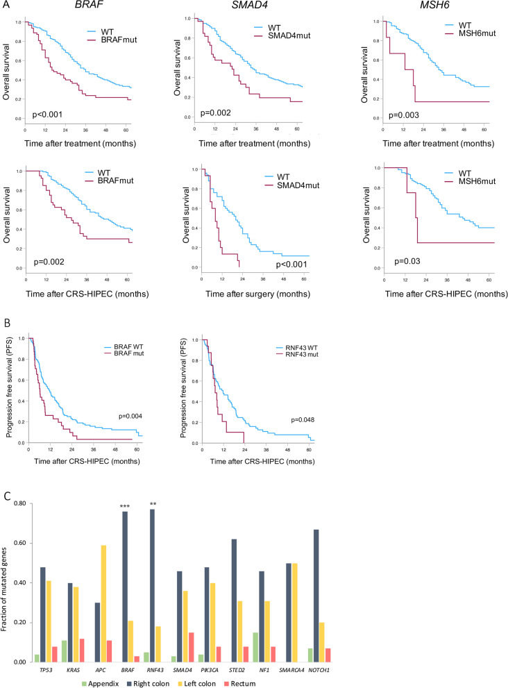
In univariable analyses, BRAF mutation was strongly associated with inferior OS (median OS: 16 vs 36 mo, p < 0.001) and PFS (median PFS: 6 vs 12 mo, p = 0.004) compared to non-mutated cases. In addition, SMAD4 and MSH6 mutations were associated with inferior OS, and RNF43 mutations were also associated with shorter PFS (Fig. 2A and B, Table S3/S4). In multivariable analysis BRAF (HR: 1.99) and SMAD4 (HR: 1.57) mutations remained associated with inferior OS, in addition to PCI (HR: 1.09) and N2-stage (HR: 1.54) (Table 2). MSH6 mutations were excluded due to small number of cases (n = 6). Factors associated with PFS in multivariable analysis were age (HR: 1.02), PCI (HR: 1.06) and BRAF mutations (HR: 1.51). RNF43 mutations were excluded as they were only accounted for in a proportion of cases (106/164).
Fig. 2.
Associations between mutations and long-term outcome. A Significant findings from univariable analyses of overall survival (OS) for the total cohort (n = 230); mutated BRAF, SMAD4 and MSH6 compared to wild-type (WT) (upper panel) and for cohort subgroups; mutated BRAF (CC = 0), SMAD4 (CC ≥ 1) and MSH6 (CC = 0) compared to WT (lower panel). B Progression-free survival (PFS, n = 164). C Mutated genes associated with primary tumor location, *p < 0.05, **p < 0.01, ***p < 0.001, ****p < 0.0001. BRAF and RNF43 mutations are associated with right sided primary CRC
Table 2.
Multivariable analysis of OS and PFS
| Variable | OS (n = 230) | PFS (n = 164) | ||
|---|---|---|---|---|
| HR (95% CI) | p-value | HR (95% CI) | p-value | |
| Age (increasing) | 1.01 (0.99–1.03) | 0.25 | 1.02 (1.01–1.04) | 0.009 |
| Gender (ref female) | 1.29 (0.91–1.83) | 0.15 | 1.29 (0.89–1.89) | 0.184 |
| PCI (increasing) | 1.09 (1.07–1.12) | < 0.001 | 1.06 (1.03–1.09) | < 0.001 |
| N2-stage | 1.54 (1.06–2.23) | 0.024 | – | – |
| BRAF mutation | 1.99 (1.39–2.83) | < 0.001 | 1.51 (1.01–2.26) | 0.044 |
| SMAD4 mutation | 1.57 (1.01–2.45) | 0.045 | – | – |
BRAF mutations associated with right-sided serrated primary CRC
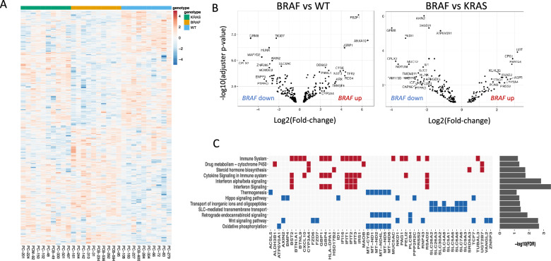
Differential gene expression analysis of BRAF mutated versus KRAS mutated and WT cases, revealed 179 and 303 differentially expressed genes (DEGs), respectively (padj < 0.1, up-regulated: 63 and 140, down-regulated: 116 and 163, Fig. 3A and B, supplementary file 6). The top ten up and down-regulated genes within each comparison are listed in Table S5 and S6. Among the top up-regulated genes in BRAF mutated compared to WT cases were ANXA10 (Annexin A10), TFF1 (Trefoil factor 1), TFF2 (Trefoil factor 2) and CTSE (Cathepsin E), which are all markers associated with BRAF mutated right sided sessile serrated primary CRC [27–30]. LY6G6D (lymphocyte antigen-6 family member G6D) was found heavily down-regulated in BRAF mutated cases compared to WT, a feature that is also associated with promoter hypermethylation and sessile serrated polyps [31]. Together with our findings of down-regulated RNF43 and ZNRF3 in BRAF-mutated PM-CRC (Figs. 3C, 4E and F, [32]) and enrichment of BRAF and RNF43 mutations in right sided primary CRC cases (Fig. 2C, supplementary file 7), previous findings that the BRAF-mutated PM-CRC are associated with right-sided serrated primary CRC are confirmed.
Fig. 3.
Differential gene expression and gene set enrichment analyses. A Heatmap of significant differential expressed genes (DEGs, p < 0.1) for individual patient samples comparing cases with mutated BRAF to mutated KRAS or WT, blue color: low expression, red color: high expression. Normalized counts from DESeq2 are used for visualization. Rows are centred and scaled. B Volcano plots showing DEGs significantly associated with BRAF mutation compared to WT (left panel) and BRAF mutation compared to KRAS mutation (right panel). C Gene set enrichment analysis showing affected signaling pathways and the DEGs involved, red markers: DEGs upregulated in BRAF mutated cases, blue markers: DEGs downregulated in BRAF mutated cases. Drug metabolism and immune signaling pathways are enriched in BRAF mutated cases, while SLC-mediated transport and Wnt signaling pathways are diminished
Fig. 4.
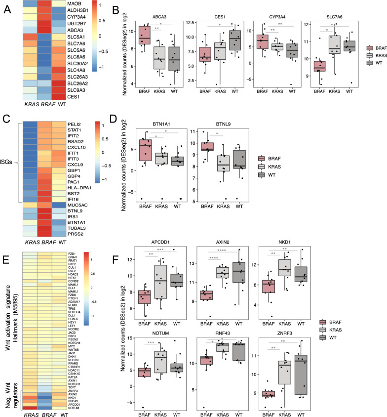
Differential expression of genes involved in drug metabolism, transport, immune signaling and Wnt signaling. Heatmaps with relative average gene expression levels within each group (BRAF, KRAS, WT) and boxplots of selected genes (indicating median, 25 and 75 percentile): A and B Altered expression of drug metabolism genes e.g. CYP3A4 and CES1 known to metabolize irinotecan, and reduced expression of SLC genes (e.g. SLC7A6) involved in transport of mitomycin C in BRAF mutated cases. C and D Increased expression of interferon stimulated genes (ISG, compared to KRAS mutated only) and BTN checkpoint molecules in BRAF mutated cases. E and F Similar expression of genes involved in the Wnt activation signature (Hallmark M5895) between the groups, however reduced expression of negative Wnt regulators in BRAF mutated cases, associated with ligand-dependent Wnt signaling. *padj < 0.1, **padj < 0.01, ***padj < 0.001, ****padj < 0.0001. Mean normalized read counts (DESeq2) in log2 scale were centred and scaled for visualization in heatmaps (A, C, E)
GSEA—Altered drug metabolism and transport in BRAF-mutated PM-CRC
GSEA of DEGs identified down-regulation of a range of SLC (solute carrier) transmembrane transporters in BRAF-mutated cases (Fig. 3C, Table S7). Expression of these transporters, which are important for the uptake of key cytotoxic drugs, was reduced compared to KRAS-mutated and WT cases (SLC5A1, SLC6A6, and SLC7A6), and compared to WT only (although a trend was also seen compared to KRAS-mutated cases (SLC30A2, SLC5A6, SLC26A3, SLC26A2, SLC9A3, SLC4A8), Fig. 4A and B). Of particular interest in this cohort was down-regulation of SLC7A6 (validated: Figure S6) which is associated with uptake of MMC, used in HIPEC treatment of the patients in this study [33]. In addition, another transporter that regulates drug efflux from the cells [34], the ATP-binding Cassette transporter ABCA3, was over-expressed in the BRAF-mutated cases compared to the other subgroups (Fig. 4B and Table S6). ABCA3 expression is associated with poor survival and multidrug resistance in Leukemia cells [35] and may have similar functions in BRAF-mutated colorectal cancer.
GSEA also revealed up-regulation of genes involved in drug metabolism, including irinotecan metabolism, in BRAF-mutated cases versus WT (Fig. 3C, Table S7): CYP3A4 (Cytochrome P450 enzymes 3A4, validated: Figure S6), UGT2B7 (UDP-Glucuronosyltransferase-2B7), MAOB (Monoamine Oxidase B) and ALDH3B1 (Aldehyde Dehydrogenase 3 Family Member B1). CYP3A4 was also found to be elevated in BRAF-mutated cases compared to KRAS-mutated (Fig. 4A and B). In addition, CES1 (Carboxylesterase 1) was down-regulated in the BRAF-mutated cases compared to WT.
Immune signaling in BRAF-mutated PM-CRC
Genes that play a role in the immune system were up-regulated in BRAF-mutated cases (Fig. 3C). The interferon (IFN)-stimulated genes RSAD2 (radical S-adenosyl methionine domain containing 2), GBP1 and GBP4 (Guanylate Binding Protein 1 and 4), BST2 (Bone Marrow Stromal Cell Antigen 2), IFIT1, IFIT2 and IFIT3 (interferon-induced protein with tetratricopeptide repeats 1, 2 and 3), HLA-DPA1 (Major Histocompatibility Complex, Class II, DP Alpha 1), CXCL10 (C-X-C motif chemokine ligand 10) and IRS1 (Insulin receptor substrate-1) were enriched compared to the KRAS-mutated subgroup (Fig. 4C). Moreover, the immune checkpoint molecule BTN1A1 (Butyrophilin subfamily 1 member, validated: Figure S6) was found up-regulated in BRAF-mutated cases compared to the other two subgroups (Fig. 4C and D). BTNL9 (Butyrophilin-Like 9, validated: Figure S6) was also significantly higher expressed in BRAF-mutated compared to KRAS-mutated tumors, and a similar trend was seen for WT cases. The gel-forming mucin MUC5AC was also found to be highly expressed in BRAF mutated cases compared to the two other subgroups, although only significant towards KRAS mutated cases.
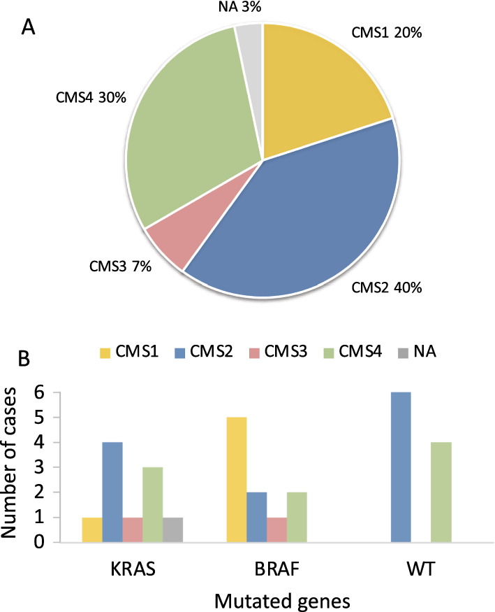
The PM-CRC gene expression data was classified according to the colorectal consensus molecular subtypes, resulting in 40% CMS2 (canonical), 30% CMS4 (mesenchymal), 20% CMS1 (immune), 7% CMS3 (metabolic) and 3% unclassified (NA) (Fig. 5A, supplementary file 8). The BRAF-mutated cases were enriched with the immune subtype, CMS1, while KRAS-mutated and WT cases contained a mixture of CMS2 and CMS4 (Fig. 5B).
Fig. 5.

Consensus molecular subtype (CMS) classification. A Distribution of CMS subtypes in PM-CRC (n = 30). CMS1 and CMS4 are enriched compared to pCRC. B Distribution of CMS subtypes in PM-CRC mutational subgroups (KRAS, BRAF, WT). BRAF mutated cases are enriched with CMS1
GSEA—ligand-dependent WNT activation in BRAF-mutated PM-CRC
GSEA revealed reduced expression of genes involved in Wnt signaling in BRAF-mutated cases compared to KRAS-mutated and WT (Fig. 3C, Table S7). To investigate whether the Wnt pathway was less activated in BRAF-mutated cases, we applied the hallmark Wnt activation signature (MSigDB M5895) on our data, and found broadly similar gene expression levels across the subgroups, indicating equal Wnt activation (Fig. 4E). The discrepancies lay mainly within the negative Wnt regulators and down-regulation of RNF43 and ZNRF3, located at the cell surface, and AXIN2 (validated: Figure S6), NKD1, APCDD1, and NOTUM, involved in negative feedback regulation, in the BRAF-mutated cases compared to the other subgroups (Fig. 4E and F). The low expression of negative feedback regulators is previously associated with ligand-dependent Wnt signaling in RNF43/RSPO aberrated CRC [36], and consistent with our findings that BRAF and RNF43 mutations/RSPO fusions often co-occur. Reduced expression of RNF43 is also in line with the presence of nonsense and frameshift mutations (Fig. 1B).
Discussion
In this cohort of PM-CRC cases, BRAF and RNF43 were 3–8 times more frequently mutated (27% and 16%, respectively) compared to previous reports from analyses of liver, 9% and 3%, and lung metastases, 6% and 2%, respectively [23, 37]. In contrast, the APC mutation frequency (29%) was low compared to previous reports from primary CRC (75%), colorectal liver metastases (82%), and lung metastases (86%) [23]. BRAF and RNF43 mutations frequently co-occurred and were associated with right-sided serrated primary tumors, in line with previous studies in CRC [32]. The differences observed between the metastatic locations suggest that the combination of BRAF and RNF43 mutations are associated with metastasis to the peritoneal cavity. BRAF mutations in CRC are associated with more aggressive disease and poor outcome through associations with pathological features (poorly differentiated tumors, tumor budding), advanced disease stage at the time of diagnosis, and peritoneal metastasis [38]. In vitro, BRAF mutations have been connected to enhanced ability of migration and invasion of CRC cell lines [39]. RNF43 mutations have also been associated with aggressive tumor biology, and in BRAF mutated patient derived organoids, RNF43 mutations were recently suggested to have a key role in promoting metastasis in animal models [40, 41]. Taken together, the marked differences between the metastatic sites with high abundance of BRAF and RNF43 mutations in PM may contribute to explain the inferior survival in PM-CRC.
In addition to the inherently aggressive biology of BRAF-mutated CRC, poor response to anti-cancer therapy could contribute to poor OS. MMC was the drug used for HIPEC in this study, and sensitivity to MMC would therefore be a key requirement for HIPEC efficacy. Our findings revealed down-regulation of several SLC transmembrane transporters in BRAF-mutated tumors. These molecules regulate uptake of cytotoxic drugs [33], and of particular interest, SLC7A6, which regulates uptake of MMC was strongly down-regulated in BRAF-mutated cases. To instigate cell killing, MMC must be taken up by the tumor cells, and reduced cellular uptake could therefore impair the efficacy of HIPEC. If validated on the protein level and through functional studies, this finding would suggest that other drugs should be considered for HIPEC in BRAF-mutated cases. The BRAF-mutated tumors also exhibited increased expression of several metabolic enzymes involved in the intracellular processing of anti-cancer drugs, which may lead to drug resistance and poor clinical efficacy. Another key drug in the management of mCRC is the topoisomerase1-inhibitor irinotecan. Irinotecan is converted to its active form (SN-38) by the intracellular enzyme CES1 [42], which was markedly down-regulated in the BRAF-mutated cases. In parallel, CYP3A4, another key enzyme which inactivates irinotecan, was up-regulated in the BRAF-mutated subgroup [42, 43], further potentially contributing to irinotecan resistance. Because follow-up after CRS-HIPEC is administered locally, details regarding irinotecan administration to patients in this study were not available. However, based on current oncological management of mCRC, it is reasonable to assume that a large proportion of patients were offered irinotecan-containing therapy as part of subsequent palliative systemic treatment for recurrent disease. Taken together, our findings reveal molecular changes pointing towards potential novel resistance mechanisms pertaining to two commonly administered drugs to patients with PM-CRC. These findings also provide a strong argument for determining the mutational status of BRAF early in the course of PM-CRC treatment, and ideally up-front of CRS-HIPEC.
Targeting BRAF V600E-mutated CRC using BRAF inhibitors alone has not been very effective, likely due to feedback activation of MAPK signaling through EGFR [44]. However, the BEACON clinical trial showed prolonged survival for mCRC patients when treated with BRAF inhibitors in combinations with EGFR and MEK inhibitors [45]. Although this treatment strategy could be an option for BRAF-mutated PM-CRC, caution should be taken as CYP3A4, discussed above, is also known to metabolize several BRAF and EGFR inhibitors, such as Vemurafenib [46], Encorafenib [47] and Erlotinib [48], and might reduce the efficacy of the drugs.
In contrast to pCRC, where ligand independent (Li) activation of the Wnt signaling pathway is dominant (in 85% of cases) mainly due to APC mutation [36], ligand-dependent (LD) Wnt activation was the principal mode of Wnt activation in the BRAF-mutated subgroup. At the genomic level, BRAF and APC mutations were almost mutually exclusive in our cohort, while BRAF mutations frequently co-occurred with RNF43 mutations or RSPO fusions that are dependent on Wnt ligand for Wnt activation. In addition, low expression of negative Wnt regulators associated with LD activation [36], were found in all BRAF-mutated cases subjected to transcriptome analyses, including when RNF43/RSPO aberrations were not detected. These findings suggest that RNF43/RSPO aberrations are more commonly co-occurring with BRAF mutations than was documented in our study. Collectively, these results point to the possibility of targeting LD-Wnt signaling in BRAF-mutated PM-CRC. With the target present in the cell membrane, LD signaling is thought to be more easily “druggable” than Li-Wnt activation, where the target is located intracellularly [49]. A class of drugs that are being extensively explored in this context are the porcupine inhibitors (e.g. LGK974), which prevent secretion of the Wnt ligand from signaling cells. Such inhibitors have been effective in in vitro and in vivo models with RNF43 and RSPO aberrations [49] and are currently being investigated in several clinical trials (NCT01351103, NCT03447470, NCT03507998). Interestingly, low or absent AXIN2 expression, which we found to be reduced in BRAF-mutated cases, has been suggested as a biomarker for selecting patients for treatment with porcupine inhibitors [36]. Hence, these results suggest that targeting LD-Wnt signaling could be beneficial in BRAF-mutated PM-CRC, possibly using AXIN2 expression as a biomarker for treatment selection which is more feasible than finding RNF43/RSPO aberrations through RNA-sequencing.
The majority of tumors in this cohort were microsatellite stable (MSS), with only 7% being MSI, which is in line with previous findings in mCRC (5–7% MSI cases) [50]. We and others have suggested that the negative prognostic impact of BRAF mutations could be counteracted by MSI status [51, 52], but although MSI cases were enriched within the BRAF-mutated subgroup, they still accounted for only 15% of the cases. Based on the frequency of MSI cases, immune checkpoint inhibitors currently in clinical use therefore do not seem to be an obvious treatment option in BRAF-mutated PM-CRC. In this context, the strong up-regulation of the newly discovered immune checkpoint molecules BTN1A1 and BTNL9 is very interesting. Although the T cell receptor for these molecules is still unknown, studies have shown that they inhibit CD4+ and CD8+ T cell proliferation and reduce production of IL-2 [53, 54]. An antibody against BTN1A1 (hSTC810) is already being studied in clinical trials (NCT05231746) [55] and if efficacious, BTN1A1 could represent a novel immunotherapy target in BRAF-mutated PM-CRC. The increased immune signaling in BRAF-mutated PM-CRC is also in line with enrichment of the immune subtype CMS1 in the BRAF-mutated cases. Collectively, these results indicate that BRAF-mutated PM-CRC have persistent immune signaling leading to increased levels of BTN immune checkpoint molecules that should be further explored as a possible novel therapeutic target for this particular patient subgroup.
A challenge, which is relevant in most studies when consecutive biobanking of surgical specimens is involved, was the inability to retrieve data from all eligible patients in this national cohort, as we ended up reporting data from 230 of 607 patients operated for PM-CRC. Prior to 2013, biobanking was anecdotal, while from 2013, a conclusion could be reached for more than half of eligible patients. In many cases, surgeons failed to collect tissue, the tissue tumor content was inadequate, or the sample failed subsequent quality control. There was no bias related to patient consent, but patients with low tumor burden and good response to neoadjuvant treatment may have been less likely to have their tumors sampled for research purposes (the surgeon prioritizing routine hisptopathology), and the samples would also be less likely to contain sufficient tumor tissue for subsequent analysis. Thereby, the mutational profiles could in principle be more representative of patients with high-volume disease and inferior chemotherapy response than of cases with very low tumor burden. Another limitation is related to the use of two targeted panels for mutation analysis, because a broader gene panel became available during the time period when the analyses were conducted, and the mutation status of some genes was therefore less extensively characterized (94/230 cases). The study cohort included patients with PM-CRC undergoing CRS and MMC-based HIPEC. While CRS is still standard of care for low-volume resectable PM, the use of HIPEC remains controversial after the failure of oxaliplatin-based HIPEC to improve outcomes in a randomized trial in oxaliplatin-pretreated patients [56]. MMC-based HIPEC has not been similarly studied in a randomized trial, and its value is thereby not fully clarified; however, our data would suggest that efficacy may be inferior in BRAF-mutated cases.
Overall, this study shows that BRAF mutations are frequent in PM-CRC, often co-occurring with RNF43 or RSPO aberrations. This combination of abnormalities could lead to a more aggressive phenotype that partly may explain the particularly poor prognosis associated with BRAF mutations. Another contributor to poor prognosis may be altered drug metabolism and transport causing resistance to anti-cancer drugs MMC and irinotecan. Two potential novel therapeutic approaches were identified, suggesting the use of inhibitors to target LD-Wnt activation and specific targeting of the BTN immune checkpoints.
Supplementary Information
Acknowledgements
The authors acknowledge the important contributions from our User Panel, composed of patients, care takers and allied healthcare professionals, to the execution of our research, in particular with respect to public reporting and communication.
Abbreviations
- PM-CRC
Peritoneal metastasis from colorectal cancer
- HIPEC
Hyperthermic intraperitoneal chemotherapy
- MMC
Mitomycin C
- CRC
Colorectal cancer
- mCRC
Metastatic colorectal cancer
- CRS
Cytoreductive surgery
- PCI
Peritoneal cancer index
- SNV
Single nucleotide variation
- CNV
Copy number variation
- HS
Ion AmpliSeq™ Cancer Hotspot panel
- Onc
Oncomine Comprehensive panel v3
- IGV
Integrative Genomics Viewer
- GSEA
Gene set enrichment analysis
- CMS
Consensus molecular subtype
- OS
Overall survival
- PFS
Progression-free survival
- MSI
Microsatellite instable
- MSS
Microsatellite stable
- HR
Hazard ratio
- WT
Wild-type
- DEGs
Differentially expressed genes
Author contributions
KF, AT and CLA has conceived, designed and supervised the study. KF and CLA has drafted the manuscript. CLA and CK has created the figures. All coauthors contributed to respective parts of conducting experiments, data acquisition, analysis, and interpretation. The manuscript is read and approved by all coauthors.
Funding
Funding for this study was provided by the Norwegian Cancer Society.
Availability of data and materials
All data that supports the findings of this study and that do not compromise the privacy of research participants, are available as supplementary data. Sensitive data, such as raw sequencing and clinical data, are stored in the European Genome-Phenome Archive (EGA) data repository (accession number: EGAD50000000593).
Declarations
Ethics approval and consent to participate
The study was approved by the Norwegian Ethics Committee (ID# s-07160b) and written informed consent has been obtained from the patients.
Consent for publication
Not applicable.
Competing interests
The authors declare that they have no competing interests.
Footnotes
Publisher's Note
Springer Nature remains neutral with regard to jurisdictional claims in published maps and institutional affiliations.
C. Lund-Andersen and A. Torgunrud have contributed equally to this work.
C. Kanduri and V. J. Dagenborg have contributed equally to this work.
References
- 1.Guend H, Patel S, Nash GM. Abdominal metastases from colorectal cancer: intraperitoneal therapy. J Gastrointest Oncol. 2015;6(6):693–698. doi: 10.3978/j.issn.2078-6891.2015.078. [DOI] [PMC free article] [PubMed] [Google Scholar]
- 2.Franko J, et al. Prognosis of patients with peritoneal metastatic colorectal cancer given systemic therapy: an analysis of individual patient data from prospective randomised trials from the Analysis and Research in Cancers of the Digestive System (ARCAD) database. Lancet Oncol. 2016;17(12):1709–1719. doi: 10.1016/S1470-2045(16)30500-9. [DOI] [PubMed] [Google Scholar]
- 3.Lund-Andersen C, et al. Omics analyses in peritoneal metastasis-utility in the management of peritoneal metastases from colorectal cancer and pseudomyxoma peritonei: a narrative review. J Gastrointest Oncol. 2021;12(Suppl 1):S191–S203. doi: 10.21037/jgo-20-136. [DOI] [PMC free article] [PubMed] [Google Scholar]
- 4.Stein MK, et al. Comprehensive tumor profiling reveals unique molecular differences between peritoneal metastases and primary colorectal adenocarcinoma. J Surg Oncol. 2020;121(8):1320–1328. doi: 10.1002/jso.25899. [DOI] [PMC free article] [PubMed] [Google Scholar]
- 5.Hamed AB, et al. Impact of primary tumor location and genomic alterations on survival following cytoreductive surgery and hyperthermic intraperitoneal chemoperfusion for colorectal peritoneal metastases. Ann Surg Oncol. 2023;30(7):4459–4470. doi: 10.1245/s10434-023-13463-x. [DOI] [PMC free article] [PubMed] [Google Scholar]
- 6.Lenos KJ, et al. Molecular characterization of colorectal cancer related peritoneal metastatic disease. Nat Commun. 2022;13(1):4443. doi: 10.1038/s41467-022-32198-z. [DOI] [PMC free article] [PubMed] [Google Scholar]
- 7.Hallam S, et al. The transition from primary colorectal cancer to isolated peritoneal malignancy is associated with an increased tumour mutational burden. Sci Rep. 2020;10(1):18900. doi: 10.1038/s41598-020-75844-6. [DOI] [PMC free article] [PubMed] [Google Scholar]
- 8.Nadler A, McCart JA, Govindarajan A. Peritoneal carcinomatosis from colon cancer: a systematic review of the data for cytoreduction and intraperitoneal chemotherapy. Clin Colon Rectal Surg. 2015;28(4):234–246. doi: 10.1055/s-0035-1564431. [DOI] [PMC free article] [PubMed] [Google Scholar]
- 9.Esquivel J, et al. American Society of Peritoneal Surface Malignancies opinion statement on defining expectations from cytoreductive surgery and hyperthermic intraperitoneal chemotherapy in patients with colorectal cancer. J Surg Oncol. 2014;110(7):777–778. doi: 10.1002/jso.23722. [DOI] [PubMed] [Google Scholar]
- 10.Yang H, Wang K. Genomic variant annotation and prioritization with ANNOVAR and wANNOVAR. Nat Protoc. 2015;10(10):1556–1566. doi: 10.1038/nprot.2015.105. [DOI] [PMC free article] [PubMed] [Google Scholar]
- 11.Patro R, et al. Salmon provides fast and bias-aware quantification of transcript expression. Nat Methods. 2017;14(4):417–419. doi: 10.1038/nmeth.4197. [DOI] [PMC free article] [PubMed] [Google Scholar]
- 12.Srivastava A, et al. Alignment and mapping methodology influence transcript abundance estimation. Genome Biol. 2020;21(1):239. doi: 10.1186/s13059-020-02151-8. [DOI] [PMC free article] [PubMed] [Google Scholar]
- 13.Bourgon R, Gentleman R, Huber W. Independent filtering increases detection power for high-throughput experiments. Proc Natl Acad Sci USA. 2010;107(21):9546–9551. doi: 10.1073/pnas.0914005107. [DOI] [PMC free article] [PubMed] [Google Scholar]
- 14.Hebenstreit D, et al. RNA sequencing reveals two major classes of gene expression levels in metazoan cells. Mol Syst Biol. 2011;7:497. doi: 10.1038/msb.2011.28. [DOI] [PMC free article] [PubMed] [Google Scholar]
- 15.Scrucca L, et al. mclust 5: clustering, classification and density estimation using gaussian finite mixture models. R J. 2016;8(1):289–317. doi: 10.32614/RJ-2016-021. [DOI] [PMC free article] [PubMed] [Google Scholar]
- 16.Love MI, Huber W, Anders S. Moderated estimation of fold change and dispersion for RNA-seq data with DESeq2. Genome Biol. 2014;15(12):550. doi: 10.1186/s13059-014-0550-8. [DOI] [PMC free article] [PubMed] [Google Scholar]
- 17.Raudvere U, et al. g:Profiler: a web server for functional enrichment analysis and conversions of gene lists (2019 update) Nucleic Acids Res. 2019;47(W1):W191–W198. doi: 10.1093/nar/gkz369. [DOI] [PMC free article] [PubMed] [Google Scholar]
- 18.Guinney J, et al. The consensus molecular subtypes of colorectal cancer. Nat Med. 2015;21(11):1350–1356. doi: 10.1038/nm.3967. [DOI] [PMC free article] [PubMed] [Google Scholar]
- 19.Eide PW, et al. CMScaller: an R package for consensus molecular subtyping of colorectal cancer pre-clinical models. Sci Rep. 2017;7(1):16618. doi: 10.1038/s41598-017-16747-x. [DOI] [PMC free article] [PubMed] [Google Scholar]
- 20.Velasco A, et al. Multi-center real-world comparison of the fully automated Idylla microsatellite instability assay with routine molecular methods and immunohistochemistry on formalin-fixed paraffin-embedded tissue of colorectal cancer. Virchows Arch. 2021;478(5):851–863. doi: 10.1007/s00428-020-02962-x. [DOI] [PMC free article] [PubMed] [Google Scholar]
- 21.Poplawski A, Binder H. Feasibility of sample size calculation for RNA-seq studies. Brief Bioinform. 2018;19(4):713–720. doi: 10.1093/bib/bbw144. [DOI] [PubMed] [Google Scholar]
- 22.Wu H, Wang C, Wu Z. PROPER: comprehensive power evaluation for differential expression using RNA-seq. Bioinformatics. 2015;31(2):233–241. doi: 10.1093/bioinformatics/btu640. [DOI] [PMC free article] [PubMed] [Google Scholar]
- 23.Yaeger R, et al. Clinical sequencing defines the genomic landscape of metastatic colorectal cancer. Cancer Cell. 2018;33(1):125–136.e3. doi: 10.1016/j.ccell.2017.12.004. [DOI] [PMC free article] [PubMed] [Google Scholar]
- 24.Tsukiyama T, et al. Molecular role of RNF43 in canonical and noncanonical Wnt signaling. Mol Cell Biol. 2015;35(11):2007–2023. doi: 10.1128/MCB.00159-15. [DOI] [PMC free article] [PubMed] [Google Scholar]
- 25.Seshagiri S, et al. Recurrent R-spondin fusions in colon cancer. Nature. 2012;488(7413):660–664. doi: 10.1038/nature11282. [DOI] [PMC free article] [PubMed] [Google Scholar]
- 26.Stransky N, et al. The landscape of kinase fusions in cancer. Nat Commun. 2014;5:4846. doi: 10.1038/ncomms5846. [DOI] [PMC free article] [PubMed] [Google Scholar]
- 27.Tsai JH, et al. Aberrant expression of annexin A10 is closely related to gastric phenotype in serrated pathway to colorectal carcinoma. Mod Pathol. 2015;28(2):268–278. doi: 10.1038/modpathol.2014.96. [DOI] [PubMed] [Google Scholar]
- 28.Jahan R, et al. Odyssey of trefoil factors in cancer: diagnostic and therapeutic implications. Biochim Biophys Acta Rev Cancer. 2020;1873(2):188362. doi: 10.1016/j.bbcan.2020.188362. [DOI] [PubMed] [Google Scholar]
- 29.Gala MK, et al. TFF2-CXCR4 axis is associated with BRAF V600E colon cancer. Cancer Prev Res. 2015;8(7):614–619. doi: 10.1158/1940-6207.CAPR-14-0444. [DOI] [PMC free article] [PubMed] [Google Scholar]
- 30.Caruso M, et al. Over-expression of cathepsin E and trefoil factor 1 in sessile serrated adenomas of the colorectum identified by gene expression analysis. Virchows Arch. 2009;454(3):291–302. doi: 10.1007/s00428-009-0731-0. [DOI] [PubMed] [Google Scholar]
- 31.Caruso FP, et al. Lymphocyte antigen 6G6D-mediated modulation through p38alpha MAPK and DNA methylation in colorectal cancer. Cancer Cell Int. 2022;22(1):253. doi: 10.1186/s12935-022-02672-1. [DOI] [PMC free article] [PubMed] [Google Scholar]
- 32.De Palma FDE, et al. The molecular hallmarks of the serrated pathway in colorectal cancer. Cancers. 2019;11(7):1017. doi: 10.3390/cancers11071017. [DOI] [PMC free article] [PubMed] [Google Scholar]
- 33.Girardi E, et al. A widespread role for SLC transmembrane transporters in resistance to cytotoxic drugs. Nat Chem Biol. 2020;16(4):469–478. doi: 10.1038/s41589-020-0483-3. [DOI] [PMC free article] [PubMed] [Google Scholar]
- 34.Alketbi L, et al. The role of ATP-binding cassette subfamily A in colorectal cancer progression and resistance. Int J Mol Sci. 2023;24(2):1344. doi: 10.3390/ijms24021344. [DOI] [PMC free article] [PubMed] [Google Scholar]
- 35.Chapuy B, et al. Intracellular ABC transporter A3 confers multidrug resistance in leukemia cells by lysosomal drug sequestration. Leukemia. 2008;22(8):1576–1586. doi: 10.1038/leu.2008.103. [DOI] [PubMed] [Google Scholar]
- 36.Kleeman SO, et al. Exploiting differential Wnt target gene expression to generate a molecular biomarker for colorectal cancer stratification. Gut. 2020;69(6):1092–1103. doi: 10.1136/gutjnl-2019-319126. [DOI] [PMC free article] [PubMed] [Google Scholar]
- 37.Ostrup O, et al. Molecular signatures reflecting microenvironmental metabolism and chemotherapy-induced immunogenic cell death in colorectal liver metastases. Oncotarget. 2017;8(44):76290–76304. doi: 10.18632/oncotarget.19350. [DOI] [PMC free article] [PubMed] [Google Scholar]
- 38.Fanelli GN, et al. The heterogeneous clinical and pathological landscapes of metastatic Braf-mutated colorectal cancer. Cancer Cell Int. 2020;20:30. doi: 10.1186/s12935-020-1117-2. [DOI] [PMC free article] [PubMed] [Google Scholar]
- 39.Makrodouli E, et al. BRAF and RAS oncogenes regulate Rho GTPase pathways to mediate migration and invasion properties in human colon cancer cells: a comparative study. Mol Cancer. 2011;10:118. doi: 10.1186/1476-4598-10-118. [DOI] [PMC free article] [PubMed] [Google Scholar]
- 40.Matsumoto A, et al. RNF43 mutation is associated with aggressive tumor biology along with BRAF V600E mutation in right-sided colorectal cancer. Oncol Rep. 2020;43(6):1853–1862. doi: 10.3892/or.2020.7561. [DOI] [PMC free article] [PubMed] [Google Scholar]
- 41.Bugter JM, et al. RNF43 mutations facilitate colorectal cancer metastasis via formation of a tumour-intrinsic niche. bioRxiv. 2023 doi: 10.1101/2022.12.22.521159. [DOI] [Google Scholar]
- 42.Ma MK, McLeod HL. Lessons learned from the irinotecan metabolic pathway. Curr Med Chem. 2003;10(1):41–49. doi: 10.2174/0929867033368619. [DOI] [PubMed] [Google Scholar]
- 43.Santos A, et al. Metabolism of irinotecan (CPT-11) by CYP3A4 and CYP3A5 in humans. Clin Cancer Res. 2000;6(5):2012–2020. [PubMed] [Google Scholar]
- 44.Sahin IH, Klostergaard J. BRAF mutations as actionable targets: a paradigm shift in the management of colorectal cancer and novel avenues. JCO Oncol Pract. 2021;17(12):723–730. doi: 10.1200/OP.21.00160. [DOI] [PubMed] [Google Scholar]
- 45.Kopetz S, et al. Encorafenib, binimetinib, and cetuximab in BRAF V600E-mutated colorectal cancer. N Engl J Med. 2019;381(17):1632–1643. doi: 10.1056/NEJMoa1908075. [DOI] [PubMed] [Google Scholar]
- 46.Zhang W, Heinzmann D, Grippo JF. Clinical pharmacokinetics of vemurafenib. Clin Pharmacokinet. 2017;56(9):1033–1043. doi: 10.1007/s40262-017-0523-7. [DOI] [PubMed] [Google Scholar]
- 47.Wollenberg L, et al. A phase I, single-center, open-label study to investigate the absorption, distribution, metabolism and excretion of encorafenib following a single oral dose of 100 mg [(14) C] encorafenib in healthy male subjects. Pharmacol Res Perspect. 2023;11(5):e01140. doi: 10.1002/prp2.1140. [DOI] [PMC free article] [PubMed] [Google Scholar]
- 48.Ling J, et al. Metabolism and excretion of erlotinib, a small molecule inhibitor of epidermal growth factor receptor tyrosine kinase, in healthy male volunteers. Drug Metab Dispos. 2006;34(3):420–426. doi: 10.1124/dmd.105.007765. [DOI] [PubMed] [Google Scholar]
- 49.Kleeman SO, Leedham SJ. Not all Wnt activation is equal: ligand-dependent versus ligand-independent Wnt activation in colorectal cancer. Cancers. 2020;12(11):3355. doi: 10.3390/cancers12113355. [DOI] [PMC free article] [PubMed] [Google Scholar]
- 50.Motta R, et al. Immunotherapy in microsatellite instability metastatic colorectal cancer: current status and future perspectives. J Clin Transl Res. 2021;7(4):511–522. [PMC free article] [PubMed] [Google Scholar]
- 51.Offermans K, et al. Association between mutational subgroups, Warburg-subtypes, and survival in patients with colorectal cancer. Cancer Med. 2023;12(2):1137–1156. doi: 10.1002/cam4.4968. [DOI] [PMC free article] [PubMed] [Google Scholar]
- 52.Larsen SG, et al. Impact of KRAS, BRAF and microsatellite instability status after cytoreductive surgery and HIPEC in a national cohort of colorectal peritoneal metastasis patients. Br J Cancer. 2022;126(5):726–735. doi: 10.1038/s41416-021-01620-6. [DOI] [PMC free article] [PubMed] [Google Scholar]
- 53.Herrmann T, Karunakaran MM. Butyrophilins: gammadelta T cell receptor ligands, immunomodulators and more. Front Immunol. 2022;13:876493. doi: 10.3389/fimmu.2022.876493. [DOI] [PMC free article] [PubMed] [Google Scholar]
- 54.Smith IA, et al. BTN1A1, the mammary gland butyrophilin, and BTN2A2 are both inhibitors of T cell activation. J Immunol. 2010;184(7):3514–3525. doi: 10.4049/jimmunol.0900416. [DOI] [PubMed] [Google Scholar]
- 55.Lee S, Shin S, Howie L, et al. 740 A first-in-human trial of hSTC810 (anti-BTN1A1 Ab), a novel immune checkpoint with a mutually exclusive expression with PD-1/PD-L1, in patients with relapsed/refractory solid tumors. J ImmunoTher Cancer. 2022;10:A772–A772. [Google Scholar]
- 56.Quenet F, et al. Cytoreductive surgery plus hyperthermic intraperitoneal chemotherapy versus cytoreductive surgery alone for colorectal peritoneal metastases (PRODIGE 7): a multicentre, randomised, open-label, phase 3 trial. Lancet Oncol. 2021;22(2):256–266. doi: 10.1016/S1470-2045(20)30599-4. [DOI] [PubMed] [Google Scholar]
Associated Data
This section collects any data citations, data availability statements, or supplementary materials included in this article.
Supplementary Materials
Data Availability Statement
All data that supports the findings of this study and that do not compromise the privacy of research participants, are available as supplementary data. Sensitive data, such as raw sequencing and clinical data, are stored in the European Genome-Phenome Archive (EGA) data repository (accession number: EGAD50000000593).


