Abstract
Most humans and Old World nonhuman primates are infected for life with Epstein-Barr virus (EBV) or closely related gammaherpesviruses in the same lymphocryptovirus (LCV) subgroup. Several potential strategies for immune evasion and persistence have been proposed based on studies of EBV infection in humans, but it has been difficult to test their actual contribution experimentally. Interest has focused on the EBV nuclear antigen 1 (EBNA1) because of its essential role in the maintenance and replication of the episomal viral genome in latently infected cells and because EBNA1 endogenously expressed in these cells is protected from presentation to the major histocompatibility complex class-I restricted cytotoxic T-lymphocyte (CTL) response through the action of an internal glycine-alanine repeat (GAR). Given the high degree of biologic conservation among LCVs which infect humans and Old World primates, we hypothesized that strategies essential for viral persistence would be well conserved among viruses of this subgroup. We show that the rhesus LCV EBNA1 shares sequence homology with the EBV and baboon LCV EBNA1 and that the rhesus LCV EBNA1 is a functional homologue for EBV EBNA1-dependent plasmid maintenance and replication. Interestingly, all three LCVs possess a GAR domain, but the baboon and rhesus LCV EBNA1 GARs fail to inhibit antigen processing and presentation as determined by using three different in vitro CTL assays. These studies suggest that inhibition of antigen processing and presentation by the EBNA1 GAR may not be an essential mechanism for persistent infection by all LCV and that other mechanisms may be important for immune evasion during LCV infection.
Epstein-Barr virus (EBV), a gammaherpesvirus in the lymphocryptovirus (LCV) subgroup, is widespread in the human population, where it is carried as a latent infection in peripheral blood B cells and as a lytic infection in the oropharynx (28). The latent B-cell infection appears to be critical for viral persistence, based on studies of viral clearance after total body irradiation in bone marrow transplant patients and of viral maintenance in patients treated with acyclovir to inhibit lytic replication (7, 37). Normal, immunocompetent humans develop EBV-specific cytotoxic T-lymphocyte (CTL) responses which target both lytically infected cells and latently infected growth-transformed B cells and yet fail to eliminate the virus from the body (28).
There are several potential mechanisms whereby EBV may evade the immune system and maintain persistent B-cell infection. First, EBV can downregulate the typical repertoire of latent gene expression normally associated with growth transformation in EBV-infected B-lymphoblastoid cell lines (LCLs) in vitro (29). Thus, of the six nuclear proteins (EBV nuclear antigens [EBNAs]) and three latent membrane proteins (LMPs) found in LCL cells, studies of viral RNA species detectable in circulating B cells in vivo suggest that viral antigen expression in these cells is limited at most to one of the nuclear antigens, EBNA1, selectively expressed from the BamHI Q promoter (Qp) and one of the membrane proteins, LMP2 (22, 34). By contrast the viral promoters which drive expression of all six EBNA mRNA transcripts in LCL cells (the BamHI Wp/Cp promoters) are silenced in circulating B cells in vivo. As a result, the EBNA3A, -3B, and -3C proteins, which form the immunodominant targets for the latent antigen-specific CTL response, are not expressed, and the infected cells may therefore avoid CTL recognition. Second, the EBNA1 protein, the only viral antigen that is absolutely required for maintenance of the viral episome in latently infected cells, is itself protected from presentation to CTLs through the conventional pathway of major histocompatibility complex (MHC) class I antigen processing. Thus, Masucci and colleagues showed that a large glycine-alanine repeat (GAR) domain in EBNA1 that is not required for the protein’s plasmid maintenance and transcriptional transactivation ability can inhibit antigen presentation when placed in cis with CTL epitopes (17). Protection from CTL recognition appears to be due to the ability of the GAR to inhibit the proteosomal degradation of indicator proteins into which the repeat domain is introduced (18). Despite this protective mechanism, Blake et al. have found that CD8+ CTLs with specificity for EBNA1-derived epitopes are naturally generated in vivo (2). However, when isolated and grown in vitro, these CTLs only recognize target cells endogenously expressing a GAR-deleted form of EBNA1 and not targets expressing the wild-type protein, confirming the protective role of the GAR domain. Interestingly, full-length EBNA1 can be recognized by these CTLs if the protein is provided exogenously to antigen-presenting cells, suggesting that the EBNA1-specific CTL response may have been stimulated in vivo by exogenous protein release from lysed EBV-infected cells and presented again via a cross-priming pathway.
How important each of these potential immune escape mechanisms is to the phenomenon of EBV persistence remains a matter of conjecture. It is likely, however, that key elements of immune evasion will have been conserved throughout LCV evolution since these viruses all appear to establish very similar interactions with their Old World primate hosts. For example, in a recently developed animal model involving rhesus LCV, experimental infection of naive rhesus monkeys results in asymptomatic persistent infection in the peripheral blood and oropharynx just as is seen in EBV-positive humans (23). Furthermore, all LCVs analyzed to date have a genome structure that is highly homologous to EBV and a similar repertoire of latent genes which appear, from recent studies on the EBNA2 and LMP homologues, to have conserved function even where there is considerable local sequence divergence (5, 6, 19). In the context of strategies for viral persistence mentioned above, a homologue of the Qp EBNA1 promoter has been identified in rhesus LCV, suggesting that the capacity for promoter switching and concomitant downregulation of most latent cycle antigens will be shared by all members of the LCV subgroup (30). In addition, recent studies on the EBNA1 homologue in baboon LCV have demonstrated not only conservation of the plasmid maintenance-transcriptional transactivation properties of the protein but also the presence of an internal GAR-like domain (38). In the current study, we have cloned the rhesus LCV EBNA1 homologue and then gone on to test whether the rhesus and baboon GAR-like domains could inhibit MHC class I-restricted antigen presentation.
MATERIALS AND METHODS
Cell lines and peptides.
S594 is a baboon LCV (referred to elsewhere as cercopithicine herpesvirus 12 or herpesvirus papio)-infected B-cell line derived by spontaneous growth from baboon peripheral blood lymphocytes (26). LCL8664 is a rhesus LCV (cercopithecine herpesvirus 15)-infected B-cell line derived from a retro-orbital B-cell lymphoma in a rhesus monkey (27). Mamu A*01-positive rhesus monkey fibroblast cells were derived from skin biopsies of animals with the Mamu A*01 MHC class I allele. B95-8 is a marmoset B-cell line infected with EBV (human herpesvirus 4) from a patient with infectious mononucleosis. Standard human LCLs were generated by EBV (B95.8 strain) transformation of peripheral blood B cells from donors of known human lymphocyte antigen (HLA) type. All LCLs were maintained in RPMI containing 10% (vol/vol) fetal calf serum, 100 IU of penicillin per ml, 100 μg of streptomycin per ml, and 2 mM l-glutamine. Human 143B TK−, BS-C-1, and CV-1 cell lines were kindly provided by Geoffrey Smith (University of Oxford, Oxford, United Kingdom) and maintained in Dulbecco modified Eagle medium supplemented as described above. Peptides were synthesized by standard fluorenylmethoxycarbonyl chemistry (Alta Bioscience, University of Birmingham, Birmingham, United Kingdom) and dissolved in dimethyl sulfoxide (DMSO), and their concentration was determined by biuret assay.
Genomic DNA cloning.
Genomic DNA from LCL8664 cells and S594 cells were digested in turn with SalI and HindIII, respectively, and cloned into pBluescript. Simian LCV EBNA1 was identified by hybridization with a DNA fragment containing the B95-8 EBV EBNA1 open reading frame (ORF). Filters were washed at 50°C with 0.5% SSC (1 × SSC is 0.15 M NaCl plus 0.015 M sodium citrate) and 1% sodium dodecyl sulfate (SDS). Nucleotide sequencing of the identified DNA fragments showed that the baboon LCV HindIII DNA fragment, Khind7, was homologous to the EBV genome from nucleotides 95360 to 100009. The rhesus LCV SalI DNA fragment, RS8, was homologous with the B95-8 EBV genome from nucleotides 105632 to 113286. Both DNA fragments span the EBV EBNA1 open reading frame (nucleotides 107950 to 109880). The nucleotide sequence of baboon LCV EBNA1 ORF matched the previously published sequence (38), and the rhesus LCV EBNA1 sequence has been submitted to GenBank (CHU93909).
Plasmids.
EBNA1 expression vectors were constructed by using the pSG5 plasmid (Stratagene). A 2.2-kb PstI fragment from RS8, coding for rhesus LCV EBNA1, was blunt ended and cloned into the BamHI site of plasmid pSG5 to generate pSG5-Pst22. B95-8 EBV EBNA1 ORF was isolated by an XhoII digest of BamHI K DNA and similarly blunt-end cloned into pSG5. Expression of human and simian LCV EBNA1s in pSG5 was confirmed by in vitro translation and by transient transfection in cos-1 cells and immunoblotting with human, rhesus monkey, and baboon immune sera (data not shown).
The amplicon plasmid, BSAII, contains the EBV lytic origin of replication (ori-lyt), the EBV packaging and cleavage signals within the terminal repeats, the EBV latent origin of replication (ori-P) for episomal maintenance, and the hygromycin phosphotransferase gene (36).
Insertion of the simian LCV GARs into EBV EBNA1 in place of the EBV GAR was achieved by using the plasmid p1813-E1dGA, which contains a form of EBNA1 deleted for the GAR (2). The baboon LCV GAR (baGAR) was derived from the plasmid p701, which contains a baboon LCV genomic fragment spanning the EBNA1 homologue (38), by isolating a 186-bp HinPI-TaqI fragment, blunt ending, and cloning into the SmaI site of pUC-1813 (14). The baGAR was then re-isolated as a 185-bp RsaI-BfaI fragment and blunt ended. This fragment was then inserted into the BspEI (treated with Klenow) site of p1813-E1dGA; this corresponds to EBV B95.8 strain genomic coordinate 108160. This construct was sequenced to confirm the reading frame of the inserted baGAR. A 1,995-bp HindII fragment was then excised, corresponding to the E1baGAR coding region, and cloned into the SmaI site of the vaccinia virus transfer vector pSC11. The GAR of the rhesus LCV EBNA1 (rhGAR) was isolated from pSG5-Pst22 by PCR. A 267-bp product was amplified with the following primers: forward primer 5′-GTTGGCTccggAAAGGTGGCACTG and reverse primer 5′-ACTACCTCCggaTGCCGCGGCCTC; BspEI sites (underlined) were generated by introducing point mutations within the primers (shown in lowercase letters). The PCR product was cut with BspEI and cloned into the corresponding site within p1813-E1dGA. This construct was sequenced to confirm the sequence of the PCR product and reading frame of the rhGAR. A 2,052-bp HindII fragment was then excised, corresponding to the E1rhGAR coding region, and cloned into the SmaI site of the vaccinia transfer vector pRB21, generating the plasmid pRB-E1RhGAR.
Rhesus LCV EBNA1 containing a simian immunodeficiency virus (SIV) gag epitope (amino acids 181 to 190: CTPYDINQML [1, 21]) and FLAG (DYKDDDK) epitopes were generated by introducing the recombinant sequences in frame into the unique BamHI site of rhesus LCV EBNA1. To generate a FLAG epitope (DYKDDDDK), the oligonucleotides 5′-ATAGGATCCCAGATGCTGGACTAC-3′ (BAM-Flg-FAMP) and 5′-ATAAGATCTCTTGTCGTCGTCGTCCTTGTAGTCCAGCATCTG-3′ (Bgl-Flg-RAMP) were annealed. One picogram of each annealed primer pair was end filled with the Klenow fragment of Escherichia coli DNA polymerase and used as a template for PCR by using the same oligonucleotides that generated the template. Amplified DNA fragments were blunt ended, phosphorylated with T4 DNA kinase, and self-ligated to form concatamers by using T4 DNA ligase. The concatamers were digested with BamHI and BglII, and the low-molecular-weight DNA was isolated and ligated to the BamHI site of pSG5-Pst22, generating the constructs pSG5-rhE1flg. Rhesus LCV EBNA-1 containing a gag-FLAG epitope (CTPYDINQMLDYKDDDK; pSG5-rhE1gag) was constructed in a similar manner. Deletion of the GAR domain from FLAG and gag-FLAG EBNA1s were generated by an NcoI/BamHI digestion and religation of blunt ends, generating the plasmids pSG5-rhE1dGAR and pSG5-rhE1dGARgag. The NcoI/BamHI deletion maintains the reading frame, FLAG, and gag and deletes amino acids 41 to 270 containing the GAR domain. RhE1/N-gag was constructed by amplifying the amino terminus of rhesus LCV EBNA-1 with a 5′ primer containing the gag epitope (5′-CAGGAATTCCTGCACCCCGTACGACTAAACCAAATGCTTTCCGACGGAAGG GGCCCG) and cloning this PCR fragment in frame with the amino-terminal FLAG epitope in pcDNA FLG (33). The remainder of the EBNA-1 coding sequence was added by cloning the BstXI/XhoI fragment of RS-8 into the FLAG vector with the amino terminus of EBNA-1. All DNA constructs were sequenced and verified for expression by transient transfection into BJAB cells, followed by immunoblotting of cell lysates with M2 anti-FLAG monoclonal antibody. The epitope tagged EBNA1s were excised and cloned into the BamHI site of the adenovirus vector, pAd-lox (8).
Viruses.
A recombinant vaccinia virus expressing E1baGAR (vaccE1baGAR) was generated according to standard protocols (32). A vaccinia virus expressing E1rhGAR (vaccE1rhGAR) was constructed based on the protocol of Blasco and Moss (3). Briefly, after infection of CV-1 cells with the virus vRB12, cells were then transfected with the plasmid pRB-E1rhGAR. Recombinant virus was selected based on large-plaque phenotype when plaqued on BS-C-1 cells. After several rounds of plaque purification, recombinant virus was amplified and the titers were determined on BS-C-1 cells. For both viruses, expression of protein of the expected size was confirmed by Western blot analysis. A total of 106 BS-C-1 cells were infected with virus at a multiplicity of infection (MOI) of 10 and incubated for 15 h. Cells were harvested, and 2 × 10⁵ cell equivalents were subjected to SDS–7.5% polyacrylamide gel electrophoresis. Protein was transferred to nitrocellulose and detected with the EBNA1-specific monoclonal antibody 1H4-1 and by using a chemiluminescence detection protocol. The recombinant vaccinia viruses vaccB*3501, vaccE1dGA, vaccEBNA1, and vaccEBNA3A have been described previously (2, 16, 25).
Recombinant adenoviruses were generated as described by Hardy et al. (8). Briefly, recombinant pAdLox vectors were cotransfected with Psi-5 adenovirus DNA by calcium phosphate precipitation into cre-expressing 293 cells, cre-8. Primary supernatants containing recombinant adenoviruses were plaque purified and amplified on 293 cells. Expression was verified by immunoblotting infected 293 cells with the M2, anti-FLAG monoclonal antibody.
EBNA1-dependent plasmid maintenance and replication.
A total of 5 × 10⁶ exponentially growing BJAB cells were cotransfected by electroporation with 10 μg of control pSG5 or a recombinant EBNA1 expression plasmid with 1 μg of BSAII for plasmid maintenance studies or a PUC plasmid with the EBV ori-p for plasmid replication studies. For plasmid maintenance studies, cells were allowed to recover for 24 h after electroporation, counted for viability by trypan blue exclusion, and plated by limiting dilution in 96-well plates in RPMI supplemented with 10% fetal calf serum and 400 μg of hygromycin per ml. The medium was replaced after 7 days, and at 14 days the plates were scored for the number of wells with hygromycin-resistant cell growth. Assays for plasmid replication studies were performed as previously described (38) with the following modifications. Hirt DNA extracts were prepared from cells 3 days after transfection. Then, 1 μg of Hirt DNA was digested with 20 U of DpnI for 12 h at 37°C. DpnI-resistant DNA was quantitated by real-time PCR by using Syber Green as described by the manufacturer (Perkin-Elmer/Applied Biosystems). Next, 50 ng of DpnI-digested Hirt DNA was amplified with EBV ori-p primers (BC-4843 5′-ACACCTTACTGTTCACAACTCAGCA-3′ and BC-4948 5′-TTAGTCACAAGGGCAGTGGCT-3′) in duplicate and quantitated against a standard curve derived by log dilutions of the ori-p plasmid in 10 μg of yeast tRNA per ml starting at 2 × 10⁶ copies/reaction.
CTL clones and chromium release assays.
EBV EBNA1-specific CTL clones restricted through HLA B*3501 (EBNA1 minimal epitope HPVGEADYFEY; amino acids 407 to 417) and HLA A*0203 (EBNA1 minimal epitope VLKDAIKDL; amino acids 574 to 582) were generated and maintained as described previously (2). Chromium release assays were carried out as follows. Target cells were infected with the appropriate recombinant vaccinia virus for 90 min at an MOI of 10, followed by incubation for another 15 h. Targets were then labelled with 50 to 100 μCi of (51Cr)O4 for 90 min, washed, and incubated with CTL at known effector-to-target (E:T) ratios in a standard 5-h chromium release assay. The percentage of specific lysis was calculated as follows: (release by CTL − spontaneous release) × 100/(total release in 1% SDS − spontaneous release). Where peptides were used, target cells were coincubated with peptide at a concentration of 2 × 10⁻⁸ M or with DMSO alone.
CTL lines specific for the Mamu-A*01-restricted gag CTL epitope CTPYDINQML were isolated and maintained as previously described (1). Target cells consisted of autologous primary fibroblast lines (Mamu-A*01+), which had been pretreated with recombinant human interferon gamma (800 IU/106 cells; Genzyme) for 48 h and then infected with recombinant adenoviruses at an MOI of 500 PFU/cell. Fibroblasts were infected with recombinant adenoviruses for 48 h prior to the CTL assay. Fibroblasts infected with wild-type adenovirus were used as a negative control, and fibroblasts infected overnight with recombinant vaccinia virus expressing SIV-gag served as a positive control. Target cells were labeled overnight with 51Cr (DuPont NEN, Wilmington, Del.) at 100 μCi per 106 cells. Standard 51Cr release assays were then carried out as previously described (12, 13), and the percent specific cytotoxicity was calculated as described above. The spontaneous release of target cells was <25% in all assays.
RESULTS
Rhesus LCV EBNA1 coding sequence.
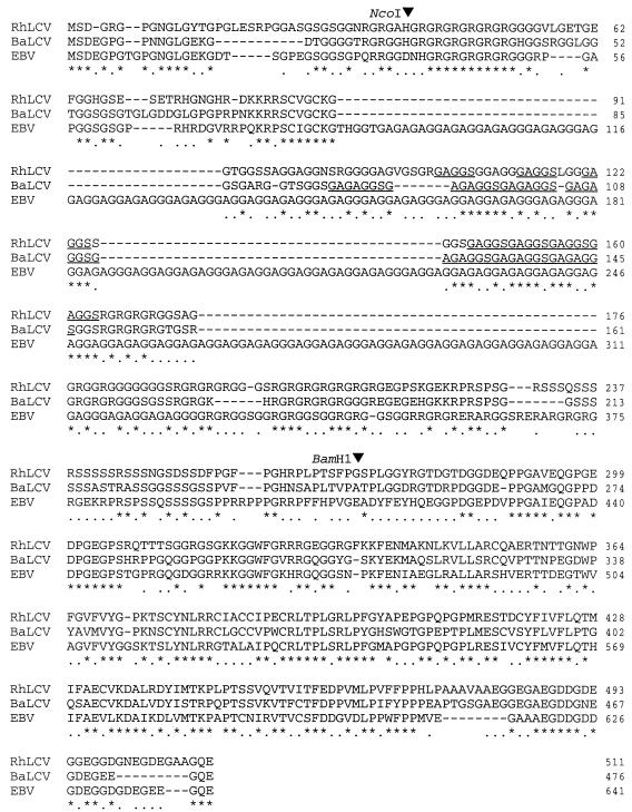
The rhesus LCV EBNA1 gene was isolated from a 7.6-kb SalI fragment of rhesus LCV DNA and encodes for 511 amino acids compared to 476 and 641 amino acids for the baboon LCV and EBV (B95.8) EBNA1 proteins, respectively. The three sequences are aligned in Figure 1. First, it should be noted that the difference in size between the rhesus LCV and EBV EBNA1 is due almost entirely to a much smaller GAR domain in the rhesus LCV EBNA1. Where the EBV EBNA1 contains 84 repeats of a G1–3A peptide over 252 amino acids, the rhesus LCV EBNA1 contains four perfect repeats of a GAGGS motif preceded by three GAGGS repeats interspersed with 12 additional amino acids forming 7 glycine/alanine-rich repeats within a 47-amino-acid stretch. The baboon LCV EBNA1 contains seven perfect repeats of a similar GAGAGGS motif (38). In Fig. 1, the repeat domains of the rhesus and baboon LCV EBNA1s are underlined to highlight the difference in sizes. In all three species, the GAR domain is flanked on both sides by GR-rich regions. The sequence alignment is also notable for a highly conserved KKRRSCVGCKG sequence at the amino-terminal side of the GAR and a serine-rich domain at the carboxy-terminal side of the GAR. The remainder of the EBNA1 carboxy terminus containing the DNA binding and dimerization domain is relatively well conserved with 63 and 53% amino acid identities between EBV and the rhesus or baboon LCVs, respectively.
FIG. 1.
Amino acid alignment of rhesus LCV, baboon LCV, and EBV EBNA1. Similar or identical residues in all three proteins are indicated by an asterisk and in two proteins by a period. GAR motifs in rhesus and baboon LCVs are underlined. BamHI and NcoI restriction enzyme sites used for C-terminal epitope insertion and deletion of the GAR in rhesus EBNA1 are highlighted.
Rhesus LCV EBNA1 supports EBV ori-p-dependent plasmid maintenance and replication.
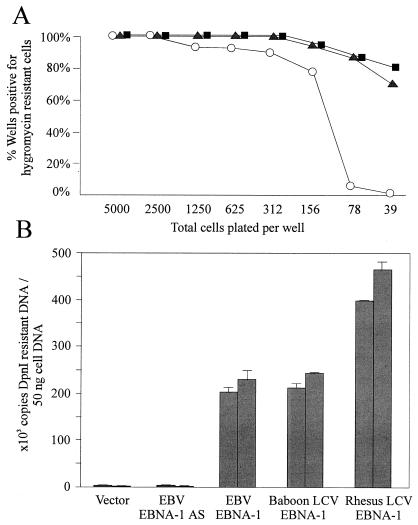
We tested whether the rhesus LCV EBNA1 could support EBV ori-p-dependent plasmid maintenance by cotransfecting an EBV EBNA1, rhesus LCV EBNA1, or a vector control expression vector into the EBV-negative B-cell line, BJAB, with an EBV ori-p and hygromycin resistance gene containing plasmid, BSAII. The relative efficiency of plasmid maintenance was measured by the frequency of hygromycin-resistant cell growth. A high frequency of hygromycin-resistant cells was demonstrated in cells cotransfected with BSAII and either EBV EBNA1 or rhesus LCV EBNA1. In a representative experiment shown in Fig. 2A, 100% (96 of 96) of the wells cotransfected with either type of EBNA1 and plated to as few as 312 cells/well were positive for hygromycin-resistant growth, and there was even a high frequency of hygromycin-resistant growth in wells seeded at 39 cells/well (81 and 73% for EBV and rhesus LCV EBNA1, respectively). In contrast, the frequency of hygromycin-resistant growth in vector control-cotransfected cells was already below 100% in wells seeded at 1,250 cells per well and was almost undetectable at a seeding of 39 cells per well.
FIG. 2.
Rhesus LCV EBNA1 supports ori-p-dependent plasmid maintenance and replication. (A) Frequency of hygromycin-resistant growth after cotransfection of hygromycin phosphotransferase containing BSAII plasmid with EBV EBNA1 (■), rhesus LCV EBNA1 (▴), or vector control (○) is shown. (B) Replication of ori-p plasmid DNA in eukaryotic cells is shown as the copy number of DpnI-resistant DNA in Hirt extracts from BJAB cells cotransfected with an ori-p plasmid and vector control, a construct with EBV EBNA1 in the antisense (AS) orientation, and EBV EBNA1, baboon LCV EBNA1, and rhesus LCV EBNA1 expression constructs. Results from two representative experiments are shown as the mean and the standard deviation of duplicate PCR measurements.
In order to measure more directly the effect of rhesus LCV EBNA1 on ori-p-dependent plasmid replication and maintenance, the amount of ori-p plasmid DNA replicated in eukaryotic cells was measured by quantitating DpnI-resistant DNA 3 days after EBNA1 or vector control cotransfection. As shown in Fig. 2B, the rhesus LCV EBNA1 supported ori-p-dependent replication as well as, or slightly better, than EBV and baboon LCV EBNA1. These results confirm that this DNA clone encodes a functionally active rhesus LCV EBNA1 and that EBNA1-dependent episomal maintenance and replication is well conserved among the LCVs.
Effects of simian GAR domains on antigen presentation.
We then carried out two series of experiments to ask whether the GAR domains found in rhesus and baboon LCVs were able to reproduce the inhibition of antigen processing shown by the homologous domain in EBV.
(i) Experiments on EBV EBNA1 with simian GAR inserts.
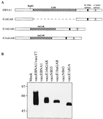
In this series of experiments the rhesus and baboon GARs were separately cloned into the EBV EBNA1 sequence in place of the latter’s natural GAR domain. The insertion site of the simian GARs corresponds to amino acid position 71 of EBV EBNA1, slightly upstream (19 amino acids) of the start of the natural GAR location. A schematic representation of these constructs is shown in Fig. 3A. We were interested at this point to know whether the chimeric constructs thus produced could be expressed from a recombinant vaccinia virus vector since earlier work had shown that the presence of the EBV EBNA1 GAR sequence in any construct was incompatible with the production of a viable vaccinia virus recombinant by conventional techniques (25). It was therefore significant that recombinant viruses expressing the rhesus GAR-containing and the baboon GAR-containing EBNA1 chimeras were obtained without difficulty by using the standard transfer vectors. Figure 3B shows an immunoblot of protein extracts from cells infected either with the EBNA1/rhesus GAR recombinant (vaccE1rhGAR) or with the EBNA1/baboon GAR recombinant (vaccE1baGAR) probed with a monoclonal antibody 1H4-1 that recognizes a unique EBV EBNA1 epitope that lies C terminal to the inserted GAR domains. This confirms the expression of chimeric proteins of the expected size, one larger than EBV EBNA1 lacking its natural GAR domain (see vaccE1dGAR) and smaller than wild-type EBV EBNA1. Note that here “wild-type” EBNA1 was cloned under a T7-inducible vector to allow its expression from a vaccinia virus recombinant (vaccEBNA1/vacc T7) and actually appears as a 75-kDa full-length species and multiple breakdown products due to partial excision of GAR sequences during vaccinia virus vector replication (2).
FIG. 3.
(A) Schematic representation of EBV EBNA1, EBV EBNA1-deleted for the GAR domain (E1dGAR), and the EBV EBNA1/rhesus or baboon GAR chimeras (E1rhGAR and E1baGAR). The BspEI site within EBV EBNA1 used for insertion of the rhesus and baboon GARs is shown, as is the location of the B*3501-restricted epitope (HPVGEADYFEY) and the A*0203-restricted epitope (VLKDAIKDL) within EBV EBNA1. (B) Western blot of recombinant vaccinia viruses expressing EBV EBNA1 (vacc [EBNA1]) and chimeric EBNA1s containing the rhesus LCV EBNA1 GAR (vaccE1rhGAR), baboon LCV GAR (vaccE1baGAR), or no GAR (vaccE1dGAR). BS-C-1 cells were infected with recombinant vaccinia viruses for 15 h, and extracts of 2 × 10⁵ cell equivalents were then probed with the EBNA1-specific monoclonal antibody 1H4-1. EBV EBNA1 expression is driven by a T7 promoter and requires coinfection with a T7-expressing vaccinia virus (vaccT7). vaccNB55 and vaccNB43 are control viruses with E1rhGAR and E1baGAR cloned in the reverse orientation.
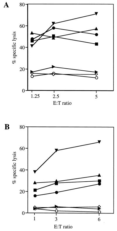
These viruses were then used to infect human LCLs of the appropriate HLA type, and the cells were tested as targets in cytotoxicity assays with human CTL clones with defined specificities for epitopes in the EBV EBNA1 sequence. Figure 4A shows the results obtained with an HLA-B*3501-restricted clone recognizing the EBV EBNA1 epitope 407-417, HPVGEADFYEY. The targets expressing the EBNA1/rhesus GAR chimera or the EBNA1/baboon GAR chimera were both recognized and killed at least as well as targets expressing the GAR-deleted EBNA1 protein and almost as well as uninfected targets pulsed with the epitope peptide. In contrast, targets expressing the wild-type EBV EBNA1 protein showed the same background levels of nonspecific lysis as did the uninfected cells or the cells infected with an irrelevant vaccinia virus recombinant (vaccEBNA3A). These results were confirmed in several independent cytotoxicity assays with EBV EBNA1 epitope 407-417-specific CTL clones. Furthermore, Fig. 4B shows parallel data from an experiment with HLA-A*0203-positive LCL targets and a CTL clone specific for the HLA-A*0203-restricted EBV EBNA1 epitope 574-582, VLKDAIKDL. Again, the results make it clear that replacing the native GAR domain in EBNA1 with simian GARs does not abrogate the processing and presentation of EBNA1 epitopes in human cells.
FIG. 4.
CTL recognition of EBV EBNA1/rhesus GAR and EBV EBNA1/baboon GAR chimeric proteins endogenously expressed in a human B-cell background. (A) HLA B*3501-positive LCL infected with recombinant vaccinia viruses expressing EBNA1/T7 (▸), EBNA1 deleted of the GAR domain (●), EBNA1/rhGAR (▴), EBNA1/baGAR (■), or EBNA3A (◊) were used as targets for EBV EBNA1 epitope 407-417-specific B35-restricted CTL effectors. (B) HLA A*0203-positive LCL infected with recombinant vaccinia viruses as in panel A were used as targets for EBNA1-specific A*0203-restricted EBV EBNA1 epitope 574-582 CTL effectors. For each graph, LCL pulsed with the cognate epitope peptide (▾) or DMSO (○) are shown as controls. Results are displayed as the percent specific lysis at the indicated effector/target ratio (E:T).
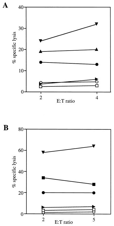
To test whether inhibitory effects of simian GAR inserts might nevertheless be apparent in cells of the natural host species, the above experiments were extended to use simian LCLs as targets. In this case we again used as effectors human B*3501-restricted CTL clones specific for the EBV EBNA1 epitope 407-417 and now provided the appropriate B*3501 restricting allele by coinfecting the target cells with a vaccinia virus B*3501 recombinant (vacc B*3501). Figure 5A presents the results of an experiment conducted in the rhesus target LCL 278. Clearly, targets coexpressing both B*3501 and the EBNA1/rhesus GAR chimeric protein or EBNA1 deleted of the GAR domain were specifically recognized by the EBNA1-specific CTL clone, as were vacc B*3501-infected peptide-loaded targets, whereas targets coexpressing B*3501 and wild-type EBNA1 were not. The same pattern of results was obtained in the parallel experiment with the baboon target LCL S594 (Fig. 5B). In this case, coexpression of B*3501 and of the EBNA1/baboon GAR chimeric protein in a baboon cell background again resulted in clear CTL recognition. In both instances, expression of EBNA1 deleted of the GAR domain or either the EBNA1/rhesus or baboon chimera alone did not sensitize target cells for CTL recognition (data not shown).
FIG. 5.
CTL recognition of EBV EBNA1/rhesus GAR and EBV EBNA1/baboon GAR chimeric proteins endogenously expressed in a simian B-cell background. LCL of rhesus (A) or baboon (B) origin were used as target cells in CTL assays with EBV EBNA1 epitope 407-417-specific B35-restricted CTL effectors. The human HLA B*3501 allele was provided by coinfection with a recombinant vaccinia virus expressing the HLA B35 heavy chain. Target cells were infected with the recombinant vaccinia virus vaccB*3501 (□), or coinfected with vaccB*3501 and vaccE1dGAR (●), vaccE1rhGAR (▴), or vaccE1baGAR (■), and vaccEBNA1/vaccT7 (▸). For controls, simian LCLs were infected with vaccB*3501 and pulsed with the EBV EBNA1 epitope 407-417 peptide (HPVGEADYFEY) (▾) or DMSO (○). Results are displayed as the percent specific lysis at the indicated effector/target ratio (E:T).
(ii) Experiments on rhesus EBNA1 with a CTL epitope insert.
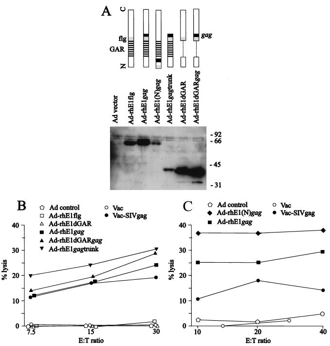
In the final series of experiments, we used both target and effector cells of rhesus origin and tested the ability of the rhesus EBNA1 protein to present an inserted CTL epitope. The Mamu A*01-restricted epitope sequence from the SIV gag gene was inserted, along with a FLAG tag for antibody detection, either C terminal to the rhesus GAR domain (rhE1/C-gag) and therefore mimicking the EBV EBNA1 epitope 407-417 location in the EBV EBNA1 or N terminal to the rhesus GAR domain (rhE1/N-gag) and therefore mimicking the epitope insertion site used by Levitskaya et al. (17). A C-terminal epitope insertion was also introduced into a GAR-deleted form of rhesus EBNA1 (rhE1dGAR/C-gag). These various constructs and appropriate epitope-negative controls were used to generate adenovirus recombinants capable of expressing the relevant chimeric proteins in Mamu A*01-positive rhesus fibroblasts. Figure 6A shows an immunoblot probed with a FLAG-specific antibody confirming that proteins of the appropriate size were indeed expressed in recombinant adenovirus-infected cells.
FIG. 6.
Construction of rhesus LCV EBNA1 expression vectors containing an SIV-gag epitope and presentation of the gag epitope to SIV-gag-specific CTL. (A) Schematic diagrams of the rhesus LCV EBNA1 chimeras are shown at the top highlighting the relative positions of the flag epitope (flg), SIV-gag epitope (gag) and GAR domain. Expression of the rhesus LCV EBNA1 chimeras by using recombinant adenoviruses is shown in the Western blot with a flag-specific monoclonal antibody. 293 cells were infected with the indicated recombinant adenovirus; 48 h later cells were harvested, and extracts were probed with a FLAG-specific antibody. (B and C) SIV-gag-specific CTL activity against Mamu A*01 fibroblast targets infected with recombinant adenoviruses expressing rhesus LCV EBNA1 chimeras or B-cell targets infected with recombinant vaccinia virus expressing wild-type SIV-gag is shown. Constructs containing the SIV-gag epitope are shown as solid symbols and constructs without the SIV-gag epitope are shown as open symbols. Results are displayed as the percent specific lysis at the indicated effector/target ratio (E:T).
The same targets were then tested for CTL recognition by a Mamu A*01-restricted CTL clone specific for the SIV-gag epitope. As controls, the same target cells were infected with a vaccinia virus recombinant expressing SIV-gag or with a control vaccinia virus. As shown in Fig. 6B and C, there was specific CTL recognition not just of vacc-SIV-gag-infected targets but also of all targets expressing epitope-positive versions of the rhesus EBNA1 protein. Importantly, these included cells expressing full-length rhesus EBNA1 with epitope insertions either C-terminal (rhE1/C-gag; Fig. 6B and C) or N-terminal (rhE1/N-gag; Fig. 6C) of the rhesus GAR domain. All control proteins lacking the relevant epitope sequence never sensitized the cells to lysis. Thus, in a system that used effector and target cells from the same species, there was no evidence for any cis-mediated effect of rhesus EBNA1 on epitope processing and/or presentation.
DISCUSSION
LCVs from human and nonhuman primate hosts share the ability to transform B lymphocytes to permanent growth in vitro yet persist as a latent infection in the B-lymphoid system of the host. Studies of the latent, transformation-associated genes of the baboon and rhesus LCVs suggest that the mechanisms used to transform B lymphocytes have been well conserved throughout this virus subgroup. For example, the baboon LCV EBNA2 homologue interacts with the CBF1/RBP-Jk transcriptional factor and transactivates transcription in a way similar to the EBV EBNA2 (19). Likewise, the rhesus and baboon LCV LMP1 homologues can interact with human tumor necrosis factor alpha receptor-associated factors and induce NF-κB activity in human B cells, despite considerable sequence divergence from EBV LMP1 in the carboxy terminus (6). Indeed, analysis of these LMP1 homologues identified the minimal PXQXT/S TRAF-binding motif in the proximal carboxy-terminal activation region of these molecules and revealed strong sequence conservation of the distal activation region, both of which are important for EBV-induced B-cell immortalization (6, 10, 11). In a similar way, comparisons between the human and nonhuman LCVs may identify important mechanisms underpinning viral persistence in the immune host.
In that context, EBNA1 is likely to be important for viral persistence both in vitro and in vivo since the protein has a well-documented role in the maintenance of viral episomes in latently infected growth-transformed B cells. These plasmid maintenance and replication functions have been well conserved in both the rhesus and baboon LCV EBNA1s (Fig. 2; see also reference 38), and the EBNA1 carboxy terminus containing the DNA binding domain is well conserved (Fig. 1), as might be expected for an essential function common to all LCVs. The relatively well conserved sequences flanking the GAR also suggest important functions for this region of EBNA1, and these regions correlate well with the highly charged regions that have been identified as important for DNA linking properties of EBNA-1 (20). Indeed, EBNA1 is one of the most well-conserved rhesus LCV latent infection genes, perhaps reflecting the fundamental importance of plasmid maintenance (percent amino acid identities between rhesus LCV and EBV for: EBNA-1, 56%; EBNA-2, 29%; EBNA-3A, 34%; EBNA-3B, 35%; EBNA-3C, 35%; LMP1, 29%; LMP2A, 57 and 38%, first exon [5, 6, 35]).
The central role played by EBNA1 in virus genome maintenance suggests that there may be circumstances in vivo where virus persistence does not require the full panoply of latent-growth-transforming proteins (i.e., where it does not require virus-driven proliferation), yet a minimum level of EBNA1 protein needs to be maintained. In fact, this might explain the evolutionary importance of the Qp promoter, providing a means of expressing EBNA1 in the absence of those other nuclear antigens (EBNAs 2, 3A, 3B, 3C, and LP) that in latent-growth-transforming infections are cotranscribed with EBNA1 from the BamHI Cp/Wp promoters. It is therefore interesting to note that homologues of Qp have been identified in baboon and rhesus LCVs, with the same interferon response factor regulatory and EBNA1-autoregulatory elements seen in EBV, implying that promoter switching and the transcriptional control of latency programs is generally important for LCV persistence (30). This might be directly testable in the rhesus monkey model by generating mutant rhesus LCV incapable of selective EBNA1 expression due to a Qp deletion.
The present study is concerned with the likely evolutionary importance of the EBNA1 GAR domain. In studies with EBV EBNA1, this domain is known not to be essential for the genome maintenance function of the protein and indeed is not required for EBV-induced B-cell transformation in vitro (15). Yet GAR-like motifs have been conserved in the EBNA1 molecules of rhesus and baboon LCVs, suggesting some other important function. The finding that the EBV GAR domain offers endogenously expressed EBNA1 protection from CD8+ CTL recognition provided a likely candidate for such a function, namely, an ability of cells in which EBNA1 is selectively expressed to avoid all EBV-specific CTL recognition. Such an ability may be important at particular stages of the virus life cycle in vivo. Indeed, other herpesviruses, such as herpes simplex virus and cytomegaloviruses (9, 39), encoding gene products which interfere with the HLA class I antigen processing pathway, have provided logical precedents for an EBV protein with some analogous evasion function. Against this background, the present finding (Fig. 4 to 6) that the baboon and rhesus LCV EBNA1 GARs do not show the same protective effect on antigen processing as the EBV EBNA1 GAR is surprising.
We have considered the possibility that our findings are an artifact, either of the viral isolates being studied or of the in vitro assays employed, but we believe that this is unlikely. The EBNA1 GARs from two different species of naturally occurring simian LCLs were studied, and there is no reason to believe that these are both abnormal mutant isolates. The baboon LCV was isolated as a spontaneous B-cell line from the cultured lymphocytes of an otherwise-healthy animal (26), much as standard EBV isolates are rescued from asymptomatic carriers. The rhesus LCV was isolated from a virus-positive B cell lymphoma arising in an immunosuppressed rhesus monkey (27), and inoculation of this virus into immunocompetent animals has demonstrated experimentally that it is fully capable of establishing an asymptomatic persistent infection in vivo (23).
It is also worth noting that the rhesus and baboon GAR domains, with lengths of 72 and 52 amino acids, respectively, are significantly shorter than the 252-amino-acid GAR sequence found in the prototype EBV strain B95.8. Indeed, the shortest GAR length in any naturally rescued EBV isolate is of the order of 100 amino acids (4). However, GAR repeat size is a stable characteristic of any one EBV strain and in our experience does not change with long-term serial passage of the virus-carrying LCL in vitro. We think it very unlikely, therefore, that the shorter GAR domain size seen in simian LCVs is an artifact introduced by in vitro isolation or passage. Furthermore, studies on minimalized versions of the EBNA1 GAR domain suggest that a sequence of as few as 17 amino acid residues is sufficient to confer protection in cis from proteosomal cleavage (18). If this assay faithfully reflects the ability of such a small repeat also to protect the protein from CTL detection, then we would anticipate that the LCV GAR domains being tested in the experiments should have been long enough to express any protective potential.
Finally, the lack of immune protection mediated by the simian GAR domains was apparent in several different experimental situations involving different combinations of indicator antigens, target cell backgrounds, and effector CTLs. Thus, the simian GARs were first inserted into EBV EBNA1 replacing the endogenous GAR domain, and then the EBNA1/simian GAR chimeras were tested for the presentation of two native EBNA1 epitopes to human CTL clones. There was no inhibition of CTL detection in a system where EBNA1’s own GAR domain is clearly protective (Fig. 4 and 5). This did not reflect some species-specific requirement, since recognition of the B*3501-restricted EBV EBNA1 epitope 407-417 was still observed when the above EBNA1/simian GAR chimeras were expressed along with HLA-B*3501 in rhesus or baboon target cells (Fig. 5). Equally important, the protective effect of EBV’s native GAR domain was observed just as strongly in these simian cell backgrounds as in human cells. In a final set of experiments which recapitulate the type of epitope insertions first used to demonstrate EBV EBNA1’s protective capacity, an indicator epitope (in this case from SIV-gag) was introduced into the full-length rhesus EBNA1 at both the N-terminal and C-terminal ends of the natural GAR domain, and in either situation it was efficiently processed for recognition in assays where both effector and target cells were of rhesus origin.
These experiments do not argue against an immune protective role for the EBV EBNA1 GAR. Indeed, the effect of the domain on endogenous antigen processing via the conventional MHC class I pathway has now been well documented in a number of studies (2, 17, 24). However, in showing that the homologous GAR domains of rhesus and baboon LCVs have not acquired this capacity, our experiments call into question whether immune evasion per se is the primary function of the GAR. In this context the mere fact that EBV establishes persistence in resting lymphocytes in the memory B-cell population may be sufficient to afford these cells immunological protection, since resting cells do not express the costimulatory molecules upon which immune T-cell activation depends and are also likely to have a much-reduced antigen-presenting capacity compared to activated proliferating B lymphoblasts. On this basis, not only “self-protected” molecules such as EBNA1 but also potentially immunogenic viral antigens such as LMP2A, thought to be expressed in the EBV-positive reservoir of resting B cells, could perhaps be sustained without alerting the CTL response.
It may be, therefore, that the immune evasion capability of EBV EBNA1 is a relatively recent acquisition in evolutionary terms, perhaps as a byproduct of a more fundamental and more widely conserved property of the LCV GAR domain. One possibility, hinted at in the results of a recent biochemical study, is that the GAR might offer EBNA1 a more general protection from proteolysis rather than specific proteosomal breakdown (31). Such a scenario puts the emphasis on LCV’s achieving stability of the viral genome maintenance protein rather than its immunological silencing.
ACKNOWLEDGMENTS
N.W.B. and A.M. contributed equally to this work.
We thank John Yates (Roswell Park Institute) for providing the plasmid p701 containing the HVP EBNA1 gene and B. Moss (National Institutes of Health) for providing plasmid pRB21 and vaccinia virus vRB12.
This work was funded by the Cancer Research Campaign and by the Medical Research Council, United Kingdom, by grants from the U.S. Public Health Service (CA68051 and CA65319), and by support to the New England Regional Primate Research Center (USPHS P51RR00168).
REFERENCES
- 1.Allen T M, Sidney J, del Guercio M F, Glickman R L, Lensmeyer G L, Wiebe D A, DeMars R, Pauza C D, Johnson R P, Sette A, Watkins D I. Characterization of the peptide binding motif of a rhesus MHC class I molecule (Mamu-A*01) that binds an immunodominant CTL epitope from simian immunodeficiency virus. J Immunol. 1998;160:6062–6071. [PubMed] [Google Scholar]
- 2.Blake N, Lee S, Redchenko I, Thomas W, Steven N, Leese A, Steigerwald-Mullen P, Kurilla M G, Frappier L, Rickinson A. Human CD8+ T cell responses to EBV EBNA1: HLA class I presentation of the (Gly-Ala)-containing protein requires exogenous processing. Immunity. 1997;7:791–802. doi: 10.1016/s1074-7613(00)80397-0. [DOI] [PubMed] [Google Scholar]
- 3.Blasco R, Moss B. Selection of recombinant vaccinia viruses on the basis of plaque formation. Gene. 1995;158:157–162. doi: 10.1016/0378-1119(95)00149-z. [DOI] [PubMed] [Google Scholar]
- 4.Falk K, Gratama J W, Rowe M, Zou J Z, Khanim F, Young L S, Oosterveer M A, Ernberg I. The role of repetitive DNA sequences in the size variation of Epstein-Barr virus (EBV) nuclear antigens, and the identification of different EBV isolates using RFLP and PCR analysis. J Gen Virol. 1995;76:779–790. doi: 10.1099/0022-1317-76-4-779. [DOI] [PubMed] [Google Scholar]
- 5.Franken M, Annis B, Ali A, Wang F. 5′ coding and regulatory region sequence divergence with conserved function of the Epstein-Barr virus LMP2A homolog in herpesvirus papio. J Virol. 1995;69:8011–8019. doi: 10.1128/jvi.69.12.8011-8019.1995. [DOI] [PMC free article] [PubMed] [Google Scholar]
- 6.Franken M, Devergne O, Rosenzweig M, Annis B, Kieff E, Wang F. Comparative analysis identifies conserved tumor necrosis factor receptor-associated factor 3 binding sites in the human and simian Epstein-Barr virus oncogene LMP1. J Virol. 1996;70:7819–7826. doi: 10.1128/jvi.70.11.7819-7826.1996. [DOI] [PMC free article] [PubMed] [Google Scholar]
- 7.Gratama J W, Oosterveer M A, Zwaan F E, Lepoutre J, Klein G, Ernberg I. Eradication of Epstein-Barr virus by allogeneic bone marrow transplantation: implications for sites of viral latency. Proc Natl Acad Sci USA. 1988;85:8693–8696. doi: 10.1073/pnas.85.22.8693. [DOI] [PMC free article] [PubMed] [Google Scholar]
- 8.Hardy S, Kitamura M, Harris-Stansil T, Dai Y, Phipps M L. Construction of adenovirus vectors through Cre-lox recombination. J Virol. 1997;71:1842–1849. doi: 10.1128/jvi.71.3.1842-1849.1997. [DOI] [PMC free article] [PubMed] [Google Scholar]
- 9.Hengel H, Flohr T, Hammerling G J, Koszinowski U H, Momburg F. Human cytomegalovirus inhibits peptide translocation into the endoplasmic reticulum for MHC class I assembly. J Gen Virol. 1996;77:2287–2296. doi: 10.1099/0022-1317-77-9-2287. [DOI] [PubMed] [Google Scholar]
- 10.Izumi K M, Kaye K M, Kieff E D. The Epstein-Barr virus LMP1 amino acid sequence that engages tumor necrosis factor receptor associated factors is critical for primary B lymphocyte growth transformation. Proc Natl Acad Sci USA. 1997;94:1447–1452. doi: 10.1073/pnas.94.4.1447. [DOI] [PMC free article] [PubMed] [Google Scholar]
- 11.Izumi K M, Kieff E D. The Epstein-Barr virus oncogene product latent membrane protein 1 engages the tumor necrosis factor receptor-associated death domain protein to mediate B lymphocyte growth transformation and activate NF-kappaB. Proc Natl Acad Sci USA. 1997;94:12592–12597. doi: 10.1073/pnas.94.23.12592. [DOI] [PMC free article] [PubMed] [Google Scholar]
- 12.Johnson R P, Glickman R L, Yang J Q, Kaur A, Dion J T, Mulligan M J, Desrosiers R C. Induction of vigorous cytotoxic T-lymphocyte responses by live attenuated simian immunodeficiency virus. J Virol. 1997;71:7711–7718. doi: 10.1128/jvi.71.10.7711-7718.1997. [DOI] [PMC free article] [PubMed] [Google Scholar]
- 13.Kaur A, Daniel M D, Hempel D, Lee-Parritz D, Hirsch M S, Johnson R P. Cytotoxic T-lymphocyte responses to cytomegalovirus in normal and simian immunodeficiency virus-infected rhesus macaques. J Virol. 1996;70:7725–7733. doi: 10.1128/jvi.70.11.7725-7733.1996. [DOI] [PMC free article] [PubMed] [Google Scholar]
- 14.Kay R, McPherson J. Hybrid pUC vectors for addition of new restriction enzyme sites to the ends of DNA fragments. Nucleic Acids Res. 1987;15:2778. doi: 10.1093/nar/15.6.2778. [DOI] [PMC free article] [PubMed] [Google Scholar]
- 15.Lee M A, Diamond M E, Yates J L. Genetic evidence that EBNA-1 is needed for efficient, stable latent infection by epstein-barr virus. J Virol. 1999;73:2974–2982. doi: 10.1128/jvi.73.4.2974-2982.1999. [DOI] [PMC free article] [PubMed] [Google Scholar]
- 16.Lee S P, Constandinou C M, Thomas W A, Croom-Carter D, Blake N W, Murray P G, Crocker J, Rickinson A B. Antigen presenting phenotype of Hodgkin Reed-Sternberg cells: analysis of the HLA class I processing pathway and the effects of interleukin-10 on Epstein-Barr virus-specific cytotoxic T-cell recognition. Blood. 1998;92:1020–1030. [PubMed] [Google Scholar]
- 17.Levitskaya J, Coram M, Levitsky V, Imreh S, Steigerwald-Mullen P M, Klein G, Kurilla M G, Masucci M G. Inhibition of antigen processing by the internal repeat region of the Epstein-Barr virus nuclear antigen-1. Nature. 1995;375:685–688. doi: 10.1038/375685a0. [DOI] [PubMed] [Google Scholar]
- 18.Levitskaya J, Sharipo A, Leonchiks A, Ciechanover A, Masucci M G. Inhibition of ubiquitin/proteasome-dependent protein degradation by the Gly-Ala repeat domain of the Epstein-Barr virus nuclear antigen 1. Proc Natl Acad Sci USA. 1997;94:12616–12621. doi: 10.1073/pnas.94.23.12616. [DOI] [PMC free article] [PubMed] [Google Scholar]
- 19.Ling P D, Ryon J J, Hayward S D. EBNA-2 of herpesvirus papio diverges significantly from the type A and type B EBNA-2 proteins of Epstein-Barr virus but retains an efficient transactivation domain with a conserved hydrophobic motif. J Virol. 1993;67:2990–3003. doi: 10.1128/jvi.67.6.2990-3003.1993. [DOI] [PMC free article] [PubMed] [Google Scholar]
- 20.Mackey D, Middleton T, Sugden B. Multiple regions within EBNA1 can link DNAs. J Virol. 1995;69:6199–6208. doi: 10.1128/jvi.69.10.6199-6208.1995. [DOI] [PMC free article] [PubMed] [Google Scholar]
- 21.Miller M D, Yamamoto H, Hughes A L, Watkins D I, Letvin N L. Definition of an epitope and MHC class I molecule recognized by gag-specific cytotoxic T lymphocytes in SIVmac-infected rhesus monkeys. J Immunol. 1991;147:320–329. [PubMed] [Google Scholar]
- 22.Miyashita E M, Yang B, Lam K M, Crawford D H, Thorley-Lawson D A. A novel form of Epstein-Barr virus latency in normal B cells in vivo. Cell. 1995;80:593–601. doi: 10.1016/0092-8674(95)90513-8. [DOI] [PubMed] [Google Scholar]
- 23.Moghaddam A, Rosenzweig M, Lee-Parritz D, Annis B, Johnson R P, Wang F. An animal model for acute and persistent Epstein-Barr virus infection. Science. 1997;276:2030–2033. doi: 10.1126/science.276.5321.2030. [DOI] [PubMed] [Google Scholar]
- 24.Mukherjee S, Trivedi P, Dorfman D M, Klein G, Townsend A. Murine cytotoxic T lymphocytes recognize an epitope in an EBNA-1 fragment, but fail to lyse EBNA-1-expressing mouse cells. J Exp Med. 1998;187:445–450. doi: 10.1084/jem.187.3.445. [DOI] [PMC free article] [PubMed] [Google Scholar]
- 25.Murray R J, Kurilla M G, Brooks J M, Thomas W A, Rowe M, Kieff E, Rickinson A B. Identification of target antigens for the human cytotoxic T cell response to Epstein-Barr virus (EBV): implications for the immune control of EBV-positive malignancies. J Exp Med. 1992;176:157–168. doi: 10.1084/jem.176.1.157. [DOI] [PMC free article] [PubMed] [Google Scholar]
- 26.Rabin H, Neubauer R H, Hopkins R F d, Nonoyama M. Further characterization of a herpesvirus-positive orangutan cell line and comparative aspects of in vitro transformation with lymphotropic old world primate herpesviruses. Int J Cancer. 1978;21:762–767. doi: 10.1002/ijc.2910210614. [DOI] [PubMed] [Google Scholar]
- 27.Rangan S R, Martin L N, Bozelka B E, Wang N, Gormus B J. Epstein-Barr virus-related herpesvirus from a rhesus monkey (Macaca mulatta) with malignant lymphoma. Int J Cancer. 1986;38:425–432. doi: 10.1002/ijc.2910380319. [DOI] [PubMed] [Google Scholar]
- 28.Rickinson A B, Kieff E. Epstein-Barr virus. In: Knipe D, Fields B, Howley P, editors. Virology. Philadelphia, Pa: Raven Press; 1996. pp. 2397–2446. [Google Scholar]
- 29.Rowe M, Rowe D T, Gregory C D, Young L S, Farrell P J, Rupani H, Rickinson A B. Differences in B cell growth phenotype reflect novel patterns of Epstein-Barr virus latent gene expression in Burkitt’s lymphoma cells. EMBO J. 1987;6:2743–2751. doi: 10.1002/j.1460-2075.1987.tb02568.x. [DOI] [PMC free article] [PubMed] [Google Scholar]
- 30.Ruf I K, Moghaddam A, Wang F, Sample J. Mechanisms that regulate Epstein-Barr virus EBNA-1 gene transcription during restricted latency are conserved among lymphocryptoviruses of Old World primates. J Virol. 1999;73:1980–1989. doi: 10.1128/jvi.73.3.1980-1989.1999. [DOI] [PMC free article] [PubMed] [Google Scholar]
- 31.Sharipo A, Imreh M, Leonchiks A, Imreh S, Masucci M G. A minimal glycine-alanine repeat prevents the interaction of ubiquitinated I κB α with the proteasome: a new mechanism for selective inhibition of proteolysis. Nat Med. 1998;4:939–944. doi: 10.1038/nm0898-939. [DOI] [PubMed] [Google Scholar]
- 32.Smith G L. Gene expression by vaccinia virus vectors. In: Davison A, Elliot R, editors. Molecular virology: a practical approach. Oxford, United Kingdom: IRL Press; 1993. pp. 257–284. [Google Scholar]
- 33.Sylla B S, Hung S C, Davidson D M, Hatzivassiliou E, Malinin N L, Wallach D, Gilmore T D, Kieff E, Mosialos G. Epstein-Barr virus-transforming protein latent infection membrane protein 1 activates transcription factor NF-κB through a pathway that includes the NF-κB-inducing kinase and the IκB kinases IKKα and IKKβ. Proc Natl Acad Sci USA. 1998;95:10106–10111. doi: 10.1073/pnas.95.17.10106. [DOI] [PMC free article] [PubMed] [Google Scholar]
- 34.Tierney R J, Steven N, Young L S, Rickinson A B. Epstein-Barr virus latency in blood mononuclear cells: analysis of viral gene transcription during primary infection and in the carrier state. J Virol. 1994;68:7374–7385. doi: 10.1128/jvi.68.11.7374-7385.1994. [DOI] [PMC free article] [PubMed] [Google Scholar]
- 35.Wang, F., H. Jiang, P. Rivailler, P. Ling, A. Gordadze, and Y. Cho. Unpublished observations.
- 36.Wang F, Li X, Annis B, Faustman D L. Tap-1 and Tap-2 gene therapy selectively restores conformationally dependent HLA class I expression in type I diabetic cells. Hum Gene Ther. 1995;6:1005–1017. doi: 10.1089/hum.1995.6.8-1005. [DOI] [PubMed] [Google Scholar]
- 37.Yao Q Y, Ogan P, Rowe M, Wood M, Rickinson A B. Epstein-Barr virus-infected B cells persist in the circulation of acyclovir-treated virus carriers. Int J Cancer. 1989;43:67–71. doi: 10.1002/ijc.2910430115. [DOI] [PubMed] [Google Scholar]
- 38.Yates J L, Camiolo S M, Ali S, Ying A. Comparison of the EBNA-1 proteins of Epstein-Barr virus and herpesvirus papio in sequence and function. Virology. 1996;222:1–13. doi: 10.1006/viro.1996.0392. [DOI] [PubMed] [Google Scholar]
- 39.York I A, Roop C, Andrews D W, Riddell S R, Graham F L, Johnson D C. A cytosolic herpes simplex virus protein inhibits antigen presentation to CD8+ T lymphocytes. Cell. 1994;77:525–535. doi: 10.1016/0092-8674(94)90215-1. [DOI] [PubMed] [Google Scholar]


