Abstract
Our knowledge of the form of lateralized sleep behavior, known as unihemispheric slow wave sleep (USWS), seen in all members of the order Cetacea examined to date, is described. We trace the discovery of this phenotypically unusual form of mammalian sleep and highlight specific aspects that are different from sleep in terrestrial mammals. We find that for cetaceans sleep is characterized by USWS, a negligible amount or complete absence of rapid eye movement (REM) sleep, and a varying degree of movement during sleep associated with body size, and an asymmetrical eye state. We then compare the anatomy of the mammalian somnogenic system with what is known in cetaceans, highlighting areas where additional knowledge is needed to understand cetacean sleep. Three suggested functions of USWS (facilitation of movement, more efficient sensory processing and control of breathing) are discussed. Lastly, the possible selection pressures leading to this form of sleep are examined, leading us to the suggestion that the selection pressure necessitating the evolution of cetacean sleep was most likely the need to offset heat loss to the water from birth and throughout life. Aspects such as sentinel functions and breathing are likely to be proximate evolutionary phenomenon of this form of sleep.
Keywords: Evolution, Rapid eye movement sleep, Slow wave sleep, Unihemispheric sleep, Thermoregulation, Laterality, Vigilance
1. Introduction
That sleep is an essential part of the life of animals is well known; indeed, it has been demonstrated that under some conditions lack of sleep is fatal (Rechtschaffen et al., 1983; Shaw et al., 2002). Despite this, the answer to the function of sleep, at its most basic level, has not been discovered, i.e. we cannot yet say why animals need to sleep. Sleep, or sleep-like, behavior has been observed and measured in many animal species, both vertebrate and invertebrate, but the exact phylogenetic boundaries defining the occurrence of sleep, and types of sleep, have not yet been determined. In the class Mammalia, all studies directed towards observing sleep have found it, in varying amounts and with different sleep topographies, e.g. some mammals have more rapid eye movement (REM) sleep than others (e.g. Zepelin et al., 2005; Siegel et al., 1999). The studies of sleep amongst various mammalian species have yet to reveal any specific or definitive links related to phylogeny, phenotype, life history, or environment; however, it has been seen that immaturity at birth is related to increased amounts of REM sleep, and that larger body size is related to a lower total sleep time in land mammals (e.g. Zepelin et al., 2005; Siegel, 2003, 2005; Lesku et al., 2006), but these trends are not strongly predictive.
The present review focuses on the study of sleep in cetaceans, a group of mammals that, arguably, face an unusual, or even extreme, environment in which mammalian sleep must occur. Studies of cetacean sleep have led to one of the most unusual findings to date in respect of sleep in mammals, this being the unihemispheric nature of slow waves (Mukhametov, 1987; Mukhametov et al., 1977; Mukhametov and Polyakova, 1981; Ridgway, 2002; Lyamin et al., 2004), i.e. cetaceans have the slow waves in one half of their brain at a time, while the other half of the brain has low voltage activity. This asymmetrical EEG state occurs in all cetaceans studied to date, with each half of the brain exhibiting approximately 4 h of SWS per day in the bottlenose dolphin, Tursiops truncatus (the most intensively studied species). Added to this unusually lateralized sleep pattern has been the lack of finding of any distinct form of REM sleep in the Cetacea (Mukhametov, 1988, 1995), i.e. the polygraphic signs of REM sleep, if it exists, are rare or unclear. These findings have made the study of sleep in whales and dolphins one of the most interesting topics in terms of the life history of cetaceans and more generally for mammalian sleep evolution.
In the present review we outline the history of the discovery of lateralized SWS in cetaceans, examining the hints derived from early behavioral observations through to the definitive polygraphic recordings of sleep. We then detail what has been found in these and several more recent studies of cetacean sleep to build as complete a picture as possible of cetacean sleep phenomenology. Following this we compare what is known about the anatomy and physiology of the somnogenic system from representative mammals (mostly rats, cats, monkeys and humans) with what is known in cetaceans, highlighting areas where our knowledge in cetaceans is deficient and areas of interest for future studies. Finally, we examine the various hypotheses forwarded regarding the evolution of cetacean sleep phenomenology, and attempt to define what selection pressures may have been contributing or underlying factors in the positive evolutionary selection of this phenotypically unusual form of mammalian sleep.
2. History of finding USWS in cetaceans
2.1. Lilly’s predictions
John Lilly, who pioneered early experimental physiological studies of dolphins, proposed several theories regarding dolphin physiology, including specific proposals regarding sleep in the dolphin, and in doing so generated many novel ideas (Lilly, 1964). Lilly proposed that respiration in dolphins was controlled at the thalamo-cortical level of the CNS rather than the respiratory centers located in the medulla and at the pontomedullary junction, surmising that dolphins and porpoises “lack our unconscious automatic, self-sustained breathing”, having respiration that is “almost if not fully voluntary”. Lilly also proposed that bottlenose dolphins do not breathe while asleep, suggesting that they wake up in order to surface for each breath. Lilly further noted, significantly, that dolphins sleep with one eye closed and one eye open, and thought that this may “assure that the animal is always scanning his environment with at least half of his afferent inputs” (presumably visual as dolphins have not been observed to echolocate during rest). This proposal led Lilly to the concept of unihemispheric slow-wave sleep (USWS) being a possibility for dolphins (Lilly, 1964).
The first detailed analysis of Lilly’s predictions was conducted by McCormick (1969). McCormick presented controlled mixtures of CO2, O2 and NO2 and discovered that respiration in dolphins can be driven by peripheral and central chemoreceptors in a manner not dissimilar to other mammals. McCormick concluded that the loss of respiration in dolphins prior to the loss of consciousness during induction of an anaesthetic state may be due to certain neuromuscular problems (the control of intercostal and diaphragm muscles) rather than a shift of a brain-stem function to the thalamo-cortical level as theorized by Lilly. McCormick concluded that: “the respiration in the porpoise can be automatic or can be brought under cortical control, just as in other mammals”. Further contradicting Lilly, McCormick (1969) also made observations in dolphins that appeared to be asleep saying that: “the animal floats at the surface with blowhole exposed for respiration, and both eyes remain closed for periods as long as one hour.” However, this study is often overlooked, and the idea that dolphins are voluntary breathers persists. The second proposal of Lilly (that dolphins must awaken to breathe) was also unsupported by further experimentation, as Mukhametov and colleagues showed that dolphins are able to breathe while having USWS (see Section 2.3). On the other hand, Lilly was correct in predicting that dolphins can sleep with one eye open and have unihemispheric sleep.
2.2. The first electrophysiological study of sleep in cetaceans
Serafetinides et al. (1972) were the first to perform polygraphic recording (EEG, EMG and EOG) on a cetacean (a 4.5 year-old, 450 kg, 3 m long captive-raised female pilot whale). This study was most likely inspired by Lilly’s ideas as they aimed to examine the degree of hemispheric EEG coherence in the cetacean brain. To record EEG they used standard 18-gauge hypodermic needles inserted into the blubber above the skull, but they managed only one night of successful recording. The authors described “frequency (asynchrony) and voltage (asymmetry) discrepancies between” the EEG in the two cerebral hemispheres, which had an alternating character and tended to be replaced by bilateral low voltage activity during arousal. Thus, the “bioelectrical discordance between the two hemispheres” was interpreted in this study as the state of “relative relaxation” (or quiet waking) but not sleep. They suggested that the EEG asymmetry was induced by the asymmetry in sensory input from the two eyes.
2.3. Mukhametov and colleagues’ studies and definitions
In 1973, Mukhametov and Supin first recorded EEG in 4 bottlenose dolphins during a summer of fieldwork on the Black Sea coast near the village of Bolshoi (Big), Utrish. In 1976 the Utrish Marine Station of the Severtsov Institute of Ecology and Evolution of the Russian Academy of Sciences was founded, 12 km from Bolshoi near the village of Small Utrish (40 km from the city of Novorossiysk) by the Black Sea in Russia where further studies were conducted. Mukhametov and colleagues modified the electrodes and the implantation procedure first used by Lilly and subsequently by Bullock and colleagues (Lilly, 1958, 1964; Bullock et al., 1968). The bottlenose dolphins were studied in 5 m × 5 m × 1.3 m outdoor pools filled with seawater, using the traditional wire/harness technique employed in sleep research. In 1975 they published their first paper (Mukhametov and Supin, 1975, in Russian), where they stated that: “occasionally asynchronous development of EEG synchronization or desynchronization was observed” (translated from Russian by the authors). However, for the most part they concentrated on the absence of the polygraphic features of REM sleep, the ability of dolphins to display asymmetrical synchronization of the EEG while swimming, and that the dolphins did not need to be bilaterally awake to breath. In 1976 and 1977 they summarized the data obtained from 9 bottlenose dolphins (Mukhametov et al., 1976, 1977), focusing on “the unusual interhemispheric asymmetry of the brain functional states” in dolphins. Mukhametov and colleagues proposed that the function of the desynchronized hemisphere during USWS may: (1) support the maintenance of breathing; (2) be to perform sentinel functions; or (3) the desynchronized hemisphere may be in the state of paradoxical sleep (PS). Mukhametov (1984, 1985) forwarded a definition of USWS, this being “a state of the brain in which slow wave EEG patterns are recorded in one hemisphere (alternatively in the right or the left one) while EEG-desynchronization is recorded simultaneously in the other hemisphere”. There are “frequent episodes … when both hemispheres clearly show two different forms of synchronization, such as delta waves of maximal amplitude in one hemisphere and slow waves of low amplitude in the other”. Such a state of distinct interhemispheric asymmetry of slow wave EEG cannot be regarded as unihemispheric slow wave sleep in the strictest sense. Nevertheless, it clearly differs from the symmetrical “bihemispheric” sleep that characterizes all terrestrial mammals that have been studied. By 1997 sleep in three species of cetaceans had been studied when Mukhametov published his most extensive review on sleep in the bottlenose dolphin. It was a chapter for the book entitled “Black sea bottlenose dolphin” published by the Russian Academy of Sciences (Mukhametov et al., 1997). Beginning in the year 2000, research at the Utrish Marine Station continued as a collaborative effort between Utrish Dolphinarium Ltd. (Russia) and the Sleep Research Center of UCLA (Lyamin et al., 2002a, 2003, 2004, 2005a,b).
The phenotypically unusual features of cetacean sleep were always considered adaptive and related to the aquatic environment. It was on this theoretical basis that Mukhametov and colleagues began to study sleep in other aquatic mammals. Since 1981 sleep has been studied electrophysiologically in six pinniped species (Mukhametov et al., 1984, 1985; Lyamin, 1993, 2004; Lyamin et al., 1993, 2002c; Lyamin and Mukhametov, 1998), two species of manatees, Trichechus spp. (Sokolov and Mukhametov, 1982; Mukhametov et al., 1992), in the walrus, Odobenus rosmarus (Lyamin et al., 2006b), and behavioral sleep has been examined in the sea otter, Enhydra lutris (Lyamin et al., 2000), Baikal seal, Phoca sibrica (Nazarenko et al., 2001), and hippopotamus, Hippopotamus amphibius (Lyamin and Siegel, 2005). The physiological studies indicated that interhemispheric EEG asymmetry, which in some cases resembles USWS, is not only a feature typical of cetacean sleep. Along with bilateral symmetrical sleep, interhemipsheric EEG asymmetry during SWS was recorded in all pinnipeds belonging to the family Otariidae (Lyamin et al., 2002c), in one young walrus belonging to the family Odobenidae (Lyamin et al., 2006b), and in one specimen of the Amazonian manatee, Trichechus inungius (Mukhametov et al., 1992). The fur seal (Callorhinus ursinus, the most extensively studied pinniped) displays a great variability in the expression of EEG asymmetry during SWS both in water and land. While fur seals stay on land, their sleep is primarily bihemispheric (Lyamin and Chetyrbok, 1992; Lyamin and Mukhametov, 1998; Mukhametov et al., 1985). While seals sleep in water the asymmetry significantly increases, largely resembling USWS in cetaceans. USWS was not found in seals belonging to the family Phocidae (Mukhametov et al., 1984; Lyamin et al., 1993; Castellini et al., 1994).
3. Behavioral and physiological aspects of cetacean sleep
Several observational studies of dolphins have attempted to describe aspects of what was assumed to be sleep (Lilly, 1964; McCormick, 1969, 2007; Flanigan, 1974a,b, 1975a,b,c; Mukhametov and Lyamin, 1997; Goley, 1999; Gnone et al., 2001; Sekiguchi and Kohshima, 2003). All the available descriptions of resting behavior of bottlenose dolphins in the wild are primarily anecdotal (e.g. Klinowska, 1986; Shane et al., 1986; Hanson and Defran, 1993; Mann and Smuts, 1999), as the animals were usually observed from a substantial distance and typically only during daylight hours. Moreover, rest behavior of dolphins traditionally receives little attention in comparison to, for example, the extensive studies of sound production, echolocation, and other active behaviors.
To date sleep has been studied physiologically in 5 species of cetaceans, including the pilot whale (Globicephala spp.), bottlenose dolphin (Tursiops truncatus), harbor porpoise (Phocoena phocoena), Amazonian river dolphin (Inia geoffrensis) and beluga whale (Delphinapterus leucas) (Serafetinides et al., 1972; Mukhametov, 1984, 1987, 1995; Mukhametov and Polyakova, 1981; Mukhametov et al., 1977, 1988, 1988; Ridgway, 2002; Lyamin et al., 2002a, 2004, 2005b). All studies indicated that SWS, with pronounced interhemispheric EEG asymmetry or USWS, was the main form of sleep in all species. Episodes of high amplitude bilateral deep (delta) EEG were never recorded for more than a few seconds in dolphins, porpoises and whales during natural sleep.
3.1. EEG stages
Bottlenose dolphins have served as the subjects in the great majority of the cetacean EEG studies. Mukhametov et al. (1997) reviewed the physiological data obtained from 31 bottlenose dolphins (males and females, aged from 1 to 24 years) studied between 1973 and 1996. While most of these dolphins were studied in small shallow pools, several successful attempts were made to record EEG in freely moving dolphins in an open sea enclosure using AM telemetry. More recently, Ridgway (2002) reported the presence of EEG asymmetry in a single bottlenose dolphin using radio telemetry. Lyamin et al. (2005b) recently studied 4 additional bottlenose dolphins using both traditional wire cables and a specially designed digital recorder. With respect to other cetacean species, physiological data is also available for three young (2–5 years old) male harbor porpoises (Mukhametov and Polyakova, 1981), one calf (several weeks old) and one young male Amazonian river dolphin (Mukhametov, 1987), and one adult 540 kg, 3.2 m long, male beluga whale (Lyamin et al., 2002a, 2004).
In all studied dolphins and porpoises, a low-amplitude high frequency EEG characterized waking, while slow waves (1–3 Hz), and occasionally spindles (10–14 Hz), were characteristic of slow wave sleep. This is similar to the physiological studies of EEG stages in terrestrial mammals. While low and intermediate voltage slow waves (usually defined as EEG slow waves and spindles that exceed the waking EEG amplitude by at least 1.5 times) can occur in two hemispheres concurrently, high voltage EEG of the maximal amplitude is practically never recorded bilaterally under normal conditions (Figs. 1 and 2). It is important to emphasize that in most of the epochs scored as bilateral SWS the difference in the amplitude and frequency of the EEG between the two hemispheres was also easily recognized visually. Spectral analysis performed in one beluga and three bottlenose dolphins revealed that the EEG spectral power in the range of 1–4 Hz never approached the maximal values (that were recorded during USWS) in each hemisphere at the same time (Fig. 2). USWS was recorded in immature dolphins and in a calf that was only a few weeks old (the teeth had not as yet erupted) (Mukhametov, 1987) indicating that USWS is present in dolphins from a young age and likely occurs in newborns.
Fig. 1.
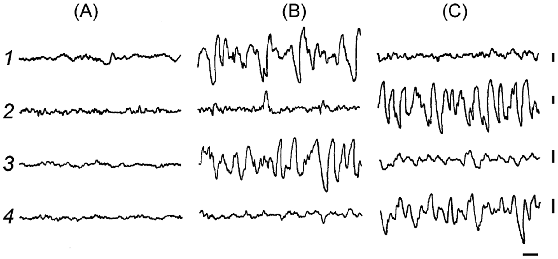
EEG recorded from two cortical hemispheres and two thalamus during waking (A), unihemispheric slow wave sleep in the right (B) and left (C) hemispheres in a bottlenose dolphin. Recording: 1—right cortex, 2—left cortex, 3—right thalamus and 4—left thalamus. Calibration 1 s and 200 μm (modified from Supin and Mukhametov, 1986).
Fig. 2.
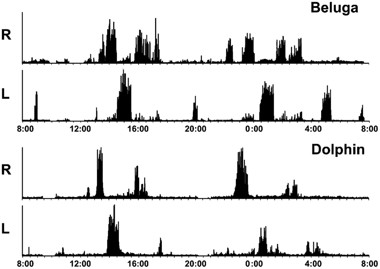
EEG slow wave power in the range of 1.2–4 Hz in the right (R) and left (L) hemispheres in a bottlenose dolphin and beluga recorded over 24 h.
3.2. REM sleep
3.2.1. Behavioral features of REM sleep
Despite many studies, no credible evidence of REM sleep in dolphins or any other EEG instrumented cetacean has been obtained. The absence of such evidence may result from the restraining conditions under which EEG recordings have been conducted and the insufficient periods of recording. However, muscle jerks and eyelid movements have been documented in behavioral observations in several cetacean species, including bottlenose dolphins (Flanigan, 1974a; Mukhametov and Lyamin, 1994, 1997), Amazon river dolphins (Oleksenko et al., 1996), belugas (Flanigan, 1974b, 1975c; Lyamin et al., 2002b), killer whales, Orcinus orca (Flanigan, 1975a; Lyamin et al., 2003), white-sided dolphins (Flanigan, 1975b; Nelson and Lein, 1994), Commerson’s dolphins (Shpak et al., in press) and in a 1-year old gray whale calf, Eschrichtius robustus (Lyamin et al., 2000, 2001). The quantitative data indicates that the number of muscle jerks and their intensity varies significantly between these species (Table 1).
Table 1.
Number of muscle jerks in cetaceans
| Cetacean species | Age | Number of jerks | Reference |
|---|---|---|---|
| Gray whale | One 1-year old calf | 45 over 6 days | Lyamin et al. (2000) |
| Bottlenose dolphin | Three adult males and one adult female | 15–98 per day | Mukhametov and Lyamin (1997) |
| Amazon River dolphin | Two adults | 26 over 3 days in female, a few/night in male | Oleksenko et al. (1996) |
| Beluga | One adult male | 144 ± 24, average over 6 nights | Lyamin et al. (2002a,b,c) |
| Killer whale | One 1–4 months old calf | <29 single and <10 serial per night | Lyamin and Siegel (unpublished data) |
| Killer whale | One adult female | 1–6 (2.4 average) per night over 4 nights | Lyamin and Siegel (unpublished data) |
| Commerson’s dolphin | Three adult males | 13, 31 and 35/3 days | Shpak et al. (in press) |
In spite of the quantitative species variability, there are several qualitative similarities: jerks are frequently related to resting behavior (floating or slow swimming); they occur more often at night and in the morning; some jerks follow each other (sometimes associated with eye movements) creating a series of jerks and twitches; a significant proportion of jerks (particularly serial jerks) are followed by arousal; and there is some indication for periodicity of jerks across nightly rest periods. Based on these characteristics, muscle jerks in cetaceans fit some behavioral features of REM sleep in terrestrial mammals. On the other hand, a significant proportion of these jerks occur in the beginning of rest periods and during waking, raising the question of whether they are actually indicative of REM sleep. The number of jerks varied considerably between individuals with, for example, one dolphin having 15 per day whereas another had 98 per day. Moreover, in humans and terrestrial mammals, muscle jerks cannot be considered a sufficient indicator of REM sleep as a variety of motor events are present throughout sleep, particularly during the transition from waking to sleep.
Penile erections are a feature of REM sleep in many species of terrestrial mammals. In bottlenose dolphins many erections (more than 80%) begin and end while hanging at the surface and occasionally during circular swimming (8–18%). The total number of erections varied in different dolphins from 2 to 31 per 24-h period and lasted between 4 and 102 s (Mukhametov and Lyamin, 1997). In one Commerson’s dolphin we documented 5 erections over a 3-day observation period, 4 of them occurring during slow swimming. On two occasions the erection coincided with jerks and once between two jerks separated by 4 min (Shpak et al., in press). The few erections that we documented in a killer whale calf born in 2001 at SeaWorld, San Diego, occurred during floating at the surface (Lyamin and Siegel, unpublished data). However, in all studied dolphins and killer whales erections also occur during active behavior. Therefore, without EEG recording, erections cannot be considered definitive features of REM sleep in cetaceans.
On the basis of existing data, one cannot exclude the possibility that REM sleep is absent in cetaceans. Another possibility to consider is whether REM sleep may take a form in dolphins that has escaped detection. Earlier we suggested that REM sleep might exist in cetaceans in a modified form without its typical features (muscle jerks, body twitches and rapid eye movements), or as very short episodes reminiscent of avian REM sleep (Mukhametov, 1995; Mukhametov et al., 1997; Lyamin et al., 2002b). Even if some muscle jerks represent short episodes of REM sleep in dolphins and whales, the total amount of REM sleep would be among the smallest of any previously studied mammal and/or is uniquely sensitive to disturbance and adaptation. Therefore, simultaneous EEG recording and visual observations need to be conducted and with correlated behavioral events (muscle jerks, erections, rapid eye movements) to draw a more definite conclusion about the presence or absence of REM sleep in cetaceans.
3.2.2. No polygraphic features of REM sleep
Shurley et al. (1969) reported seeing one REM sleep episode in one out of three non-consecutive nights of EEG recording and visual observations in a single pilot whale. This study is the only physiological report of REM sleep in cetaceans. The authors stated that: “REM sleep occurred exactly 60 min after sleep onset, for 6 min, with marked loss of tonus of trunk muscle, and one time only”.
Despite long periods of electrophysiological recording of sleep, no such episodes have been observed in any of the four physiologically studied cetacean species examined by Mukhametov (1984, 1995), Mukhametov et al. (1977, 1988), Lyamin et al. (2002a, 2004). In 1997 Mukhametov and colleagues listed the parameters they attempted to monitor in bottlenose dolphins to answer the question on the presence or absence of REM sleep in cetaceans. This list included the EEG of both cerebral hemispheres, both lateral geniculate bodies and both hippocampi, the electromyogram (EMG) of the nuchal and extraocular muscles, the electrocardiogram (ECG), respiration rate, brain temperature, and behavior. The nuchal musculature EMG could be very low and even briefly atonic during both bilateral unambiguous waking and USWS (Mukhametov and Supin, 1975); this may be because the antigravity musculature tone is not as highly expressed in cetaceans as in terrestrial mammals. The EOG voltage in dolphins was low and even when recorded was not helpful to identify eye movements that could be considered related to REM sleep. The eye state can be monitored via video cameras but continuous monitoring is possible only if the animals are limited in their swimming (i.e. restrained), as we did in our recent studies in one beluga whale and one bottlenose dolphin (Lyamin et al., 2002a,b, 2004). However, this procedure might disrupt REM sleep if present, unless a long period of adaptation is performed beforehand. There were successful implantations of EEG electrodes (based on evident evoked responses to light flashes) into the lateral geniculate bodies in two dolphins, however, no PGO spikes were recorded during normal waking, sleep or in response to reserpine injections (Mukhametov et al., 1997), and in the case of reserpine the dose was so high that it affected the normal behavior of the dolphin. In the cat reserpine administration induces PGO spikes similar to those in REM (e.g., Jouvet et al., 1965), but clear PGO spikes have never been reported in the dog and clear lateral geniculate PGO spikes have not been recorded in the rat, even though both species clearly have REM sleep; however, PGO spikes have been recorded in the rat locus coeruleus during REM sleep (Kaufman and Morrison, 1981). These data indicate that dolphins may have PGO spikes as rats do despite their absence in the geniculate nucleus. One attempt was made to implant two electrodes in the hippocampus but no theta activity was registered (Mukhametov et al., 1997), this result may be related to the small size of the hippocampus in cetaceans (Jacobs et al., 1979; Manger, 2006), making exacting implantation of electrodes difficult, and the small size perhaps not forming theta activity of large enough voltage for recording. The inter-breath interval in dolphins can last as long as several minutes (Mukhametov et al., 1997; Mukhametov and Lyamin, 1997), rendering irregularities in breathing not useful in identifying REM sleep in dolphins as it is in terrestrial mammals or pinnipeds (e.g., Lyamin et al., 1993, 2002c). ECG is easily recorded in dolphins, however the normally long but variable respiratory intervals with a normal respiratory cardiac arrhythmia complicates the detection of heart rate irregularities associated with REM sleep (Mukhametov et al., 1997). Complete immobility is rare in dolphins, with some animals swimming continuously 24 h per day. Muscular jerks were occasionally noted in implanted dolphins in studies done prior to 1998, but they were not correlated with other physiological changes associated with REM sleep in terrestrial mammals. Adaptation to the experimental conditions was another factor considered by Mukhametov and colleagues that may be related to the lack of observation of REM sleep in dolphins. However, no REM sleep was observed, even when the dolphins had been recorded up to 4 weeks continuously—these dolphins exhibited normal sleep patterns and were in good health. Based on these studies, Mukhametov et al. (1988, 1997) concluded that: (1) “although the negative results do not prove the absence of REM sleep in dolphins, it seems very likely that paradoxical sleep is absent in its classical form in adult bottlenose dolphins”; and (2) that REM sleep can be present in dolphins but in “a significantly modified form compared to that in terrestrial mammals which makes it difficult to identify”.
Mukhametov and colleagues looked for the most well known physiological and behavioral correlates of REM sleep traditionally used to identify this stage of sleep in terrestrial mammals. We would like to make two comments that we feel are of importance. First, the traditional criteria used to identify REM sleep are “designed” for “long” REM sleep episodes which last minutes but not seconds. In birds REM sleep episodes only last seconds and are not always accompanied by marked postural changes, making identification of REM sleep a difficult task in birds (Amlaner and Ball, 1994). It then follows that if dolphins have REM sleep in the form of short episodes it would be very difficult to detect by the traditional criteria. Second, the adaptation factor, or the time which the animals are given to become accustomed to the experimental environment prior to recording, is extremely important. Fur seals, which display “normal” amounts of REM sleep on land (3.5–6.2%; Lyamin and Mukhametov, 1998), may stay in the water with a minimal amount of REM sleep (0.2–1.2%) for as long as 45 days without showing any significant REM sleep rebound when they return to land (Lyamin et al., 1996). Therefore, an adaptation period of 2–3 weeks after the implantation of electrodes may not be enough time for dolphins to display REM sleep. However, if this explanation turns to be true, then the dolphins’ ability to go without REM sleep for several weeks is a remarkable feature of their sleep, as no other animals are capable of this.
3.3. Quantitative data
Quantitative data on the sleep parameters of studied cetaceans is provided in Table 2. Dolphins have slow waves in one hemisphere from 34% to 57% of 24 h. The smallest amount of sleep time is the average for six adult bottlenose dolphins. These data were collected from dolphins swimming in shallow pools with seawater (bottlenose dolphins and harbor porpoises) or fresh water (Amazonian river dolphins), or restricted in their movements (one beluga and one bottlenose dolphin), using the traditional tethered recording system with low noise cables. The longest total sleep time was shown by the youngest of the studied dolphins (Amazonian river dolphin), which was only a few weeks old at the time of recording (Mukhametov, 1987). However, in this study the recording started right after the surgery (to implant EEG electrodes) before which the dolphin was given diazepam, know to induce USWS (see below). The only studied beluga was restricted in its movement in order to videotape the state of eyes (Lyamin et al., 2002a, 2004). The data presented in Table 2 indicates that USWS is the main form of cetacean sleep irrespective of age, sex and experimental conditions. USWS represents from 70 to 90% of the total sleep time. The remaining time (10–30% of total sleep time) is occupied by episodes of highly asymmetrical but unambiguous bilateral SWS (high voltage SWS in one hemisphere and low voltage SWS in the other), and bilateral low amplitude SWS. On average, in six bottlenose dolphins, episodes of sleep in one hemisphere lasted 42 ± 10 min ranging between 3.5 and 131.5 min. The number of episodes in one hemisphere varied between 2 and 12 and averaged 5 ± 1 per day. In one beluga whale 26 episodes of unihemispheric and asymmetrical SWS were scored over 2 days of continuous recording. The lengths of the episodes varied between 10 and 81 min with an average of 44 ± 4 min. In Amazonian river dolphins, USWS episodes could be as long as 1 h (Mukhametov, 1987). In all cetaceans studied episodes of USWS tended to occur in the two hemispheres alternatively.
Table 2.
Characteristic of sleep in Cetaceans
| Bottlenose dolphina | Harbor porpoiseb | Amazon river dolphinc | Belugad | |
|---|---|---|---|---|
| Number of animals | 6 | 1 | 1 | 1 |
| Sex | 3 males, 3 females | Male | Male | Male |
| Age | Adults | 2 years | Calf (several weeks) | Adult |
| Amount of (% of 24-h) | ||||
| Total SWS timee | 33.9 | 46.4 | 57.0 | 43.0 |
| SWS left hemisphere | 20.4 | 28.6 | 31.0 | 25.5 |
| SWS in right hemisphere | 17.5 | 33.1 | 42.0 | 28.5 |
| Composition of SWS (% of TST) | ||||
| Low amplitude USWS | 51.0 | 48.5 | 35.1 | 46.8 |
| High amplitude USWS | 37.2 | 18.5 | 36.8 | 21.3 |
| Asymmetrical SWS | 4.4 | 10.6 | 14.0 | 14.7 |
| Low amplitude bilateral SWS | 7.4 | 22.4 | 14.0 | 17.0 |
| High amplitude bilateral SWS | 0.0 | 0.0 | 0.0 | 0.2 |
The total amount of SWS in all cetaceans was less than the amount of SWS in the left hemisphere and in the right hemisphere calculated separately because cetaceans have USWS and episodes of bilateral sleep with EEG asymmetry.
The average amount of time each cerebral hemisphere spends in sleep differs between individuals in any given 24 h period. For instance, in the bottlenose dolphins this difference was evident in all 24 h sessions and could be as much as 4-fold during a single day (Mukhametov, 1985; Mukhametov et al., 1977). This is very unlike terrestrial mammals, i.e. people do not get 2 h sleep one day then 8 h the next, unless sleep deprived. However, when the total sleep time and the duration of USWS in each hemisphere were averaged over several days, the difference in the amount of time each hemisphere is asleep becomes less significant. Therefore, it is likely that neither individual nor population based hemispheric lateralization in the amount of sleep is characteristic of bottlenose dolphins.
3.4. Topography of sleep
Simultaneous recordings from the dorsal part of the bottlenose dolphin cerebral cortex (parietal, occipital and occasionally frontal) showed some regional differences in the EEG from different locations. However, each dolphin hemisphere works as “a functionally independent unit”, meaning that the differences between two brain hemispheres were always more clearly expressed compared to the regional EEG differences within one hemisphere (Mukhametov et al., 1997). A similar situation was observed in harbor porpoises (Mukhametov and Polyakova, 1981). In a few dolphins, EEG was recorded from the two hemispheres and dorsal thalamic areas at the same time. As with the cortical EEG, the electrical activity in the thalamus could be subdivided into the same three stages (desynchronization, low and high amplitude slow waves). In all cases the EEG changes in the ipsilateral cortex and thalamus were parallel (Fig. 1). Based on these observations, it was hypothesized that USWS in dolphins is not only a cortical phenomenon, but appears to involve at a minimum the entire cortical–thalamic system (Supin et al., 1978).
3.5. Sleep and movement
3.5.1. Constant (continuous) swimming
Continuous swimming was found to be characteristic of several species of porpoises and small dolphins such as Dall’s porpoise, Phocoenoides dalli (McCormick, 1969), the Indian River dolphin, Platinista gangetica (Pilleri, 1979), the Spinner dolphin, Stenella longirostris (Norris and Dohl, 1980), the harbor porpoise (Mukhametov et al., 1981; Oleksenko and Lyamin, 1996), Commerson’s dolphin, Cephalorhynchus commersonii (Shpak et al., in press), and common dolphins, Delphinus spp. (Ridgway, unpublished observations). EEG was recorded in only one species of “continuously swimming dolphins”—the harbor porpoise (Mukhametov and Polyakova, 1981). This study showed that porpoises displayed unihemispheric slow waves while swimming (Table 2).
An interesting phenomenon is continuous swimming in bottlenose dolphin and killer whale neonates and their mothers (Lyamin et al., 2003, 2005a, 2006a, 2007). Although in aquaria adult bottlenose dolphins spend a significant portion of time floating (presumably sleeping at the surface) and killer whales (and other whales and dolphins) rest while floating and lying on the bottom of pools (Fig. 3), these behaviors were not observed in cetacean mothers and their calves for several months postpartum. As the calves aged, the time spent floating at the surface gradually increased both in mothers and calves. During this time mothers shorten the inter-breath interval compared to the mean adult values, however the calves frequently surfaced alone when the mother’s inter-breath intervals were longer than 1 min. During these instances the mother always continued swimming, forcing the neonate to track her movements and catch up. This behavior is unlikely to be compatible with sustained periods of sleep. While we do not know exactly what the physiological mechanisms triggering continuous swimming in cetacean mothers are, the ability to remain active and responsive after birth has several advantages for newborn cetaceans. Motor activity may reduce predation and may help to maintain body temperature until their body mass has increased and insulating blubber develops.
Fig. 3.
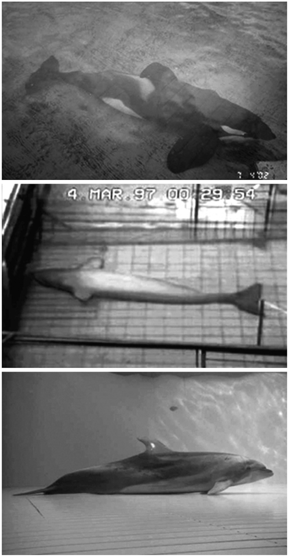
Rest on the bottom of pools in a captive killer whale (photo by O.I. Lyamin), beluga (from Lyamin et al., 2002b) and bottlenose dolphin (from Sekiguchi and Kohshima, 2003).
Continuous swimming is a unique feature and is compatible with the EEG slow waves that correlate with sleep in terrestrial mammals. Swimming in dolphins is necessary for coming to the surface for breathing and maintaining the body postures that are required to perform a series of respiratory acts. Swimming is also necessary to counteract water currents. This is particularly relevant for some river dolphins, which live in continuously moving water and must swim from birth. Small dolphins (e.g. porpoises) also swim continuously, 24-h per day, even while in aquaria in pools with still water, potentially reflecting an adaptation to life in the open sea, where even small waves may disrupt their posture and position. Large cetaceans can be minimally active at the surface when the ocean is calm, however they are also often observed slowly swimming in one direction (e.g., killer whales; Ford, 2002). It appears that USWS allows dolphins to accommodate aspects of sleep and swimming simultaneously. It seems logical to suggest that USWS enables continuous motion through activation of brain centres involved in controlling co-ordinated movement.
The postpartum absence of “rest” behavior occurs during a period when continuous long distance migration may occur in the wild, often for a period of weeks or months. The postpartum period is a time in which the neonate is most vulnerable to predation from conspecifics and sharks and therefore must stay alert, responsive and in proximity to its mother. These necessary characteristics of this state call into question the subsequent suggestion of Gnone et al. (2006) and Sekiguchi et al. (2006) that this behavior be called sleep.
In our work we have noted that eye opening in two cetacean species (bottlenose dolphin and beluga whale) is linked to low voltage EEG states in the contralateral hemisphere (Lyamin et al., 2004). In our report documenting continuous postpartum activity in cetaceans, we observed that the dolphin neonates and their mothers generally had both eyes open when they surfaced, which happened at a mean interval of less than one minute in the neonate (Lyamin et al., 2007), indicating bilateral EEG desynchrony. Brief periods of unihemispheric EEG synchrony may have occurred during swimming periods between breaths. However, such short periods of sleep are not restorative in humans (Bonnet, 1989) and sleep interruption at comparable intervals in the rat is lethal (Rechtschaffen and Bergmann, 2002). Thus the continuous postpartum activity seen in cetaceans differs radically from the sleep behavior in all land mammals, which have maximal sleep and inactivity in the postpartum period. These issues are discussed in greater detail elsewhere (Lyamin et al., 2006a, 2007; Siegel, 2008).
3.5.2. Rest while swimming slowly
In aquaria, stereotypic slow swimming (also called “resting swimming”) along the enclosure perimeter may occupy a significant portion of time in bottlenose dolphins (Lilly, 1964; Flanigan, 1974a; Marino and Stowe, 1997; Mukhametov and Lyamin, 1994, 1997; Gnone et al., 2001; Sekiguchi and Kohshima, 2003) and in other cetacean species (e.g., Flanigan, 1975a,b,c; Mukhametov, 1984; Goley, 1999; Shpak et al., in press). Most studies report the prevalence of a counter-clockwise swimming bias in bottlenose dolphins (e.g., Flanigan, 1974a; Ridgway, 1990; Sobel et al., 1994; Mukhametov et al., 1997; however, see also, Stafne and Manger, 2004 showing a clockwise bias in Southern hemisphere dolphins). In the wild, Hawaiian spinner dolphins (Stenella longirostris) and dusky dolphins (Lagenorhynchus obscurus) were observed slowly swimming and apparently sleeping for a portion of time in tight echelon formations in shallow waters (Norris and Dohl, 1980; Würsig and Würsig, 1980). Killer whales, in the ocean, rest while slowing swimming in tight formation, synchronizing their breathing and diving (Ford, 2002).
In earlier studies, no behavioral correlates of sleep were found in the bottlenose dolphin, Amazon River dolphin and harbour porpoise (Mukhametov, 1987; Mukhametov and Polyakova, 1981; Mukhametov et al., 1977). However, the parameters of dolphin behavior were recorded only visually. Our recent study (Shpak et al., in press) of behavior in Commerson’s dolphins (one of the smallest cetaceans) showed that these dolphins spent 96–99% of 24-h exhibiting circular swimming, but throughout the day swimming speed varied. The dolphins displayed obvious periods of slow swimming circling 3–5 times at the bottom of the pool during one breath interval and even literally stopped in the water column. Occasionally they assumed a “tail-up” position for several seconds or even turned completely over, resuming swimming in 2–5 s. It is likely that the described episodes of slow swimming represented the deepest form of rest or sleep in Commerson’s dolphins.
Gangetic dolphins (Platanista gangetica) were also observed to never stop moving in tanks, typically swimming on their sides and touching the floor with one flipper. Sound recording showed that the dolphins generated echolocation clicks during the entire day with interruptions lasting from 4 to 60 s (Pilleri, 1979). The total length of all these silent pauses was about 7 h over a 24-h period, which is within the range of “normal” sleep duration in mammals. The sound recording indicates that if sleep occurs, it must be greatly fragmented beyond what has been reported in any land mammal.
3.5.3. Rest while floating at the surface
Another common behavior in bottlenose dolphins, traditionally considered rest and sleep, is floating or hanging at the surface (McBride and Hebb, 1948; McCormick, 1969, 2007; Ridgway, 1972; Flanigan, 1974a). These authors observed that bottlenose dolphins float for considerable periods of time, remaining almost motionless at the surface except for periodic breaths and slow movements of the tail flukes. Spencer et al. (1967) observed similar floating behavior in killer whales and suggested that the behavior “may represent sleeping.” Mukhametov and Lyamin (1997) observed that bottlenose dolphins performed minimal movements with their flippers and tail fluke at this time; however, were not completely immobile for longer than a few seconds (see also McCormick, 1969; Flanigan, 1974a). One eye may be closed at this time (both eyes are closed simultaneously only rarely) (Flanigan, 1974a; Gnone et al., 2001; Sekiguchi and Kohshima, 2003). Killer whales can be completely immobile for about 1 h or even longer while floating at the surface (Lyamin et al., 2003). A gray whale calf also showed immobile hanging at the surface in a shallow channel (Lyamin et al., 2001). While resting it clasped both pectoral fins tightly to the body while the tail fluke touched the bottom of the pool and was motionless. These episodes occurred mostly at night and lasted from 3 to 98 min (Lyamin et al., 2001).
Floating at the surface is also characteristic of large whales in the wild (Gray, 1927). Some observations indicate that southern right whales and Mediterranean fin whales lie motionless at the surface (Cassini and Vila, 1990; Ricciardi et al., 2001). Sperm whales may alternate long dives with periods at the surface, especially in daylight (Watkins et al., 1999). Humpback whales frequently “log” at the surface at high latitudes in feeding grounds during calm water conditions. When they rest on the surface their respiratory rate is low and any motion other than that related to breathing is not observed. Occasionally it is possible to closely approach these whales with a boat without them moving; however, if they are alarmed they dive for a long time and emerge far from the place they rested showing evident indication of behavioral awakening (Robbins et al., 1998; P.J. Clapham and D.K. Mattila, personal communication). It is also interesting to note that the Pygmy and Dwarf Sperm whales, the two smallest species of sperm whales inhabiting tropical waters worldwide, are known to spend a lot of time lying motionless at the surface with the back and head exposed. In ancient Japan, these whales were called uki-kujira, or “floating whales” (Reeves et al., 2002).
Recently, Miller et al. (2008) observed sperm whale behavior at sea suggesting that the whales were asleep with both brain hemispheres unlike the unihemispheric sleep seen in dolphins and other whales mentioned above. The behavior that Miller et al. (2008) show is not new to the cetacean literature. Gray (1927) showed similar behavior in the Greenland whale (Balaena mysticetus). Further, Nishiwaki (1962) shows such resting behavior from areal observations of sperm whales (see his Fig. 2), though Nishiwaki does not claim the behavior represents sleep. Bottlenose dolphins and belugas that show floating “sleep behavior” as well as bottom “sleep behavior” (e.g., McCormick, 1969; Lyamin et al., 2002a,b; Sekiguchi and Kohshima, 2003) also show unihemispheric sleep by EEG measures. At the same time, they do not have high voltage bilateral SWS and have a very small amount of low voltage bilateral SWS (see Section 3.1). Miller et al. (2008) tried to claim that sperm whales were fundamentally different but the behavioral data they show cannot prove this, even eye closure data is not available from the Miller et al. study.
3.5.4. Rest and sleep on the pool bottom and at depth
Rest on the pool bottom (Fig. 3) has been described in captive bottlenose dolphins at various facilities (McCormick, 1969, 2007; Lyamin et al., 1999; Sekiguchi and Kohshima, 2003). The total duration of this behavior varies between 0.2 and 5.4% of 24-h (average 2.0 ± 1.2% of 24-h for 4 dolphins) and individual episodes never lasted longer than 4 min (average 1.7 ± 0.1, Lyamin et al., 1999). McCormick (1969) described “cat-napping on the bottom” with one or both eyes open or blinking alternately and the animal may be responsive to movement of observers or other animals. Sekiguchi and Kohshima (2003) documented this behavior in 14 adult dolphins (on average 4.1% of recording time) excluding a one year-old calf and considered this behavior as rest. Interestingly, Howard et al. (2006), have recently used the bispectral index and found unihemispheric changes in bottlenose dolphins resting quietly on a rubber pad out of the water. In the same out-of-water situation, diazepam induced unihemispheric slow waves were recorded with needle electrodes by Ridgway et al. (2006b).
In a recent study (Lyamin et al., 2005b) we recorded EEG from two bottlenose dolphins using portable digital recorders. This procedure allowed the dolphins to freely swim in a more spacious pool (9 m × 4 m × 1.3 m). The behavior of the dolphins was continuously videotaped and synchronized with the EEG over 4 and 6 continuous days. As with the previous studies, these two dolphins slept mostly during floating at the surface or slow swimming along the enclosure. However, for the first time we documented 4 episodes of USWS in one bottlenose dolphin while it lay quietly on the bottom of the pool, these episodes lasting from 150 to 212 s. Those episodes followed each other and were interrupted by arousals when the dolphin surfaced to breath. This data, collected with a digital recorder, along with the available behavioral data, indicates plasticity in the sleep behavior of dolphins, specifically their ability to display USWS while lying on the bottom in addition to that seen while slowly swimming or floating at the surface.
Lying on the pool bottom has also been observed in other cetacean species (Fig. 3). For instance, this behavior occupied on average 45% of a 24-h period in one beluga, with the longest single episode lasting about 12 min (Shpak et al., 2001; Lyamin et al., 2002b). Rest on the bottom occupied 13% of 24-h in a one-year-old gray whale calf (Lyamin et al., 2001). It is interesting, that this behavior was observed in the gray whale calf at the age of several weeks, shortly after it was caught and put in the pool after becoming separated from its mother (G. Sumich, personal communication). Lying on the bottom of the pool is also characteristic of all adult killer whales currently residing at SeaWorld, San Diego (Lyamin et al., 2003, 2005a). Single episodes of rest on the bottom for these killer whales lasted between 3 and 7 min. However, this behavior was not reported in two other killer whales observed at the Vancouver aquarium 30 years earlier (Flanigan, 1975a). There is some indication that belugas and bottlenose dolphins adopt this behavior from each other when they reside together (Lyamin et al., 2002b), but this question has not yet been studied. There is anecdotal evidence on the presence of lying on the sea bottom in wild whales (P. Clapham, personal communication). All this data indicates that rest on the bottom (and most likely sleep) is a common behavior both in aquaria and in wild large cetaceans. As mentioned above humpback whales “log” at the surface and perform long dives always emerging for breathing at the same place. Underwater observations indicate that they are frequently motionless during these long dives (Robbins et al., 1998; D. Mattila, P. Clapham, personal communication) suggesting that this may also be a resting behavior.
To conclude, sleep may occur in cetaceans during slow swimming, floating at the surface, or resting on the bottom of pools or shallow marine floors, exhibiting a degree of variance in sleep postures and behavior. But this is the case only for middle size and large size cetaceans, as porpoises and some small dolphins seem to be “obligate” swimmers and can sleep only while swimming (Lyamin and Siegel, 2006). This is likely due to thermoregulatory and buoyancy reasons (Pillay and Manger, 2004; Lyamin et al., 2004, 2005a) to be discussed later.
3.5.5. Correlation between EEG and movement
There are specific differences in the sleep behavior of different cetaceans. Bottlenose dolphins sleep mostly while swimming or floating at the surface (e.g., Mukhametov et al., 1977, 1997), while harbor porpoises (Mukhametov and Polyakova, 1981; Oleksenko and Lyamin, 1996) and Amazon River dolphins (Mukhametov, 1984; Oleksenko et al., 1996) never float (rest) at the surface. Harbor porpoises swim under the water with a speed of about 50 m/min 24 h/day and surface only for breathing, which is not necessarily accompanied by bilateral EEG arousal (desynchronized EEG) or any visually detectable reduction of the amplitude of the EEG in the synchronized hemisphere when they are asleep. Most bottlenose dolphins and all harbor porpoises studied at the Utrish Marine Station in Russia swam in a counter-clockwise direction (Sobel et al., 1994); however, the Commerson’s dolphins (Shpak et al., in press) and the Amazon River dolphins always circled in a clockwise direction (Oleksenko et al., 1996). This means that USWS occurs and alternates in the two hemispheres without any relationship with the direction of swimming. An observational study of 8 presumably sleeping dolphins in South Africa demonstrated that all dolphins swam in a predominantly clockwise direction during all slow swimming periods, strengthening the concept that USWS and swimming direction are not related to cerebral EEG states (Stafne and Manger, 2004), with swimming direction being related to some other as yet unexplained features of the cetacean habitat.
Visual observations on dolphins and porpoises did not reveal any correlation between the pattern of EEG in the two hemispheres and their motion. Waking can be detected visually only when the dolphins and porpoises were eating fish, swimming fast, playing with an object, or rubbing against the walls. When floating at the surface bottlenose dolphins can be awake or asleep, as they were almost never completely immobile for a period of time longer than several seconds, paddling their tail fluke and flippers to stabilize their posture, eliminating the ability to judge whether they were awake or asleep based on visual criteria alone (Mukhametov et al., 1997). All behavioral states (bilateral waking, low and high voltage USWS and BSWS) in harbor porpoises occurred during stereotypic counter-clockwise circular swimming (Mukhametov and Polyakova, 1981).
3.6. Unilateral eye closure/asymmetrical eye state
It was proposed by Lilly that dolphins sleep with one eye closed and one eye open to visually monitor the environment, and he attracted the attention of biologists and the general public to this interesting characteristic of dolphin behavior. The first electrophysiological studies (Serafetinides et al., 1972; Mukhametov et al., 1977; Mukhametov and Supin, 1975) confirmed that dolphins could indeed have one eye open during sleep without waking immediately; however, no correlation was found between the state of the eye and the pattern of EEG in either of the two hemispheres. Subsequent visual observations have confirmed that this behavior is common in bottlenose dolphins (e.g., Mukhametov and Lyamin, 1994, 1997; Gnone et al., 2001; Sekiguchi and Kohshima, 2003). Resting behavior with an asymmetrical eye state has also been documented in several other cetacean species, including the Amazon River dolphin (Oleksenko et al., 1996), white-sided dolphin (Goley, 1999), beluga whale (Lyamin et al., 2002b), gray whale (Lyamin et al., 2001) and killer whale (personal communication with trainers of SeaWorld in San Diego).
When presenting visual stimuli to the open eye while observing the behavioral response and any changes in the EEG of implanted dolphins, it was shown that a behavioral response may, or may not, be elicited, irrespective of the pattern of EEG in the hemisphere contralateral to the open eye (Supin et al., 1978). It was therefore concluded that: “unilateral eye closure and unilateral sleep in dolphins are two independent phenomena and there appears to be no link between these two processes” (Supin et al., 1978). However, after more data collection, Mukhametov et al. (1997) made “a preliminary conclusion” that the dolphin eye, contralateral to the hemisphere with the high voltage (delta) slow wave EEG, was usually closed.
Lilly (1964) proposed that the open eye may ensure the dolphin is always scanning the environment. However, the fact that one eye is open does not necessarily mean that the animal is capable of detecting changes in the environment. Mukhametov and Lyamin (1997) presented a contrasting black-and-white “chessboard” of squares during episodes of resting swimming to two dolphins. As one might expect, there was no behavioral response if the chessboard was presented in the visual field of the closed eye. On the contrary, when the chessboard was presented to a unilaterally opened eye, the behavioral response (such as startling, bursting swimming or orientation response) was always marked and rapid. This reaction was independent of the degree of closure/opening of the eye and was similar for the right and left eyes. This data indicates that the open eye in slowly swimming dolphins can perform a sentinel function.
Recently Goley (1999) studied sleep behavior in a group of four captive pacific white-sided dolphins. While resting, these animals swam in echelon usually with one eye open. When they switched positions, they also switched the state of eyes in such a way, that the open eye was always directed toward the schoolmates. Similar observations were made in bottlenose dolphins (Gnone et al., 2001). Lilly (1964) speculated that dolphins need to keep one eye open to scan the environment, but Goley’s data (1999) indicate that the open eye in dolphins allows them to maintain visual contact with group members.
As we have shown above, even some earlier electrophysiological data (Serafetinides et al., 1972; Mukhametov et al., 1997; see also Ridgway, 2002) suggested that a relationship between the EEG in the two hemispheres and the state of the eyes is present in cetaceans, but the nature of the eyelid position during these states had not been established. This was because of the difficulty in reliably documenting the position of the eyelids in swimming dolphins. Testing the suggestion of Serafetinides et al. (1972) that the EEG asymmetry was induced by the asymmetry in sensory input from the two eyes, Ridgway (2002) covered the dolphin’s eyes one at a time for 20 min intervals and found that merely eliminating visual input did not result in EEG changes in the opposite brain hemisphere (Ridgway, 2002). To document the relationship between sleep and eye state in cetaceans, we recorded EEG from both hemispheres and videotaped the state of the two eyes in a beluga and bottlenose dolphin while they were restricted in their movement (Lyamin et al., 2002a, 2004). Movement of the animals was limited by harnesses, so that the animals could swim in place for a distance of about 50 cm but not around the entire pool. This allowed us to observe on a monitor and videotape in real time the state of both eyes using several cameras. As shown in Fig. 4, polygraphic recording undoubtedly showed a clear relationship between the state of EEG in two hemispheres and the state of two eyes. In both animals during USWS and asymmetrical SWS, the eye contralateral to the more deeply sleeping hemisphere was usually closed (40% and 52% of the total sleep time in the contralateral hemisphere in the beluga, respectively for the left and right eyes; 55% and 60% in the dolphin) or in an intermediate state (46% and 31% in the beluga; 37% and 35% in the dolphin). Simultaneously, the eye contralateral to the waking hemisphere was open or in an intermediate state (95–98% of the time). However, regardless of the general association between the sleeping hemisphere and the state of two eyes in cetaceans, brief changes in the state of one eye were not necessarily accompanied by parallel changes of the EEG in the contralateral hemisphere in the studied dolphin and beluga. As shown in Fig. 4A, the beluga could briefly open the eye contralateral to the sleeping hemisphere for 10–40 s. These episodes were frequently accompanied by a decrease in the amplitude of slow waves in the sleeping hemisphere but did not lead to complete awakening. The next example, shown in Fig. 4B, illustrates that the eye contralateral to the sleeping hemisphere could open a minimum of 60 s prior to complete bilateral awakening. Therefore, while EEG and the state of the eyes were highly correlated over long time periods in this whale, EEG and eye position could be independent over short time periods.
Fig. 4.
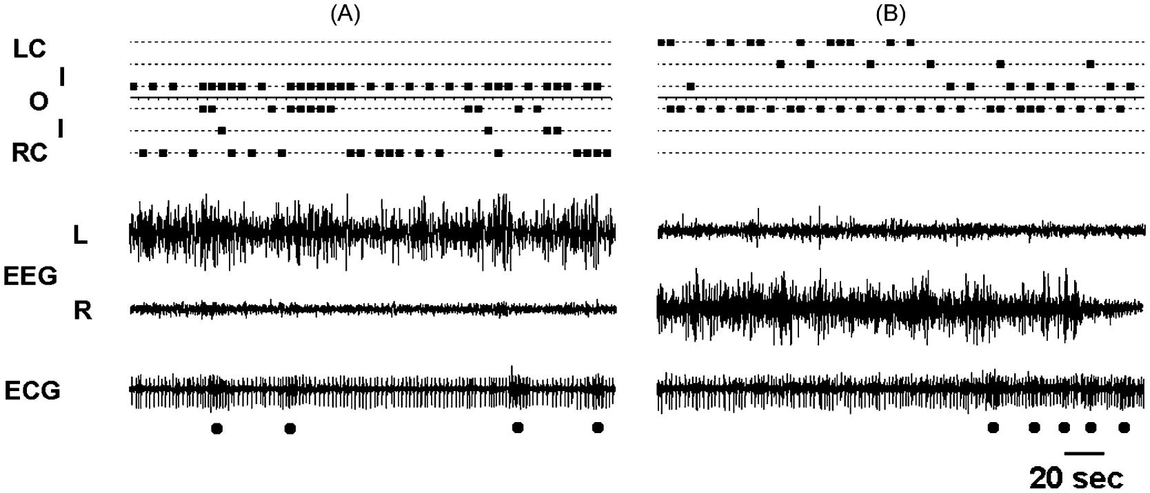
Relationship between eye state and EEG in a beluga. (A and B) Episodes of unihemispheric sleep in the left (L) and right (R) hemispheres, respectively, paralleled with continuous noting of the state of two eyes (L—left; R—right). The state of two eyes was scored as open (O), closed (C; LC—left closed; RC—right closed) and intermediate (I). ECG—electrocardiogram. Black dots below ECG mark breaths. Note: (1) that the eye contralateral to the waking hemisphere did not close during the entire episode of sleep in the opposite hemisphere, and that (2) slow wave EEG did not change immediately with opening of the opposite eye.
Further analysis (Fig. 5) indicated that the opening of two eyes in the dolphin and beluga was highly correlated with waking (79% of the time when the two eyes were open in the dolphin and 80% in the beluga). The epochs with only one eye closed while the other eye was open indicated sleep in 80% of the cases in the dolphin and 91% in the beluga. Moreover, 74% of these epochs in the dolphin and 80% in the beluga represented right or left side USWS or asymmetrical SWS. Unilateral eye closure in both animals was associated with up to about twice as much high voltage USWS and asymmetrical SWS (54% in the dolphin and 56% in the beluga) than low voltage USWS (20% and 24%, respectively; scoring of low and high voltage USWS and asymmetrical SWS is described in Lyamin et al., 2004). Both eyes were rarely closed in the dolphin and beluga (2.0% of the all recorded eye states in the dolphin and 1.6% in the beluga). Bilateral eye closure in the dolphin indicated low or high voltage USWS (75% of the bilateral closure time) in equal frequency while in the beluga this eye state mostly represented waking (49%) or low voltage USWS (32%). Three other combinations of the two eye states (open–intermediate, intermediate–intermediate, intermediate–closed) were not specifically related to any of the particular behavioral states we discuss here.
Fig. 5.
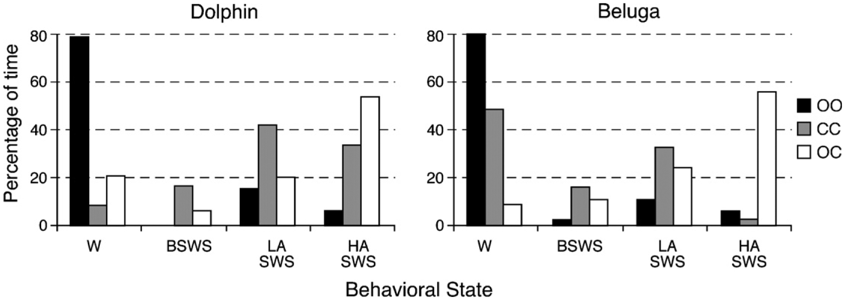
Association between eye state and behavioral stage in a bottlenose dolphin and in a beluga. The height of bars is proportional to the time spent in waking (W), bilateral slow wave sleep (BSWS), low and high asymmetrical amplitude slow wave sleep (LASWS and HASWS, respectively) as a percentage of the given eye state (OO—both eyes open, CC—both eyes closed and OC—one eye open and another eye closed).
While unilateral eye opening or closure is not always strictly associated with changes in the EEG of the contralateral hemisphere on a second by second basis, a clear relationship between these two phenomena occurs over longer time intervals. This should not be considered a contradiction to the link between the pattern of EEG and the state of the eyes in cetaceans. Rather, it provides us with a clue to the mechanisms underlying this association and to the “active” role of cortex in these events (it suggests that USWS is not a consequence of eye closure). The data also indicates that this association is substantial enough to roughly estimate sleep duration based on the condition of the two eyes. However, it does not allow reliable discrimination of waking from low voltage (light) USWS or bilateral symmetrical low voltage SWS on the basis of the eye state alone.
Maintaining visual contact during sleep is of great importance to cetacean mothers and their newborn calves. Only about 43% of killer whale calves survive the first year of their lives, because they are killed by sharks and aggressive killer whale males or because they lose their mothers and starve (Ford, 2002). Considering that killer whales are amongst the top predators in the ocean, maintenance of close contact would be of even greater importance for mothers and calves of other cetacean species. Studies of two captive bottlenose dolphin mother calf pairs showed that dolphin mothers surfaced predominantly with both eyes open (91–100% of the time; Fig. 6) regardless of time of day during the entire period of observations (Lyamin et al., 2005a, 2007). The calves also surfaced more often with both eyes open; however, the percentage was substantially less compared to that in their mothers (39–90% of the time, depending on the calf’s age). In the remaining cases the calves surfaced with one eye open while the other eye was in an intermediate state (on average, from 6–39% in two calves, depending on the age and time of day) or closed (on average 1.5–10% of the time). Moreover, the eye of the calf that was directed toward its mother was observed to be open more frequently (91–100% of the of the time observed) compared to the eye directed away from the mother (35–93% of the observations). Additionally, when bottlenose dolphin calves changed their positions next to their mothers, they always changed the state of their eyes (Fig. 6), as seen in adult white-side dolphins (Goley, 1999). Taking into account the association between eye state and unihemispheric sleep in dolphins and belugas (Lyamin et al., 2004), this would be a way to alternate sleep in the two hemispheres for the calves while circling in the same direction (counter-clockwise in this case). The available data now indicates that rest (and sleep) with one eye open is characteristic of many (if not all) cetacean species. The open eye does appear to have a sentinel function, specifically to allow the maintenance of visual contact with conspecifics.
Fig. 6.
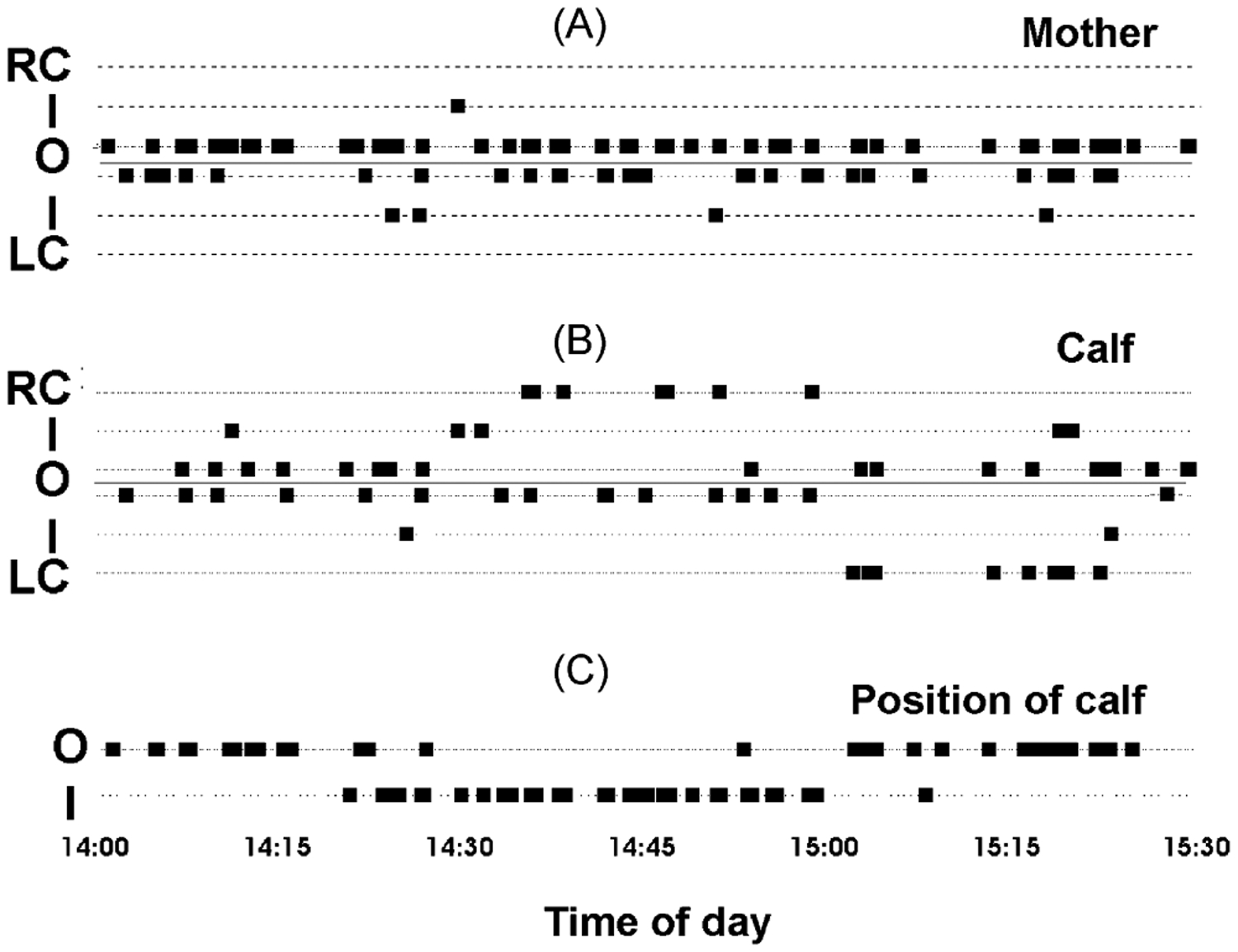
Simultaneous recording the state of two eyes in a bottlenose dolphin mother showing that calf’s open eye is generally directed at the mother (A) and calf (B) and the position of the calf (C) during swimming in a counter-clockwise direction. Each mark represents the state of visible eye (R—right, L—left; O—open, C—closed and I—intermediate) or the position of calf next to the mother (I—inner or O—outer) during echelon swimming.
3.7. Arousal thresholds
Moruzzi and Magoun (1949) demonstrated that high-frequency electrical stimulation of the brainstem lead to a diffuse widespread activation of the entire cortex. One dolphin was implanted with electrodes for electrical stimulation in the pontine tegmentum (Supin and Mukhametov, 1986). Arousal thresholds during high voltage USWS were measured in response to electrical stimulation (1 ms, 50 Hz, 5 s trains) of the site that was ipsilateral or contralateral to the “sleeping” hemisphere and was expressed as the voltage required to induce EEG arousal. These experiments showed that the intensity of stimuli required to induce bilateral EEG arousal was 1.4–1.7 times larger if high voltage USWS was present in the ipsilateral hemisphere compared to when USWS was present contralateral to the stimulation electrode. This data indicates a lateralization of the ascending activating brainstem systems in the dolphin related to arousal and wakefulness. No other EEG studies have been conducted to estimate arousal thresholds to sensory stimuli (e.g. acoustic or visual) during different types of SWS in cetaceans.
3.8. Brain temperature
Recording of brain temperature using implanted thermistors in parallel with cortical EEG in four dolphins and one harbor porpoise revealed two major types of fluctuations in the dolphin brain temperature (Kovalzon and Mukhametov, 1983). These temperature fluctuations were mainly similar to those observed in the rat (Kovalzon, 1973). The first type was usually seen in waking dolphins and characterized by slow shifts (up to 1 °C) occurring in both hemispheres simultaneously. These large temperature variations strongly correlated with changes in the level of activity and alertness (floating, swimming, eating, etc) of the dolphin. The second type appeared as more rapid smaller amplitude fluctuations (order of 10−1 to 10−2 °C) that correlated with changes in the amplitude of EEG. The transition from bilaterally desynchronized EEG (waking) to unilateral SWS was associated with smaller decrease in the temperature of the sleeping hemisphere (“by one or several tenths of a centigrade”; Kovalzon and Mukhametov, 1983), but in the hemisphere exhibiting waking EEG the temperature remained constant. Spontaneous, or evoked, awakening was accompanied by a gradual increase in the temperature (10−1 to 10−2 °C) of only the sleeping hemisphere. Thus, temperature fluctuations in dolphin brain were seen to occur unihemispherically.
3.9. BSWS and USWS deprivation
Bilateral and unilateral sleep deprivation in dolphins was performed for periods ranging from 35 to 119 h (Supin et al., 1978; Supin and Mukhametov, 1986; Oleksenko et al., 1992). Typically a one-day baseline recording was followed by a USWS (1–6 days), or bilateral SWS (1–3 days) deprivation period followed by a recovery day. Dolphins were awakened each time they entered USWS in the deprived hemisphere or in either of two hemispheres, respectively. This procedure effectively eliminated high voltage (or delta) sleep while the amount of low amplitude sleep remained unchanged. While bilateral sleep deprivation largely induced an increase in high voltage sleep in both hemispheres, unihemispheric deprivation, in most cases, led to rebound in “delta” sleep time in the deprived hemisphere only. Although rebound was evident in most dolphins, the response was highly variable, with some animals showing minimal recovery of lost sleep after almost 2 days of bilateral sleep deprivation. Moreover, there was no correlation between amount of time lost during bilateral or unilateral sleep deprivation and the amount of slow wave sleep in each hemisphere during the recovery period. This variability is quite unlike the effects of sleep deprivation reported in humans and other terrestrial mammals (Rechtschaffen and Bergmann, 2002; Rechtschaffen et al., 1989; Bonnet, 2000; Dinges et al., 2005). The rebound effect following USWS deprivation was maximally expressed during the first hours of recovery. The first allowed USWS episodes lasted for up to 3 h (Supin and Mukhametov, 1986), which is almost 1 h longer than that seen during baseline conditions. Bilateral and unilateral sleep deprivation significantly increased the tendency for USWS to alternate in two hemispheres in dolphins. Thus, under baseline conditions episodes of USWS in the two hemispheres alternated in 67% of cases while after bilateral and unilateral sleep deprivation the alternation increased on average to 87% and 80%, respectively. This study revealed that there is a vital need for sleep in each hemisphere in dolphins and the absence or deficit of sleep in one hemisphere cannot be substituted with sleep in the other hemisphere.
3.10. Pharmacological studies
The effect of certain drugs on sleep in dolphins has also been studied. Dolphins were shown to be very sensitive to sodium pentobarbital. Changes in the EEG (low amplitude bilateral SWS) became evident at a dose of 6 mg/kg (intramuscular) and occurred simultaneously in all sites recorded across the cortical mantle. At doses of 12 mg/kg and higher, the EEG was characterized by presence of high voltage, bilateral, slow waves. The increase in the amplitude of the EEG in the two hemispheres was accompanied by a cessation of breathing (Supin et al., 1978).
Diazepam (or valium, a benzodiazepine with CNS depressant properties) administered intramuscularly at doses ranging from 0.5 to 2.0 mg/kg rapidly induced USWS (Mukhametov et al., 1997; Ridgway et al., 2006b), which progressively became higher voltage and deeper. At higher doses (>2.0 mg/kg) USWS became bilateral high voltage SWS. It needs to be emphasized that the response to diazepam in bottlenose dolphins varied considerable between individuals, appeared to depend on the alertness of the animal, and the level of adaptation to the experimental conditions. For instance, in one case even a dose of 5 mg/kg did not induce EEG slow wave activity. A difference between pentobarbital and diazepam induced bilateral high voltage SWS, was that dolphins were still capable of breathing by themselves when administered diazepam. However, it was noted that immediately prior to each breath the amplitude of the EEG in one or both hemispheres decreased, such as that the dolphin exhibited either USWS, low amplitude bilateral SWS, or bilateral waking (Mukhametov et al., 1997). These observations indicated a marked difference between bilateral SWS induced by barbiturates and benzodiazepines, however both had an inhibitory effect on respiration in dolphins.
Based on these two experiments the conclusion was made that autonomous breathing in dolphins is not compatible with bilateral high voltage sleep and that the “active state of at least one hemisphere” is required. This may be due to the complexity of movements and reflexes required to perform each breath. According to Mukhametov et al. (1997), this observation may also explain the absence of REM sleep in cetaceans in its traditional form, as a loss of muscle tone typically coincides with REM sleep in terrestrial mammals.
McCormick (1969, 2007) observed that a small dose of a neuroleptic tranquilizer trifluromeprazine (a phenothiazine derivative) resulted in bottlenose dolphins resting virtually immobile at the surface of the water with both eyes closed and the dolphin being insensitive to light or acoustic stimuli. McCormick suggested that the animal was in bihemispheric sleep however no electrophysiological observations were possible. Body and brain temperature changes may have been key to the behavior observed. Phenothiazine derived tranquilizers are dopamine antagonists. With dolphins in water, these tranquilizers cause a drop in body temperature that may be progressive and life threatening (Ridgway, 1972). In one illustrative case, a dolphin in water of about 12 °C lost control of breathing, 35 min after an injection of 20 mg of Acepromazine (a phenothiazine derivative). Deep rectal temperature at this time was 28 °C. An endotracheal tube was inserted and the animal placed on a respirator and warmed for 3 h as body temperature gradually climbed to 37.2 °C when the dolphin began to breathe without assistance and was returned to its pool (Ridgway, 1972). It is possible that the tranquilization observed by (McCormick, 1969, 2007) caused a less extreme reduction in body and brain temperature resulting in a bilateral sleep state. Such a state could likely be reversed with dopamine injections.
3.11. Distribution of sleep across the day and nighttime
Visual observations revealed a circadian rhythmicity in captive bottlenose dolphins (Mukhametov and Lyamin, 1997; Sekiguchi and Kohshima, 2003). The rest stage was characterized by a bimodal pattern and present mostly at night and, less frequently, afternoon. A similar distribution of sleep time was observed in captive bottlenose dolphins in a quite different situation during electrophysiological experiments (Mukhametov et al., 1988, 1997). Feeding of dolphins in the first half of the day and in the evening, and human activity in the facility certainly affected the final 24-h distribution of behavioral states. However, what is interesting is that the hourly amount of sleep began progressively declining after 3 a.m., when there was no disturbance to dolphins (Mukhametov et al., 1988). Our recent data obtained from two bottlenose dolphins instrumented with digital recorders and freely swimming in more spacious pools (compared to the earlier experiments) indicates that periods of sleep in dolphins may be equally present both during the night and daytime (Lyamin et al., 2005b). In the wild in some locations bottlenose dolphins were found to be most active (displaying acoustic activity, feeding, socializing, or travel) in the early morning and late afternoon (Norris and Dohl, 1980; Saayman et al., 1973) and less active at night (Mate et al., 1995). In other places dolphins are active at night as well as during the day or even more active (mostly feeding and social behavior, but not traveling) during nocturnal periods (Shane et al., 1986; Klinowska, 1986). Therefore, it is quite reasonable to suggest that a bimodal distribution of the rest state (or sleep in electrophysiological experiments) with two maximums of activity at night and in the afternoon is distinctive of dolphins when the animals are not disturbed. In these cases dolphin display an afternoon rest/sleep period. In the wild, the circadian activity of bottlenose dolphins appears to be defined by many external factors, first of all availability of prey fish, seasonal and diurnal migrations, tidal currents, weather conditions, etc. (e.g., Klinowska, 1986; Shane et al., 1986; Bearzi and Politi, 1999). Dolphins were shown to be able to maintain continuous auditory vigilance for five days without any signs of sleep deprivation (Ridgway et al., 2006a). During the studies there was a distinct diurnal rhythm in response time to auditory target stimuli with longer responses occurring during nighttime hours.
4. Anatomy and physiology of the mammalian somnogenic system: a comparison with cetaceans
4.1. Neuronal groups involved in the control of the sleep–wake cycle
Within the mammalian brain there are several neuronal groups that have been implicated in the control of the sleep–wake cycle, and many of these have been identified anatomically such that they can be recognized with immunohistochemical stains. These neuronal groups are found in two broad regions, the first comprising those neurons in the basal forebrain and hypothalamus, the second comprising those groups located in a cluster at the ponto-medullary junction of the brainstem. Many of these neuronal groups project broadly throughout the brain, but some of these groups have very specific innervation patterns of importance to the regulation of the sleep–wake cycle. In terms of the anatomical structure of the brain, it can be seen that all these groups are found in the portion of the brain that develops from the embryonic basal plate, thus they are located in the ventral portion of the brain, inferior to the hypothalamic sulcus–cerebral aqueduct–sulcus limitans–central canal of spinal cord continuum that differentiates the adult derivatives of the basal and alar plates found in neural development (Puelles, 2001). The second basic anatomical feature of interest for these neuronal groups is that they appear to be restricted in the location of the neuronal bodies to the identifiable neuromeric segments of the developing brain. Those neurons in the basal forebrain and hypothalamus correspond to the adult derivative of prosomeric segments 3–6, and those in the ponto-medullary junction to the isthmic neuromere and rhombomeres 1–3 (Puelles, 2001). These two groups, distinct in the adult brain, are also separated in the developing brain by the mesencephalic neuromere and prosomeres 1 and 2, indicating a telencephalic origin for the rostral group of sleep–wake regulating neurons and a brainstem origin for the caudal group. This segmental organization is of importance in understanding the comparative aspects of these neuronal groups across species, both in determining homologies and in determining specific differences in structural organization that may lead to differing functional expressions of sleep patterns.
4.1.1. GABAergic neurons of the basal forebrain and anterior hypothalamus
The GABAergic sleep active neurons in the basal forebrain and anterior hypothalamus of mammals are unusual in that many are maximally active during non-REM sleep, but are less active during REM sleep and minimally active in waking, and are the most potent sleep-promoting neurons in the brain (Szymusiak, 1995; Szymusiak et al., 2001; Siegel, 2004). Many of these neurons also contain galanin. These neurons increase their discharge rate at sleep onset and continue to release GABA during sleep, thereby inhibiting the neurons involved in arousal, e.g. direct or indirect inhibition of the cholinergic neurons of the basal forebrain and histaminergic neurons of the posterior hypothalamus to deactivate the cerebral cortex. The non-REM on GABAergic neurons in the preoptic region of the hypothalamus are also thermosensitive and increase their discharge rate when warmed.
Sleep active GABAergic neurons in the basal forebrain and anterior hypothalamus of the cetacean brain have not been identified, but this region of the cetacean brain does not differ dramatically in comparison to other mammals, thus they are likely to be found if looked for. The probable GABAergic neurons of this region in the cetacean brain may be of great interest in understanding the mechanisms of USWS. Firstly, it appears likely that the discharge of these neurons in each hemisphere will be complementary, i.e. those on one side of the brain will be maximally active during SWS in that hemisphere, while those on the other side will be less active. This asynchronous activation would promote the hemispheric asynchrony of the EEG during SWS. The preoptic neurons that are thermosensitive are also of interest in terms of cetacean sleep. For example, if one hemisphere of the cetacean brain was slightly warmer than the other hemisphere after a day of being awake, it is possible that this may trigger that hemisphere to be the first to go into SWS. This possibility might be tested during recording of EEG by over-stimulating one hemisphere of the cetacean brain throughout the day and recording which hemisphere went into SWS first (see description of USWS deprivation above).
Another interesting possibility is that these neurons may act as a trigger for the switching of hemispheric EEG activity, as the temperature of the brain remains constant in the awake hemisphere, but drops in the sleeping hemisphere during USWS (see above). It is conceivable that the longer a hemisphere is awake, the greater the temperature in this hemisphere, even if this is only by a very small amount, and this increased temperature may initiate the activity of the non-REM on cells and thus induce SWS in that hemisphere. The other hemisphere, having been asleep for some time, and thus having cooled down (see above), would have a decreased discharge rate of these neurons, leading to cortical arousal. The presence, absence, or atypical presence of these neurons could be determined using immunohistochemical techniques on appropriately fixed tissue, providing insights into the nature of the mechanisms controlling USWS in cetaceans.
Studies of diazepam induced USWS (Ridgway et al., 2006b) using single photon emission tomography (SPECT) and positron emission tomography (PET) have suggested that cerebral blood flow reduction may be a controlling factor in the temperature reduction observed by Kovalzon and Mukhametov (1983) during USWS. Brain temperature may be influenced by three factors: (1) the temperature of blood flowing to the brain; (2) the rate of cerebral blood flow; and (3) the metabolic heat production of neurons and glia. Reduced cerebral blood flow and therefore reduced glucose supply likely will affect regional brain temperature and metabolic heat production. Furthermore, these factors may impact GABAA receptor sensitivity to diazepam such that a reciprocal effect between the hemispheres could be created so that the active or “non-sleeping” hemisphere would have a raised threshold for sleep until receptor sensitivity changes (Ridgway et al., 2006b).
4.1.2. Cholinergic neurons of the basal forebrain and anterior hypothalamus
Within the basal forebrain and the hypothalamus are several cholinergic neuronal groups. These include the islands of Calleja, the olfactory tubercle, nucleus accumbens, nucleus basalis, the diagonal band of Broca, and those in the antero-lateral, dorsal and ventral parts of the hypothalamus (Woolf, 1991; Maseko et al., 2007). These cholinergic neurons embody one of the main arousal systems of the forebrain, and their widespread projections through the forebrain, releasing acetylcholine upon discharge, activates the cerebral cortex and other structures, and contributes to the desynchronized EEG of waking and REM sleep. During SWS these neurons are inhibited by the GABAergic neurons in the vicinity (see above).
These cholinergic groups have been identified in all mammals studied (Maseko et al., 2007), but they have not been investigated in the cetacean brain. Complete descriptions of the anatomy of the basal forebrain of cetaceans appear to be lacking in the literature, except for the brief mention of some nuclear groups, but these have only been studied with Nissl stain, making the identification tenuous, but similar to other mammals. The hypothalamus is readily identified, but due to the small size of the fornix, it is difficult in Nissl stained sections to demarcate hypothalamic regions in the cetaceans. The hypothalamus appears small, relative to the size of the dorsal thalamus (Kruger, 1966), but does have a typical mammalian appearance, apart perhaps from a reduced olfactory component.
The projections of these neurons in the typical mammal would be expected to be primarily ipsilateral, but there might be a component that crosses the midline, perhaps through the anterior commissure, to create hemispheric EEG coherence during wake and REM sleep. It would be of interest to examine the extent of any potential commissural pathway of these cholinergic neurons in bihemispheric sleeping mammals, and then compare this with cetaceans. Given the small size of the anterior commissure in cetaceans (see below), it might be predicted that the cetaceans would lack any commissural projections of the cholinergic system of the basal forebrain, this lack potentially aiding hemispheric independence in the arousal of the cerebral cortex as seen in USWS.
4.1.3. Hypocretin (or orexin) containing neurons of the middle hypothalamus
The hypocretin containing neuronal group is found in the middle of the hypothalamus, situated between the sleep-active neurons of the anterior hypothalamus and the wake-active histamine neurons of the posterior hypothalamus in the mammals studied to date (Gerashchenko and Shiromani, 2004). These neurons project strongly upon many of the other arousal systems of the brain, including specifically the histaminergic, cholinergic, noradrenergic and serotonergic neurons, and appear to have a driving effect on these systems (Gerashchenko and Shiromani, 2004). This neuronal group is also involved in the facilitation of muscle tone via the projections to the brainstem (Kiyashchenko et al., 2002; Lai and Siegel, 1990; Peyron et al., 1998). Our own unpublished immunohistochemical studies have revealed the presence of hypocretin neurons in the hypothalamus of the bottlenose dolphin and they appear similar to those seen in other mammals. Further studies, for example quantification, may reveal specific differences in this neuronal population that may relate to cetacean USWS.
4.1.4. Histaminergic neurons of the posterior hypothalamus
Those neurons that produce histamine are localized to the posterior hypothalamus in mammals and project widely throughout the CNS. It has been demonstrated that these histaminergic neurons are involved in promoting wakefulness, and inhibition of the activity of these neurons by the GABAergic neurons of the basal forebrain promotes sleepiness (John et al., 2004; Saper et al., 2001). It has also been demonstrated that the tonic activity of these neurons with those of the noradrenergic and serotonergic systems combines with the activity of the cholinergic system to maintain the arousal of the forebrain and EEG desynchrony. Histaminergic neurons have not yet been identified in the cetacean brain. Again, due to the lateralized nature of sleep in cetaceans, there may be certain anatomical differences in the projections and connections of the histaminergic neurons, related to the lateralized arousal. One would suspect that during USWS, those histaminergic neurons in the hemisphere in SWS would be discharging at a very low rate—potentially inhibited by the GABAergic neurons of the basal forebrain, while those in the awake hemisphere would be discharging tonically to promote wakefulness and EEG desynchrony. Immunohistochemical identification of this neural group in cetaceans, and any specific differences that may be found, would be an important aspect of understanding the lateralized cetacean sleep phenomenology.
4.1.5. Cholinergic neurons of the midbrain and dorsal pons
The cholinergic neurons related to the sleep–wake cycle in this region of the brain are found in the lateral dorsal tegmental nucleus (LDT) and the pedunculopontine tegmental nucleus (PPN), LDT being found within the periaqueductal gray matter, and PPN in the adjacent pontine tegmentum (Woolf, 1991; Maseko et al., 2007). These neurons are active during wake and REM sleep, some discharging tonically in these states, and all discharging minimally during SWS (Siegel, 2000). These neurons are thus involved in the production of the desynchronized EEG of waking and REM sleep (Steriade et al., 1990). This is achieved by the release of acetylcholine in the diencephalon, blocking the oscillatory discharge of the thalamic mechanisms responsible for EEG spindles and slow waves (McCormick, 1989; Steriade et al., 1993). A bursting discharge of a subgroup of these neurons is involved in the generation of the pontine-geniculate-occipital (PGO) spikes characteristic of REM sleep in many mammals (Steriade et al., 1990). Some of these neurons are selectively active in REM sleep and are involved in the suppression of muscle tone during this state (Sakai and Koyama, 1996), presumably preventing the acting out of dreams.
This cholinergic population has not yet been identified in the cetacean brain. Given that the cetaceans appear to lack, or have minimal REM sleep, it is possible that these neurons could be less in number than one might expect given the size of the cetacean brain. It would be reasonable to think that those cholinergic neurons involved in suppression of movement during REM sleep in other mammals may actually be absent, or greatly reduced in number in the cetaceans. These neurons may also play a role in the level of arousal in the different hemispheres during USWS, as potentially enlarged commissural pathways may exist. These neurons, which lie caudal to the posterior commissure (see below) are ideally located to control global EEG patterns in the sleeping cetacean through projections to the diencephalon, and thus it would be of great interest to identify them immunohistochemically, and trace their major projections.
4.1.6. GABAergic neurons of the midbrain and dorsal pons
GABAergic neurons are found throughout the tegmentum of the midbrain and dorsal pons. It has been seen that some of these GABAergic neurons are involved in the inhibition of the activity of the serotonergic and noradrenergic neuronal populations in the brainstem (Nitz and Siegel, 1997a,b). These GABAergic neurons have not been identified in the cetacean brain, but are of interest in terms of the possible lack of REM sleep.
4.1.7. Noradrenergic neurons of the locus coeruleus complex
The locus coeruleus complex in mammals is found at the level of the pons, and is readily demarcated with immunohistochemistry using tyrosine hydroxylase (e.g. Manger et al., 2003; Maseko et al., 2007). These noradrenalin-producing neurons are for the most part inactive during REM sleep and it has been suggested that the inactivity of these neurons during REM sleep is related to the loss of muscle tone (John et al., 2004; Siegel et al., 1991; Lai et al., 2001). During waking these neurons discharge regularly and tonically, slowing in discharge rate during the initial phases of SWS and becoming silent in REM sleep (Siegel, 2000).
The locus coeruleus complex of the bottlenose dolphin has been described in detail (Manger et al., 2003). Briefly, the subdivisions of this complex that were found include the A6 dorsal subdivision, or locus coeruleus proper, located within the periaqueductal gray matter of the pons, and A6 ventral (locus coeruleus alpha), A7 (subcoeruleus), and A5 (fifth arcuate nucleus) subdivisions located within the dorsal pontine tegmentum. The distribution, nuclear subdivision and cellular morphology of this complex does not indicate any specific specializations that may have been anticipated due to the large size or lateralized sleep phenomenology of the cetacean brain. However, given the role of the locus coeruleus in the control of muscle tone, and the relationship of the discharge of these neurons to sleep states in other mammals, it was postulated that the neurons of the locus coeruleus would be continuously discharging in the cetaceans (Manger et al., 2003), though the frequency of discharge may differ between the left and right side of the brain (Ridgway et al., 2006b). This continuous discharge would enable the maintenance of muscle tone for the sleeping cetacean, which would be necessary given that cetaceans are often active during sleep.
A second potential effect of the continuous release of noradrenalin into the cetacean central nervous system would be a continuous stimulation of glia to increase their metabolic rate (Magistretti et al., 1993; Pellerin et al., 1997; Subbarao and Hertz, 1990). Cetaceans have a high glia:neuron ratio, thus in combination with the comparative excess of noradrenalin that we hypothesize, the metabolic activity of cetacean glia could be far higher than that of other mammals. One effect of this potentially increased glial metabolism would be production of heat (Manger, 2006). This proposed mechanism is supported by measures of the brain temperature during sleep. It has been seen (see above) that the sleeping hemisphere cools during the SWS period whereas the temperature of the awake hemisphere remains constant. It is possible that the neurons of the locus coeruleus ipsilateral to the sleeping hemisphere will slow their discharge rate during USWS, thus lessening the amount of noradrenalin stimulating glial metabolism in that hemisphere, lowering the temperature. Upon awakening, the temperature of the hemisphere increases, this potentially being associated with an increased, or waking, discharge rate of locus coeruleus neurons. This proposed hemispheric mismatch in the rate of discharge of locus coeruleus neurons has gained recent experimental support through observations with functional imaging of sleeping dolphins (Ridgway et al., 2006b).
A final point of interest relates to the decussation of the ascending locus coeruleus projections. It has been seen that a dense fascicle of tyrosine hydroxylase immunopositive axons, presumably arising from the locus coeruleus, cross the midline through the posterior commissure (Manger et al., 2004; see below). It is possible this ascending decussation is in some way related to the unihemispheric nature of SWS in the cetacean, but it is difficult to determine in what manner.
4.1.8. Serotonergic neurons of the brainstem raphe
The serotonergic neurons of the brainstem of most mammals can be divided into a midbrain/pontine group, commonly called the rostral cluster and a medullary group commonly referred to as the caudal cluster (Bjarkam et al., 1997; Maseko et al., 2007). The rostral cluster is composed of the dorsal raphe (made up of several subdivisions located in the pontine periaqueductal gray matter) the caudal linear nucleus, the median raphe nucleus and the B9 (no anatomical nomenclature) subdivisions located throughout the pontine tegmentum in a pararaphe position. This rostral cluster sends projections to the telencephalic hemispheres (Törk, 1990). The caudal cluster is composed of three pararaphe nuclei, the raphe obscurus nucleus, raphe magnus nucleus, and the raphe pallidus nucleus, all found within the medullary tegmentum and sending projections to the spinal cord (Törk, 1990). These neurons exhibit a similar discharge pattern in relation to the sleep–wake cycle as the neurons of the locus coeruleus, i.e. tonically active during wake, slowing during SWS and near minimal discharge during REM sleep (Siegel, 2004). The pontine, or rostral cluster, of serotonergic neurons have been implicated in gating, inhibiting or disinhibiting aspects of REM sleep, specifically in the regulation of PGO spikes (Siegel, 2000). The serotonergic neurons may also play a role in the maintenance of arousal, in regulating muscle tone, and suppression of phasic events during waking (Wu et al., 2004).
Serotonergic neurons have not been identified in the cetacean brain, but it is likely that immunohistochemistry would reveal this neuronal population as they have been found in all mammals studied to date (Maseko et al., 2007). The morphology, number and projection pathways of these would be of interest in terms of cetacean sleep. Given their role in the regulation of muscle tone it can be surmised that those groups projecting to the spinal cord, the nuclei of the caudal cluster, may be increased in neuron number. Also, the lack of REM sleep in the cetaceans might indicate the specific loss of a particular group of serotonergic neurons related to the control of phasic events during REM sleep. Alternatively, it may be that brief inactivity of these neurons underlies the phasic muscular events in cetacean sleep such as the jerks observed that have been theoretically linked to the muscle twitches of REM sleep (e.g. Mukhametov et al., 1997; Lyamin et al., 2002b).
4.1.9. Summary and future directions
Very little is actually known about the anatomy or the physiology of the neuronal groups involved in the control of sleep in the cetacean brain, with only the locus coeruleus complex having been identified at present (Manger et al., 2003). Importantly, many of these neuronal groups are readily identifiable using immunohistochemical procedures. The ability to identify, quantify, and trace the major projection pathways of these neuronal groups using post-mortem tissue and immunohistochemical techniques should lead to a greater understanding of the neural basis of cetacean sleep control and regulation, and may provide clues to the function and evolution of this unusual sleep phenomenology, as well as provide important clues regarding the neural basis of sleep control in bi-hemispheric sleeping mammals. Of great interest is the relationship of these neuronal groups to the major commissures of the brain (see below) and to each other in comparison to known bihemispheric sleepers. Undertaking physiological recording of these neurons during the sleep–wake cycle of the cetacean is presently not possible, making the anatomical identification of these neuronal groups from post-mortem tissue all the more important. It is likely that these neuronal groups will be very similar to those seen in other mammals, and thus, very accurate quantification across mammalian species and pathway tracing in the cetaceans is crucial to our developing understanding of cetacean sleep. Such differences as the total neuronal number relative to brain size, and the number of decussating fibres are likely to provide crucial hints.
Another aspect of the identification of these neurons relates to the potential identification of the neurons that trigger sleep phases. While it is known that the neuronal groups described above play important roles in the control, timing and regulation of mammalian sleep, it is still presently unknown if any of these groups of neurons are the “executive” controller of sleep, wake, SWS, or REM sleep onset. It is probable that at least a subset of these neurons have internal mechanisms (such as clock genes involved in the control of circadian rhythm) responsive to the specific needs of sleep (whatever they may actually be). Once stimulated by this need for sleep, or indeed wake, these mechanisms may be activated, changing the discharge rate of this subset of neurons, and thus initiating a cascade of neuronal events in these associated neurons causing a change in the global state of the brain. If such intracellular mechanisms are found in bihemispheric sleepers, and if these are found in a specific subset of neurons that are immunohistochemically identifiable, this information could be readily transferred, through anatomical investigation, to the unihemispheric sleeping cetacean to gain a better insight into the mechanisms of cetacean sleep phenomenology. Alternatively, if anatomical differences exist between the cetaceans and other mammals in certain “sleep-related” cell groups, but not others, these differences may yield important clues in our understanding of sleep generation mechanisms.
4.2. Commissures
Due to the lateralized nature of sleep phenomenology in the cetaceans, it is of interest to examine what is known of the commissural systems of the central nervous system, how these may relate to cetacean sleep, and how they relate to sleep in other, presumably bihemispheric sleeping mammals. The commissural fibers of the central nervous system allow for communication, transfer, and coherent processing of information to occur between the left and right sides of the brain. There are many commissures found in the brains of mammals, the three major ones concerned with sleep include the anterior commissure, corpus callosum and posterior commissure, as these are involved in either interhemispheric communication or interhemispheric coherence.
4.2.1. The anterior commissure
This commissure, in general mammalian terms, is composed of fibers that connect the olfactory bulbs, the olfactory tuberculum, the hippocampus, the amygdala (direct projections and those passing through the stria terminalis), and a significant portion of the cerebral cortex of the temporal lobe. In eutherian (placental) mammals this commissure is not large in comparison to that seen in non-eutherian mammals and non-mammalian vertebrates (that lack a corpus callosum), but its size is still significant and visible in gross dissection. The anterior commissure is rarely mentioned in descriptions of the cetacean central nervous system, and when mentioned is described as small (e.g. Wilson, 1933), or “incipient” (Buhl and Oelschläger, 1988). The small size of this commissure is perhaps not surprising, given the lack of olfactory bulbs in the odontocetes, and very small olfactory bulbs in mysticetes, the small olfactory tuberculum, the small hippocampus and the small temporal lobe in all cetacean species studied to date (see Manger, 2006, for review). In this sense, any information transfer or hemispheric coherence achieved by the anterior commissure in most mammals may not be strongly supported in the brain of the cetaceans. The reduced size of the anterior commissure in cetaceans may thus contribute to hemispheric independence.
4.2.2. The corpus callosum
Stryker and Antonini (2001) state: “Perhaps because of its prominence in midline sections of the human brain, the corpus callosum, one of the few structures identifiable by even the slowest student, has attracted wide interest throughout history”. The corpus callosum is the largest of the commissural pathways in eutherian mammals, and the study of its anatomy and function has led to an extensive literature (e.g. Innocenti, 1986; Jeeves, 1994; Gazzaniga, 2000; Olivares et al., 2001). The most comprehensive study of this structure in cetaceans was undertaken by Tarpley and Ridgway (1994), where it was reported that the corpus callosum in the Delphinidae had a smaller relative size compared to brain mass in comparison other eutherian mammals. Furthermore, the size of the corpus callosum increased more slowly than increases in cetacean brain size, and there were no apparent sex differences in size. The reduced size of the corpus callosum, in comparison to total brain size and volume of the cerebral cortex in cetaceans, may be related to the low density of neurons in the cetacean cerebral cortex (see Manger, 2006 for review), as the majority of callosally projecting neurons in other mammals are found in layers 3 and 5 of the cerebral cortex (Innocenti, 1986). The reduced size of the corpus callosum may also be related to the hemispheric independence in the sleeping cetacean, a smaller connection resulting in a lowering of coherent activity between the two hemispheres.
The role of the corpus callosum in determining the coherence of the EEG activity in the two cerebral hemispheres during the various phases of sleep has not been studied intensely, but certain clues have been provided by studies of callosal agenesis and callosotomy patients in humans, and by transection studies in animals. Nielsen et al. (1994) examined sleep architecture and EEG interhemispheric coherence in humans that had undergone callosotomy, or did not develop a corpus callosum (agenesis). Those subjects with callosal agenesis spent more time in stages 3 + 4 sleep than controls and less time in stage 2 sleep. Similar changes in sleep times were found for subjects that had undergone partial or full callosotomies. The callosal agenesis subjects had more REM sleep periods per night than controls and these REM sleep periods were shorter, indicating a faster REM sleep periodicity, but a similar amount of total REM sleep was found in subjects with callosal agenesis and controls. These findings indicated to Nielsen et al. (1994) that the corpus callosum was involved in the propagation of slow waves, creating interhemispheric coherence of the slow waves. This was supported by examining the global interhemispheric coherence of the EEG during the sleep wake cycle, where it was seen that during stages 2, 3 + 4 and REM sleep, but not during wakefulness, that the interhemispheric coherence was lower in those subjects with callosal agenesis in comparison to controls. Similar decreased interhemispheric coherence was found in patients with full callosotomies, while the interhemispheric coherence was decreased only in those regions affected by the partial callosotomy in those subjects with some corpus callosum remaining.
The role of the corpus callosum in interhemispheric EEG synchronization during SWS was recently examined in mice with congenital callosal dysgenesis (Vyazovskiy et al., 2004). The level of coherence in acallosal mice was lower than in the mice with only partial callosal dysgenesis. The difference between mice with congenital callosal dysgenesis and control mice was observed over the entire 0.5–25 Hz frequency range during SWS and in all frequencies except for the high delta and low theta band (3–7 Hz) in REM sleep and in waking. The results show that EEG synchronization between the hemispheres in sleep and waking in mice is mediated to a large part by the corpus callosum as in humans.
A specific study of the activity of the fibers of the corpus callosum in the cat during the sleep–wake cycle (Berlucchi, 1965) demonstrated that these fibers were most active during wake, slowed in activity during SWS and were minimally active during REM sleep. When the corpus callosum was sectioned in cats and the EEG of homologous cortical areas recorded and compared, it was seen that the slow waves were not synchronized as is seen in normal cats during SWS (Berlucchi, 1966). This bilateral asynchrony of the EEG was also seen in cats with callosal agenesis. Despite this, the onset of differing EEG states, for example from REM sleep to waking, SWS to REM sleep, etc., were synchronous in the two hemispheres (Berlucchi, 1966). Berlucchi (1966, p. 356) thus concluded that: “the corpus callosum is essential for the maintenance of the fine bilateral correspondence of the EEG waves…”.
It is of special interest to note the study of Michel and Roffwarg (1967) who performed complete longitudinal sections of the brainstem on the cat. In 6 out of 11 cats with the longest sections, the authors observed “alternating and bilaterally asynchronous appearances of SWS in the 2 hemispheres”, so “either hemisphere might exhibit the spindles and slow waves before the other, though in some cats one side generally predominated in this initiation”. During bilaterally desynchronized EEG periods the cats were awake, however, during periods of asynchrony of the slow wave EEG they were always behaviorally asleep.
These findings are of interest in comparison to the sleep phenomenology of cetaceans, in that they resemble, in part, the situation in the cetaceans. The lack of interhemispheric coherence with a reduced, sectioned, or absent, corpus callosum is consistent with many of the findings of EEG studies in cetaceans. Further, the increased number and shorter bouts of REM sleep may indicate that REM sleep in cetaceans has been reduced to very short, but more frequent bouts, such as has been suggested from studies of muscle jerks in sleeping cetaceans (Lyamin et al., 2001, 2002b). The callosal studies of humans and cats are not a direct reflection, or homolog, of the situation observed in cetaceans; however, the indicative parallels are important in understanding the mechanisms of cetacean sleep phenomenology, and in this case indicate that the reduced size of the corpus callosum in cetaceans may play a significant role in the extremely low interhemispheric coherence of cetacean SWS sleep and the polygraphically cryptic REM sleep if it exists. The longitudinal section experiments conducted in cats also suggest that EEG asymmetry in dolphins may result from anatomically distinctive features of the cetacean brainstem (see below).
4.2.3. The posterior commissure
During examination of the locus coeruleus and substantia nigra of the bottlenose dolphin (Manger et al., 2003, 2004), it was noted that the posterior commissure was extremely large, up to 8 mm diameter, and that this commissure had an altered anatomical location compared to that in non-cetacean mammals. The large posterior commissure of cetaceans had been mentioned previously in many studies; however, no special attention was paid to this anatomical variance (e.g. Breathnach, 1960).
In order to compare the anatomy of the cetacean posterior commissure with that of other mammals, the anatomy of the posterior commissure of 86 mammalian species was examined (see Table 3). The specimens were obtained from a variety of collections of sectioned mammalian brains, including those of the authors, that of Jansen in The Department of Anatomy, University of Oslo, Norway and those housed in the Comparative Mammalian Brain Collections of the University of Wisconsin, Michigan State University and The National Museum of Health and Medicine collected and sectioned by Meyer, Johnson and Welker (www.brainmuseum.org). The majority of the specimens examined were of adult animals, however, in both the Comparative Mammalian Brain Collections and Jansen collection in Oslo there were several gross and sectioned specimens of developing brains from cetaceans (see Table 4). The sections examined were stained for Nissl substances (with thionin or cresyl violet) and a variety of myelin stains depending on in which collection they were housed. The sections were examined microscopically and macroscopically and photographed. The sectioned specimens were examined from the level of the epithalamus to the inferior colliculus, thus encompassing the habenular commissure, the posterior commissure, the commissure of the superior colliculus, and the commissure of the inferior colliculus.
Table 3.
Species examined for posterior commissure morphology
| Taxa (Scientific name) | Common names | Posterior Commissure type | Collection |
|---|---|---|---|
| Order Monotremata | |||
| Ornithorhynchus anatinus | Platypus | General mammalian | Author’s |
| Tachyglossus aculeatus | Echidna | General mammalian | Author’s |
| Order Polyprotodonta | |||
| Didelphis virginiana | Virginia opossum | General mammalian | Johnson |
| Marmosa murina | Cuica, murine opossum | General mammalian | Johnson |
| Philander opossum | Four-eyed opossum | General mammalian | Johnson |
| Caluromys lanatus | Woolly opossum | General mammalian | Johnson |
| Sminthopis murina | Common dunnart | General mammalian | Johnson |
| Antechinus flavipes | Yellow-footed marsupial mouse | General mammalian | Johnson |
| Dasyurus viverrinus | Quoll | General mammalian | Johnson |
| Sarcophilus Harrissi | Tasmanian devil | General mammalian | Johnson |
| Isoodon obesulus | Short-nosed bandicoot | General mammalian | Johnson |
| Perameles nasuta | Long-nosed bandicoot | General mammalian | Johnson |
| Order Diprotodonta | |||
| Petaurus breviceps | Sugar glider | General mammalian | Johnson |
| Pseudocheirus peregrinus | Ringtail possum | General mammalian | Johnson |
| Schoinobates volans | Dusky or greater glider | General mammalian | Johnson |
| Trichosurus vulpecula | Brush-tailed possum | General mammalian | Johnson |
| Vombatus ursinus | Common wombat | General mammalian | Johnson |
| Potorous tridactylus | Potoroo,/rat kangaroo | General mammalian | Johnson |
| Setonix brachyurus | Quokka | General mammalian | Johnson |
| Thylogale billardieri | Tasmanian pademelon | General mammalian | Johnson |
| Macropus eugenii | Tammar wallaby | General mammalian | Johnson |
| Macropus rufus | Red kangaroo | General mammalian | Johnson |
| Order Paucituberculata | |||
| Caenolestes obscurus | Raton runcho | General mammalian | Johnson |
| Lestoros inca | Raton runcho peruano | General mammalian | Johnson |
| Order Edentata | |||
| Myrmecophaga tridactyla | Giant anteater | General mammalian | Welker |
| Dasypus novemcinctus | Nine-banded armadillo | General mammalian | Meyer |
| Choloepus hoffmanni | Two-toed sloth | General mammalian | Meyer |
| Order Lagomorpha | |||
| Lepus americanus | Jack rabbit | General mammalian | Welker |
| Oryctolagus cuniculus | Old World rabbit | General mammalian | Welker |
| Order Insectivora | |||
| Erinaceus europaeus | Hedgehog | General mammalian | Welker |
| Order Scandentia | |||
| Tupaia glis | Tree shrew | General mammalian | Meyer |
| Order Primates | |||
| Macaca mulatta | Rhesus macaque | General mammalian | AFIP |
| Pongo pygmaeus, | Orang utan | General mammalian | Johnson |
| Cercocebus torquatus | Sooty mangabey | General mammalian | Meyer |
| Pan troglodytes | Chimpanzee | General mammalian | Johnson |
| Order Chiroptera | |||
| Macroderma gigas | Australian Ghost bat | General mammalian | Author’s |
| Pteropus poliocephalus | Grey-headed flying fox | General mammalian | Author’s |
| Order Rodentia | |||
| Hydrochaeris hydrochaeris | Capybara | General mammalian | Welker |
| Erethizon dorsatum | North American porcupine | General mammalian | Meyer |
| Castor canadiensis | Beaver | General mammalian | Welker |
| Marmota marmota | Marmot | General mammalian | Welker |
| Marmota sp. | Groundhog | General mammalian | Meyer |
| Marmota sp. | Woodchuck | General mammalian | Meyer |
| Ondatra zibethicus | Muskrat | General mammalian | Welker |
| Gerbillus gerbillus | Gerbil | General mammalian | Welker |
| Order Cetacea | |||
| Tursiops truncatus | Bottlenose dolphin | Cetacean type | AFIP/author’s |
| Phocaenoides dalli | Dall’s porpoise | Cetacean type | AFIP |
| Phocoena phocoena | Harbor porpoise | Cetacean type | Jansen |
| Kogia breviceps | Pygmy sperm whale | Cetacean type | Author’s |
| Cephalorhynchus commersonii | Commerson’s dolphin | Cetacean type | Author’s |
| Orcinus orca | Killer whale | Cetacean type | Author’s |
| Lagenorhynchus albirostris | White-beaked dolphin | Cetacean type | Jansen |
| Megaptera novaeangliae | Humpback whale | Cetacean type | Jansen |
| Balaenoptera musculus | Blue whale | Cetacean type | Jansen |
| Balaenoptera physalis | Fin whale | Cetacean type | Jansen |
| Order Proboscidea | |||
| Loxodonta africana | African elephant | Greatly enlarged | Meyer |
| Order Perissodactyla | |||
| Equus burchelli | Grant’s zebra | General mammalian | Meyer |
| Order Artiodactyla | |||
| Sus scrofa | Domestic pig | General mammalian | Welker |
| Camelus dromedarius | Dromedary camel | General mammalian | Welker |
| Ovis aries | Domestic sheep | General mammalian | Welker |
| Odocoileus virginianus | White-tailed deer | General mammalian | Meyer |
| Lama guanicoe | Guanaco | General mammalian | Meyer |
| Order Carnivora | |||
| Ursus arctos | Kodiak brown bear | General mammalian | Welker |
| Ursus maritimus | Polar bear | General mammalian | Johnson |
| Canis familiaris | Domestic dog | General mammalian | Welker |
| Taxidea taxus | North American badger | General mammalian | Welker |
| Felis catus | Domestic cat | General mammalian | Johnson |
| Panthera tigris | Bengal tiger | General mammalian | Welker |
| Panthera leo | African lion | General mammalian | Meyer |
| Felis concolor | Puma, mountain lion | General mammalian | Welker |
| Panthera pardus | Leopard | General mammalian | Welker |
| Felis pardina | Spanish lynx | General mammalian | Meyer |
| Genetta genetta | Common genet | General mammalian | Meyer |
| Felis pardalis | Ocelot | General mammalian | Meyer |
| Procyon lotor | Raccoon | General mammalian | Welker |
| Bassariscus astutus | Ringtail cat | General mammalian | Welker |
| Bassaricyon gabbi | Olingo | General mammalian | Welker |
| Mephitis mephitis | Skunk | General mammalian | Johnson |
| Mustela putorius | Ferret | General mammalian | Johnson |
| Callorhinus ursinus | Northern fur seal | General mammalian | Author’s |
| Eumetropias jubatus | Northern sea lion | General mammalian | Author’s |
| Zalophus californianus | California sea lion | General mammalian | Author’s |
| Phoca vitulina | Harbor seal | General mammalian | AFIP |
| Odobenus rosmarus | Walrus | General mammalian | Author’s |
| Order Hyracoidea | |||
| Procavia capensis | Rock hyrax | General mammalian | Welker |
| Order Sirenia | |||
| Trichechus manatus | West Indian manatee | General mammalian | Welker |
Table 4.
Developing specimens of cetaceans examined for posterior commissure morphology
| Species | Common name | Age–specimen length | Specimen preparation | Collection |
|---|---|---|---|---|
| Phocaenoides dalli | Dall’s porpoise | Fetal specimen | Histological | AFIP |
| Tursiops truncatus | Bottlenose dolphin | Newborn specimen | Histological | AFIP |
| Phocoena phocoena | Harbor porpoise | 16 cm | Histological | Jansen |
| 125 cm | Histological | Jansen | ||
| Balaenopterus physalis | Fin whale | 9 cm | Gross dissection | Jansen |
| 10.5 cm | Histological | Jansen | ||
| 21 cm | Histological | Jansen | ||
| 24 cm | Gross dissection | Jansen | ||
| 26 cm | Gross dissection | Jansen | ||
| 30 cm | Histological | Jansen | ||
| 80 cm | Histological | Jansen | ||
| Balaenoptera musculus | Blue whale | 5.3 cm | Histological | Jansen |
| 17 cm | Histological | Jansen | ||
| 35 cm | Histological | Jansen | ||
| 60 cm | Histological | Jansen | ||
| 80 cm | Histological | Jansen | ||
| 85 cm | Gross dissection | Jansen | ||
| 150 cm | Gross dissection | Jansen | ||
| 200 cm | Gross dissection | Jansen | ||
| 210 cm | Histological | Jansen | ||
| 248 cm | Gross dissection | Jansen | ||
| 250 cm | Gross dissection | Jansen |
4.2.3.1. The generalized mammalian posterior commissure.
The morphology of the posterior commissure in most mammalian species is quite similar. Of the 86 species examined only the cetaceans demonstrated a significantly altered anatomy. The posterior commissure is generally observed to be a small bundle of heavily myelinated fibres, located between the superior colliculus and the epithalamus, in the dorsal aspect of the pretectal region of the mammalian brain. The sources of the fibres of the posterior commissure are from various regions of the brainstem and diencephalon and include fibres from the ventral nucleus of the lateral geniculate body, from the pretectum and accessory optic system, from the spinal cord, the gracile and cuneate nuclei and the cerebellum, as they make their way to the contralateral pretectal nuclei and thalamus (Nieuwenhuys et al., 1998). In addition to these projections, it has been shown in the rat that approximately 10% of the ascending axons of the locus coeruleus cross through the posterior commissure to join the ascending contralateral dorsal pathway of the locus coeruleus (Lindvall and Björklund, 1974; Jones et al., 1977; Jones and Moore, 1977).
In the myelin stained sections we observed in the various mammalian species (Fig. 7), the posterior commissure was found to lie between the habenular commissure and the commissure of the superior colliculus. The fibres entering the posterior commissure formed large fascicles surrounding the dorso-lateral aspect of the peri-aqueductal gray matter, coalescing to form the commissure proper. These fibres appear to be ascending fibres, and give the posterior commissure a dorsally curved appearance (the middle of the commissure being the highest point). The posterior commissure formed the roof of the most posterior portion of the third ventricle, anterior to the cerebral aqueduct. The position of this commissure varied slightly amongst species, but it retained the same topographical relationships to the third ventricle, cerebral aqueduct, habenular commissure and commissure of the superior colliculus in all species. The diameter of the commissure was larger in the larger brained species examined, but the proportion that this diameter occupied in relation to the remainder of the pretectal region appears to be constant across species (Fig. 7).
Fig. 7.
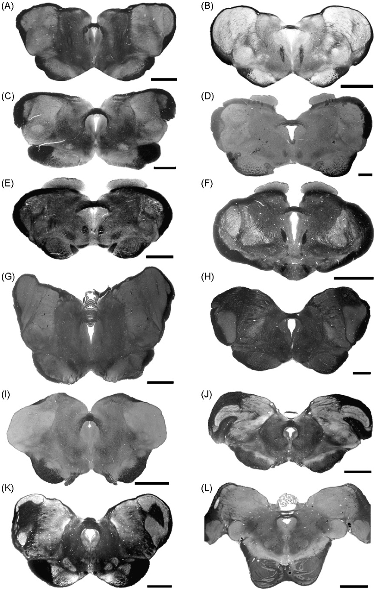
Photomicrographs of myelin stained coronal brain sections demonstrating the morphology of the posterior commissure in a variety of mammalian species. (A) Wombat (Vombatus ursinus) scale bar = 3 mm. (B) Tammar wallaby (Macropus eugenii) scale bar = 3 mm. (C) Giant anteater (Myrmecophaga tridactyla) scale bar = 3 mm. (D) Marmot (Marmota marmota) scale bar = 1 mm. (E) Woodchuck (Marmota monax) scale bar = 2 mm. (F) Jack rabbit (Lepus americanus) scale bar = 3 mm. (G) Zebra (Equus burchelli) scale bar = 5 mm. (H) Sheep (Ovis aries) scale bar = 3 mm. (I) North American badger (Taxidea taxus) scale bar = 3 mm. (J) Leopard (Panthera pardus) scale bar = 5 mm. (K) American black bear (Ursus americanus) scale bar = 5 mm. (L) Chimpanzee (Pan troglodytes) scale bar = 5 mm. All photomicrographs in this plate are taken from sections housed within the Comparative Mammalian Brain Collections of the University of Wisconsin, Michigan State University and The National Museum of Health and Medicine.
4.2.3.2. The cetacean posterior commissure.
As mentioned above, the cetacean posterior commissure is extremely large, that of the bottlenose dolphin have a diameter of around 5 mm at the midline. This is easily visible with the naked eye in gross dissections (Figs. 8A and 9A). While at first glance the posterior commissure of the cetacean appears to be located in an unusual position, it actually retains the general relationships found for this structure in most mammals. It is found caudal to the habenular commissure and anterior to the commissure of the superior colliculus (Figs. 8B and 9A), and it forms the roof of the posterior portion of the third ventricle, anterior to the consolidation of the cerebral aqueduct (Figs. 8C and 9A). Despite this, the portion of the roof of the third ventricle formed by the posterior commissure is not contiguous with that portion of the midbrain tectum formed by the superior colliculi where the anterior most portion of the cerebral aqueduct is expressed. Thus, there is an additional opening of the third ventricle at its postero-superior margin in the cetaceans as compared with other mammalian species. Dissections to date indicate that this opening is covered by the microscopic (or absent) pineal gland (Oelschläger et al., 2008) with probable membranous extensions of the arachnoid mater sealing this opening to complete the posterior roof of the third ventricle. In one dissection we undertook in the bottlenose dolphin, we found a large pineal gland in this region (Fig. 9A), but it should be noted that this specimen had been pregnant for 12 months prior to death and that this is likely to be the cause of the anomalously large pineal gland (e.g. Bishnupuri and Haldar, 2000; Haldar et al., 2006). There are reports of pineal glands in other cetaceans (Phocoena—Behrman, 1990; Megaptera—Gersh, 1938), but these were also small in size.
Fig. 8.
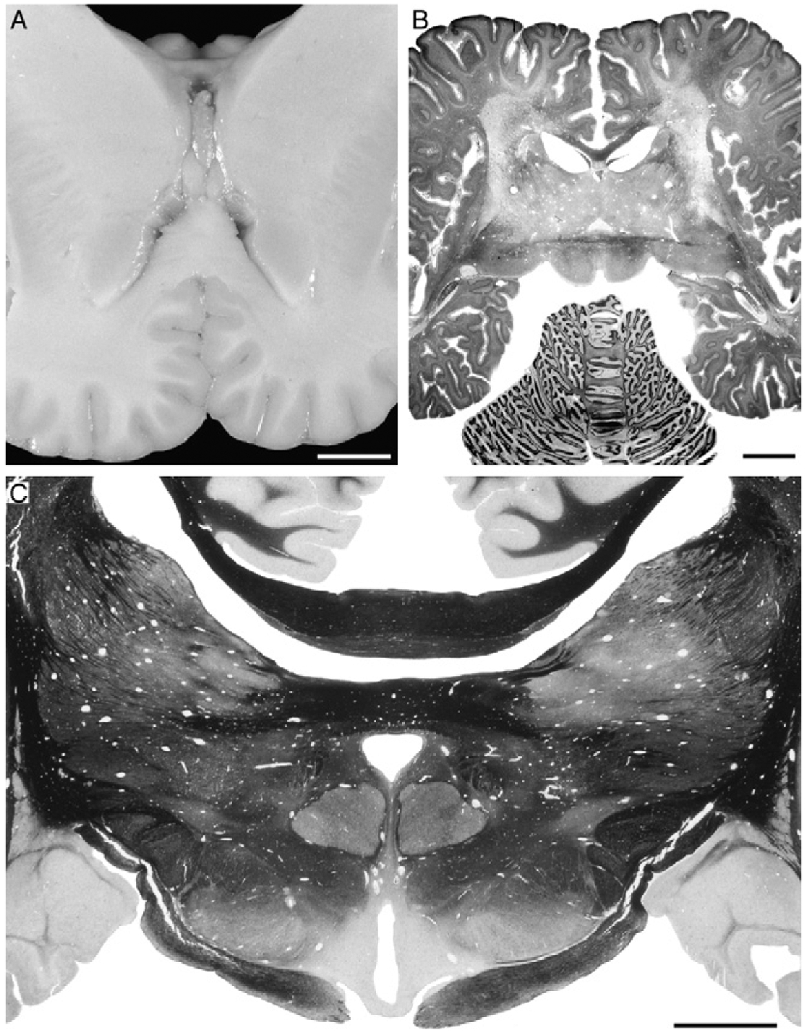
(A) Photograph of gross coronal slice through the brain of a Commerson’s or piebald dolphin (Cephalorhynchus commersonii), showing the enlarged posterior commissure situated immediately anterior to the superior colliculi, and dorsal to the septal nuclei. The coronal plane in cetaceans is different to other mammals due to the maintenance of the cephalic flexure in the adult (from the collection of Sam Ridgway). scale bar = 1 cm. (B) Photomicrograph of a horizontal section through the brain of an adult bottlenose dolphin (Tursiops truncatus) stained for both Nissl substance and myelin. The myelin dense, horizontally projecting posterior commissure can be seen anterior to the superior colliculi investing into the gray matter of the dorsal thalamus. scale bar = 1 cm. (C) Photomicrograph of a coronal section through the brain of an adult bottlenose dolphin (T. truncatus) stained for myelin. Note the thickness of the posterior commissure, lying immediately ventral to a slightly thicker corpus callosum, and projecting horizontally into the gray matter of the dorsal thalamus. Scale bar = 1 cm. The photomicrographs in this plate are taken from sections housed within the Comparative Mammalian Brain Collections of the University of Wisconsin, Michigan State University and The National Museum of Health and Medicine.
Fig. 9.
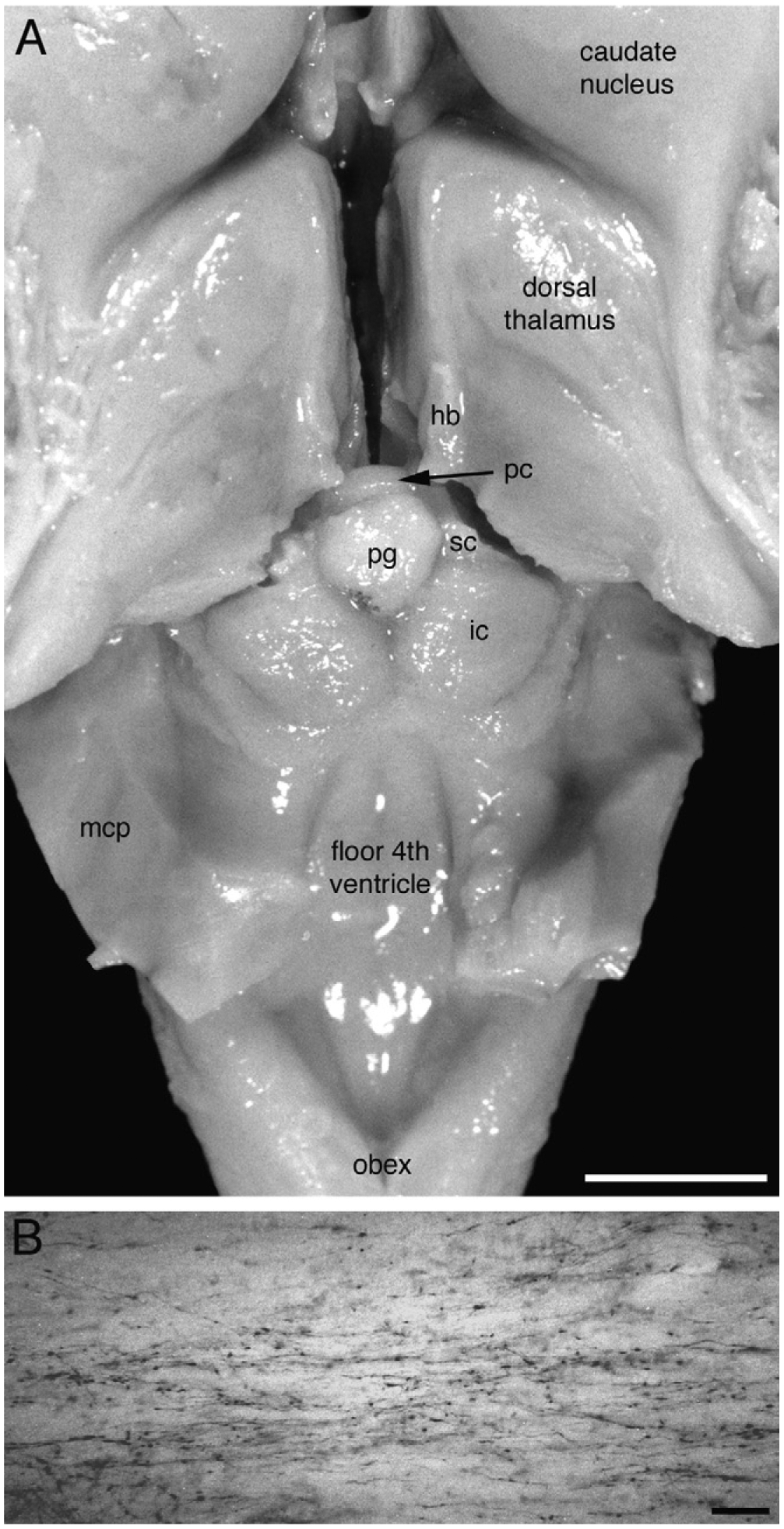
(A) Gross dissection of the bottlenose dolphin brain with the third and fourth ventricles revealed. This dissection demonstrates the location of the posterior commissure (pc) in relation to the habenular trigones (hb), the superior colliculi (sc), pineal gland (pg) and the inferior colliculi (ic). mcp—middle cerebellar peduncle. Scale bar = 1 cm. It should be noted here that the dolphin dissected in this preparation died while heavily pregnant and that in the normal situation the pineal gland is microscopic or absent in cetaceans (Oelschläger et al., 2008, see text for further details). (B) Photomicrograph of tyrosine hydroxylase immunopositive axons coursing through the posterior commissure of the bottlenose dolphin. Scale bar = 100 μm.
The most intriguing anatomical aspect of this enlarged posterior commissure is the direction in which the incoming fascicles coalesce to form the commissure. In the cetacean, these fascicles are quite long and horizontally oriented, investing deeply into the nuclei of the dorsal thalamus. This orientation is dramatically different to that seen in other mammalian species, where the fascicles project ventrally into the brainstem and not the dorsal thalamus. At present it is unclear where the fibres that form the enlarged cetacean posterior commissure arise from, but it is likely that the constituents of the posterior commissure found in other mammals form part of it. In our previous immunohistochemical studies with tyrosine hydroxylase (TH) (Manger et al., 2003, 2004) we found that large fascicles of TH immunoreactive axons formed a significant portion of the commissure (Fig. 9B). From our own observations on TH immunoreactive axons in the posterior commissure of other mammals, and those published in studies of the rat brain (Lindvall and Björklund, 1974; Jones and Moore, 1977), it appears that these axons are more numerous in the dolphin, but a full quantitative analysis will be required to verify the qualitative impression we have.
Various physiological reflexes have fibre pathways that cross through the posterior commissure. One of these is the consensual light reflex. In order to test if the cetaceans maintain this pathway, we examined the response of the bottlenose dolphin to unilateral visual stimulation of the eye in darkness and measured the area of the pupil. A bottlenose dolphin was placed on a rubber pad and kept moist in a room that could be darkened to the extent that humans could not observe any light in the room. With dim lights on, we placed a black, opaque hood containing an infrared (IR) light source at 850 nm wavelength and IR camera over one eye at a distance of 20 cm so that the pupil size could be measured. The hood was formed so that it fit tightly against the dolphin’s head and light stimuli could not impinge on the hooded eye. Another camera and light source was placed to view the non-hooded eye at 20 cm. Both IR cameras were connected to a video recorder so that both eye pictures could be played back synchronously on the same video monitor in a separate room. After the experimental setup was in place, the room lights were turned off, and both pupils dilated naturally to full size (1–1.6 cm2). After the pupils were dilated, the second eye (without the hood) was stimulated with a Grass flasher at 10 flashes per second. Measurement of the time course of pupil size changes indicated similar size changes even though only one eye was stimulated with light (Fig. 10). The experiment was repeated several times, stimulating first the left eye, observing the constriction of the pupils, followed by stopping the unilateral light stimulus, observing the recover of pupil size, then moving the hood to the right eye and stimulating that eye. This experiment revealed the presence of the consensual light reflex in the dolphin studied, indicating that at least this pathway, found in the posterior commissure of several mammals studied, forms part of the posterior commissure of cetaceans.
Fig. 10.
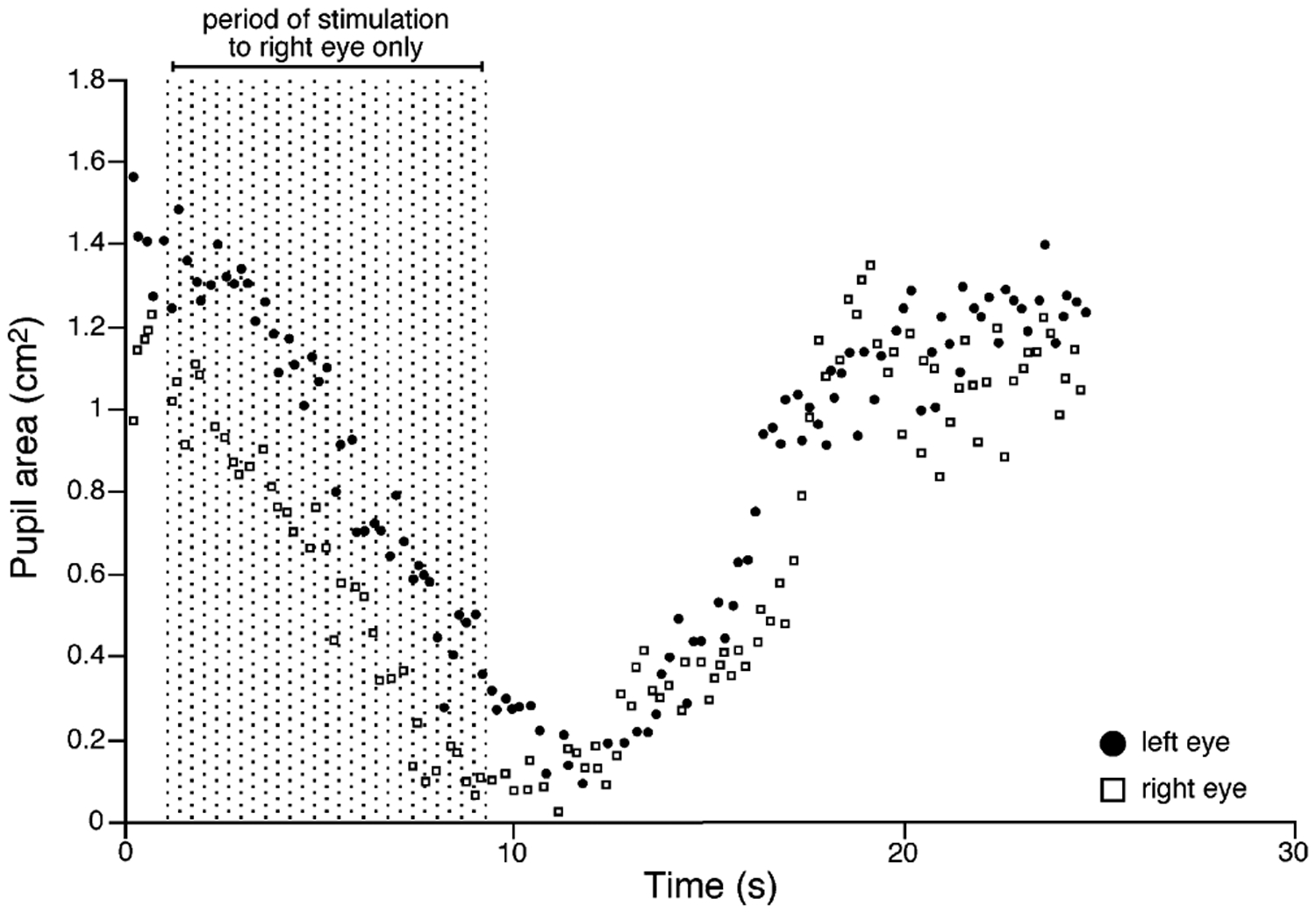
Diagram depicting the results of the experiment testing the consensual light reflex in the bottlenose dolphin. Note that as the right eye only is stimulated by light (the left light being hooded to prevent stimulation) the pupil of the right eye decreases in area and the pupil of the left eye also decreases. This response is typical of the consensual light reflex in mammals that is mediated by pathways crossing through the posterior commissure.
Because of the apparent independence of the cetacean cerebral hemispheres, it is tempting to speculate that some of the fibres of the posterior commissure may arise from the thalamo-cortical system. It would be of great interest to examine the connectivity of this structure and how this may relate to hemispheric independence, studies that may elucidate questions not only regarding cetaceans but also questions surrounding studies of perceptual rivalry in humans (Pettigrew, 2001).
The only study known to the authors that has described the potential function of the posterior commissure in the sleep–wake cycle is that of Berlucchi (1966) in the cat. In this study, along with complete sectioning of the more anterior commissures, Berlucchi sectioned the posterior commissure and examined the effect of this section on the EEG of the two cerebral hemispheres. He found that after sectioning the posterior commissure, the transition between different sleep–wake phases, such as REM sleep to waking, or SWS to REM sleep, were not coincident in the two hemispheres, that there was a mismatch in the transition with one hemisphere lagging in the state change compared with the other. This mismatching lasted up to 10 days, after which the mismatch disappeared. Berlucchi concluded from these observations that (p. 356): “the sleep–wakefulness cycle in the two separated hemispheres is controlled by the lower brainstem.” This study emphasizes the potential importance of anatomy and connectivity of the unusual posterior commissure in the cetaceans, and indicates its importance in the study of cetacean sleep phenomenology.
4.2.3.3. The development of the cetacean posterior commissure.
We were fortunate enough to be able to observe and photograph many of the embryonic and fetal baleen whale specimens collected by Jan Jansen and housed at the Department of Anatomy, University of Oslo, Norway (Fig. 11). This collection consists of both gross anatomical specimens as well as several sectioned and stained whale embryos. We were able to observe developing whales from a length of 5.3 to 248 cm from fin and blue whales (Table 3). The first readily identifiable posterior commissure was found in a 10.5 cm fin whale (Fig. 11A). It should be noted here that for both fin and blue whales, the birth length is between 6 and 7 m, and the adult length ranges from 21 to 33 m. Judging from the relative comparative development of this specimen, it approximates stage 21 of the rhesus monkey embryo (Gribnau and Geijsberts, 1985) and the 14-day rat embryo (Coggeshall, 1964), where in both species the posterior commissure is evident. Thus, the timing of first appearance of the posterior commissure, at least in the baleen whales, appears to approximate the same developmental stage as seen in other mammals. At this stage in the blue whale, the posterior commissure forms a large curved fasciculus forming the roof of the posterior portion of the developing third ventricle (Fig. 11A). The fibres that constitute the commissure appear to arise ventrally from the developing midbrain and pretectal region.
Fig. 11.
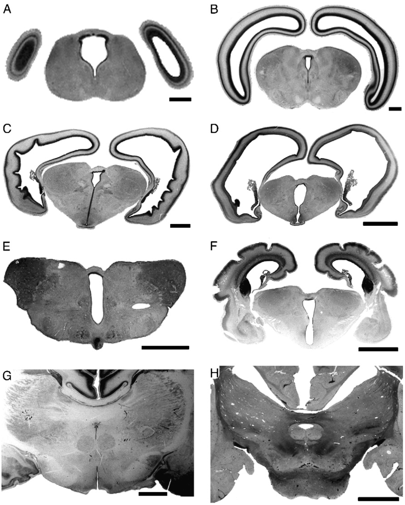
Photomicrographs of embryonic and fetal brain sections at the level of the developing posterior commissure in a series of mysticete (A–E) and odontocete (F–H) cetaceans. (A) 10.5 cm long fin whale (Balaenoptera physalus) scale bar = 1 mm. (B) 17 cm long blue whale (Balaenoptera musculus) scale bar = 1 mm. (C) 21 cm long fin whale, scale bar = 2 mm. (D) 30 cm long fin whale, scale bar = 5 mm. (E) 80 cm long blue whale, scale bar = 5 mm. The adult length of the fin whale ranges from 21 to 25 m, with birth lengths of 6–7 m, and for blue whales, the adult length ranges from 26–33.6 m, with birth lengths being 6–7 m. (F) 16 cm long Harbor porpoise (Phocoena phocoena), adult length ranges from 145–200 cm, with birth lengths ranging from 70–75 cm, scale bar = 5 mm. These sections were stained for Nissl substance and are housed in the Jan Jansen Whale Brain Collection in the Department of Anatomy, University of Oslo, Norway. (G) Fetal Dall’s porpoise (Phocoenoides dalli), unknown length and age, section stained for Nissl substance, scale bar = 3 mm. (H) New born bottlenose dolphin (T. truncatus), approximate length of 100 cm, adult T. truncatus lengths range from 2 to 3.8 m, section stained for myelin, scale bar = 1 cm. The early development of the posterior commissure is similar to that of other eutherian species, with changes in morphology occurring late in development. The last two photomicrographs in this plate are taken from sections housed within the Comparative Mammalian Brain Collections of the University of Wisconsin, Michigan State University and The National Museum of Health and Medicine.
In the histological preparations of a 17 cm long blue whale, the posterior commissure had attained a form that appears to be readily comparable to that seen in the various adult mammals studied (Figs. 11B and 7). By this we mean that the fascicles forming the posterior commissure in this specimen appear to coalesce from the ventral region of the pretectal and midbrain areas, forming a distinct arc on the dorso-lateral surface of the developing peri-aqueductal gray matter. However, in the larger specimens observed this morphology changes, whereby the fascicles become progressively more horizontal in attitude, and appear to coalesce from within the gray matter of the developing dorsal thalamus (Fig. 11C–E). Thus, by 80 cm in length, the blue and fin whales have attained a morphology of the posterior commissure that is closely reminiscent of the adult form seen in both odontocete and mysticete cetaceans.
The developing Harbor porpoise observed was 16 cm in length (adult length up to 200 cm, birth length 70–75 cm) and displayed the fully horizontal form of the posterior commissure (Fig. 11F), as did the fetal Dall’s porpoise (Fig. 11G) and the newborn bottlenose dolphin (Fig. 11H). Our observations indicate that during the early, or embryonic, stage, the cetacean posterior commissure appears to develop along the generalized mammalian lines. This indicates that the fibre pathways crossing through this commissure in the cetacean are likely to be similar to those observed in other mammals (see above discussion of the consensual light reflex). Interestingly, at later stages of development this commissure may be enlarged by other fibres, arising from different locations that cross the midline. The horizontal attitude of these additional fibres, added in later development, possibly indicates that they arise from the thalamo-cortical region of the telencephalon.
While not being a specific target of investigation, two prior studies have noted the timing of development of the posterior commissure in cetaceans. Oelschläger and Kemp (1998) in a study of the development of the brain of the sperm whale noted that the posterior commissure was visible in the 14.4 mm embryo. In contrast to this, the anterior commissure was only visible in the 47 mm embryo, and the corpus callosum rudiment visible in the 66 mm embryo. This developmental sequence of appearance of the various major commissures reflects that seen in other mammals (e.g. Gribnau and Geijsberts, 1985), but does indicate a somewhat precocious development for the posterior commissure. Similarly, in their study of the developing brain of the harbour porpoise, Buhl and Oelschläger (1988) found what was described as an “extended posterior commissure” in the 10 mm embryo, with the anterior commissure and the corpus callosum appearing in the 24 mm embryo.
From these studies we can conclude that the early development of the commissural system in the cetaceans most likely follows similar patterns to that seen in other eutherian mammals. There is the hint that the posterior commissure develops at a slightly earlier stage, and judging from the present findings and those of others, is enlarged very early on. Given the alteration in morphology of the posterior commissure during maturation of the embryo, it is also possible that additional fibres, from a source not normally contributing to the fibres of this commissure, are added later in development and that these may play a major role in the lack of coherence in the EEG of the sleeping cetacean. The posterior commissure appears to be a fruitful region for future experimentation in the anatomy of the cetacean brain as it relates to the patterns of sleep.
4.2.3.4. The otariidae/sirenian posterior commissure.
Given the observations of the cetacean posterior commissure described above, it was of interest to closely examine the morphology of the posterior commissure in pinnipeds and sirenians. In the pinnipeds, it is only the Otariidae that exhibit interhemispheric EEG asymmetry comparable to USWS in dolphins (Lyamin and Chetyrbok, 1992; Lyamin and Mukhametov, 1998). The Phocidae do not exhibit any lateralization in the EEG during sleep (Lyamin, 1993; Lyamin et al., 1989, 1993; Mukhametov et al., 1984; Castellini et al., 1994). The only studied individual of the Amazonian manatee has been shown to spend up to 25% of total sleep time in USWS (Mukhametov et al., 1992). Despite the presence of USWS in fur seals and sea lions and probably in manatees, no distinct structural alteration could be observed in the pinnipeds and sirenians examined (Fig. 12), these species exhibiting what could be described as a typical mammalian posterior commissure. Given the “normal” sized corpus callosum in these pinnipeds (and the very large corpus callosum in sirenians) and the current observations on the posterior commissure, the pinnipeds and sirenians may have an entirely different system controlling hemispheric asynchrony as compared with cetaceans.
Fig. 12.
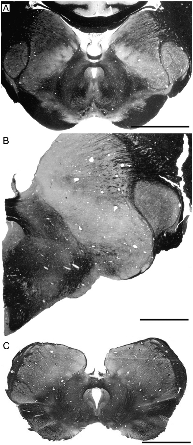
Photomicrographs of coronal sections stained for myelin in two species of pinniped and one sirenian, demonstrating the morphology of the posterior commissure. (A) Northern fur seal (Callorhinus ursinus), a member of the Otariidae family and a presumed unihemispheric sleeper when in water. Note the generally typical mammalian appearance of the posterior commissure. (B) Harbor seal (Phoca vitulina), a member of the Phocidae family and a presumed bihemispheric sleeper. Note again the typical mammalian appearance of the posterior commissure. (C) Florida manatee (Trichechus manatus), a species that exhibits both unihemispheric and bihemispheric sleep. Note again the typical mammalian appearance of the posterior commissure. Scale bar = 1 cm, applies to all three photomicrographs. All photomicrographs in this plate are taken from sections housed within the Comparative Mammalian Brain Collections of the University of Wisconsin, Michigan State University and The National Museum of Health and Medicine.
5. Evolution of cetacean sleep phenomenology
As described above, cetacean sleep is characterized by unihemispheric slow wave sleep, alternating in approximately 1-h intervals between hemispheres, a very small amount (or complete lack) of REM sleep, the ability to sleep during swimming and with only one eye closed at a time. Even though large cetaceans (both odontocetes and mysticetes) spend a lot of time sleeping while immobile at the surface or under water, it is very likely that all representatives of the order (large and small) have slow waves while maintaining continuous movement, thus maintaining muscle tone. These features combined provide a description of a phenotypically unusual form of mammalian sleep, and leads us, and others, to speculate as to the causes that may have produced such a result. Unfortunately, sleep behavior, anatomy and physiology, do not fossilize, thus we cannot rely heavily on the fossil record to elucidate the matter (although hints emanating from knowledge of fossil endocasts and the paleoclimate may be useful); however we can examine a number of modern cetacean species, and by doing so attempt to reconstruct the evolutionary trajectory leading to the modern sleep form through theoretical parsimony. Other mammalian species must also be examined as the comparative context may reveal features relevant to the evolution of the unusual form of sleep in cetaceans.
The first, and most important, point to be made here is that it appears that all cetaceans exhibit the same form of sleep (however, the single baleen whale studied, the young gray whale at Sea World was not studied physiologically but only behaviorally and no beaked whales or sperm whales have been studied). There are no individual species examined to date that have not shown clear signs of the cetacean typical pattern of sleep as determined in the early studies of dolphin sleep physiology by Mukhametov (1987, 1988, 1995), Mukhametov and Polyakova (1981), Mukhametov and Lyamin (1994) and Mukhametov et al. (1977). In this sense, the cetaceans are the first mammalian order examined in which some predictability in the form of sleep can be observed, i.e. there will be USWS and little to no REM sleep in all cetacean species. The second important point to be made here concerns the lack of REM sleep. It has been shown previously that the more immature a mammal at birth, the greater the amount of REM sleep as an adult (Zepelin et al., 2005; Siegel et al., 1998, 1999; Siegel, 2003). At birth the brain weight of the neonate bottlenose dolphin is approximately 42.5% of the adult brain weight (Ridgway and Brownson, 1984), while that of Phocoena phocoena is 36.18% (Hofman, 1983). In comparison, the human brain weighs approximately 29.6% of the adult brain weight (Hofman, 1983). For other mammals, there is a range of data available: Erinaceus europaeus—8.94%; Vulpes vulpes—8.39%; Elephas indicus—31.69%; Rattus norvegicus—11.86%; Mus musculus—18%; Tupaia glis—16.56%; Elephantulus intufi—59.03%; Lemur catta—39.9%; Macaca mulatta—68.55%; Pongo pygmaeus—37.6% (Hofman, 1983). This data would indicate that less maturation of the brain is required in the neonate cetacean compared to many, but not all, mammalian species (indeed the brain is quite heavily myelinated at birth in the cetacean, see Fig. 11), and thus the cetacean brain appears to be more mature at birth than most mammals, and accordingly one would expect less REM sleep in the adult cetacean, but this is not a complete explanation of the apparent lack of REM sleep. The third point is that unusually expressed (striking) interhemispheric EEG asymmetry appears to be a characteristic feature of sleep of all seals belonging to the family Otariidae (Mukhametov et al., 1985; Lyamin and Chetyrbok, 1992; Lyamin and Mukhametov, 1998; Lyamin et al., 2002c) and in a much lesser extent of some birds (Rattenborg et al., 1999, 2001). Therefore, these data need to be taken into account while considering the factors, which led to the evolution of the present pattern of cetacean sleep. Briefly, the first group of data indicates that fur seals generally display more USWS while sleeping in water. When fur seals sleep in water they usually float on their sides, holding one front and two hind flippers in the air. The front flipper in the water constantly paddles. The hemisphere contralateral to the paddling flipper is usually more desynchronized (awake) than the ipsilateral hemisphere suggesting that the motion during sleep in fur seals requires cortical control from the more awake hemisphere (Lyamin and Mukhametov, 1998). This data suggests an association between motion and sleep in cetaceans and otariids. Lyamin and Mukhametov (1998) proposed that the functional significance of USWS in fur seals is to optimize thermoregulation via reducing heat loss and to allow a regular pattern of breathing. On the other hand, recent EEG studies indicated that brief (lasting 1–5 s) unilateral eye opening is also recorded during SWS in the fur seal (Lyamin et al., 2004). The episodes of sleep, which were accompanied by asymmetrical eye opening usually correlated with interhemispheric slow wave EEG asymmetry. Moreover, the hemisphere contralateral to the briefly opening eye was awake or in a state of low amplitude SWS. The hemisphere contralateral to the closed eye was in a state of deeper SWS similar to that described in the bottlenose dolphin and in the beluga (Lyamin et al., 2002a, 2004). It is important to emphasize that episodes of sleep with asymmetrical eye states were recorded in fur seals both on land and in water. Therefore, detection of predators and maintenance of visual contact with conspecifics could be another adaptive advantage of USWS in fur seals (and likely in other Otariids; Lyamin and Mukhametov, 1998; Lyamin et al., 2004). A similar function was proposed for episodes of unilateral eye closure and associated EEG asymmetry during SWS in birds (Rattenborg et al., 1999, 2000).
The comparative observations of mammalian and cetacean sleep leads to three possible conclusions regarding the evolution of cetacean sleep phenomenology: (1) the form of sleep observed in extant cetacean species evolved de novo in their most recent common ancestor; or (2) the forebears of the most recent common ancestor of all modern cetacean species may have exhibited at least some form of sleep phenomenology that could have led to what is observed in extant species; or (3) cetacean sleep phenomenology evolved convergently in several cetacean lineages, minimally in the last common ancestor of odontocetes and the last common ancestor of mysticetes, but potentially several more times. This last possibility does not appear parsimonious given the current state of knowledge, i.e. all cetaceans appear to show the same sleep phenomenology, but it is theoretically possible and cannot be completely disregarded at present. The ancestors of modern cetaceans appear to have been species of the early cetaceans known as the Archaeocetes, a fully aquatic member of the Dorudontidae family that lived approximately 35 mya (Fordyce, 2002; Price et al., 2005). The ancestors of the Archaeocete were several amphibious members of the Pakicetidae family that lived approximately 52 mya (Oelschläger, 1987; Thewissen et al., 2001).
Central to our understanding of the evolution of cetacean sleep phenomenology is an understanding of its current function or current adaptive value, i.e. why do extant cetaceans sleep in the way they do? By examining this question, and coming to a reasonable, logical conclusion, we may then be able to examine the historical record of cetaceans and determine a time when this sleep phenomenology may have emerged and postulate potential selection pressures that underlie its evolution. There are currently five proposals that have been forwarded that may account for the modern functionality of cetacean sleep. To sleep in the manner cetaceans do: (1) services the need to come to the surface regularly to breathe during periods of deep sleep (Mukhametov and Polyakova, 1981; Mukhametov, 1985; Mukhametov et al., 1997); (2) services the need for continuous motion and maintains the position of the individual cetacean in the water column (Mukhametov, 1987, 1988; Mukhametov et al., 1997); (3) maintains coherence of the pod during a time when they are not in auditory contact (Goley, 1999); (4) allows the individual to scan at least half the environment for predators or at least maintain some degree of vigilance (Lilly, 1964; Rattenborg et al., 2000; Lyamin et al., 2004; Ridgway et al., 2006a); and (5) allows significant thermogenesis to occur (Manger et al., 2003; Pillay and Manger, 2004; Manger, 2006). We discuss each of these by examining their current utility and by examining if they may serve as a potential selection pressure in the evolution of cetacean sleep phenomenology.
5.1. The necessity to breathe
It is well known that the dolphin respiratory system is vulnerable to anaesthetics. Dolphin autonomic breathing ceases at the very early stages of anaesthesia, possibly even before they lose the ability to process sensory information. This situation is understandable as the air-breathing aquatic mammal must have a precisely coordinated respiratory action to prevent inhaling water into the lungs while breathing. While anaesthesia in dolphins deepens, the most complex physiological processes are impaired first. Long periods of bilateral high voltage EEG slow wave activity of maximal amplitude are never recorded in dolphins during normal sleep. These episodes can be recorded only occasionally during long breathing pauses and they usually last less than several seconds. As mentioned earlier, diazepam may induce sustained periods of bilateral high voltage slow waves in the EEG; however, it appears that bilateral high voltage delta waves and respiration are not compatible in dolphins. The dolphin fails to respire during bilateral EEG delta waves while lying in slings with the blowhole above the water surface, even if it can move and is trying to take a breath by moving its head and body. Apneas become longer at these times, lasting several minutes, and breaths occur only after the bilateral delta waves in the EEG have disappeared. When a breath is taken, only bilateral desynchronized, bilateral low voltage slow waves or unilateral high voltage EEG patterns (or USWS) have been recorded. After the breath is taken, BSWS develops rapidly. These data strongly suggest that USWS is necessary for autonomic breathing in dolphins during the resting or sleeping periods and indicates that one of the primary proximate functions of USWS is to maintain breathing.
As we mentioned in Section 3.5.1 continuous swimming is a unique feature and it is fully compatible with all forms of dolphin sleep. Therefore, it was further suggested that swimming in dolphins is necessary for coming to the surface for breathing and maintaining the body postures that are required to perform respiratory acts. Swimming is also necessary to counteract water currents and waves. This is particularly relevant for some river dolphins, which live in continuously moving water and must swim from birth. Continuous swimming of small dolphins (e.g., porpoises) in captivity can be explained by their adaptation to life in the open sea, where even small waves disrupt their postures and positions. Captive large cetaceans can be immobile floating at depth or resting on the bottom of pools for several minutes. These behaviors are observed in the wild when the ocean is quiet. On the other hand, floating at the surface should not be a very common behavior in the wild because of waves and frequent storms, which may last several days or weeks. Therefore, the ability to swim continuously appears to be a key characteristic of all cetaceans. It seems reasonable to suggest that USWS enables continuous motion through maintained activation of certain brain centers involved in controlling coordinated movement.
Surfacing to breathe must also occur during sleep in the other fully aquatic marine mammals, for instance in Phocidae seals. The majority of these seals inhabit Arctic and Antarctic seas, which are frozen in winter. These seals can hold their breath for longer periods, some for more than 1 h, and sleep under water (at depth and on the bottom of pools and under the ice in the wild). Four phocid species have been studied to date (Ridgway et al., 1975; Mukhametov et al., 1984; Lyamin et al., 1993; Castellini et al., 1994) and no episodes of USWS have been observed in three species from which recordings were taken from both hemispheres. These seals are immobile during apneas while sleeping in bilateral symmetrical SWS and REM sleep and they wake up to surface for breathing. These data indicate that during the course of evolution, the problem of sleeping and breathing simultaneously has been resolved by different physiological and behavioral adaptations.
There are some clear similarities and differences between the patterns of SWS in Otariids and Cetaceans. First of all, breathing and bilateral delta sleep is fully compatible in fur seals both on land and in water. Fur seals are also capable of autonomic breathing during a deep stage of anaesthesia as with terrestrial mammals. Secondly, interhemispheric EEG asymmetry during asymmetrical and USWS in fur seals in water is less expressed than in dolphins. Finally, EEG asymmetry in fur seals in water is accompanied by a lateralized sleep posture and paddle activity of only one flipper, while in dolphins USWS occurs during continuous swimming. It can be suggested that continuous motion may require maintained activation of larger portions of the brain than the paddling movement of one flipper.
To summarize, (1) the breathing hypothesis is strongly supported from the experiments with diazepam described above and (2) USWS appears to overcome the problem of the needs to surface to breathe and to swim continuously for the sleeping cetacean. However, one may argue that during evolution there was clearly another solution to the problem of breathing while sleeping in the water, this being breath-holding. This may be a simpler solution to implement in terms of the requisite evolutionary changes required (of both genotype and phenotype), than evolving USWS as observed in cetaceans. Based on this one may question a strict association between USWS, continuous swimming and regular breathing both in aquatic mammals in general and in cetaceans.
5.2. Sentinel functions of cetacean sleep—predator avoidance, position in the water column and pod coherence
Here we discuss these three potential factors in the evolution of cetacean sleep phenomenology together as much of the data and observations applicable to each of these factors are similar and all can be grouped under the rubric of “sentinel functions”. Many cetaceans are social animals, therefore continuous monitoring of the immediate surrounds to maintain social bonds and position in the water relative to each other is likely to be a significant factor in the life history of extant cetaceans. Moreover, the need to be aware of predators in the surrounding environment is another proposal that appears to be of utility for the sleeping cetacean. A recent study has documented for the first time that dolphins can maintain continuous auditory vigilance for five days (120 h) without any sleep rebound or physiological signs of sleep deprivation. There was a prominent diurnal pattern present in the target response time and an apparent diurnal pattern in the respiratory rates. The dolphins were able to maintain a high level of vigilance even during nighttime periods of slowed target response time when they appeared to be resting or sleeping (Ridgway et al., 2006a).
The hypothesis that dolphins sleep with one eye open to detect predators was proposed by Lilly (1964) following his description of unilateral eye closure in the apparently sleeping dolphin. As we mentioned earlier, to date asymmetrical eye closure has been documented in at least 5 cetacean species (in 4 odontocetes and in 1 mysticete). The association between the sleeping/waking hemisphere and closed/open contralateral eye was observed in two cetacean species and this data provide support for the hypothesis that USWS in cetaceans provides a sentinel function.
While it is possible that the open eye may scan the environment beyond the pod to detect predators or aggressive conspecifics, there is no experimental data supporting this concept and in many cetacean habitats the water is practically opaque so the auditory sense is primary. It is not known whether a dolphin can use echolocation during USWS; however as mentioned above dolphins can maintain continuous auditory vigilance. The ecological significance of the predator pressure in the lives of whales and dolphins is difficult to assess but it is considered significant by many researchers (e.g. Patterson et al., 1998; Balance, 2002; Weller, 2002). A range of dolphins, porpoises and whales are hunted by killer whales, and at least four other cetacean species (false killer whales, bottlenose dolphins, pygmy killer whales and short-finned pilot whales) have been observed hunting or harassing other cetaceans. Sharks represent another predatory threat to some populations of dolphins (reviewed by Weller, 2002). It has been hypothesized that large whales may undergo their annual migrations to tropical waters in order to reach calving grounds in areas of lower killer whale density (Balance, 2002).
Maintenance of pod coherence appears to be part of the typical pattern observed for sleeping cetaceans in their natural environment (Norris and Dohl, 1980; Würsig and Würsig, 1980). It has been proposed that schooling evolved in dolphins primarily as an adaptive mechanism against predators (Balance, 2002) and this schooling may prove useful during the sleep period as an anti-predator action. On the other hand, most animals are under some threat of predation, whether awake or asleep. One may argue that while sleeping with one eye open, especially in a group, may lower this risk in the exposed environment of cetaceans, this potential problem has not led to a high number of terrestrial vertebrates evolving this type of sleep (Rattenborg et al., 2000); however, most terrestrial mammals use secure “sleeping sites”, which give them protection from predators during sleep.
It appears that in terrestrial mammals only grazing ungulates (artiodactyls and perissodactyls) living in open spaces can in part be compared with cetaceans in terms of the vulnerability to predation. In the wild, ungulates (e.g. antelopes) sleep in groups and it appears that all animals in the herd are never asleep at the same time. This phenomenon is similar to “corporate vigilance” in birds (e.g. Amlaner and Ball, 1994; Lima et al., 2005). Sleep in ungulates has not been studied in enough detail to be sure that they do not have USWS; but slow wave EEG appears to occur bilaterally in the Siberian musk deer (Sokolov et al., 1988). These animals showed bilaterally symmetrical SWS while standing or lying, but were always recumbent when entering REM sleep. Another related example is the apparent absence of the asymmetrical eye state during sleep in the hippopotamus in a zoo. It is known that in the wild the hippopotamus calves are vulnerable to predation from crocodiles. The hippopotamus mother is known to be very protective of its calf; however, only bilateral eye opening was observed in a behaviorally sleeping hippopotamus female that had a 3-month old calf. These data suggest that this hippopotamus had bilaterally symmetrical SWS as in all previously studied terrestrial mammals (Lyamin and Siegel, 2005). It is clear that further physiological recording of sleep needs to be conducted in ungulates; however, currently there is not enough evidence to suggest that ungulates could utilize USWS mechanisms to monitor predators. Thus it is possible to conclude that predation pressure or maintenance of social bonds may have led to the evolution of “corporate vigilance” or bilateral SWS with low arousal thresholds and frequent arousals in ungulates, but not USWS as seen in cetaceans.
The experimental data collected in pinnipeds may be useful when trying to understand how the pattern of sleep in cetaceans has evolved, as pinnipeds face similar ecological pressures. Unilateral eye closure and USWS has been documented in two species of fur seals (Otariidae) and in the one walrus (Odobenidae) that has been studied. The authors know of no reports detailing the presence of asymmetrical eye closure in the true seals (Phocidae). Therefore, for some currently unknown reason, while the predation pressure in all pinnipeds seems to be comparable (but not yet quantitatively evaluated), USWS has evolved in the in otariids but not in phocids. Phocids are better divers, can hold their breath for much longer periods, and spend more time at depth compared to otariids. Some phocids may be at sea constantly for weeks or months and dive almost continuously when in the water (Stewart, 2002). This would be an effective strategy (along with minimizing time at the surface) both to hunt for prey and minimize the risk of predation, which most often occurs when seals surface for breathing. This would also eliminate the need for USWS and allow the presence of REM sleep in the range of normal variation observed in terrestrial mammals, if the phocids were forced to inhabit the water continuously (elephant seals spend up to 8 months in the water). The difference in sleep patterns between the otariids and the phocids indicates that either the different strategies to reduce predation risk played a role, or that predation risk really does not play a major role in sleep evolution in the pinnipeds.
Unilateral eye closure has been documented in some birds (29 species from 13 orders) but not in others (reviewed by Rattenborg et al., 2000). Three bird species (mallards, pigeons and seagulls) showed an association between interhemispheric EEG asymmetry and the open/closed eye in a manner similar to that seen in bottlenose dolphins, belugas and fur seals. However, the extent of EEG asymmetry recorded in birds was much smaller than that recorded in dolphins and seals (Lyamin et al., 2002a,c, 2004). The most important result of these studies is that mallards spent more time with one eye closed while at the edge of a group, and the open eye was directed away from the center of the sleeping group (Rattenborg et al., 1999). Similarly, pigeons showed a preference for directing the open eye away from the wall at the back of their cage and towards the open room (Rattenborg et al., 2001). Rattenborg et al. (1999) propose that this is an effect related to the possibility of predation.
The position of an individual cetacean in the pod and coordinated swimming with other dolphins in a group of sleeping cetaceans has been analyzed with respect to the problem of pod-coherence by Goley (1999). She observed that for the most part, the open eye of the sleeping cetacean was directed to its neighbor, unlike the situation seen in birds (see above). Goley also described that the sleeping cetaceans changed position in the pod at regular intervals to maintain the open eye (presumably the one contralateral to the awake hemisphere) on the remainder of the pod. Similar, although anecdotal, observations were made in bottlenose dolphins (Gnone et al., 2001; Sekiguchi and Kohshima, 2003). The results of studies in dolphins do not appear to negate Lilly’s proposal, as the open eye may be directed to the surrounding environment through the group; however, the data presented here seems to emphasize that the sentinel or vigilance (e.g. Lima et al., 2005) function of the asymmetrical eye state may allow the sleeping cetacean to stay in formation with the others of its pod more so than to monitor the broader physical characteristics of the environment where the auditory sense would be more useful. In our recent study (Lyamin et al., 2005a, 2007) we also showed that dolphin mothers and their calves continuously monitor their position relative to each other and this occurs both during active wake and slow swimming which is frequently associated with unilateral eye closure.
It also appears very likely that auditory (and perhaps tactile) processing can occur during USWS in the cetaceans. It is highly unlikely that a dolphin can maintain auditory vigilance continuously for 120 h and exhibit no rest rebound or signs of sleep deprivation without employing USWS (Ridgway et al., 2006a). Almost certainly sensory processing could be useful in maintaining pod coherence and predator detection. To conclude, it is reasonable to suggest that the terrestrial ancestor of the modern cetaceans had bilateral SWS as all terrestrial mammals do. However, when spending longer periods in the water, the semiaquatic ancestor of the modern day whales (e.g. a form of Ambulocetus, Thewissen et al., 2001) would have benefited from being able to be half asleep and half awake at the same time to avoid the predators that lived in the ocean about 40–50 mya. These ancestors of whales most likely had to sleep at the surface, where aquatic mammals are most vulnerable to predation. Therefore, it is clear that USWS and the absence/reduction of REM sleep would minimize the time during which cetaceans are not able to some extent monitor the surrounding environment, thus sentinel functions per se appear to address both aspects of the unusual sleep phenomenology of cetaceans; however, it does appear likely that there are additional reasons for the evolution of cetacean sleep mechanisms.
5.3. Thermogenesis
One of the defining features of “mammalness” is the ability to generate heat from within the body, and regulate body and brain temperature independently, for the most part, from the environmental temperature. Cetaceans, being mammals, also generate their own heat, and achieve this with mechanisms that do not differ from those of other mammals, i.e. no thermogenetic mechanisms specific to cetaceans that are not found in other mammals have been identified (Castellini, 2002; but see Manger, 2006 regarding glia). The aquatic niche of cetaceans may be fairly described as a thermally challenging environment, as conductive heat loss in water is approximately 90 times greater than that in an aerial environment at the same ambient temperatures, and this conductive heat loss challenge appears to relate to the actual size of neonate cetaceans (Downhower and Blumer, 1988). The conductive heat loss during movement in the water (or the “water chill factor”), which in the smaller cetaceans is almost constant, will increase the rate of heat loss dramatically.
It has been proposed that the anatomy, and probable physiology, of the locus coeruleus in conjunction with the unihemispheric nature of SWS and lack of REM sleep in cetaceans may provide a basis for increased thermogenesis during sleep (Manger et al., 2003; Manger, 2006). Further, it has been demonstrated that a balance between heat loss (surface area) and heat generation (movement) can be seen in resting/sleeping bottlenose dolphins (Pillay and Manger, 2004). These studies have shown that thermogenesis is related to sleep patterns in the cetaceans, but a full explanation of how thermogenesis may relate to this sleep pattern in cetaceans has not yet been provided, nor how it may relate to the evolution of cetacean sleep (although it has been partially described in Manger, 2006).
The mechanism proposed for the relationship of thermogenesis to the sleep pattern of cetaceans, this pattern being USWS, little to no REM sleep and continuous movement, is depicted in Fig. 13. The first point of importance is that the temperature of the water will act as an environmental pressure upon the cetaceans. There are three morphophysiological characteristics (possible adaptations?) underlying the physiological changes required for increased thermogenesis during sleep in the cetaceans. These include a potentially continuous production of noradrenalin by the cells of the locus coeruleus (Manger et al., 2003), an increased amount of glial cells throughout the central nervous system (Garey and Leuba, 1986; Hawkins and Olszewski, 1957; Manger, 2006; Reichenbach, 1989), and a modified arterial circulation (McFarland et al., 1979). These three characteristics have direct physiological consequences of importance during sleep, including increased noradrenalin production, increased glia metabolism, constant muscle tonus, and shunting of warm and cold blood.
Fig. 13.
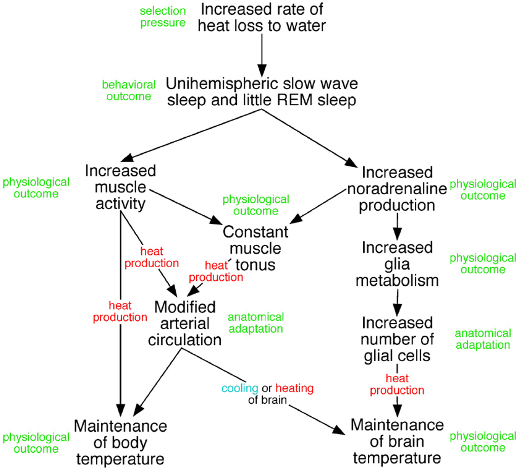
Diagrammatic representation of the possible manner in which cetacean sleep phenomenology may interact with the anatomy and physiology of the cetacean body and brain to maintain body and brain temperature in the thermally challenging aquatic environment.
The temperature of the mammalian brain and body are regulated separately (Gisolfi and Mora, 2000), but it is also quite probable that body temperature can influence brain temperature under certain circumstances (but see examples of artiodactyl temperature regulation). Because of this thermoregulation/thermogenesis dichotomy, the maintenance of body temperature and brain temperature must be treated separately although the mechanisms affecting temperature regulation in both structures overlap to an extent.
In terms of maintaining body temperature, the first important, and perhaps even simplistic point, is that the increased muscular activity associated with USWS will enable heat production. In terrestrial mammals, muscular activity can provide up to 70% of the heat required by the body (Gisolfi and Mora, 2000). This situation in cetaceans is different to that found in bihemispheric sleeping mammals, as during bihemispheric slow wave sleep there is no significant muscular activity. Thus, the movement of the unihemispherically sleeping cetacean induces heat production during a time when no significant heat production via muscular activity would occur if they were to sleep bihemispherically (Pillay and Manger, 2004; Lyamin et al., 2005a).
The second way in which heat can be generated to maintain body temperature relates to the possible physiology of the neurons of the locus coeruelus (Manger et al., 2003; Ridgway et al., 2006b). One physiological outcome of activity of the neurons of the locus coeruleus is the facilitation of the maintenance of basal muscle tone (see above; also Manger et al., 2003). Basal muscle tone accounts for approximately 30% of the heat production required by the mammalian body (Gisolfi and Mora, 2000), accounting for heat production in the larger cetaceans that do not move a great deal during sleep and that also have a far smaller surface area to volume ratio reducing heat loss.
The neurons of the locus coeruleus, in bihemispheric sleeping mammals, are active during waking, slow their discharge during SWS, and discharge minimally during REM sleep (see earlier description of locus coeruleus). This discharge pattern is of interest to compare to cetacean sleep. As there is little to no REM sleep, it is possible that there will be no time throughout the day when the neurons of the locus coeruleus are not discharging, and thus not releasing noradrenlin. It has been demonstrated that increasing the amount of noradrenalin in the CNS leads to suppression of REM sleep (reviewed in Mallick et al., 2002). In terms of USWS, as one hemisphere is awake, it will be likely that the neurons of the ipsilateral locus coeruleus will be discharging maximally, and the locus coeruleus neurons contralateral to the awake hemisphere will be discharging slower (Manger et al., 2003; Ridgway et al., 2006b). This situation will reverse when the hemispheres switch roles. This likely scenario means that at no time will the dolphin have a period when there is little to no production of noradrenalin; however, the amount released in each hemisphere may differ during USWS. Thus, we can speculate that in cetaceans, basal muscle tone will be continuously facilitated by this release of noradrenalin, maintaining heat production. This muscular activity and continuous basal muscle tone will produce heat throughout the life of the cetacean, from birth onwards (Lyamin et al., 2005a, 2007). A combination of this continuous muscular heat production, with the modified arterial circulation of the cetaceans (McFarland et al., 1979), will allow heat to be shunted where needed for warming the body, or cooling it down.
As mentioned above, the temperature of the brain is regulated separately from that of the body in mammals. It is our contention here that the nature of cetacean sleep combined with anatomical changes and correlated physiological changes allows maintenance of brain temperature in the thermally challenging aquatic environment. Previous studies have demonstrated that the crucial mechanism for the production of heat in the central nervous system is the metabolic activity of glia (Donhoffer, 1980; Szelényi, 1998). Other studies have shown that the metabolic activity of glia is substantially increased in the presence of noradrenalin (Magistretti et al., 1993; Pellerin et al., 1997; Subbarao and Hertz, 1990), and that glia are a major central target of the noradrenalin producing neurons of the locus coeruleus (Stone and Ariano, 1989). These observations of the action of noradrenalin in the normal mammalian brain may be extrapolated to the cetacean brain.
As mentioned, the hypothesized discharge pattern of the neurons of the locus coeruleus of the cetaceans will lead to more noradrenalin in the CNS of cetaceans. This will potentially increase the rate of glial metabolism in the CNS of cetaceans, and thus increase heat production. The increased amount of glia in the CNS of cetaceans, combined with the increased noradrenalin will also substantially increase overall glial heat production. It is proposed that these CNS mechanisms will provide enough heat to maintain the temperature of the brain under the thermal challenge of heat loss to the water (Manger, 2006).
This mechanism is supported by the data measuring the temperature of the dolphin brain during USWS (see above). In the awake hemisphere, the temperature remains constant, this hemisphere being in receipt of “normal” cetacean amounts of noradrenalin. The temperature of the hemisphere in SWS drops over time, this hemisphere being in receipt of less noradrenalin due to a slowing in the discharge rate of locus coeruleus neurons during SWS (Ridgway et al., 2006b), thus the metabolic rate of glia will decrease as will the production of heat. Upon awakening, or switching of the global EEG pattern, the cooler hemisphere that had been in SWS warms up due to the increased discharge rate of locus coeruleus neurons infusing the hemisphere with more noradrenalin, increasing glial metabolism and thus increasing heat production.
Based on this foregoing discussion, we conclude that it is highly likely that significant thermogenesis occurs during sleep in cetaceans. In other mammals, sleep is a time when the thermogenetic mechanisms are relaxed, the body cooling during SWS, and during REM sleep there appears to be minimal thermogenesis throughout the body (McGinty and Szymusiak, 1990; McGinty et al., 2001; Nakao et al., 1999; Parmeggiani, 2003). The alterations seen in the pattern of sleep for cetaceans, and the major thermal challenge provided by the aquatic environment, all point to thermogenesis as being an important factor underlying the altered sleep patterns of cetaceans. Moreover, this proposal is in line with all our observations of the different features of the cetacean brain that are related to sleep patterns, and can be extended to solitary cetaceans, something the previous proposals have not addressed.
As thermogenesis appears to be an important factor in the present adaptive utility of cetacean sleep, is it possible to determine if there was a time in cetacean evolutionary history when water temperature was a significant environmental pressure that may be related to the evolution of this sleep pattern? The first fully aquatic cetacean ancestors were the Archaeoceti (Fordyce and Barnes, 1994; Thewissen et al., 2001; Fordyce, 2002). These species lived in the warm, shallow, nutrient-rich Tethys sea (Fordyce and Barnes, 1994). This equatorial sea may be thought of as somewhat similar to the Arabian Sea or Red Sea with water temperatures mostly above 26 °C. In this sense, the temperature of the water may not have been as significant an environmental pressure in the lives of the Archaeocete. Modern sirenians, that exhibit a form of USWS and reduced REM sleep, are restricted in their distribution to water temperatures above 19 °C (Anderson, 1986; Husar, 1977, 1978; Lefebvre et al., 1989). Thus, it is possible that the Archaeocete either exhibited bihemispheric sleep, or had a sleep pattern somewhat reminiscent of the modern sirenians. The sirenians evolved at the same time, in the same Tethys sea as the Archaeocete (Savage, 1976). Another possibility to consider is that even in warm (26 °C) water the first amphibious cetacean ancestor Ambulocetus natans, which had an estimated body weight in the range of 140–720 kg (Thewissen et al., 1996; Gingerich, 1998), would still lose heat if it needed to stay (and sleep) immobile in water. Ambulocetus, being in the size range of modern pinnipeds and sirenians, would need to maintain motion to offset heat loss the same way small cetaceans, fur seals and sirenians do at present. Thus, it is possible that USWS, or a form of USWS reminiscent of the situation seen in either fur seals or manatees, evolved at an earlier stage of cetacean evolution when the ancestors were still semi-aquatic animals.
While these ancestors to the odontocete and mysticete cetaceans may have had a sleep pattern not dissimilar to the sirenians or pinnipeds, the extant cetacean sleep pattern is very different, in that there is little to no REM sleep, and the bouts of SWS are for the most part consistently unihemispheric. The transition of the Archaeocete to modern cetacean fauna coincided with the closure of the Tethys sea by the suturing of India to the Asian landmass and of Africa to the European landmass (Fordyce and Barnes, 1994; Fordyce, 2002). This eliminated the warm, shallow, nutrient rich environment inhabited by the Archaeocete. This period also coincided with a global cooling of oceanic temperatures by approximately 6 °C (Whitmore, 1994). Thus, this faunal transition is coincident with major changes in the thermal relations of the oceanic aquatic habitat of cetaceans. This transition is also characterized by a major change in the relative proportions and absolute size of the cetacean brain (Marino et al., 2004; Manger, 2006).
These major environmental and gross neuroanatomical changes indicate a point in cetacean evolutionary history where thermal challenges would have been a significant evolutionary pressure. The concurrence of thermal and anatomical changes indicate that the Archaeocete—modern cetacean faunal transition is likely to have been the period when the sleep patterns evident in modern cetaceans would have evolved. It also indicates that while thermogenesis has a clear proximate utility in modern cetaceans, it is reasonable to suggest that thermogenesis is likely to be the ultimate, or evolutionary cause, of the evolution of modern cetacean sleep phenomenology. The high levels of activity in newborn cetaceans relative to adults of the same species is also consistent with this hypothesis (Lyamin et al., 2005a). This pattern is the reverse of that seen in land mammals.
As a final point here, it should be emphasized that phocid seals do not show USWS, but otariid seals do (see above mentioned references). This difference may also be potentially explained with thermogenesis in mind, as it is well known that phocid seals have a thicker blubber layer than that of the otariid seals (Reijnders et al., 1993), thus potentially allowing them greater thermal insulation and obviating the need for USWS for thermal compensation. However, this potential explanation needs to be explored more fully, as the composition of the lipids in the blubber of the two pinniped groups may also differ systematically, potentially enhancing the insulative properties of phocid blubber over otariid blubber.
6. Summary and conclusions
We hypothesize that three factors – the need to come to the surface to breathe, more efficient monitoring of the environment and thermogenesis – may have been important in the evolution of the observable cetacean sleep phenomenology (sleeping while in motion, USWS, absence of REM sleep, and sleeping with one eye open). The need to come to the surface to breathe is an obvious life-sustaining requirement for any aquatic mammal. This need might make the physiological process of breathing more complicated and incompatible with deep bilateral slow wave sleep. Alternation of sleep between the two brain hemispheres appears to be an outcome of this contradiction. The risk of predation could be an important factor in the very early days of cetacean aquatic evolution when their ancestors started living in the shallow warm waters of the Tethys sea. At this stage they were not large pelagic animals and possibly lacked the abilities to hold their breath for long period and to dive to depths sufficient enough to avoid potential predators. Therefore consistent motion, monitoring of the environment for predators and conspecifics to maintain group coherence may have been pressures with enough significance to lead to the form of sleep seen in modern cetaceans. The third alternative is the need to maintain heat production in the face of the thermal challenge of living in water. Being mammals, cetaceans are under a substantial pressure to avoid heat loss, and the altered form of sleep seen in extant cetaceans is consistent with the ability to increase heat production. It is also consistent with known changes in the environment of the cetaceans over time. The last two proposals indicate that the altered form of cetacean sleep is likely to have evolved, either fully or partially expressed, in the amphibious or fully aquatic ancestors of modern cetaceans. At present it is impossible to separate either proposal, except on theoretical grounds, and it may even be possible that both played a part in the evolution of extant cetacean sleep phenomenology.
Acknowledgments
The work presented here was supported by the National Science Foundation (NSF-0234687), National Institute of Health (NS 42947), the Medical Research Service of the Department of Veterans Affairs, Utrish Dolphinarium Ltd. and the South African National Research Foundation.
References
- Amlaner CJ, Ball NJ, 1994. Avian sleep. In: Kryger MH, Roth T, Dement WC (Eds.), Principles and Practice of Sleep Medicine. second ed. Saunders, Philadelphia, pp. 81–94. [Google Scholar]
- Anderson PK, 1986. Dugongs of Shark Bay, Australia—seasonal migration, water temperature and forage. Natl. Geogr. Res 2, 473–490. [Google Scholar]
- Balance LT, 2002. Cetacean ecology. In: Perrin WF, Würsig B, Thewissen JGM (Eds.), Encyclopedia of Marine Mammals. Academic Press, New York, pp. 208–214. [Google Scholar]
- Bearzi G, Politi E, 1999. Diurnal behavior of free-ranging bottlenose dolphins in the Kvarneric (Northern Adriatic sea). Mar. Mamm. Sci 15, 1065–1097. [Google Scholar]
- Behrman G, 1990. The pineal organ (epiphysis cerebri) of the harbor porpoise (Phocoena phocoena, Linne, 1758). Aquat. Mamm 16, 96–100. [Google Scholar]
- Berlucchi G, 1965. Callosal activity in unrestrained, unanesthetized cats. Arch. Ital. Biol 103, 623–635. [PubMed] [Google Scholar]
- Berlucchi G, 1966. Electroencephalographic studies in “split brain” cats. Electroenceph. Clin. Neurophysiol 20, 348–356. [DOI] [PubMed] [Google Scholar]
- Bishnupuri KS, Haldar C, 2000. Profile of organ weights and plasma concentrations of melatonin, estradiol and progesterone during gestation and post-parturition in female Indian palms squirrel Funambulus pennanti. Indian J. Exp. Biol 38, 974–981. [PubMed] [Google Scholar]
- Bjarkam CR, Sørensen JC, Geneser FA, 1997. Distribution and morphology of serotonin-immunoreactive neurons in the brainstem of the New Zealand white rabbit. J. Comp. Neurol 380, 507–519. [PubMed] [Google Scholar]
- Bonnet MH, 1989. Infrequent periodic sleep disruption: effects on sleep, performance and mood. Physiol. Behav 45, 1049–1055. [DOI] [PubMed] [Google Scholar]
- Bonnet MH, 2000. In: Kryger MH, Roth T, Dement WC (Eds.), Sleep Deprivation. W.B. Saunders, Philadelphia, pp. 53–71. [Google Scholar]
- Breathnach AS, 1960. The cetacean central nervous system. Biol. Rev 35, 187–230. [Google Scholar]
- Buhl EH, Oelschläger HA, 1988. Morphogenesis of the brain in the harbour porpoise. J. Comp. Neurol 277, 109–125. [DOI] [PubMed] [Google Scholar]
- Bullock TH, Grinnell AD, Ikezono F, Kameda K, Katsuki Y, Nomoto M, Sato O, Suga N, Yanagisava K, 1968. Electrophysiological studies of the central auditory mechanisms in cetaceans. Z. Vergl. Physiol 59, 117–156. [Google Scholar]
- Cassini MH, Vila BL, 1990. Cluster analysis of group types in southern right whale (Eubalaena australis). Mar. Mamm. Sci 6, 17–24. [Google Scholar]
- Castellini M, 2002. Thermoregulation. In: Perrin WF, Würsig B, Thewissen JGM (Eds.), Encyclopedia of Marine Mammals. Academic Press, New York, pp. 1245–1250. [Google Scholar]
- Castellini MA, Milsom WK, Berger RJ, Costa DP, Jones DR, Castellini JM, Rea LD, Bharma S, Harris M, 1994. Patterns of respiration and heart rate during wakefulness and sleep in elephant seal pups. Am. J. Physiol 266, R863–R869. [DOI] [PubMed] [Google Scholar]
- Coggeshall RE, 1964. A study of diencephalic development in the albino rat. J. Comp. Neurol 122, 241–269. [DOI] [PubMed] [Google Scholar]
- Dinges DF, Rogers NL, Baynard MD, 2005. Chronic sleep deprivation. In: Kryger MH, Roth T, Dement WC (Eds.), Principles and Practice of Sleep Medicine. W.B. Saunders, Philadelphia, pp. 67–76. [Google Scholar]
- Donhoffer Sz., 1980. Homeothermia of the Brain. Akadémiai Kiadó, Budapest. [Google Scholar]
- Downhower JF, Blumer LS, 1988. Calculating just how small a whale can be. Nature 335, 675.3173490 [Google Scholar]
- Flanigan WF, 1974a. Nocturnal behavior of captive small cetaceans. I: The bottlenose dolphin, Tursiops truncatus. Sleep Res. 3, 84. [Google Scholar]
- Flanigan WF, 1974b. Nocturnal behavior of captive small cetaceans. II: The beluga whale, Delphinapterus leucas. Sleep Res. 3, 85. [Google Scholar]
- Flanigan WF, 1975a. More nocturnal observations of captive small cetaceans. I: The killer whale, Orcinus orca. Sleep Res 4, 139. [Google Scholar]
- Flanigan WF, 1975b. More nocturnal observations of captive small cetaceans. II: The pacific white-sided dolphin, Lagenorhynchus obliquidens. Sleep Res. 4, 140. [Google Scholar]
- Flanigan WF, 1975c. More nocturnal observations of captive small cetaceans. III: Further study of the beluga whale, Delphinapterus leucas. Sleep Res. 4, 141. [Google Scholar]
- Ford JKB, 2002. Killer whale (Orcinus orca). In: Perrin WF, Würsig B, Thewissen JGM (Eds.), Encyclopedia of Marine Mammals. Academic Press, New York, pp. 669–676. [Google Scholar]
- Fordyce RE, 2002. Cetacean evolution. In: Perrin WF, Würsig B, Thewissen JGM (Eds.), Encyclopedia of Marine Mammals. Academic Press, New York, pp. 214–220. [Google Scholar]
- Fordyce RE, Barnes LG, 1994. The evolutionary history of whales and dolphins. Annu. Rev. Earth Planet. Sci 22, 419–455. [Google Scholar]
- Garey LJ, Leuba G, 1986. A quantitative study of the neuronal and glial numerical density in the visual cortex of the bottlenose dolphin: evidence for a specialized subarea and changes with age. J. Comp. Neurol 247, 491–496. [DOI] [PubMed] [Google Scholar]
- Gazzaniga MS, 2000. Cerebral specialization and interhemispheric communication: does the corpus callosum enable the human condition? Brain 123, 1293–1326. [DOI] [PubMed] [Google Scholar]
- Gerashchenko D, Shiromani PJ, 2004. Different neuronal phenotypes in the lateral hypothalamus and their role in sleep and wakefulness. Mol. Neurobiol 29, 41–59. [DOI] [PubMed] [Google Scholar]
- Gersh I, 1938. Note on the pineal gland of the humpback whale. J. Mammal 19, 477–480. [Google Scholar]
- Gingerich PD, 1998. Paleobiological perspectives on Mesonychia, Archaeoceti, and the origin of whales. In: Thewissen JGM (Ed.), The Emergence of Whales. Evolutionary Patterns in the Origin of Cetaceans (Advances in Vertebrate Paleobiology) Plenum Press, New York, pp. 423–449. [Google Scholar]
- Gisolfi CV, Mora F, 2000. The Hot Brain. Survival, Temperature and the Human Body. MIT Press, Cambridge. [Google Scholar]
- Gnone G, Benoldi C, Bonsignori B, Fognani P, 2001. Observations of rest behaviours in captive bottlenose dolphins (Tursiops truncatus). Aquat. Mamm 27, 29–33. [Google Scholar]
- Gnone G, Moriconi T, Gambini G, 2006. Sleep behaviour: activity and sleep in dolphins. Nature 441, E10–E11. [DOI] [PubMed] [Google Scholar]
- Goley PD, 1999. Behavioral aspects of sleep in pacific white-sided dolphins (Lagenorhynchus obliquidens, Gill 1865). Mar. Mamm. Sci 15, 1054–1064. [Google Scholar]
- Gray RW, 1927. The sleep of whales. Nature 119, 636. [Google Scholar]
- Gribnau AAM, Geijsberts LGM, 1985. Morphogenesis of the brain in staged rhesus monkey embryos. In: Beck F, Hild W, Ortmann R, Pauly JE, Schiebler TH (Eds.), Advances in Anatomy Embryology and Cell Biology, vol. 91. Springer-Verlag, Berlin, pp. 1–63. [DOI] [PubMed] [Google Scholar]
- Haldar C, Yadav R, Alipreeta A, 2006. Annual reproductive synchronization in ovary and pineal gland function of femal short-nosed fruit bat, Cynopterus sphinx. Comp. Biochem. Physiol. A Mol. Integr. Physiol 144, 395–400. [DOI] [PubMed] [Google Scholar]
- Hanson MT, Defran RH, 1993. The behavior and feeding ecology of the Pacific Coast bottlenose dolphin, Tursiops truncatus. Aquat. Mamm 19, 127–142. [Google Scholar]
- Hawkins A, Olszewski J, 1957. Glia/nerve index for cortex of the whale. Science 126, 76–77. [DOI] [PubMed] [Google Scholar]
- Howard RS, Finneran JJ, Ridgway SH, 2006. BIS monitoring of unihemispheric effects in dolphins. Anesth. Anal 103, 626–632. [DOI] [PubMed] [Google Scholar]
- Hofman MA, 1983. Evolution of brain size in neonatal and adult placental mammals: a theoretical approach. J. Theor. Biol 105, 317–332. [DOI] [PubMed] [Google Scholar]
- Husar SL, 1977. Trichechus inunguis. Mammalian Species, vol. 72. 4 pp. [Google Scholar]
- Husar SL, 1978. Trichechus senegalensis. Mammalian Species, vol. 89. 3 pp. [Google Scholar]
- Innocenti GM, 1986. General organization of the callosal connections in the cerebral cortex. In: Jones EG, Peters A (Eds.), Cerebral Cortex, vol. 5. Plenum Press, New York, pp. 291–353. [Google Scholar]
- Jacobs MS, McFarland WL, Morgane PJ, 1979. The anatomy of the brain of the bottlenose dolphin (Tursiops truncatus). Rhinic lobe (Rhinencephalon): The archicortex. Brain Res. Bull 4 Suppl. 1, 1–108. [DOI] [PubMed] [Google Scholar]
- Jeeves MA, 1994. Callosal agenesis—a natural split brain overview. In: Lassonde M, Jeeves MA (Eds.), Callosal Agenesis. Plenum Press, New York, pp. 285–299. [Google Scholar]
- John J, Wu MF, Boehmer LN, Siegel JM, 2004. Cataplexy-active neurons in the hypothalamus: implications for the role of histamine in sleep and waking behavior. Neuron 42, 619–634. [DOI] [PMC free article] [PubMed] [Google Scholar]
- Jones BE, Moore RY, 1977. Ascending projections of the locus coeruleus in the rat. II: Autoradiographic study. Brain Res. 127, 23–53. [PubMed] [Google Scholar]
- Jones BE, Halaris AE, McIlhany M, Moore RY, 1977. Ascending projections of the locus coeruleus in the rat. I: Axonal transport in central noradrenaline neurons. Brain Res. 127, 1–21. [DOI] [PubMed] [Google Scholar]
- Jouvet M, Jeannerod M, Delorme F, 1965. Organization of the system responsible for phase activity during paradoxal sleep. C. R. Seances Soc. Biol. Fil 159, 1599–1604. [PubMed] [Google Scholar]
- Kaufman LS, Morrison AR, 1981. Spontaneous and elicited PGO spikes in rats. Brain Res. 9, 61–72. [DOI] [PubMed] [Google Scholar]
- Klinowska M, 1986. Diurnal rhythms in cetaceans—a review. Rept. Int. Whal. Comm 8, 75–88. [Google Scholar]
- Kiyashchenko LI, Mileykovskiy BY, Maidment N, Lam HA, Wu MF, John J, Peever J, Siegel JM, 2002. Release of hypocretin (orexin) during waking and sleep states. J. Neurosci 22, 5282–5286. [DOI] [PMC free article] [PubMed] [Google Scholar]
- Kovalzon VM, 1973. Brain temperature variations during natural sleep and arousal in white rats. Physiol. Behav 10, 667–670. [DOI] [PubMed] [Google Scholar]
- Kovalzon VM, Mukhametov LM, 1983. Temperature fluctuations of the dolphin brain corresponding to unihemispheric slow-wave sleep. J. Evol. Biochem. Physiol 18, 222–224. [Google Scholar]
- Kruger L, 1966. Specialized features of the cetacean brain. In: Norris KS (Ed.), Whales, Dolphins and Porpoises. University of California Press, Los Angeles, pp. 232–253. [Google Scholar]
- Lai YY, Siegel JM, 1990. Muscle tone suppression and stepping produced by stimulation of midbrain and rostral pontine reticular formation. J. Neurosci 10, 2727–2738. [DOI] [PMC free article] [PubMed] [Google Scholar]
- Lai YY, Kodama T, Siegel JM, 2001. Changes in monoamine release in the ventral horn and hypoglossal nucleus linked to pontine inhibition of muscle tone: an in vivo microdialysis study. J. Neurosci 21, 7384–7391. [DOI] [PMC free article] [PubMed] [Google Scholar]
- Lefebvre LW, O’Shea TJ, Rathbun GB, Best RC, 1989. Distribution, status, and biogeography of the West Indian manatee. In: Woods CA (Ed.), Biogeography of the West Indies: Past, Present and Future. Sandhill Crane Press, Gainsville, FL, pp. 567–610. [Google Scholar]
- Lesku JA, Roth TC, Amlaner CJ, Lima SL, 2006. A phylogenetic analysis of sleep architecture in mammals: the integration of anatomy, physiology, and ecology. Am. Nat 168, 441–453. [DOI] [PubMed] [Google Scholar]
- Lima SL, Rattenborg NC, Lesku JA, Amlaner CJ, 2005. Sleeping under the risk of predation. Anim. Behav 70, 723–736. [Google Scholar]
- Lilly JC, 1958. Electrode and cannulae implantation in the brain of mammals by a simple percutaneous method. Science 127, 1181–1182. [DOI] [PubMed] [Google Scholar]
- Lilly JC, 1964. Animals in aquatic environments: adaptations of mammals to the ocean. In: Dill DB (Ed.), Handbook of Physiology—Environment. American Physiology Society, Washington, DC, pp. 741–747. [Google Scholar]
- Lindvall O, Björklund A, 1974. The organization of the ascending catecholamine neuron systems in the rat brain. Acta Physiol. Scand. Suppl 412, 1–48. [PubMed] [Google Scholar]
- Lyamin OI, 1993. Sleep in the harp seal (Pagophilus groenlandica). Comparisons of sleep on land and in water. J. Sleep Res 2, 170–174. [DOI] [PubMed] [Google Scholar]
- Lyamin OI, 2004. Sleep in young steller sea lions and northern fur seals: a comparative study. In: Sea Lions of the World: Conservation and Research in the 21st Century. Abstract of the 22nd Lowell Wakefield Fisheries Symposium, Anchoradge, AK, p. 31. [Google Scholar]
- Lyamin OI, Chetyrbok IS, 1992. Unilateral EEG activation during sleep in the cape fur seal, Arctocephalus pusillus. Neurosci. Lett 143, 263–266. [DOI] [PubMed] [Google Scholar]
- Lyamin OI, Mukhametov LM, 1998. Organization of sleep in the northern fur seal. In: Sokolov VE, Aristov AA, Lisitzina TU (Eds.), The Northern Fur Seal. Systematic, Morphology, Ecology, Behavior. Nauka, Moscow, pp. 280–302. [Google Scholar]
- Lyamin OI, Siegel JM, 2005. Rest and activity states in the hippopotamuses. In: Abstract Book of the 33rd Annual Symposium of European Association for Aquatic Mammals, 15 pp. [Google Scholar]
- Lyamin OI, Siegel JM, 2006. Cetacean sleep behavior varies with body size. Sleep 29, A38. [Google Scholar]
- Lyamin OI, Oleksenko AI, Polyakova IG, 1989. Sleep and wakefulness in pups of harp seal, Pagophilus groenladnica. J. High Nerve Activity 39, 1061–1069. [Google Scholar]
- Lyamin OI, Oleksenko AI, Polyakova IG, 1993. Sleep in the harp seal (Pagophilus groenlandica). Peculiarities of sleep in pups during the first month of their lives. J. Sleep Res 2, 163–169. [DOI] [PubMed] [Google Scholar]
- Lyamin OI, Oleksenko AI, Polyakova IG, Mukhametov LM, 1996. Paradoxical sleep in northern fur seals in water and on land. J. Sleep Res 5 (Suppl. 1), 259. [Google Scholar]
- Lyamin OI, Shpak OV, Nazarenko EA, 1999. Swimming types and budget of time in bottlenose dolphins in captivity. Biologicheski vestnik (Kharkov State University) 2, 103–106. [Google Scholar]
- Lyamin OI, Manger PR, Mukhametov LM, Siegel JM, Shpak OV, 2000. Rest and activity states in a grey whale. J. Sleep Res 9, 261–267. [DOI] [PMC free article] [PubMed] [Google Scholar]
- Lyamin OI, Mukhametov LM, Siegel JM, Manger PR, Shpak OV, 2001. Resting behavior in a rehabilitating gray whale calf. Aquat. Mamm 27, 256–266. [Google Scholar]
- Lyamin OI, Mukhametov LM, Siegel JM, Nazarenko EA, Polyakova IG, Shpak OV, 2002a. Unihemispheric slow wave sleep and the state of the eyes in a white whale. Behav. Brain Res 129, 125–129. [DOI] [PMC free article] [PubMed] [Google Scholar]
- Lyamin OI, Shpak OV, Nazarenko EA, Mukhametov LM, 2002b. Muscle jerks during behavioral sleep in a beluga whale (Delphinapterus leucas L.). Physiol. Behav 76, 265–270. [DOI] [PubMed] [Google Scholar]
- Lyamin OI, Mukhametov LM, Chetyrbok IS, Vassiliev AV, 2002c. Sleep and wakefulness in the southern sea lion. Behav. Brain Res 128, 129–138. [DOI] [PubMed] [Google Scholar]
- Lyamin OI, Shpak OV, Siegel JM, 2003. Ontogenesis of rest behavior in killer whales. Sleep 26, 116. [Google Scholar]
- Lyamin OI, Mukhametov LM, Siegel JM, 2004. Relationship between sleep and eye state in Cetaceans and Pinnipeds. Arch. Ital. Biol 142, 557–568. [PMC free article] [PubMed] [Google Scholar]
- Lyamin OI, Pryaslova J, Lance V, Siegel JM, 2005a. Animal behaviour: continuous activity in cetaceans after birth. Nature 435, 1177. [DOI] [PMC free article] [PubMed] [Google Scholar]
- Lyamin OI, Kosenko PO, Lapierre JL, Vyssotski AL, Lipp HP, Mukhametov LM, Siegel JM, 2005b. Association between behavior and sleep in bottlenose dolphins. In: Abstracts of the 16th Biennial Conference on the Biology of Marine Mammals. 174 pp. [Google Scholar]
- Lyamin OI, Pryaslova J, Lance V, Siegel JM, 2006a. Sleep behaviour: sleep in continuously active dolphins. Activity and sleep in dolphins (Reply). Nature 441, E11. [DOI] [PubMed] [Google Scholar]
- Lyamin OI, Pryaslova JP, Kosenko PO, Lapierre JL, Mukhametov LM, Siegel JM, 2006b. Sleep and rest states in the walrus. In: Abstract Book of the 34th Annual Symposium of European Association for Aquatic Mammals. 14 pp. [Google Scholar]
- Lyamin OI, Pryaslova J, Kosenko PO, Siegel JM, 2007. Behavioral aspects of sleep in bottlenose dolphin mothers and their calves. Physiol. Behav 92, 725–733. [DOI] [PMC free article] [PubMed] [Google Scholar]
- Magistretti PJ, Sorg O, Yu N, Martin JL, Pellerin L, 1993. Neurotransmitters regulate energy metabolism in astrocytes: implications for the metabolic trafficking between neural cells. Dev. Neurosci 15, 306–312. [DOI] [PubMed] [Google Scholar]
- Mallick BN, Majumdar S, Faisal M, Yadav V, Madan V, Pal D, 2002. Role of norepinephrine in the regulation of rapid eye movement sleep. J. Biosci 27, 539–551. [DOI] [PubMed] [Google Scholar]
- Manger PR, 2006. An examination of cetacean brain structure with a novel hypothesis correlating thermogenesis to the evolution of a big brain. Biol. Rev. Camb. Philos. Soc 81, 293–338. [DOI] [PubMed] [Google Scholar]
- Manger PR, Ridgway SH, Siegel JM, 2003. The locus coeruleus complex of the bottlenose dolphin (Tursiops truncatus) as revealed by tyrosine hydroxylase immunohistochemistry. J. Sleep Res 12, 149–155. [DOI] [PMC free article] [PubMed] [Google Scholar]
- Manger PR, Fuxe K, Ridgway SH, Siegel JM, 2004. The distribution and morphological characteristics of catecholamine cells in the diencephalons and midbrain of the bottlenose dolphin (Tursiops truncatus). Brain Behav. Evol 64, 42–60. [DOI] [PMC free article] [PubMed] [Google Scholar]
- Mann J, Smuts BB, 1999. Behavioral development in wild bottlenose dolphin newborns (Tursiops sp.). Behavior 136, 529–566. [Google Scholar]
- Marino L, Stowe J, 1997. Lateralized behavior in two captive bottlenose dolphins (Tursiops truncatus). Zoo Biol. 16, 173–177. [Google Scholar]
- Marino L, McShea DW, Uhen MD, 2004. Origin and evolution of large brains in toothed whales. Anat. Rec. A Discov. Mol. Cell Evol. Biol 281, 1247–1255. [DOI] [PubMed] [Google Scholar]
- Maseko BC, Bourne JA, Manger PR, 2007. Distribution and morphology of cholinergic, putative catecholaminergic and serotonergic neurons in the brain of the Egyptian Rousette flying fox, Rousettus aegyptiacus. J. Chem. Neuroanat 34, 108–127. [DOI] [PubMed] [Google Scholar]
- Mate BR, Rossbach KA, Nieukirk SL, Wells RS, Irvine AB, Scott MD, Read AJ, 1995. Satellite-monitored movements and dive behavior of a bottlenose dolphin (Tursiops truncatus) in Tampa Bay, Florida. Mar. Mamm. Sci 11, 452–463. [Google Scholar]
- McBride AF, Hebb DO, 1948. Behavior of the captive bottlenose dolphin, Tursiops truncatus. J. Comp. Physiol. Psych 41, 111–123. [DOI] [PubMed] [Google Scholar]
- McCormick DA, 1989. Cholinergic and noradrenergic modulation of thalamocortical processing. Trends Neurosci. 12, 215–221. [DOI] [PubMed] [Google Scholar]
- McCormick JG, 1969. Relationship of sleep, respiration, and anesthesia in the porpoise: a preliminary report. Proc. Natl. Acad. Sci. U.S.A 62, 697–703. [DOI] [PMC free article] [PubMed] [Google Scholar]
- McCormick JG, 2007. Behavioral observations of sleep and anesthesia in the dolphin: implications for bispectral index monitoring of unihemispheric effects in dolphins. Anesth. Analg 104, 239–241. [DOI] [PubMed] [Google Scholar]
- McFarland WL, Jacobs MS, Morgane PJ, 1979. Blood supply to the brain of the dolphin, Tursiops truncatus, with comparative observations on special aspects of cerebrovascular supply of other vertebrates. Neurosci. Biobehav. Rev 3 (suppl. 1), 1–93. [Google Scholar]
- McGinty D, Szymusiak R, 1990. Keeping cool: a hypothesis about the mechanisms and functions of slow-wave sleep. Trends Neurosci. 13, 480–487. [DOI] [PubMed] [Google Scholar]
- McGinty D, Alam MN, Szymusiak R, Nakao M, Yamamoto M, 2001. Hypothalamic sleep-promoting mechanisms: coupling to thermoregulation. Arch. Ital. Biol 139, 63–75. [PubMed] [Google Scholar]
- Michel F, Roffwarg HP, 1967. Chronic split brainstem preparation: effect on sleep-waking cycle. Experientia 23, 126–128. [DOI] [PubMed] [Google Scholar]
- Miller PJ, Aoki K, Rendell LE, Amano M, 2008. Stereotypical resting behavior of the sperm whale. Curr. Biol 18, R21–R23. [DOI] [PubMed] [Google Scholar]
- Moruzzi G, Magoun HW, 1949. Brain stem reticular formation and activation of the EEG. Electroenceph. Clin. Neurophysiol 1, 455–473. [PubMed] [Google Scholar]
- Mukhametov LM, 1984. Sleep in marine mammals. Exp. Brain Res 8, 227–238. [Google Scholar]
- Mukhametov LM, 1985. Unihemispheric slow wave sleep in the brain of dolphins and seals. In: Inoue S, Borbely AA (Eds.), Endogenous Sleep Substrates and Sleep Regulation. Japan Societies Press, Tokyo, pp. 67–75. [Google Scholar]
- Mukhametov LM, 1987. Unihemispheric slow-wave sleep in the Amazonian dolphin, Inia geoffrensis. Neurosci. Lett 79, 128–132. [DOI] [PubMed] [Google Scholar]
- Mukhametov LM, 1988. The absence of paradoxical sleep in dolphins. In: Koella WP, Obal F, Schulz H, Visser P (Eds.), Sleep 1986. Gustav Fischer Verlag, New York, pp. 154–156. [Google Scholar]
- Mukhametov LM, 1995. Paradoxical sleep peculiarities in aquatic mammals. Sleep Res. 24A, 202. [Google Scholar]
- Mukhametov LM, Lyamin OI, 1994. Rest and active states in bottlenose dolphins (Tursiops truncatus). J. Sleep Res 3, 174. [Google Scholar]
- Mukhametov LM, Lyamin OI, 1997. The Black Sea bottlenose dolphin: the conditions of rest and activity. In: Sokolov VE, Romanenko EV (Eds.), The Black Sea Bottlenose Dolphin. Nauka, Moscow, pp. 650–668. [Google Scholar]
- Mukhametov LM, Polyakova IG, 1981. EEG investigation of sleep in porpoises (Phocoena phocoena). J. High Nerve Activity 31, 333–339. [Google Scholar]
- Mukhametov LM, Supin AY, 1975. EEG study of different behavioural states in free moving dolphin (Tursiops truncatus). J. High Nerve Activity 25, 396–401. [PubMed] [Google Scholar]
- Mukhametov LM, Supin A.Ya., Strokova IG, 1976. Interhemispheric asymmetry of cerebral functional states during sleep in dolphins. Dokl. Akad. Nauk. SSSR 229, 767–770. [PubMed] [Google Scholar]
- Mukhametov LM, Supin A.Ya., Polyakova IG, 1977. Interhemispheric asymmetry of the electroencephalographic sleep pattern in dolphins. Brain Res. 134, 581–584. [DOI] [PubMed] [Google Scholar]
- Mukhametov LM, Supin A.Ya., Polyakova IG, 1984. Sleep in Caspian seals (Phoca caspica). J. High Nerve Activity 34, 259–264. [PubMed] [Google Scholar]
- Mukhametov LM, Lyamin OI, Polyakova IG, 1985. Interhemispheric asynchrony of the sleep EEG in northern fur seals. Experientia 41, 1034–1035. [DOI] [PubMed] [Google Scholar]
- Mukhametov LM, Oleksenko AI, Polyakova IG, 1988. Quantitative characteristics of the electrocorticographic sleep stages in bottle-nosed dolphins. Neurofiziologiia 20, 532–538. [PubMed] [Google Scholar]
- Mukhametov LM, Lyamin OI, Chetyrbok IS, Vassilyev AA, Diaz R, 1992. Sleep in an Amazonian manatee, Trichechus inunguis. Experientia 48, 417–419. [DOI] [PubMed] [Google Scholar]
- Mukhametov LM, Oleksenko AI, Polyakova IG, 1997. The Black Sea bottlenose dolphin: the structure of sleep. In: Sokolov VE, Romanenko VE (Eds.), The Black Sea Bottlenose Dolphin. Nauka, Moscow, (in Russian), pp. 492–512. [Google Scholar]
- Nakao M, McGinty D, Szymusiak R, Yamamoto M, 1999. Thermoregulatory model of sleep control: losing the heat memory. J. Biol. Rhythms 14, 547–556. [DOI] [PubMed] [Google Scholar]
- Nazarenko EA, Lyamin OI, Shpak OV, Mukhametov LM, 2001. Behavioral sleep in captive Baikal seals. In: Abstracts of the 14th Biennial Conference on the Biology of Marine Mammals. 154 pp. [Google Scholar]
- Nelson DL, Lein J, 1994. Behavior pattern of two captive Atlantic white-sided dolphins, Lagenorhvnchus acutus. Aquat. Mamm 20, 1–10. [Google Scholar]
- Nielsen TA, Montplaisir J, Marcotte R, Lassonde M, 1994. Sleep, dreaming and EEG coherence patterns in agenesis of the corpus callosum: comparisons with callosotomy patients. In: Lassonde M, Jeeves MA (Eds.), Callosal Agenesis. Plenum Press, New York, pp. 109–117. [Google Scholar]
- Nieuwenhuys R, ten Donkelaar HJ, Nicholson C, 1998. The Central Nervous System of Vertebrates, vol. 3. Springer, New York. [Google Scholar]
- Nishiwaki M, 1962. Aerial photographs show sperm whales’ interesting habits. Norsk. Hvalfangst-Tidende 51, 395–398. [Google Scholar]
- Nitz D, Siegel JM, 1997a. GABA release in the locus coeruleus as a function of sleep/wake state. Neuroscience 78, 795–801. [DOI] [PMC free article] [PubMed] [Google Scholar]
- Nitz D, Siegel JM, 1997b. GABA release in the dorsal raphe nucleus: role in the control of REM sleep. Am. J. Physiol 273, R451–R455. [DOI] [PMC free article] [PubMed] [Google Scholar]
- Norris KS, Dohl TP, 1980. The structure and functions of cetacean schools. In: Herman LM (Ed.), Cetacean Behavior: Mechanisms and Functions. John Wiley and Sons, New York, pp. 211–261. [Google Scholar]
- Oelschläger HA, 1987. Pakicetus inachus and the origin of whales and dolphins (Mammalia: Cetacea). Gegenbaurs Morphol. Jahrb 133 (5), 673–685. [PubMed] [Google Scholar]
- Oelschläger HA, Kemp B, 1998. Ontogenesis of the sperm whale brain. J. Comp. Neurol 399, 210–228. [DOI] [PubMed] [Google Scholar]
- Oelschläger HA, Haas-Rioth M, Fung C, Ridgway SH, Knauth M, 2008. Morphology and evolutionary biology of the dolphin (Delphinus sp.) brain—MR imaging and conventional histology. Brain Behav. Evol 71, 68–86. [DOI] [PubMed] [Google Scholar]
- Oleksenko AI, Lyamin OI, 1996. Rest and activity states in female and baby of harbor porpoise (Phocoena phocoena). J. Sleep Res 5 (suppl. 1), 159. [Google Scholar]
- Oleksenko AI, Mukhametov LM, Polyakova IG, Supin A.Ya., Kovalzon VM, 1992. Unihemispheric sleep deprivation in bottlenose dolphins. J. Sleep Res 1, 40–44. [DOI] [PubMed] [Google Scholar]
- Oleksenko AI, Chetyrbok IS, Polyakova IG, Mukhametov LM, 1996. Rest and active states in Amazonian dolphins. In: Sokolov VE (Ed.), The Amazonian Dolphin. Nauka, Moscow, pp. 257–266. [Google Scholar]
- Olivares R, Montiel J, Aboitiz F, 2001. Species differences and similarities in the fine structure of the mammalian corpus callosum. Brain Behav. Evol 57, 98–105. [DOI] [PubMed] [Google Scholar]
- Patterson IA, Reid RJ, Wilson B, Grellier K, Ross HM, Thompson PM, 1998. Evidence for infanticide in bottlenose dolphins: an explanation for violent interactions with harbour porpoises? Proc. Biol. Sci 265, 1167–1170. [DOI] [PMC free article] [PubMed] [Google Scholar]
- Parmeggiani PL, 2003. Thermoregulation and sleep. Front. Biosci 8, s557–s567. [DOI] [PubMed] [Google Scholar]
- Pellerin L, Stolz M, Sorg O, Martin JL, Deschepper CF, Magistretti PJ, 1997. Regulation of energy metabolism by neurotransmitters in astrocytes in primary culture and in an immortalized cell line. Glia 21, 74–83. [DOI] [PubMed] [Google Scholar]
- Pettigrew JD, 2001. Searching for the switch: neural bases for perceptual rivalry alternations. Brain Mind 2, 85–118. [Google Scholar]
- Peyron C, Tighe DK, van den Pol AN, De Lecea L, Heller HC, Sutcliffe JG, Kilduff TS, 1998. Neurons containing hypocretin (orexin) project to multiple neuronal systems. J. Neurosci 18, 9996–10015. [DOI] [PMC free article] [PubMed] [Google Scholar]
- Pillay P, Manger PR, 2004. Testing thermogenesis as the basis for the evolution of cetacean sleep phenomenology. J. Sleep Res 13, 353–358. [DOI] [PubMed] [Google Scholar]
- Pilleri G, 1979. The blind Indus dolphin, Platanista indi. Endeavour 3, 45–56. [Google Scholar]
- Price SA, Bininda-Emonds ORP, Gittleman JL, 2005. A complete phylogeny of the whales, dolphins and even-toed hoofed mammals (Cetartiodactyla). Bio. Rev. Camb. Philos. Soc 80, 1–29. [DOI] [PubMed] [Google Scholar]
- Puelles L, 2001. Brain segmentation and forebrain development in amniotes. Brain Res. Bull 55, 695–710. [DOI] [PubMed] [Google Scholar]
- Rattenborg NC, Lima SL, Amlaner CJ, 1999. Facultative control of avian unihemispheric sleep under the risk of predation. Behav. Brain Res 15, 163–172. [DOI] [PubMed] [Google Scholar]
- Rattenborg NC, Amlaner CJ, Lima SL, 2000. Behavioral, neurophysiological and evolutionary perspectives on unihemispheric sleep. Neurosci. Biobehav. Rev 24, 817–842. [DOI] [PubMed] [Google Scholar]
- Rattenborg NC, Amlaner CJ, Lima SL, 2001. Unilateral eye closure and interhemispheric EEG asymmetry during sleep in the pigeon (Columba livia). Brain Behav. Evol 58, 323–332. [DOI] [PubMed] [Google Scholar]
- Rechtschaffen A, Bergmann BM, 2002. Sleep deprivation in the rat: an update of the 1989 paper. Sleep 25, 18–24. [DOI] [PubMed] [Google Scholar]
- Rechtschaffen A, Gilliland MA, Bergmann BM, Winter JB, 1983. Physiological correlates of prolonged sleep deprivation in rats. Science 221, 182–184. [DOI] [PubMed] [Google Scholar]
- Rechtschaffen A, Bergmann BM, Everson CA, Kushida CA, Gilliland MA, 1989. Sleep deprivation in the rat: X. Integration and discussion of the findings. Sleep 12, 68–87. [PubMed] [Google Scholar]
- Reichenbach A, 1989. Glia:neuron index: review and hypothesis to account for different values in various mammals. Glia 2, 71–77. [DOI] [PubMed] [Google Scholar]
- Reijnders P, Brasseur S, van der Toorn J, van der Wolf P, Boyd I, Harwood J, Lavigne D, Lowry L, 1993. Seals, Fur Seals, Sea Lions, and Walrus. IUCN Publications Unit, Cambridge, UK. [Google Scholar]
- Reeves RR, Stewart BS, Clapham PJ, Powell JA, 2002. Pygmy and dwarf sperm whales. In: Guide to Marine Mammals of the World, National Audubon Society, New York, pp. 244–247. [Google Scholar]
- Ricciardi F, Jahoda M, Azzellino A, Almirante C, 2001. The definition of behavioral categories in Mediterranean fin whales (Balaenoptera physalus) on the basis of swimming-surfacing parameters. In: Abstract of the 15th Conference of European Cetacean Society. pp. 27–28. [Google Scholar]
- Ridgway SH, 1972. Homeostasis in the aquatic environment. In: Ridgway SH (Ed.), Mammals of the Sea: Biology and Medicine. Charles C. Thomas, Springfield, pp. 590–747. [Google Scholar]
- Ridgway SH, 1990. The central nervous system of the bottlenose dolphin. In: Leatherwood S, Reeves RR (Eds.), The Bottlenose Dolphin. Academic Press, San Diego, pp. 69–97. [Google Scholar]
- Ridgway SH, 2002. Asymmetry and symmetry in brain waves from dolphin left and right hemispheres: some observations after anesthesia, during quiescent hanging behavior, and during visual obstruction. Brain Behav. Evol 60, 265–274. [DOI] [PubMed] [Google Scholar]
- Ridgway SH, Brownson RH, 1984. Relative brain sizes and cortical surface areas in odontocetes. Acta Zool. Fennica 172, 149–152. [Google Scholar]
- Ridgway SH, Harrison RJ, Joyce PL, 1975. Sleep and cardiac rhythm in the gray seal. Science 187 (4176), 553–555. [DOI] [PubMed] [Google Scholar]
- Ridgway SH, Carder D, Finneran JJ, Keogh M, Kamolnick T, Todd M, Goldblatt A, 2006a. Dolphin continuous auditory vigilance for five days. J. Exp. Biol 209, 3621–3628. [DOI] [PubMed] [Google Scholar]
- Ridgway SH, Houser D, Finneran J, Carder D, Keogh M, van Bonn W, Smith C, Scadeng M, Dubowitz D, Mattery R, Hoh C, 2006b. Functional imaging of dolphin brain metabolism and blood flow. J. Exp. Biol 209, 2902–2910. [DOI] [PubMed] [Google Scholar]
- Robbins J, Mattila DK, Palsboll PJ, Berube M, 1998. Asynchronous diving pairs of humpback whales: implications of a newly described behavior observed in the North Atlantic wintering grounds. In: Abstract of the World Marine Mammals Science Conference, Monaco, pp. 20–24. [Google Scholar]
- Saayman GS, Tayler CK, Bower D, 1973. Diurnal activity cycles in captive and free-ranging Indian Ocean bottlenose dolphins (Tursiops aduncus, Ehrenburg). Behaviour 44, 212–233. [Google Scholar]
- Sakai K, Koyama Y, 1996. Are there cholinergic and non-cholinergic paradoxical sleep-on neurons in the pons. Neuroreport 7, 2449–2453. [DOI] [PubMed] [Google Scholar]
- Saper CB, Chou TC, Scammell TE, 2001. The sleep switch: hypothalamic control of sleep and wakefulness. Trends Neurosci. 24, 726–731. [DOI] [PubMed] [Google Scholar]
- Savage RJG, 1976. Review of early Sirenia. Syst. Zool 25, 344–351. [Google Scholar]
- Sekiguchi Y, Kohshima S, 2003. Resting behaviors of captive bottlenose dolphins (Tursiops truncatus). Physiol. Behav 79, 643–653. [DOI] [PubMed] [Google Scholar]
- Sekiguchi Y, Arai K, Kohshima S, 2006. Sleep behaviour: sleep in continuously active dolphins. Nature 441, E9–E10. [DOI] [PubMed] [Google Scholar]
- Serafetinides EA, Shurley JT, Brooks RE, 1972. Electroencephalogram of the pilot whale, Globicephala scammoni, in wakefulness and sleep: lateralization aspects. Int. J. Psychobiol 2, 129–135. [Google Scholar]
- Shane SH, Wells RS, Wursig B, 1986. Ecology, behavior and social organization of the bottlenose dolphin: a review. Mar. Mamm. Sci 2, 34–63. [Google Scholar]
- Shaw PJ, Tononi G, Greenspan RJ, Robinson DF, 2002. Stress response genes protect against lethal effects of sleep deprivation in Drosophila. Nature 417, 287–291. [DOI] [PubMed] [Google Scholar]
- Shpak OV, Lyamin OI, Nazarenko EA, Mukhametov LM, 2001. Variability of swimming styles and their relationship to rest and activity states in captive white whales. In: Abstracts of the 14th Biennial Conference on the Biology of Marine Mammals. 196 pp. [Google Scholar]
- Shpak OV, Lyamin OI, Manger PR, Siegel JM, Mukhametov LM Rest and activity states in the Commerson’s dolphin (Cephalorhynchus commersonii). Zh. Evol. Biokhim. Fiziol, in press (in Russian: ). [PMC free article] [PubMed] [Google Scholar]
- Shurley JT, Serafetinides EA, Brooks RE, Elsner R, Kenney DW, 1969. Sleep in Cetaceans: I. The pilot whale, Globicephala scammoni. Psychophysiology 6, 230. [Google Scholar]
- Siegel JM, 2000. Brainstem mechanisms generating REM sleep. In: Kryger MH, Roth T, Dement WC (Eds.), Principles and Practices of Sleep Mechanisms. WB Saunders, Philadelphia, PA, pp. 112–133. [Google Scholar]
- Siegel JM, 2003. Why we sleep. Sci. Am 289, 92–97. [DOI] [PMC free article] [PubMed] [Google Scholar]
- Siegel JM, 2004. The neurotransmitters of sleep. J. Clin. Psychiatry 65 (suppl. 16), 4–7. [PMC free article] [PubMed] [Google Scholar]
- Siegel JM, 2005. Clues to the functions of mammalian sleep. Nature 437, 1264–1271. [DOI] [PMC free article] [PubMed] [Google Scholar]
- Siegel JM, 2008. Do all animals sleep? Trends Neurosci. 31, 208–213. [DOI] [PMC free article] [PubMed] [Google Scholar]
- Siegel JM, Nienhuis R, Fahringer HM, Paul R, Shiromani P, Dement WC, Mignot E, Chiu C, 1991. Neuronal activity in narcolepsy: identification of cataplexy related cells in the medial medulla. Science 252, 1315–1318. [DOI] [PMC free article] [PubMed] [Google Scholar]
- Siegel JM, Manger PR, Nienhuis R, Fahringer HM, Pettigrew JD, 1998. Monotremes and the evolution of rapid eye movement sleep. Philos. Trans. R. Soc. Lond. B Biol. Sci 353, 1147–1157. [DOI] [PMC free article] [PubMed] [Google Scholar]
- Siegel JM, Manger PR, Nienhuis R, Fahringer HM, Pettigrew JD, 1999. Sleep in the platypus. Neuroscience 9, 391–400. [DOI] [PMC free article] [PubMed] [Google Scholar]
- Sobel N, Supin A.Ya., Myslobodsky MS, 1994. Rotational swimming tendencies in the dolphin (Tursiops truncatus). Behav. Brain Res 65, 41–45. [DOI] [PubMed] [Google Scholar]
- Sokolov VE, Mukhametov LM, 1982. Electrophysiological study of the sleep on the manatee, Trichechus manarts. J. Evol. Biochem. Physiol 18, 191–193. [Google Scholar]
- Sokolov VE, Mukhametov LM, Prihodko VI, Polyakova IG, 1988. Electrophysiological study of sleep in the Siberian musk deer. Dokl. Akad. Nauk. SSSR 302 (4), 1005–1009. [Google Scholar]
- Spencer MP, Gornall TA, Poulter TC, 1967. Respiratory and cardiac activity of killer whales. J. Appl. Physiol 22, 974–981. [DOI] [PubMed] [Google Scholar]
- Stafne G, Manger PR, 2004. Predominance of clockwise swimming during rest in Southern Hemisphere dolphins. Physiol. Behav 82, 919–926. [DOI] [PubMed] [Google Scholar]
- Steriade M, Pare D, Datta S, Oakson G, Curro Dossi R, 1990. Different cellular types in mesopontine cholinergic nuclei related to ponto-geniculo-occipital waves. J. Neurosci 10, 2560–2579. [DOI] [PMC free article] [PubMed] [Google Scholar]
- Steriade M, McCormick DA, Sejnowski TJ, 1993. Thalamocortical oscillations in the sleeping and aroused brain. Science 262, 679–685. [DOI] [PubMed] [Google Scholar]
- Stewart BS, 2002. Diving behavior. In: Perrin WF, Würsig B, Thewissen JGM (Eds.), Encyclopedia of Marine Mammals. Academic Press, New York, pp. 333–339. [Google Scholar]
- Stone AA, Ariano MA, 1989. Are glial cells targets of the central noradrenergic system? A review of the evidence. Brain Res. Rev 14, 297–309. [DOI] [PubMed] [Google Scholar]
- Stryker MP, Antonini A, 2001. Factors shaping the corpus callosum. J. Comp. Neurol 433, 437–440. [DOI] [PubMed] [Google Scholar]
- Subbarao KV, Hertz L, 1990. Noradrenaline induced stimulation of oxidative metabolism in astrocytes but not in neurons in primary cultures. Brain Res. 527, 346–349. [DOI] [PubMed] [Google Scholar]
- Supin A.Ya., Mukhametov LM, Ladygina TF, Popov VV, Mass AM, Polyakova IG, 1978. Electrophysiological Studies of the Brain of Dolphin. Nauka, Moscow. [Google Scholar]
- Supin A.Ya., Mukhametov LM, 1986. Some mechanisms of the unihemispheric slow wave sleep in dolphins. In: Sokolov VE (Ed.), Electrophysiology of Sensory Systems in Marine Mammals. Nauka, Moscow, pp. 188–207. [Google Scholar]
- Szelényi Z, 1998. Neuroglia: possible role in thermogenesis and body temperature control. Med. Hypoth 50, 191–197. [DOI] [PubMed] [Google Scholar]
- Szymusiak R, 1995. Magnocellular nuclei of the basal forebrain: substrates of sleep and arousal regulation. Sleep 18, 478–500. [DOI] [PubMed] [Google Scholar]
- Szymusiak R, Steininger T, Alam N, McGinty D, 2001. Preoptic area sleep-regulating mechanisms. Arch. Ital. Biol 139, 77–92. [PubMed] [Google Scholar]
- Tarpley RJ, Ridgway SH, 1994. Corpus callosum size in delphinid cetaceans. Brain Behav. Evol 44, 156–165. [DOI] [PubMed] [Google Scholar]
- Törk I, 1990. Anatomy of the serotonergic system. Ann. N. Y. Acad. Sci 600, 9–35. [DOI] [PubMed] [Google Scholar]
- Thewissen JGM, Madar SI, Hussain ST, 1996. Ambulocetus natans, an Eocene cetacean (Mammalia) from Pakistan. Courier Forschungsinstitut Senckenberg 191, 1–86. [Google Scholar]
- Thewissen JG, Williams EM, Roe LJ, Hussain ST, 2001. Skeletons of terrestrial cetaceans and the relationship of whales to artiodactyls. Nature 413 (6853), 277–281. [DOI] [PubMed] [Google Scholar]
- Vyazovskiy V, Achermann P, Borbély AA, Tobler I, 2004. Interhemispheric coherence of the sleep electroencephalogram in mice with congenital callosal dysgenesis. Neuroscience 124, 481–488. [DOI] [PubMed] [Google Scholar]
- Watkins WA, Daher MA, DiMarzio NA, Samuels A, Wartzok D, Fristrup KM, Gannon DP, Howey PW, Maiefski RR, 1999. Sperm whale surface activity from tracking by radio and satellite tags. Mar. Mamm. Sci 15, 1158–1180. [Google Scholar]
- Weller DW, 2002. Predation on marine mammals. In: Perrin WF, Würsig B, Thewissen JGM (Eds.), Encyclopedia of Marine Mammals. Academic Press, New York, pp. 985–994. [Google Scholar]
- Whitmore FC, 1994. Neogene climate change and the emergence of the modern whale fauna of the North Atlantic Ocean. Proc. San Diego Soc. Nat. Hist 29, 223–227. [Google Scholar]
- Wilson RB, 1933. The anatomy of the brain of the whale (Balaenoptera sulfurea). J. Comp. Neurol 58, 419–480. [Google Scholar]
- Woolf NJ, 1991. Cholinergic systems in mammalian brain and spinal cord. Prog. Neurobiol 37, 475–524. [DOI] [PubMed] [Google Scholar]
- Wu MF, John J, Boehmer LB, Nguyen GB, Siegel JM, 2004. Activity of dorsal raphe cells across the sleep-waking cycle and during cataplexy in narcoleptic dogs. J. Physiol 554, 202–215. [DOI] [PMC free article] [PubMed] [Google Scholar]
- Würsig B, Würsig M, 1980. Behavior and ecology of the dusky dolphin, Lagenorhynchus obscurus, in the South Atlantic. Fishery Bull. U.S 77, 871–890. [Google Scholar]
- Zepelin H, Siegel JM, Tobler I, 2005. Mammalian sleep. In: Kryger MH, Roth T, Dement WC (Eds.), Principles and Practice of Sleep Medicine. Saunders, Philadelphia, pp. 91–100. [Google Scholar]


