PLOS ONE logoLink to PLOS ONE
. 2021 Jul 22;16(7):e0254426. doi: 10.1371/journal.pone.0254426

Constitutive activation of NF-κB inducing kinase (NIK) in the mesenchymal lineage using Osterix (Sp7)- or Fibroblast-specific protein 1 (S100a4)-Cre drives spontaneous soft tissue sarcoma

Jennifer L Davis 1,2, Roman Thaler 3,4, Linda Cox 1,2, Biancamaria Ricci 1,5, Heather M Zannit 1,5, Fei Wan 6, Roberta Faccio 1,5,7, Amel Dudakovic 3,4, Andre J van Wijnen 3,4, Deborah J Veis 1,2,7,*
Editor: Jung-Eun Kim8
PMCID: PMC8297882  PMID: 34292968

Abstract

Aberrant NF-κB signaling fuels tumor growth in multiple human cancer types including both hematologic and solid malignancies. Chronic elevated alternative NF-κB signaling can be modeled in transgenic mice upon activation of a conditional NF-κB-inducing kinase (NIK) allele lacking the regulatory TRAF3 binding domain (NT3). Here, we report that expression of NT3 in the mesenchymal lineage with Osterix (Osx/Sp7)-Cre or Fibroblast-Specific Protein 1 (FSP1)-Cre caused subcutaneous, soft tissue tumors. These tumors displayed significantly shorter latency and a greater multiple incidence rate in Fsp1-Cre;NT3 compared to Osx-Cre;NT3 mice, regardless of sex. Histological assessment revealed poorly differentiated solid tumors with some spindled patterns, as well as robust RelB immunostaining, confirming activation of alternative NF-κB. Even though NT3 expression also occurs in the osteolineage in Osx-Cre;NT3 mice, we observed no bony lesions. The staining profiles and pattern of Cre expression in the two lines pointed to a mesenchymal tumor origin. Immunohistochemistry revealed that these tumors stain strongly for alpha-smooth muscle actin (αSMA), although vimentin staining was uniform only in Osx-Cre;NT3 tumors. Negative CD45 and S100 immunostains precluded hematopoietic and melanocytic origins, respectively, while positive staining for cytokeratin 19 (CK19), typically associated with epithelia, was found in subpopulations of both tumors. Principal component, differential expression, and gene ontology analyses revealed that NT3 tumors are distinct from normal mesenchymal tissues and are enriched for NF-κB related biological processes. We conclude that constitutive activation of the alternative NF-κB pathway in the mesenchymal lineage drives spontaneous sarcoma and provides a novel mouse model for NF-κB related sarcomas.

Introduction

NF-κB signaling is best known for its pivotal roles in innate and adaptive immunity [1]. However, this transcription factor family can also regulate both physiologic and pathologic processes outside of the immune system such as in bone and skin, and has been implicated in tumorigenesis [14]. The NF-κB pathway has two principal arms, classical and alternative. Under basal conditions, the five NF-κB subunits (RelA/p65, c-Rel, RelB, p50, and p52), which dimerize to form transcription factors, remain in the cytoplasm bound to inhibitors of κB (IκBs) rendering them inactive. A multitude of stimuli (including inflammation, pathogens, and DNA damage) can trigger a rapid classical NF-κB cascade which usually resolves in minutes to hours. Classical signaling is largely mediated through TAK1 phosphorylation of the IKKα/β/γ complex resulting in release of p65-containing dimers [5]. On the other hand, a much more limited repertoire of inputs feeds into the alternative NF-κB pathway (BAFFR, LtβR, CD40, RANKL). Induction of alternative NF-κB signaling requires de novo transcription and results in much slower kinetics on the order of hours to days. The alternative NF-κB pathway is triggered exclusively through NF-κB-inducing kinase (NIK) which phosphorylates IKKα to mobilize RelB/p52-containing dimers into the nucleus [1, 6]. Both RelB and Nfkb2, the gene encoding p52, have κB binding sites in their promoters and are transcriptionally upregulated by classical NF-κB stimuli. Importantly, overexpression and/or activating mutations in NIK can drive both classical and alternative signaling arms. Thus, while activation of either the classical or the alternative NF-κB program is highly stimulus-dependent, significant crosstalk exists between these two pathways [7].

Although direct mutations in upstream kinases or DNA-binding subunits of the NF-κB pathway are relatively rare, hyperactivation of NF-κB signaling is a common feature of a variety of cancers [8, 9]. Perturbations of the NF-κB pathway are prevalent in hematological malignancies, particularly B-cell lymphomas and multiple myeloma, but also drive many solid tumors (breast, liver, lung, prostate, and others) [10]. Moreover, elevated NF-κB expression is associated with worse overall survival in solid tumors [11]. NF-κB interacts with other signaling pathways such as mTOR, and with both transcriptional coactivators (CBP) as well as transcriptional corepressors (SMRT) to spur tumor growth [12]. NF-κB is also well-documented to antagonize the classical tumor suppressor p53 pathway in multiple cancer types [3]. Less commonly, NF-κB signaling is tumor suppressive as reflected by tumor formation in mice exhibiting loss of IKKγ in liver [13] and IKKα in skin [14]. Thus, dysregulation of NF-κB, in either direction, may promote tumor proliferation.

Compared to classical NF-κB, far less studies have been published on the alternative pathway, even though the pinnacle kinase of this signaling arm, NIK, is activated in multiple cancer types, including multiple myeloma, carcinomas (pancreas, ovarian and lung) and melanoma [15, 16]. Downstream of NIK, increased expression of the alternative NF-κB subunits RelB and/or p52 has been implicated in the pathogenesis of various other carcinomas and gliomas [17, 18]. Additionally, transcriptional activity of RelB/p52 can cooperate with genome stability enzymes, such as APOBECs and the TERT catalytic subunit of telomerase, to fuel tumor growth [19, 20]. Lastly, RelB/p52 can regulate the transcriptional suppressor EZH2 which inhibits transcription of well-known tumor suppressors (p16Ink4a and p14ARF) and p53 target genes [17] in a variety of cancer types.

Alternative NF-κB pathway output is tightly controlled with multiple layers of negative feedback, all coalescing to maintain low basal NIK levels [6]. Normally, NIK is bound to TRAF2/3 and is continually K48-ubiquitinated by the E3 ligase, cIAP1/2, tagging it for proteasomal degradation. Upon signal induction, cIAP1/2 instead ubiquitinates TRAF3, leading to stabilization and accumulation of NIK. Removal of the TRAF3 binding domain of NIK stabilizes protein levels, leading to persistent elevation of alternative NF-κB signaling. Transgenic mice bearing a NIK allele lacking the TRAF3 binding motif (NT3) downstream of a loxP/Neo-STOP/loxP cassette knocked into the ROSA26 locus permit targeted expression of a constitutively active form of NIK with any Cre-recombinase driver [21]. Given the critical function of NIK in multiple myeloma [15], it was quite surprising that NT3 mice crossed to CD19-Cre (pre B-cell) presented only with B-cell hyperplasia and not lymphoma or myeloma [21]. However, other studies using CD19-Cre show that insults to either Bcl6 [22] or Notch [23], in conjunction with the NT3 transgene, did lead to oncogenic transformation. The NT3 allele expressed in T-cells caused fatal inflammation and autoimmunity, but not lymphoma [24]. NT3 expression early in the hematopoietic lineage also drove unchecked inflammation as well as bone marrow failure, but again, the pathogenesis in these mice arrested as a myeloid dysplastic syndrome rather than frank malignancy. Surprisingly, inducible global expression of NT3 delayed the onset of acute myeloid leukemia driven by another oncogene [25]. In sum, NT3 allele expression in these studies is necessary but not sufficient to promote tumor growth and may in some instances even suppress tumor formation.

Our laboratory has recently shown that constitutive activation of NIK in the osteolineage in Osterix-Cre;NT3 (Osx-Cre;NT3) mice enhances both basal and loading-induced bone formation [26]. Unexpectedly, these Osx-Cre;NT3 mice also developed malignant subcutaneous, soft tissue tumors in early adulthood. To date, there have been very limited reports of either classical or alternative NF-κB involvement in sarcomas. Balkhi and colleagues found increased NF-κB activity across a panel of human sarcoma cell lines [27]. In another study, targeted sequencing of human follicular dendritic cell sarcomas revealed a loss of the NF-κB regulatory genes (NFKBIA, CYLD, and A20) in ~38% of samples [28]. While NIK has been shown to directly interact with the transmembrane K15 protein in viral-associated Kaposi sarcoma, downstream signaling output converged on the classical subunit p65 [29]. Finally, a hybrid approach utilizing both human and murine models of undifferentiated pleomorphic sarcoma found NF-κB to be the most transcriptionally active pathway in these tumors, through crosstalk with YAP1 signaling. Moreover, inhibition of p65 was shown to reduce tumor burden [29]. Importantly, the aforementioned studies linking NF-κB and sarcoma mainly focused on the role of the classical NF-κB pathway in promoting oncogenesis.

Here, we report that conditional activation of NIK in Osx-Cre;NT3 mice, as well as in mice with alternative NF-κB expression broadly in the mesenchymal lineage, using Fibroblast-specific protein 1-Cre (FSP1-Cre;NT3), promotes tumorigenesis. Histology, immunohistochemistry and RNA-Seq analyses confirmed mesenchymal derivation. To our knowledge, this is the first report of an alternative NF-κB activation driver in sarcoma.

Materials and methods

Mice

Mice were housed communally in a temperature-controlled, pathogen-free barrier facility, with 12-hour light/dark cycles, and had ad libitum access to fresh chow and water. Mice were observed daily by Division of Comparative Medicine (DCM) staff and 1–2 times a week by laboratory personnel. Tumor-bearing animals were euthanized at a humane endpoint before tumor size surpassed ethical limits (2cm). Any other health concerns were reported promptly to the DCM veterinarian and all treatment or euthanasia recommendations followed. All protocols were approved by Institutional Animal Studies Committee at Washington University School of Medicine (ASC protocol #19–1059). Details addressing Animal Research: Reporting of In Vivo Experiments (ARRIVE) guidelines for study design, sample size, outcome measures, statistical methods, experimental animals, experimental procedures, and results are indicated in their respective sections. No animals were excluded from analyses. As only Cre-positive animals developed tumors, randomization and blinding by genotype was not possible in our studies.

The transgenic mouse lines NIKΔT3 (NT3) [21] on a C57Bl/6J background and Osx1-GFP::Cre Tet-OFF (Osx-Cre) [30] on a mixed C57Bl/6J and CD1 background were as previously described. FSP1-Cre (S100a4-Cre) [31] mice on a FVB/n background were a gift from Dr. Gregory Longmore [32]. Osx-Cre;TdT mice were generated by crossing the following lines from The Jackson Laboratory, USA: B6.Cg-Tg(Sp7-tTA,tetO-EGFP/Cre)1Amc/J (catalog #006361) and B6.Cg-Gt(ROSA)26Sortm9(CAG-tdTomato)Hze/J (TdT) (catalog #007909) [33]. All parental lines were maintained separately due to strain differences. Homozygous NT3 females were crossed to heterozygous Osx-Cre males to generate Osx-Cre;NT3 mice or to heterozygous FSP1-Cre males to generate FSP1-Cre;NT3 mice. Control (Ctrl) animals are the Cre-negative littermates from each crossing. Genotyping was performed on tail DNA using a REDExtract-N-Amp Tissue PCR kit (XNAT, Millipore Sigma, USA). Primer sequences and run conditions are listed in S1 Table. Osx-Cre;NT3 or Osx-Cre;TdT and their Ctrl littermates were fed a 200ppm doxycycline chow (Purina Test Diet #1816332–203, St. Louis, MO, USA) until weaning (P21-P22) then switched to standard chow to induce NT3 or Tdt transgene expression (Purina 5058, St. Louis, MO, USA).

Tumor latency and multiple tumor incidence

Mice were checked 1–2 times weekly for the presence of at least one palpable tumor mass and both the age and number of tumor masses per animal at this time was recorded. Multiple tumor incidence was defined as the presence of ≥2 tumors in the same animal. For a few animals, (n = 1 Osx-Cre;NT3 female and n = 3 FSP1-Cre;NT3 males) only the presence/absence of tumors was recorded, but not location or number, so these were included only in tumor latency data. No tumors were observed in Ctrl animals from either strain for ages up to 1 year. Kaplan-Meier curves were used to calculate tumor latency.

Immunohistochemistry

Tumors were excised and fixed in 10% neutral buffered formalin. Paraffin-embedding and sectioning to 4-5μm thickness was performed by the Musculoskeletal Research Center Histology and Morphometry core at Washington University. Immunohistochemistry staining was completed by the Mouse Histology and Phenotyping Laboratory at Northwestern University. Either a no primary negative (with goat primary) or IgG isotype negative (rabbit and rat primaries) was used to confirm positive signal in stained sections. The TROMA-III hybridoma product (CK19) developed by Kemler, R. was obtained from the Developmental Studies Hybridoma Bank, created by the NICHD of the NIH and maintained at The University of Iowa, Department of Biology, Iowa City, IA 52242. Antibodies are listed in S2 Table. n = 3 for each genotype, mixed male and female. Sex and location of tumors for images shown: H&E—Osx-Cre;NT3 = female trunk; FSP1-Cre;NT3 = female trunk, GFP—Osx-Cre;NT3 = female perineal; FSP1-Cre;NT3 = female perineal, RelB—Osx-Cre;NT3 = female perineal; FSP1-Cre;NT3 = female perineal, αSMA—Osx-Cre;NT3 = male facial, FSP1-Cre;NT3 = female perineal; vimentin—Osx-Cre;NT3 = female perineal, FSP1-Cre;NT3 = female perineal; CK19—Osx-Cre;NT3 = female facial, FSP1-Cre;NT3 = female perineal; CD45—Osx-Cre;NT3 = female perineal, FSP1-Cre;NT3 = female perineal; S100—Osx-Cre;NT3 = male facial, FSP1-Cre;NT3 = female perineal. Whole slide images were acquired using standard brightfield settings on a NanoZoomer 2.0 HT whole‐ slide scanner (Hamamatsu Photonics, Hamamatsu City, Shizuoka, Japan). Negative control stains on additional tumor sections are provided in S1 Fig. Sections were evaluated by Dr. Deborah J. Veis, a board-certified anatomic pathologist.

Genomic DNA recombination

Tumor, liver, and dorsal skin tissues were either stored in RNAlater (R0901, Millipore Sigma, USA) at -20C or flash frozen in liquid nitrogen and stored at -80C°. Sex and location of tumors: mixed male and female, 2 facial and 1 perineal for Osx-Cre;NT3 or 2 perineal and 1 trunk for FSP1-Cre;NT3. gDNA was extracted from tissue samples as previously described [26]. PCR cycling conditions using GoTaq polymerase (M7123, Promega, USA) are listed in S3 Table. GelRed (41003, Biotium) was added at a concentration of 1:10,000 to agarose gels for DNA visualization as well as a 1Kb Plus DNA Ladder (10787018, Invitrogen, USA) for band size determination. Gel images were captured using a ChemiGenius 2 system using an ethidium bromide filter with accompanying GeneSnap v7.12.02 software (Syngene, United Kingdom).

RNA-Seq

Tumors were excised to remove any gross overlaying tissue, and either stored at -20C° in RNAlater (R0901; Sigma, USA) or flash frozen in liquid nitrogen, and stored at -80C°. Frozen tumors were pulverized in liquid nitrogen followed by RNA extraction as previously described [26]. Samples were screened by quantitative real-time PCR (qPCR) for blood- (Hba1, Hba2, and Hbb) and monocyte-specific (CD68) markers to assess low peripheral blood content. Gene expression was normalized to the levels of Gapdh, a housekeeping gene. qPCR primer sequences are listed in S4 Table. A Nanodrop device (ND-2000; ThermoFisher, USA) and other RNA quality assessments (Bioanalyzer 2100; Agilent Technologies, USA) were used to determine RNA concentration and RNA integrity number (RIN score). Samples with sufficient RNA yields as well as RIN scores and DV200 scores were prioritized for RNA-Seq analysis as previously described [34, 35]. Final samples for analysis (n = 3 each genotype, all female, Osx-Cre;NT3 = 3 trunk or FSP1-Cre;NT3 = 1 trunk and 2 perineal) had an average RIN score of 7.7 for Osx-Cre;NT3 and 8.5 for FSP1-Cre;NT3.

RNA sequencing was performed at the Advanced Genomics Technology Center (Mayo Clinic, Rochester, MN) using an Illumina HiSeq2500 sequencer as previously described [34, 35] with the following modifications. Raw reads were trimmed and aligned to the mouse genome mm10 with Histat2 (Galaxy Version 2.1.0) using standard settings. Normalized reads per gene (read count) and statistical analyses were performed using standard settings of featureCounts [36], a read summarization program which counts, quantifies, and assigns sequencing reads for specific genomic features like mRNAs. Differential expression of genes was calculated based on the above read counts using negative binomial generalized linear models in DESeq2 [37]. Publicly available datasets were downloaded from NCBI’s Gene Expression Omnibus (GEO) (https://www.ncbi.nlm.nih.gov/geo/) [38] and checked for quality with FASTQC (Galaxy Version 0.72). Osx-Cre;NT3 and FSP1-Cre;NT3 tumors were grouped together as NT3 tumors due to their high degree of similarity on principal component analysis. Normalized reads per gene of NT3 tumors (group 1) were compared to a panel of benign tissue datasets (group 2) including non-loaded cortical bone from Ctrl and Osx-Cre;NT3 mice (GSE133212) [26], skeletal muscle (GSE83541) [39], white adipose tissue (GSE112999), mature cartilage (GSE110051), vein (GSE128870), dermal fibroblast (GSE60569) [40], blood (GSE116630), and liver (GSE92364 [41], GSE93380 [42]). Individual sample files used for each tissue type are listed in S5 Table. RNA‐Seq data for the NT3 tumors have been deposited in GEO under the accession number GSE158479. Differentially expressed genes were defined as having fold changes (FC) >1.5 and a Benjamini‐Hochberg adjusted p-value < 0.05 between group 1 and group 2 (S6 Table).

Gene ontology (GO) analysis

The 1,314 DEGs listed in S6 Table were mapped to the Mus musculus reference genome (22,265 reference IDs) using the PANTHER Overrepresentation Test (Released 20200407) for GO biological process complete (Fisher’s exact test with FDR, p<0.05). Annotation version and release date: GO Ontology database DOI: 10.5281/zenodo.3727280 Released 2020-03-23 (http://geneontology.org/). The final query for NT3 tumors included 1,022 unique IDs. Significant biological processes (Fisher’s exact test FDR adjusted p-value < 1e-3) as well as unmapped IDs (Riken clones, GM pseudogenes, and miRNAs) and IDs mapping to multiple genes are listed in S7 Table.

Statistical analysis

Except for RNA-Seq analyses, statistics were computed using either GraphPad Prism v8 software (GraphPad Software, Inc., USA) or SAS v9.4 (SAS institute Inc., USA). Normalized reads per gene were log10-transformed and compared by one-way ANOVA followed by Dunnett’s multiple comparisons test in Fig 5. For the continuous outcome of average number of tumors per mouse, a two-way ANOVA was performed including strain, sex, and strain by sex interaction followed by Sidak’s multiple comparisons test. A Fisher’s exact test was used to compare differences in single vs multiple tumor incidence within and between strains and the false discovery rate (FDR) adjustment was used to adjust p-values for the number of comparisons.

Fig 5. Tumors show strong expression of NF-κB target genes.
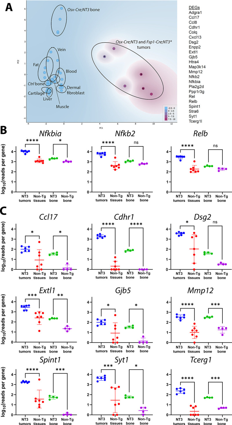
Fig 5

(A) Principal component analysis of Osx-Cre;NT3 tumors and FSP1-Cre;NT3 tumors (marked with *), Ctrl and Osx-Cre;NT3 bone, and other normal tissues utilizing the 23 genes listed. Expression of (B) NF-κB genes (Nfkbia, Nfkb2, and Relb) and (C) 9 additional NT3 transgene targets previously identified as top DEGs in NT3 bone vs Ctrl bone, each with 2 or more κB response elements. Log10(reads per gene) is shown from normalized RNA-Seq data. One-way ANOVA, followed by Dunnett’s multiple comparisons test; *, p<0.05, **p<0.01; ***, p<0.001; ****, p<0.0001.

Kaplan-Meier curves for tumor latency were compared either within or between strains using a standard log-rank (Mantel-Cox) test if the proportional hazard assumption was reasonable. If the assumption was violated, an extended Cox proportional hazards model was applied. For Osx-Cre;NT3 male and female mice, hazard ratios were calculated in two different time intervals (t<125 days or t≥125 days) by including two Heaviside functions (hv1 = 1*male if t<125, hv1 = 0 if t≥125; hv2 = 1*male if t≥125; hv2 = 0 if t<125) with assumption that the hazard ratios are constant in each time interval. Ctrl animals for each strain were considered censored at the end of the follow-up period. Values of p<0.05 were considered significant, and data are presented as mean ± SD. Sample sizes are indicated in the respective figure legends or methods section.

Results

NT3 transgenic mice develop spontaneous soft tissue tumors in the mesenchymal lineage

In the course of characterizing the bone phenotype of the Osx-Cre;NT3 mice [26], we observed spontaneous soft tissue tumors. Based on their subcutaneous location and histological appearance, we considered that a mesenchymal origin was likely, and therefore crossed the NT3 transgenic mice with a FSP1-Cre (S100a4) line [31], which predominantly shows expression in the fibroblast population of multiple organs but has also been documented in the myeloid lineage [31, 32, 4345]. Beginning at 6 weeks of age, we monitored these mice for palpable tumor masses 1–2 times per week.

Both Osx-Cre;NT3 and FSP1-Cre;NT3 mice presented with spontaneous, superficial soft tissue tumors with 100% penetrance (Fig 1A). Ctrl, Cre-negative littermates of either line, did not develop tumors during a 1-year follow-up period. Tumors from both transgenic lines were generally firm, beige, and nodular in appearance. Osx-Cre;NT3 mice had masses that were typically larger, more cystic, and predominantly (95% of males, 68% of females) found on or near the face in the submandibular region. In addition to the face, Osx-Cre;NT3 animals also presented with tumors on the trunk (15% of males, 50% of females), perineal area (5% of males, 26% of females), and rarely on the limbs (1 female, 3%) (S2A Fig). FSP1-Cre;NT3 tumors often became ulcerated and were most commonly found in the perineal region (78% of males, 100% of females). Tumor locations for FSP1-Cre;NT3 animals also included the face (78% of males, 58% of females), localized mainly anterior on the snout, as well as the trunk (11% of males, 67% of females) and on the limbs (11% of males, 25% of females) (S2A Fig). Consistent with the broader expression pattern of FSP1 in the mesenchymal lineage, we observed more tumors on average (3.57 ± 1.83 vs 1.95 ± 2.02, **p<0.0018) in FSP1-Cre;NT3 animals, in addition to an earlier onset (79 vs 129 days, ****p<0.0001) versus their Osx-Cre;NT3 counterparts. Lastly, radiographic imaging confirmed no overt bony tumors in either line that would be indicative of an osteosarcoma (S2B Fig).

Fig 1. NT3 transgenic mice develop spontaneous soft tissue tumors.
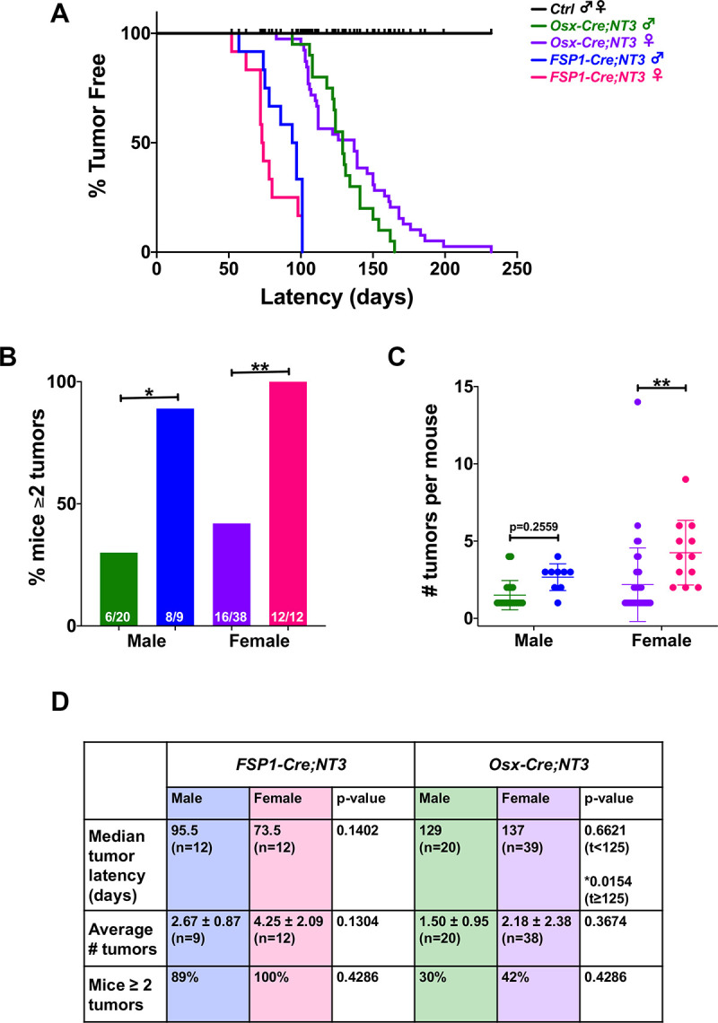
Fig 1

(A) Kaplan-Meier curves showing tumor latency by sex in each strain. (B) Comparison between strains of males or females for the percentage of mice with ≥ 2 tumors or (C) number of tumors per mouse. (D) Comparison by sex of the median tumor latency, average number of tumors per mouse, and percentage of mice with ≥ 2 tumors in Osx-Cre;NT3 and FSP1-Cre;NT3. Sample sizes are as indicated. Results are presented as mean ± SD. Standard log-rank (Mantel-Cox) test or extended (time-varying) Cox proportional hazards model for Kaplan-Meier curves. Two-way ANOVA followed by Sidak’s multiple comparisons test for numbers of tumors per mouse or Fisher’s exact test with FDR adjustment for percentage of mice with ≥ 2 tumors. *p<0.05, **p<0.01.

We next examined if there was a sex difference between the two mesenchymal Cre-driven NT3 transgenic lines. There was no interaction between sex and strain for this between-group comparison. However, the percentage of mice presenting with multiple tumors is significantly higher with FSP1-Cre in both sexes (Fig 1B). The absolute number of tumors per mouse revealed a similar upward trend in either sex in Osx-Cre;NT3 vs FSP1-Cre;NT3, but this was only statistically significant in female mice (Fig 1C). The lack of statistical significance in male animals in Fig 1C is likely due to the difference in sample sizes as there were nearly twice as many animals in the Osx-Cre;NT3 vs the FSP1-Cre;NT3 cohort.

Analysis of the sex by time interaction indicated a time-varying hazard ratio between sexes within the same strain only for Osx-Cre;NT3 mice (*p = 0.0173). A Cox proportional hazards model in two different time intervals (t<125 days or t≥125 days) was applied to test for a possible divergence in tumor latency by sex. In the first time interval (t<125 days), Osx-Cre;NT3 males had a longer tumor latency than females, but this difference was not statistically significant. However, in the second time interval (t≥125 days), Osx-Cre;NT3 males had a considerably shorter tumor latency period than females (Fig 1A and 1D). A similar tumor latency was observed between Osx-Cre;NT3 males with single vs multiple tumors (129 vs 126 days) (S2C Fig). Osx-Cre;NT3 females that presented with multiple tumors had a markedly longer median tumor latency period than females with only single tumors (161.5 days vs 112 days) (S2C Fig). Notably, neither average number of tumors, multiple tumor incidence, nor tumor latency differed significantly between male and female FSP1-Cre;NT3 mice (Fig 1D, S2C Fig). Overall, despite a mild statistical difference between male and female Osx-Cre;NT3 mice in tumor latency, we concluded that the NT3 transgene was similarly oncogenic in both sexes of either strain, and the primary differences between the strains were the shorter latency and higher number of tumors with FSP1-Cre.

Spontaneous tumors derive from NT3 transgene expressing cells

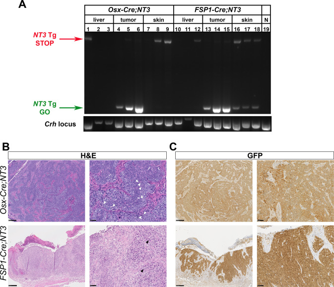
To verify NT3 transgene expression in tumor tissue, we first performed PCR on gDNA from whole tumors. Primers were designed to flank the loxP/Neo-STOP/loxP cassette [21]. In the absence of Cre-mediated recombination, the intact allele results in a larger band, inefficiently amplified in these PCR conditions (Fig 2A, NT3 Tg STOP). If the loxP/Neo-STOP/loxP cassette is excised, a smaller band is present (Fig 2A, NT3 Tg GO). Tumors from both Osx-Cre;NT3 (Fig 2A, lanes 4–6) and FSP1-Cre;NT3 (Fig 2A, lanes 13–15) mice show robust recombination of the NT3 allele. Liver samples from these same mice served as a negative control for the recombination event (Fig 2A, lanes 1–3; 10–12). A parallel PCR of the Crh, corticotropin releasing hormone, locus was used as a positive control for gDNA extraction of whole tissue preps (Fig 2A, bottom panel). Furthermore, as the location of these tumors was subcutaneous, we also checked for presence of the recombination band in whole skin tissue to see if the tumors were perhaps arising from Cre expression in dermal fibroblasts. The NT3 Tg GO product was seen in skin from FSP1-Cre;NT3 mice (Fig 2A, lanes 16–18) but not in skin from Osx-Cre;NT3 (Fig 2A, lanes 7–9) or Ctrl animals (Fig 2A, lane 19), suggesting that FSP1-Cre drives significant recombination in benign skin. FSP1-Cre is known to be expressed in dermal fibroblasts [31, 43], but was not found in keratinocytes [32]. Similarly, skin from Osx-Cre;Tdt reporter mice did not show positive Tdt expression in keratinocytes (S3A and S3B Fig). Interestingly, we did observe a rare population of Tdt+ cells in the dermis as well as in cultured dermal fibroblasts from Osx-Cre;Tdt mice (S3 Fig). These findings suggest that NT3 transgene recombination occurs in dermal fibroblasts of both strains, with the greater extent in FSP1-Cre;NT3 mice corresponding to a greater number and earlier onset of tumors.

Fig 2. Tumors derive from NT3-transgene expressing cells.

Fig 2

(A) gDNA PCR of whole tissue from liver, tumor, or skin in Osx-Cre;NT3, FSP1-Cre;NT3, or Ctrl animals. Amplification across the loxP/Neo-STOP/loxP cassette in the intact allele or after Cre-mediated recombination. NT3 Tg = NT3 transgene. N = Cre-negative, Ctrl. Crh, corticotropin releasing hormone. (B) Representative H&E staining or (C) immunohistochemistry for GFP which is expressed by the NT3 transgene as well as the Cre construct in Osx-Cre mice. (left images) 10x, scale bars = 250μM and (right images) 40x magnification, scale bars = 50μM.

H&E staining revealed a similar histologic pattern in both Osx-Cre;NT3 and FSP1-Cre;NT3 tumors. The tumors have a solid and sometimes nested growth pattern, usually with some areas of elongated, spindled cell shapes. Tumor cells are enlarged, with pleomorphic nuclei and frequent mitoses (Fig 2B). Some tumors have regional necrosis. Despite these aggressive features, tumor borders were largely pushing rather than infiltrative. As both the NT3 transgene and the Osx-Cre driver express a GFP tag, we used an antibody to GFP to immunostain the tumors. The majority of tumor cells (75–100%) are GFP-positive in both Osx-Cre;NT3 and FSP1-Cre;NT3 tumor samples (Fig 2C). The immunostained sections also highlight nests of tumor cells at the dermal-epidermal junction in many FSP1-Cre;NT3 tumors (Fig 2C, bottom right).

NT3 tumors share markers of soft tissue sarcomas

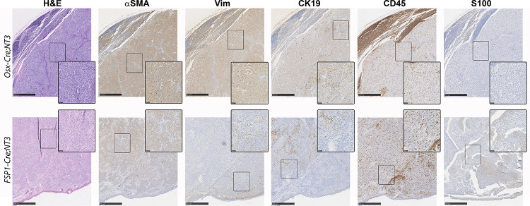
We next sought to determine the cellular origin of these spontaneous tumors using immunohistochemistry (Fig 3). Use of either the Osx-Cre or FSP1-Cre drivers targets NT3 transgene expression to the mesenchymal lineage. However, evidence for NF-κB, and particularly alternative NF-κB, in the pathogenesis of sarcomas is very limited and largely associative [46]. Nevertheless, we proceeded to investigate two well-known mesenchymal lineage markers, alpha-smooth muscle actin (αSMA) and vimentin (Vim), for which anti-mouse antibodies are available. Both Osx-Cre;NT3 and FSP1-Cre;NT3 tumors (n = 3 of each) exhibit moderate to strong αSMA staining in at least 60% of the tumor cells. Strong expression of Vim was seen in most (70–80%) tumor cells from Osx-Cre;NT3 animals, but Vim staining was restricted to the periphery in FSP1-Cre;NT3 tumors, where 20–50% of cells were weakly to moderately positive. Furthermore, staining of the epithelial marker, cytokeratin 19 (CK19), revealed a dichotomous expression pattern in these tumors with areas of positive as well as negative staining within the tumor mass. Interestingly, the CK19+ cells appeared morphologically distinct as large, spindle-shaped cells compared to the smaller, rounder tumor cells.

Fig 3. NT3 tumors have features of soft tissue sarcomas.

Fig 3

Immunohistochemical staining of representative Osx-Cre;NT3 (top) and FSP1-Cre;NT3 (bottom) tumors for hematoxylin and eosin (H&E), alpha-smooth muscle actin (αSMA), vimentin (Vim), Cytokeratin 19 (CK19), CD45, and S100 protein. Scale bars = 500μM. Insets are taken from boxed regions, with scale bars = 50μM.

Both the Osx-Cre and FSP1-Cre drivers have been previously shown to have off-target effects outside of the mesenchymal lineage, most notably in cells of hematopoietic origin [32, 33, 44, 45, 47, 48]. To this end, we assayed CD45 expression. While tumor samples from both mesenchymal Cre-drivers showed positive CD45 staining, this appeared to be restricted to the inflammatory infiltrate. FSP1-Cre;NT3 tumor samples exhibited a higher degree of this CD45 infiltrate, most likely owing their propensity to be superficially ulcerated at presentation.

As melanoma is almost always part of the diagnostic workup for a poorly differentiated subcutaneous tumor in patients, we checked for S100 expression [49]. Remarkably, both FSP1-Cre;NT3 and Osx-Cre;NT3 tumors were negative for S100 staining, except for a few individual cells that are likely skin macrophages that have taken up melanosomes from benign melanocytes. In sum, the totality of the above morphological and immunohistochemistry evidence suggests that these spontaneous tumors represent a poorly differentiated, aggressive sarcoma.

NF-κB /NF-κB -related biological processes are enriched in NT3 tumors

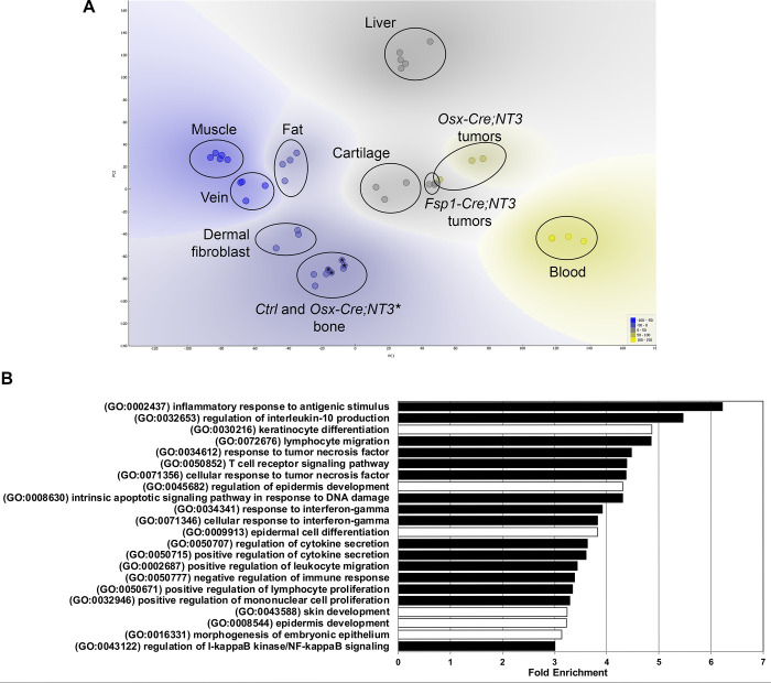
To gain insight into both the possible cellular origin and pathogenesis of these spontaneous tumors, we performed RNA-Seq analysis of tumors from both Osx-Cre;NT3 and FSP1-Cre;NT3 mice. We performed principal component analysis (PCA) of tumors and a panel of benign tissues using publicly available datasets from NCBI’s Gene Expression Omnibus (GEO) [26, 3942] (S5 Table), including cortical bone samples from Osx-Cre;NT3 and littermate Ctrl. We also included two tissues or cell types of non-mesenchymal origin (e.g. blood and liver) as biological outgroups for PCA and hierarchical clustering. As expected, tumors from both lines segregated away from both blood and liver samples (Fig 4A). Notably, the Osx-Cre;NT3 tumors clustered more closely with the FSP1-Cre;NT3 tumors than to cortical bone from Osx-Cre;NT3 mice [26]. Both Osx-Cre;NT3 and FSP1-Cre;NT3 tumors did not significantly overlap with any other mesenchymal tissue assayed (muscle, vein, fat, cortical bone, dermal fibroblast) except for mature cartilage. Anatomically, these tumors were not found near any cartilaginous sites, and histologically they did not show indications of cartilage differentiation such as lacunar morphology or matrix production. Furthermore, expression of selected cartilage and muscle marker genes was low compared to their respective benign tissues (S4 Fig). Based on this analysis, we concluded that the Osx-Cre;NT3 and FSP1-Cre;NT3 tumor expression profiles most closely match one another, suggesting a distinct, but as yet unclear, developmental origin.

Fig 4. NT3 tumors cluster together and show upregulation of NF-κB biological processes.

Fig 4

(A) Principal component analysis of Osx-Cre;NT3 tumors and FSP1-Cre;NT3 tumors vs Ctrl bone and Osx-Cre;NT3 bone (marked with an *) as well as other benign tissues—muscle, fat, vein, dermal fibroblast, mature cartilage, blood, liver. (B) Gene ontology (GO) analysis of NT3 tumors vs the same panel of tissues in (A) showing the top biological processes (BP) with fold-enrichment ≥ 3 and a Fisher’s exact FDR adjusted p-value < 1e-3. black bars = NF-κB /NF-κB related processes; white bars = skin/epithelial processes.

Due to their high degree of similarity, we grouped together Osx-Cre;NT3 and FSP1-Cre;NT3 tumors (NT3 tumors) and compared them to all remaining tissues shown in Fig 4A to generate a list of differentially expressed genes (DEGs). We further filtered the DEG list to only those genes with a fold-change (FC) >1.5 (S6 Table) as NF-κB chiefly functions as an activating transcriptional program. Moreover, the vast majority of DEGs in our previous study comparing Ctrl bone to Osx-Cre;NT3 bone showed that differences skewed strongly towards increased, and not decreased, target gene expression as a result of the NT3 transgene [26]. Inspection of the DEG list did not yield any evidence of a single oncogenic driver and/or tumor suppressor. Importantly, we did not observe abnormal expression levels of oncogenes known to lead to NF-κB activation including p53, KRAS, EGFR, or PI3K [3]. However, this lack of evidence could be due to the absence of a true matched reference for our NT3 tumors since their etiology remains unknown.

As individual genes are rarely biologically informative in isolation, we performed pathway analysis. Gene ontology (GO) analysis for biological process (BP) of the 1,314 DEG genes from above (S6 Table) revealed 509 GO BP terms, 170 of which were highly significant (FDR adjusted p-value <1e-3; S7 Table). To distill out the key pathways driving spontaneous tumor formation in Osx-Cre;NT3 and FSP1-Cre;NT3 mice, we further refined our search to those processes with a fold-enrichment ≥ 3 relative to the expected incidence in the mouse genome (Fig 4B). The most overrepresented program, with a 6.22 fold-enrichment score, was the GO term for inflammatory response to antigenic stimulus (GO:000243) while the GO term for regulation of IKK/NF-κB signaling (GO:0043122) displayed a 3-fold enrichment. Of the 22 total GO BP shown in Fig 4B, 73% (16/22 –black bars) are known NF-κB/NF-κB related terms, predominated by inflammation and immune processes. Other NF-κB related GO terms of interest include intrinsic apoptotic signaling pathway in response to DNA damage (GO:0008630) and both positive regulation of lymphocyte proliferation (GO:0050671) and positive regulation of mononuclear cell proliferation (GO:0032946), as these may indicate possible tumor cell proliferation. The remaining 27% GO BP (6/22 –white bars) represent skin development, particularly of the epidermis, which is likely contamination of overlaying skin incidental from gross tumor excision.

We next performed a gene set enrichment analysis (GSEA) focused on NF-κB related gene sets to determine if NT3 tumors and NT3 bone [26] would co-segregate, reflecting common transgene targets. However, with the large number of genes represented (~500 genes, S8 Table), the tissue signature predominated, and all bones clustered with each other and away from the tumors (S5 Fig). To more specifically focus on NT3 transgene targets, we turned to our previous comparison of gene expression in age/sex/tissue matched Osx-Cre;NT3 and Ctrl bone. PCA of the top 20 DEGs and 3 NF-κB pathway genes that were upregulated in Osx-Cre;NT3 vs Ctrl bones was performed, including all of the samples used in Fig 4A. All of the non-transgenic tissues including bone clustered together, and although the NT3-expressing tumors and NT3 bone were distinct from each other, they segregated away from this large cluster (Fig 5A). To further demonstrate NF-κB activation, we compared expression of the 3 NF-κB genes (Fig 5B), as well as several DEGs (Fig 5C), each with 2 or more κB response elements. Strikingly, the vast majority of these normal tissues had much lower levels of expression of all of these NF-κB targets, despite the varied tissues represented, and there were only rare cases in which individual tissues showed expression comparable to the NT3 tumors.

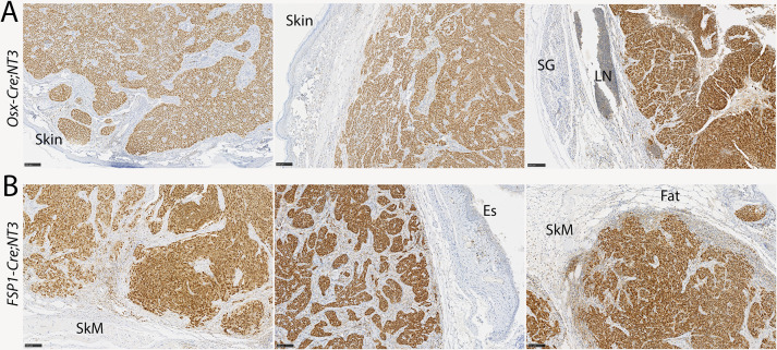
With this strong evidence of NF-κB upregulation in tumors, we turned to immunostaining to verify that the tumor cells themselves, rather than tumor stroma, show pathway activation. RelB is the principal NF-κB subunit downstream of NIK responsible for modulation of gene transcription. Strikingly, both Osx-Cre;NT3 and FSP1-Cre;NT3 mice show robust nuclear staining of RelB in all tumors (Fig 6A and 6B). Furthermore, there is a clear distinction between the uniformly RelB-positive tumor cells versus RelB-negative stromal cells. The majority of cells in adjacent benign tissues also lack nuclear RelB. Based on these gene expression and immunohistochemistry data, together with the presence of the Cre-mediated recombination product, in two distinct mouse lines, we concluded that NT3-initiated tumor formation is indeed driven by NF-κB activation.

Fig 6. The NT3 transgene activates alternative NF-κB in tumor cells.

Fig 6

Immunostaining for RelB in (A) Osx-Cre;NT3 or (B) FSP1-Cre;NT3 tumors. RelB is uniformly nuclear in malignant cells, indicating robust activation of the alternative NF-κB pathway. Few cells in adjacent benign tissues show significant staining for RelB. SG, salivary gland; LN, lymph node; SkM, skeletal muscle; Es, esophagus. Scale bars = 100μM. n = 3 each genotype.

Discussion

Although the initial discovery of NF-κB arose from observations that the v-rel viral oncogene and translocations of c-rel can be oncogenic in lymphocytes, few mutations in genes of the NF-κB pathway have clear oncogenic effects [50]. In fact, the NT3 transgene used here was first generated after multiple genetic aberrations in the alternative NF-κB pathway were identified in B cell malignancies. However, removal of the negative regulatory TRAF3-binding domain of NIK in this transgene was insufficient to cause oncogenesis in the B cell lineage [2123]. Other studies have expressed the NT3 transgene in hematopoietic stem cells, T cells, myeloid cells, intestine, pancreatic islets, and hepatocytes, without reports of malignant transformation [5156]. NF-κB expression is found both within the epidermal and dermal layers of the skin with expression in the dermis restricted to dermal fibroblasts [57]. Intact NF-κB signaling, particularly upstream kinase activation, is essential to maintain epidermal homeostasis and to prevent squamous cell carcinoma [14]. Therefore, the presentation of subcutaneous tumors in Osx-Cre;NT3 mice was unexpected. Our initial motivation in generating these conditional transgenic mice was to understand the role of alternative NF-κB in osteogenic cells, where Osx is highly expressed, but we never observed any bone tumors. Based on the location and histologic appearance, we hypothesized that the cell of origin was likely mesenchymal and in the dermis or subcutaneous soft tissues, and we were able to replicate the tumor phenotype with a Cre driver with that expression pattern, FSP1-Cre. Together, these two mouse lines displaying spontaneous tumorigenesis indicate that the loss of TRAF3 binding in NIK can be oncogenic in certain cellular contexts.

We used immunohistochemistry to attempt to classify NT3 tumors by cell of origin. Both Osx-Cre and FSP1-Cre driven NT3 tumors expressed the marker αSMA in most malignant cells, while vimentin staining was present, although more variable, in the latter colony. It is not clear why vimentin was less consistently expressed than αSMA. Nevertheless, αSMA and GFP showed a very high degree of concordance. A mesenchymal origin is also aligned with that expected from the overlap in pattern of these Cre drivers. Thus, the totality of the evidence points towards a mesenchymal origin without differentiation towards a specific lineage. The classification of poorly differentiated sarcomas is notoriously difficult, with a paucity of diagnostic antibodies for mesenchymal populations, compounded here by limited cross-reactivity of antibodies to test tumors of murine rather than human origin. The presence of a significant population of CK19+ cells in both tumor types, in populations of large and pleomorphic cells indicative of tumor rather than host cells, is somewhat confounding. CK19 expression has been described mainly in thyroid, gastroenteropancreatic and hepatobiliary tumors [58, 59]. Because of the unique combination of location, overall histology, and expression patterns of the two Cre drivers, we consider an epithelial origin unlikely. The calcium binding protein S100, a marker that is highly expressed in melanomas, was completely absent in our tumors, and this result argues against a melanocytic derivation. Although expression of both Osx-Cre and FSP1-Cre alleles has been observed in CD45+ cells [32, 33, 44, 45, 48], the absence of CD45 in the tumor cells indicates that a hematopoietic origin is unlikely. In further support of this conclusion, expression of this NT3 transgene in various hematopoietic cells did not result in malignancy [5153].

We next attempted further classification of NT3 tumors using RNA-Seq data and PCA, which suggested that the tumors are distantly related to tissues with cartilage expression signatures. However, histologic features are not consistent with a chondrogenic origin, which also seems unlikely in dermal or subcutaneous mesenchyme, and cartilage marker genes are poorly expressed. The NT3 transgene is expressed in the skin of both FSP1-Cre;NT3 and Osx-Cre;NT3 animals, but PCA analysis did not indicate shared features with normal dermal fibroblasts. As bulk RNA-Seq analysis of NT3 tumors is unlikely to uncover significant similarity to a distinct subpopulation of dermal fibroblasts, this does not rule out a dermal origin for tumorigenesis. Undoubtedly, our RNA expression analysis is limited by the datasets used for comparison. Without clear hypotheses to indicate other tissues or cells to include, further comparisons are outside the scope of this study. Such work might include filtering the data to reduce the keratinocyte signature, since the tumors do not appear to have an epidermal origin, as well as comparisons to a panel of murine mesenchymal and perhaps epithelial tumors. Finally, one intriguing possibility is a follicular dendritic cell sarcoma, which can arise from ubiquitous perivascular cells, and in humans can show aberrant NF-κB regulation and high expression of CXCL13, an alternative NF-κB driven gene [60].

Based on early studies employing overexpression, NIK was originally named for its ability to activate classical NF-κB signaling [61]. Although it was later shown to be dispensable for this function, various studies employing the constitutively activated NIK allele used here have shown induction of both classical and alternative NF-κB signaling [1]. RNA-Seq analysis of NT3-driven tumors demonstrates activation of κB-responsive genes including RelB, but it is not clear if tumorigenesis requires classical and/or alternative signaling. Therefore, additional studies utilizing mice deficient in downstream components specific to each pathway would be required to demonstrate whether one or both pathways are necessary for the observed tumorigenesis.

The variable time to emergence of the NT3 tumors, as well as a relatively small average number of tumors per mouse, suggests that a second genetic hit is likely needed in addition to NIK activation for oncogenic transformation. Previous studies have shown that Bcl6 [22] and Notch [23] collaborate with the NT3 transgene to generate tumors. Yet, pathway analysis of RNA-Seq data from NT3 tumors compared to several benign tissues failed to identify other clear oncogenic signals, including pathways such as p53 that have previously been linked to sarcomagenesis [46, 62]. Our study remains limited by the lack of a corresponding benign comparator to the cell of origin. Furthermore, chromosomal translocations are also a common feature of sarcomas, and we have not queried for this form of genomic change in the NT3 tumors.

Although we wish to emphasize that the NT3 transgene drove tumors in both sexes, with two different Cre drivers, we did observe some differences between the groups. With regard to the Cre driver, tumors in FSP1-Cre;NT3 mice of both sexes appeared with shorter latency and in greater numbers. This is most likely a direct effect of constitutive expression of the FSP1-Cre allele in dermal fibroblasts. Cre expression in Osx-Cre;NT3 mice, on the other hand, was induced postnatally in our studies, and detected only in a rare population of dermal fibroblasts. Despite subtle variations by sex in tumor latency in the Osx-Cre;NT3 mice, none were present in the FSP1-Cre;NT3 line. Many Osx-Cre;NT3 males in our colony were utilized for other experiments prior to the appearance of tumors [26] and, therefore, the number of animals between sexes in this strain was not balanced. We also had fewer FSP1-Cre;NT3 mice of both sexes, as these were generated later. The discrepancy in group size may have influenced the outcome of the sex-by-strain statistical analysis. Nevertheless, despite the fact that we previously observed sex differences in bone phenotype with modulation of the alternative NF-κB pathway [63], there appears to be little to no biologically significant effect of sex on tumorigenesis suggesting that NT3 tumors form independent of male or female sex steroids.

In summary, we describe here a new, 100% penetrant, spontaneous soft tissue tumor model in mice expressing an activated form of NIK, the apex kinase in the alternative NF-κB pathway. Tumors arose using two distinct Cre-driver strains with overlapping mesenchymal lineage expression, supporting our conclusion that this mutant NIK allele has oncogenic potential. While NF-κB activation is a feature of many types of hematopoietic and epithelial tumors, it has not previously been intimately associated with mesenchymal tumors. Our observation that this particular NIK allele seems to have oncogenic potential only in the latter cell ontogeny suggests that there is much we do not yet understand about the role of NF-κB in cancer, specifically in the sarcoma setting.

Supporting information

S1 Fig. IHC negative control staining.

(A) No primary antibody stain for anti-goat GFP-biotin antibody. (B) Rat IgG background staining for CK19 antibody. (C) Rabbit IgG background staining for αSMA, vimentin, CD45, and S100 protein antibodies. All negative control stains were performed on Osx-Cre;NT3-tumor sections. Top row for each panel is 10x and the bottom row for each panel is 40x magnification. Scale bars = 100μM.

(TIF)

S2 Fig. Additional tumor information for Osx-Cre;NT3 and FSP1-Cre;NT3 mice.

(A) Number of Osx-Cre;NT3 and FSP1-Cre;NT3 mice presenting with a tumor mass at different anatomical locations. In the case of multiple tumors within the same animal, the animal was counted once in each category for 1 or more masses at a given location. (B) Representative radiographic images revealing no overt osseous tumors in either strain. scale bar = 3mm. (C) Median tumor latency for Osx-Cre;NT3 and FSP1-Cre;NT3 animals presenting with single or multiple (≥2) tumors. Samples sizes are as indicated in each panel. Standard log-rank (Mantel-Cox) test: ****p<0.0001.

(TIF)

S3 Fig. Rare population of Tdt+ dermal fibroblasts in Osx-Cre;Tdt mice.

(A) Direct fluorescence of Tdt with DAPI counterstain in frozen skin sections from Ctrl or (B) Osx-Cre;Tdt mice. Representative 20x images shown with white arrows denoting Tdt+ cells. Scale bars = 20μm. (A’-B’) Higher magnification of boxed areas in A-B. Scale bars = 5μm. n = 2 each genotype. (C) Flow cytometry analysis for Tdt in cultured skin fibroblasts from Ctrl or Osx-Cre;Tdt mice. n = 3 each genotype. Unpaired one-tailed t-test with Welch’s correction: *p<0.05.

(TIF)

S4 Fig. Low expression of cartilage and muscle markers in Osx-Cre;NT3 and FSP1-Cre;NT3 tumors.

(A) Expression of cartilage markers or (B) muscle markers in NT3 tumors versus the respective tissue. Log10(reads per gene) is shown from normalized RNA-Seq data. Mann Whitney U test; *, p<0.05, **p<0.01.

(JPG)

S5 Fig. Tissue signature dominates over NT3 transgene signature in broad NF-kB gene list.

Principal component analysis of Osx-Cre;NT3 tumors, FSP1-Cre;NT3 tumors, Osx-Cre;NT3 bone, and Ctrl bone using genes from a gene set enrichment analysis (GSEA) of top NF-κB related gene sets listed in S8 Table.

(PNG)

S6 Fig. Raw gel image for Fig 2.

(TIF)

S1 Table. Genotyping conditions.

(XLSX)

S2 Table. List of antibodies.

(XLSX)

S3 Table. Genomic DNA recombination conditions.

(XLSX)

S4 Table. q-RT-PCR primers for peripheral blood and monocyte markers.

(XLSX)

S5 Table. GEO datasets for Osx-Cre;NT3 and FSP1-Cre;NT3 tumors and benign tissues.

(XLSX)

S6 Table. Mean normalized counts for DEG list between NT3 tumors and benign tissues.

(XLSX)

S7 Table. GO analysis for top DEGs.

(XLSX)

S8 Table. Mean normalized counts NF-κB gene sets from GSEA between NT3 tumors and NT3 bone.

(XLSX)

S1 File

(DOCX)

Acknowledgments

We wish to thank Crystal Idleburg and Samantha Coleman for their histological expertise as well as Gary London for imaging support. Katherine Gruner at Northwestern University provided valuable technical assistance with IHC.

Data Availability

All relevant data are contained within the manuscript and its Supporting Information files. RNA‐Seq data have been deposited in NCBI’s Gene Expression Omnibus (https://www.ncbi.nlm.nih.gov/geo/) under the GEO Series accession number GSE158479.

Funding Statement

This work was supported by NIAMS, National Institutes of Health Grants R01 AR052705 (to DJV), R01 AR070030 (to DJV and RF) and R01 AR066551 (to RF), R01 AR049069 (to AJvW), F31 AR068853 (to JLD), T32 AR060719 (to HZ); and NCI, National Institutes of Health, grants R01 CA235096 (to RF), and T32 CA113275 (to JLD). Histology services were provided by the Washington University Musculoskeletal Research Center Histology and Morphometry Core supported by P30 AR074992 and the Northwestern University Research Histology and Phenotyping Laboratory supported by P30 CA060553 awarded to the Robert H Lurie Comprehensive Cancer Center. Slide images were captured in the Alafi Neuroimaging Laboratory supported by S10RR027552. Additional support was provided by the Siteman Cancer Center Investment Program, St. Louis, MO to RF. Funding from Shriners Hospitals for Children was provided to DJV and RF. The funders had no role in study design, data collection and analysis, decision to publish, or preparation of the manuscript. The content is solely the responsibility of the authors and does not necessarily represent the official views of the National Institutes of Health.

References

  • 1.Zhang Q., Lenardo M. J., and Baltimore D., “30 Years of NF-κB: A Blossoming of Relevance to Human Pathobiology,” Cell, vol. 168, no. 1, pp. 37–57, Jan. 2017. doi: 10.1016/j.cell.2016.12.012 [DOI] [PMC free article] [PubMed] [Google Scholar]
  • 2.Novack D. V., “Role of NF-κB in the skeleton,” Cell Res., vol. 21, no. 1, pp. 169–182, Nov. 2010. doi: 10.1038/cr.2010.159 [DOI] [PMC free article] [PubMed] [Google Scholar]
  • 3.Xia Y., Shen S., and Verma I. M., “NF-κB, an active player in human cancers.,” Cancer Immunology Research, vol. 2, no. 9, pp. 823–830, Sep. 2014. doi: 10.1158/2326-6066.CIR-14-0112 [DOI] [PMC free article] [PubMed] [Google Scholar]
  • 4.Frederiksen A. L., Larsen M. J., Brusgaard K., Novack D. V., Knudsen P. J. T., Schrøder H. D., et al., “Neonatal High Bone Mass With First Mutation of the NF-κB Complex: Heterozygous De Novo Missense (p.Asp512Ser) RELA (Rela/p65).,” J. Bone Miner. Res., vol. 31, no. 1, pp. 163–172, Jan. 2016. doi: 10.1002/jbmr.2590 [DOI] [PMC free article] [PubMed] [Google Scholar]
  • 5.Ghosh S. and Hayden M. S., “Celebrating 25 years of NF-κB research.,” Immunol. Rev., vol. 246, no. 1, pp. 5–13, Mar. 2012. doi: 10.1111/j.1600-065X.2012.01111.x [DOI] [PMC free article] [PubMed] [Google Scholar]
  • 6.Sun S.-C., “The non-canonical NF-κB pathway in immunity and inflammation,” Nature Publishing Group, vol. 17, no. 9, pp. 545–558, Jun. 2017. doi: 10.1038/nri.2017.52 [DOI] [PMC free article] [PubMed] [Google Scholar]
  • 7.Shih V. F.-S., Tsui R., Caldwell A., and Hoffmann A., “A single NFκB system for both canonical and non-canonical signaling,” Nature Publishing Group, vol. 21, no. 1, pp. 86–102, Nov. 2010. doi: 10.1038/cr.2010.161 [DOI] [PMC free article] [PubMed] [Google Scholar]
  • 8.Kaltschmidt B., Greiner J., Kadhim H., and Kaltschmidt C., “Subunit-Specific Role of NF-κB in Cancer,” Biomedicines, vol. 6, no. 2, pp. 44–12, Jun. 2018. doi: 10.3390/biomedicines6020044 [DOI] [PMC free article] [PubMed] [Google Scholar]
  • 9.Perkins N. D., “The diverse and complex roles of NF-κB subunits in cancer,” Nat. Rev. Cancer, Jan. 2012. doi: 10.1038/nrc3204 [DOI] [PubMed] [Google Scholar]
  • 10.Gasparini C., Celeghini C., Monasta L., and Zauli G., “NF-κB pathways in hematological malignancies,” Cell. Mol. Life Sci., vol. 71, no. 11, pp. 2083–2102, Jan. 2014. doi: 10.1007/s00018-013-1545-4 [DOI] [PMC free article] [PubMed] [Google Scholar]
  • 11.Wu D., Wu P., Zhao L., Huang L., Zhang Z., Zhao S., et al., “NF-κB Expression and Outcomes in Solid Tumors,” Medicine, vol. 94, no. 40, pp. e1687–12, Oct. 2015. doi: 10.1097/MD.0000000000001687 [DOI] [PMC free article] [PubMed] [Google Scholar]
  • 12.Colomer C., Marruecos L., Vert A., Bigas A., and Espinosa L., “NF-κB Members Left Home: NF-κB-Independent Roles in Cancer,” Biomedicines, vol. 5, no. 4, pp. 26–11, Dec. 2017. doi: 10.3390/biomedicines5020026 [DOI] [PMC free article] [PubMed] [Google Scholar]
  • 13.Luedde T., Beraza N., Kotsikoris V., van Loo G., Nenci A., De Vos R., et al., “Deletion of NEMO/IKKγ in Liver Parenchymal Cells Causes Steatohepatitis and Hepatocellular Carcinoma,” Cancer Cell, vol. 11, no. 2, pp. 119–132, Feb. 2007. doi: 10.1016/j.ccr.2006.12.016 [DOI] [PubMed] [Google Scholar]
  • 14.Liu B., Xia X., Zhu F., Park E., Carbajal S., Kiguchi K., et al., “IKKα Is Required to Maintain Skin Homeostasis and Prevent Skin Cancer,” Cancer Cell, vol. 14, no. 3, pp. 212–225, Sep. 2008. doi: 10.1016/j.ccr.2008.07.017 [DOI] [PMC free article] [PubMed] [Google Scholar]
  • 15.Thu Y. M. and Richmond A., “NF-kB inducing kinase: A key regulator in the immune system and in cancer,” Cytokine Growth Factor Rev., vol. 21, no. 4, pp. 213–226, Aug. 2010. doi: 10.1016/j.cytogfr.2010.06.002 [DOI] [PMC free article] [PubMed] [Google Scholar]
  • 16.Maubach G., Feige M. H., Lim M. C. C., and Naumann M., “NF-kappaB-inducing kinase in cancer,” BBA—Reviews on Cancer, vol. 1871, no. 1, pp. 40–49, Jan. 2019. doi: 10.1016/j.bbcan.2018.10.002 [DOI] [PubMed] [Google Scholar]
  • 17.Tegowski M. and Baldwin A., “Noncanonical NF-κB in Cancer,” Biomedicines, vol. 6, no. 2, pp. 66–21, Jun. 2018. doi: 10.3390/biomedicines6020066 [DOI] [PMC free article] [PubMed] [Google Scholar]
  • 18.Giopanou I., Lilis I., Papadaki H., Papadas T., and Stathopoulos G. T., “A link between RelB expression and tumor progression in laryngeal cancer.,” Oncotarget, vol. 8, no. 69, pp. 114019–114030, Dec. 2017. doi: 10.18632/oncotarget.23109 [DOI] [PMC free article] [PubMed] [Google Scholar]
  • 19.Leonard B., McCann J. L., Starrett G. J., Kosyakovsky L., Luengas E. M., Molan A. M., et al., “The PKC/NF-κB Signaling Pathway Induces APOBEC3B Expression in Multiple Human Cancers,” Cancer Res., vol. 75, no. 21, pp. 4538–4547, Nov. 2015. doi: 10.1158/0008-5472.CAN-15-2171-T [DOI] [PMC free article] [PubMed] [Google Scholar]
  • 20.Li Y., Zhou Q.-L., Sun W., Chandrasekharan P., Cheng H. S., Ying Z., et al., “Non-canonical NF-κB signalling and ETS1/2 cooperatively drive C250T mutant TERT promoter activation,” Nat. Cell Biol., vol. 17, no. 10, pp. 1327–1338, Sep. 2015. doi: 10.1038/ncb3240 [DOI] [PMC free article] [PubMed] [Google Scholar]
  • 21.Sasaki Y., Calado D. P., Derudder E., Zhang B., Shimizu Y., Mackay F., et al., “NIK overexpression amplifies, whereas ablation of its TRAF3-binding domain replaces BAFF:BAFF-R-mediated survival signals in B cells.,” Proc Natl Acad Sci USA, vol. 105, no. 31, pp. 10883–10888, Aug. 2008. doi: 10.1073/pnas.0805186105 [DOI] [PMC free article] [PubMed] [Google Scholar]
  • 22.Zhang B., Calado D. P., Wang Z., Fröhler S., Köchert K., Qian Y., et al., “An Oncogenic Role for Alternative NF-κB Signaling in DLBCL Revealed upon Deregulated BCL6 Expression,” CellReports, vol. 11, no. 5, pp. 715–726, May 2015. doi: 10.1016/j.celrep.2015.03.059 [DOI] [PMC free article] [PubMed] [Google Scholar]
  • 23.Xiu Y., Dong Q., Fu L., Bossler A., Tang X., Boyce B., et al., “Coactivation of NF-κB and Notch signaling is sufficient to induce B-cell transformation and enables B-myeloid conversion,” Blood, vol. 135, no. 2, pp. 108–120, Jan. 2020. doi: 10.1182/blood.2019001438 [DOI] [PMC free article] [PubMed] [Google Scholar]
  • 24.Murray S. E., Polesso F., Rowe A. M., Basak S., Koguchi Y., Toren K. G., et al., “NF-κB–inducing kinase plays an essential T cell–intrinsic role in graft-versus-host disease and lethal autoimmunity in mice.,” Journal of Clinical Investigation, vol. 121, no. 12, pp. 4775–4786, Dec. 2011. [DOI] [PMC free article] [PubMed] [Google Scholar]
  • 25.Xiu Y., Dong Q., Li Q., Li F., Borcherding N., Zhang W., et al., “Stabilization of NF-kB-Inducing Kinase Suppresses MLL-AF9-Induced Acute Myeloid Leukemia,” CellReports, vol. 22, no. 2, pp. 350–358, Jan. 2018. [DOI] [PMC free article] [PubMed] [Google Scholar]
  • 26.Davis J. L., Cox L., Shao C., Lyu C., Liu S., Aurora R., et al., “Conditional Activation of NF‐κB Inducing Kinase (NIK) in the Osteolineage Enhances Both Basal and Loading‐Induced Bone Formation,” Journal of Bone and Mineral Research, vol. 34, no. 11, pp. 2087–2100, Aug. 2019. doi: 10.1002/jbmr.3819 [DOI] [PMC free article] [PubMed] [Google Scholar]
  • 27.Balkhi M. Y., Balkhi M. Y., Iwenofu O. H., Bakkar N., Ladner K. J., Chandler D. S., et al., “miR-29 acts as a decoy in sarcomas to protect the tumor suppressor A20 mRNA from degradation by HuR.,” Sci Signal, vol. 6, no. 286, p. ra63, Jul. 2013. doi: 10.1126/scisignal.2004177 [DOI] [PMC free article] [PubMed] [Google Scholar]
  • 28.Griffin G. K., Sholl L. M., Lindeman N. I., Fletcher C. D. M., and Hornick J. L., “Targeted genomic sequencing of follicular dendritic cell sarcoma reveals recurrent alterations in NF-kB regulatory genes,” Modern Pathology, pp. 1–8, Apr. 2019. [DOI] [PubMed] [Google Scholar]
  • 29.Ye S., Lawlor M. A., Rivera-Reyes A., Egolf S., Chor S., Pak K., et al., “YAP1-Mediated Suppression of USP31 Enhances NFκB Activity to Promote Sarcomagenesis.,” Cancer Res., vol. 78, no. 10, pp. 2705–2720, May 2018. doi: 10.1158/0008-5472.CAN-17-4052 [DOI] [PMC free article] [PubMed] [Google Scholar]
  • 30.Rodda S. J., “Distinct roles for Hedgehog and canonical Wnt signaling in specification, differentiation and maintenance of osteoblast progenitors,” Development, vol. 133, no. 16, pp. 3231–3244, Aug. 2006. doi: 10.1242/dev.02480 [DOI] [PubMed] [Google Scholar]
  • 31.Vassilev L. T., Vu B. T., Graves B., Carvajal D., Podlaski F., Filipovic Z., et al., “In vivo activation of the p53 pathway by small-molecule antagonists of MDM2.,” Science, vol. 303, no. 5659, pp. 844–848, Feb. 2004. doi: 10.1126/science.1092472 [DOI] [PubMed] [Google Scholar]
  • 32.Bayer S., Grither W. R., Brenot A., eLife P. H., 2019, “DDR2 controls breast tumor stiffness and metastasis by regulating integrin mediated mechanotransduction in CAFs,” elifesciences.org, May 2019. doi: 10.7554/eLife.45508 [DOI] [PMC free article] [PubMed] [Google Scholar]
  • 33.Ricci B., Tycksen E., Celik H., Belle J. I., Fontana F., Civitelli R., et al., “Osterix-Cre marks distinct subsets of CD45- and CD45+ stromal populations in extra-skeletal tumors with pro-tumorigenic characteristics,” Elife, vol. 9, pp. 7556–29, Aug. 2020. doi: 10.7554/eLife.54659 [DOI] [PMC free article] [PubMed] [Google Scholar]
  • 34.Dudakovic A., Camilleri E., Riester S. M., Lewallen E. A., Kvasha S., Chen X., et al., “High-Resolution Molecular Validation of Self-Renewal and Spontaneous Differentiation in Clinical-Grade Adipose-Tissue Derived Human Mesenchymal Stem Cells,” J. Cell. Biochem., vol. 115, no. 10, pp. 1816–1828, Aug. 2014. doi: 10.1002/jcb.24852 [DOI] [PMC free article] [PubMed] [Google Scholar]
  • 35.Bydon M., Moinuddin F. M., Yolcu Y. U., Wahood W., Alvi M. A., Goyal A., et al., “Lumbar intervertebral disc mRNA sequencing identifies the regulatory pathway in patients with disc herniation and spondylolisthesis.,” Gene, vol. 750, p. 144634, Aug. 2020. doi: 10.1016/j.gene.2020.144634 [DOI] [PubMed] [Google Scholar]
  • 36.Liao Y., Smyth G. K., and Shi W., “featureCounts: an efficient general purpose program for assigning sequence reads to genomic features,” Bioinformatics, vol. 30, no. 7, pp. 923–930, Mar. 2014. doi: 10.1093/bioinformatics/btt656 [DOI] [PubMed] [Google Scholar]
  • 37.Love M. I., Huber W., and Anders S., “Moderated estimation of fold change and dispersion for RNA-seq data with DESeq2.,” Genome Biol., vol. 15, no. 12, p. 550, 2014. doi: 10.1186/s13059-014-0550-8 [DOI] [PMC free article] [PubMed] [Google Scholar]
  • 38.Clough E. and Barrett T., “The Gene Expression Omnibus Database.,” Methods Mol. Biol., vol. 1418, pp. 93–110, 2016. doi: 10.1007/978-1-4939-3578-9_5 [DOI] [PMC free article] [PubMed] [Google Scholar]
  • 39.Charrier A., Wang L., Stephenson E. J., Ghanta S. V., Ko C.-W., Croniger C. M., et al., “Zinc finger protein 407 overexpression upregulates PPAR target gene expression and improves glucose homeostasis in mice,” AJP: Endocrinology and Metabolism, vol. 311, no. 5, pp. E869–E880, Nov. 2016. doi: 10.1152/ajpendo.00234.2016 [DOI] [PMC free article] [PubMed] [Google Scholar]
  • 40.Hamburg-Shields E., DiNuoscio G. J., Mullin N. K., Lafyatis R., and Atit R. P., “Sustained β-catenin activity in dermal fibroblasts promotes fibrosis by up-regulating expression of extracellular matrix protein-coding genes,” J. Pathol., vol. 235, no. 5, pp. 686–697, Jan. 2015. doi: 10.1002/path.4481 [DOI] [PMC free article] [PubMed] [Google Scholar]
  • 41.Al-Baghdadi R. J. T., Nikonorova I. A., Mirek E. T., Wang Y., Park J., Belden W. J., et al., “Role of activating transcription factor 4 in the hepatic response to amino acid depletion by asparaginase.,” Sci Rep, vol. 7, no. 1, pp. 1272–12, Apr. 2017. doi: 10.1038/s41598-017-01041-7 [DOI] [PMC free article] [PubMed] [Google Scholar]
  • 42.Connerney J., Lau-Corona D., Rampersaud A., and Waxman D. J., “Activation of Male Liver Chromatin Accessibility and STAT5-Dependent Gene Transcription by Plasma Growth Hormone Pulses,” Endocrinology, vol. 158, no. 5, pp. 1386–1405, Feb. 2017. doi: 10.1210/en.2017-00060 [DOI] [PMC free article] [PubMed] [Google Scholar]
  • 43.Iwano M., Plieth D., Danoff T. M., Xue C., Okada H., and Neilson E. G., “Evidence that fibroblasts derive from epithelium during tissue fibrosis.,” J. Clin. Invest., vol. 110, no. 3, pp. 341–350, Aug. 2002. doi: 10.1172/JCI15518 [DOI] [PMC free article] [PubMed] [Google Scholar]
  • 44.Le Hir M., Hegyi I., Cueni-Loffing D., Loffing J., and Kaissling B., “Characterization of renal interstitial fibroblast-specific protein 1/S100A4-positive cells in healthy and inflamed rodent kidneys,” Histochem Cell Biol, vol. 123, no. 4, pp. 335–346, Apr. 2005. doi: 10.1007/s00418-005-0788-z [DOI] [PubMed] [Google Scholar]
  • 45.Österreicher C. H., Penz-Österreicher M., Grivennikov S. I., Guma M., Koltsova E. K., Datz C., et al., “Fibroblast-specific protein 1 identifies an inflammatory subpopulation of macrophages in the liver.,” Proc Natl Acad Sci USA, vol. 108, no. 1, pp. 308–313, Jan. 2011. doi: 10.1073/pnas.1017547108 [DOI] [PMC free article] [PubMed] [Google Scholar]
  • 46.Dufresne A., Brahmi M., Karanian M., and Blay J.-Y., “Using biology to guide the treatment of sarcomas and aggressive connective-tissue tumours,” Nature Reviews Clinical Oncology, pp. 1–16, Jun. 2018. doi: 10.1038/s41571-018-0012-4 [DOI] [PubMed] [Google Scholar]
  • 47.Chen J., Shi Y., Regan J., Karuppaiah K., Ornitz D. M., and Long F., “Osx-Cre Targets Multiple Cell Types besides Osteoblast Lineage in Postnatal Mice,” PLoS ONE, vol. 9, no. 1, pp. e85161–6, Jan. 2014. doi: 10.1371/journal.pone.0085161 [DOI] [PMC free article] [PubMed] [Google Scholar]
  • 48.Kong P., Christia P., Saxena A., Su Y., and Frangogiannis N. G., “Lack of specificity of fibroblast-specific protein 1 in cardiac remodeling and fibrosis.,” Am J Physiol Heart Circ Physiol, vol. 305, no. 9, pp. H1363–72, Nov. 2013. doi: 10.1152/ajpheart.00395.2013 [DOI] [PMC free article] [PubMed] [Google Scholar]
  • 49.Ohsie S. J., Sarantopoulos G. P., Cochran A. J., and Binder S. W., “Immunohistochemical characteristics of melanoma,” J Cutan Pathol, vol. 35, no. 5, pp. 433–444, May 2008. doi: 10.1111/j.1600-0560.2007.00891.x [DOI] [PubMed] [Google Scholar]
  • 50.Xia L., Tan S., Zhou Y., Lin J., Wang H., Oyang L., et al., “Role of the NF-kB-signaling pathway in cancer,” OTT, vol. 11, pp. 2063–2073, 2018. [DOI] [PMC free article] [PubMed] [Google Scholar]
  • 51.Xiu Y., Xue W. Y., Lambertz A., Leidinger M., Gibson-Corley K., and Zhao C., “Constitutive Activation of NIK Impairs the Self-Renewal of Hematopoietic Stem/Progenitor Cells and Induces Bone Marrow Failure,” STEM CELLS, vol. 35, no. 3, pp. 777–786, Nov. 2016. doi: 10.1002/stem.2523 [DOI] [PMC free article] [PubMed] [Google Scholar]
  • 52.Polesso F., Sarker M., Anderson A., Parker D. C., and Murray S. E., “Constitutive expression of NF-κB inducing kinase in regulatory T cells impairs suppressive function and promotes instability and pro-inflammatory cytokine production.,” Sci Rep, vol. 7, no. 1, pp. 14779–16, Nov. 2017. doi: 10.1038/s41598-017-14965-x [DOI] [PMC free article] [PubMed] [Google Scholar]
  • 53.Yang C., McCoy K., Davis J. L., Schmidt-Supprian M., Sasaki Y., Faccio R., et al., “NIK stabilization in osteoclasts results in osteoporosis and enhanced inflammatory osteolysis.,” PLoS ONE, vol. 5, no. 11, p. e15383, 2010. doi: 10.1371/journal.pone.0015383 [DOI] [PMC free article] [PubMed] [Google Scholar]
  • 54.Ramakrishnan S. K., Zhang H., Ma X., Jung I., Schwartz A. J., Triner D., et al., “Intestinal non-canonical NFκB signaling shapes the local and systemic immune response.,” Nature Communications, vol. 10, no. 1, pp. 660–16, Feb. 2019. doi: 10.1038/s41467-019-08581-8 [DOI] [PMC free article] [PubMed] [Google Scholar]
  • 55.Li X., Jia L., Chen X., Dong Y., Ren X., Dong Y., et al., “Islet α-cell Inflammation Induced By NF-κB inducing kinase (NIK) Leads to Hypoglycemia, Pancreatitis, Growth Retardation, and Postnatal Death in Mice,” Theranostics, vol. 8, no. 21, pp. 5960–5971, Nov. 2018. doi: 10.7150/thno.28960 [DOI] [PMC free article] [PubMed] [Google Scholar]
  • 56.Shen H., Sheng L., Chen Z., Jiang L., Su H., Yin L., et al., “Mouse hepatocyte overexpression of NF-κB-inducing kinase (NIK) triggers fatal macrophage-dependent liver injury and fibrosis.,” Hepatology, vol. 60, no. 6, pp. 2065–2076, Dec. 2014. doi: 10.1002/hep.27348 [DOI] [PMC free article] [PubMed] [Google Scholar]
  • 57.Hinata K., Gervin A. M., Jennifer Zhang Y., and Khavari P. A., “Divergent gene regulation and growth effects by NF-κB in epithelial and mesenchymal cells of human skin,” Oncogene, vol. 22, no. 13, pp. 1955–1964, Apr. 2003. doi: 10.1038/sj.onc.1206198 [DOI] [PubMed] [Google Scholar]
  • 58.Jain R., Fischer S., Serra S., and Chetty R., “The use of Cytokeratin 19 (CK19) immunohistochemistry in lesions of the pancreas, gastrointestinal tract, and liver.,” Appl. Immunohistochem. Mol. Morphol., vol. 18, no. 1, pp. 9–15, Jan. 2010. doi: 10.1097/PAI.0b013e3181ad36ea [DOI] [PubMed] [Google Scholar]
  • 59.Cheung C. C., Ezzat S., Freeman J. L., Rosen I. B., and Asa S. L., “Immunohistochemical diagnosis of papillary thyroid carcinoma.,” Modern Pathology, vol. 14, no. 4, pp. 338–342, Apr. 2001. doi: 10.1038/modpathol.3880312 [DOI] [PubMed] [Google Scholar]
  • 60.Facchetti F. and Lorenzi L., “Follicular dendritic cells and related sarcoma,” Seminars in Diagnostic Pathology, vol. 33, no. 5, pp. 262–276, Sep. 2016. doi: 10.1053/j.semdp.2016.05.002 [DOI] [PubMed] [Google Scholar]
  • 61.Malinin N. L., Boldin M. P., Kovalenko A. V., and Wallach D., “MAP3K-related kinase involved in NF-KB induction by TNF, CD95 and IL-1,” Nature, vol. 385, no. 6616, pp. 540–544, Feb. 1997. doi: 10.1038/385540a0 [DOI] [PubMed] [Google Scholar]
  • 62.Dodd R. D., Mito J. K., and Kirsch D. G., “Animal models of soft-tissue sarcoma.,” Disease Models & Mechanisms, vol. 3, no. 9, pp. 557–566, Sep. 2010. doi: 10.1242/dmm.005223 [DOI] [PMC free article] [PubMed] [Google Scholar]
  • 63.Zarei A., Yang C., Gibbs J., Davis J. L., Ballard A., Zeng R., et al., “Manipulation of the Alternative NF-κB Pathway in Mice Has Sexually Dimorphic Effects on Bone,” JBMR Plus, vol. 3, no. 1, pp. 14–22, Aug. 2018. doi: 10.1002/jbm4.10066 [DOI] [PMC free article] [PubMed] [Google Scholar]

Decision Letter 0

Jung-Eun Kim

19 Mar 2021

PONE-D-20-37281

Constitutive Activation of NF-κB Inducing Kinase (NIK) in the Mesenchymal Lineage using Osterix (Sp7)- or Fibroblast-Specific Protein 1 (S100a4)-Cre Drives Spontaneous Soft Tissue Sarcoma

PLOS ONE

Dear Dr. Veis,

Thank you for submitting your manuscript to PLOS ONE. After careful consideration, we feel that it has merit but does not fully meet PLOS ONE’s publication criteria as it currently stands. Therefore, we invite you to submit a revised version of the manuscript that addresses the points raised during the review process.

Please submit your revised manuscript by May 03 2021 11:59PM. If you will need more time than this to complete your revisions, please reply to this message or contact the journal office at plosone@plos.org. When you're ready to submit your revision, log on to https://www.editorialmanager.com/pone/ and select the 'Submissions Needing Revision' folder to locate your manuscript file.

Please include the following items when submitting your revised manuscript:

  • A rebuttal letter that responds to each point raised by the academic editor and reviewer(s). You should upload this letter as a separate file labeled 'Response to Reviewers'.

  • A marked-up copy of your manuscript that highlights changes made to the original version. You should upload this as a separate file labeled 'Revised Manuscript with Track Changes'.

  • An unmarked version of your revised paper without tracked changes. You should upload this as a separate file labeled 'Manuscript'.

If you would like to make changes to your financial disclosure, please include your updated statement in your cover letter. Guidelines for resubmitting your figure files are available below the reviewer comments at the end of this letter.

If applicable, we recommend that you deposit your laboratory protocols in protocols.io to enhance the reproducibility of your results. Protocols.io assigns your protocol its own identifier (DOI) so that it can be cited independently in the future. For instructions see: http://journals.plos.org/plosone/s/submission-guidelines#loc-laboratory-protocols

We look forward to receiving your revised manuscript.

Kind regards,

Jung-Eun Kim

Academic Editor

PLOS ONE

Journal Requirements:

When submitting your revision, we need you to address these additional requirements.

1. Please ensure that your manuscript meets PLOS ONE's style requirements, including those for file naming. The PLOS ONE style templates can be found at

https://journals.plos.org/plosone/s/file?id=wjVg/PLOSOne_formatting_sample_main_body.pdf and

https://journals.plos.org/plosone/s/file?id=ba62/PLOSOne_formatting_sample_title_authors_affiliations.pdf

2.  PLOS ONE now requires that authors provide the original uncropped and unadjusted images underlying all blot or gel results reported in a submission’s figures or Supporting Information files. This policy and the journal’s other requirements for blot/gel reporting and figure preparation are described in detail at https://journals.plos.org/plosone/s/figures#loc-blot-and-gel-reporting-requirements and https://journals.plos.org/plosone/s/figures#loc-preparing-figures-from-image-files. When you submit your revised manuscript, please ensure that your figures adhere fully to these guidelines and provide the original underlying images for all blot or gel data reported in your submission. See the following link for instructions on providing the original image data: https://journals.plos.org/plosone/s/figures#loc-original-images-for-blots-and-gels.

In your cover letter, please note whether your blot/gel image data are in Supporting Information or posted at a public data repository, provide the repository URL if relevant, and provide specific details as to which raw blot/gel images, if any, are not available. Email us at plosone@plos.org if you have any questions.

[Note: HTML markup is below. Please do not edit.]

Reviewers' comments:

Reviewer's Responses to Questions

Comments to the Author

1. Is the manuscript technically sound, and do the data support the conclusions?

The manuscript must describe a technically sound piece of scientific research with data that supports the conclusions. Experiments must have been conducted rigorously, with appropriate controls, replication, and sample sizes. The conclusions must be drawn appropriately based on the data presented.

Reviewer #1: Partly

Reviewer #2: Yes

Reviewer #3: Yes

**********

2. Has the statistical analysis been performed appropriately and rigorously?

Reviewer #1: Yes

Reviewer #2: Yes

Reviewer #3: Yes

**********

3. Have the authors made all data underlying the findings in their manuscript fully available?

The PLOS Data policy requires authors to make all data underlying the findings described in their manuscript fully available without restriction, with rare exception (please refer to the Data Availability Statement in the manuscript PDF file). The data should be provided as part of the manuscript or its supporting information, or deposited to a public repository. For example, in addition to summary statistics, the data points behind means, medians and variance measures should be available. If there are restrictions on publicly sharing data—e.g. participant privacy or use of data from a third party—those must be specified.

Reviewer #1: Yes

Reviewer #2: Yes

Reviewer #3: Yes

**********

4. Is the manuscript presented in an intelligible fashion and written in standard English?

PLOS ONE does not copyedit accepted manuscripts, so the language in submitted articles must be clear, correct, and unambiguous. Any typographical or grammatical errors should be corrected at revision, so please note any specific errors here.

Reviewer #1: Yes

Reviewer #2: Yes

Reviewer #3: Yes

**********

5. Review Comments to the Author

Please use the space provided to explain your answers to the questions above. You may also include additional comments for the author, including concerns about dual publication, research ethics, or publication ethics. (Please upload your review as an attachment if it exceeds 20,000 characters)

Reviewer #1: Manuscript Number: PONE-D-20-37281

In this research article, Davis et al. reported a mouse model in which NF-�B inducing kinase (NIK) was constitutively activated under the control of Osx or FSP1 promoter. In these mice, soft tissue sarcoma developed with the incidence of tumors higher for the FSP1-promoter controlled condition than the Osx-promoter mediated transgenic background. The authors demonstrated that tumor cells exhibited nuclear RelB staining and that tumors were mesenchymal cells based on immune-staining of cancer tissues with aSMA or vimentin antibody. They also presented RNA-sequencing data to demonstrate that NF-kB pathway is activated in tumor tissues. The authors’ main conclusion is that constitutive NIK activation in mesenchymal cells is responsible for sarcoma development. It is certainly interesting that when NIK mutant that cannot bind to TRAF3 is expressed under the control of the FSP1-promoter, the incidence of sarcoma is quite high. However, from current data, it is unclear if unrestrained activation of NIK is indeed responsible for sarcoma formation. Empirical evidence is not robust enough to make this claim. Although NT3 transgene has been shown to become ligand-independent in transgenic mice (under the control of different promoters), current paper should provide sufficient evidence to substantiate the claim that constitutive NIK activation drives sarcoma.

First, the authors claimed that NIK is activated in tumor cells. Nevertheless, the only line of evidence for this argument is nuclear RelB in tumor tissues. RelB staining in tumor tissues only without staining data in normal tissues is not sufficient to deem NIK constitutively active in these tumors. To substantiate that NIK is indeed active in sarcoma and that it is responsible for cancer phenotype, the authors should preform additional experiments (details below). In addition, they should perform a systematic analysis of the expression level of genes that are known to be regulated by NIK using their RNA-sequencing data. Second, it is not entirely clear if NIK activity is responsible for tumor development. To make such an argument, NIK’s activity has to be disrupted or writing should be revised to accurately reflect the data. Third, assuming that NT3 is responsible for cancer development, it is not clear sarcoma development is as a result of NIK activation in cells of mesenchymal lineage or other cell types (such as immune cells). The activity of FSP1 promoter is not specific to fibroblasts. A considerable percentage of other cell types are also positive for FSP1 (Kong et al., 2013). The authors also cited this as a pitfall however, their current data did not address this concern. Please see below for detailed explanations.

Major Issues

1) Nuclear RelB suggests activation of the non-canonical NF-kB pathway. However, without evaluating RelB staining in tissues that do not express NT3 transgene, it is hard to conclude the extent to which constitutive NIK activation contributes to nuclear RelB. I understand based on the information provided by the authors that sarcoma can develop throughout the body but in some defined regions. The authors should perform RelB staining using tissues from these regions or cells positive for FSP1/Osx derived from control mice (mice with either NT3 alone or Cre alone) which do not express NT3 transgene. It is reassuring to see that stromal cells are negative for RelB in Figure 3 however, without knowing the identity of those cells it is hard to make a conclusion about nuclear RelB level in cells that do not express NT3 transgene. In addition, the authors should use at least another independent approach to confirm that the non-canonical pathway is active. For example, they could determine the processing of p100 to p52. Another suggestion is to perform a systematic analysis of expression of genes that are known to be regulated by NIK using their RNA seq to provide further evidence for high NIK activity in tumor cells.

The observation that RelB is nuclear does not exclude the possibility that the canonical NF-kB pathway is also active in NT3 genetic background. It is established that NIK can activate both canonical and alternative pathways (Staudt 2010). Thus, the possibility that both pathways may contribute to tumor development still exists. Evaluating the status of the canonical pathway activation would be helpful to address this question. The presence of nuclear RelB in tumor cells is not sufficient for an argument that tumor development is driven by the non-canonical pathway. To make such a claim, the authors should disrupt the activation of the non-canonical pathway and demonstrate reduced propensity for tumor formation or at least provide sufficient evidence that the non-canonical pathway is more active in tumor cells compared to healthy tissues along with the status for the canonical pathway. In the case where evidence is lacking, the authors should appropriately revise the arguments and include careful discussion of the canonical and non-canonical pathway contribution to tumorigenesis.

2) The authors acknowledged that FSP1 promoter is not restricted only to fibroblasts. To address this concern, they stained tumor tissues with CD45 and concluded that CD45 staining is mostly from immune infiltrates. However, this experiment alone does not resolve the issue. Mesenchymal marker staining and CD45 staining data only demonstrate that most tumor cells are of mesenchymal origin. It does not provide evidence to support the idea that cancer arose from NT3 expressing mesenchymal cells. How do we know that tumor development is a result of NT3 expression in fibroblasts but not due to the expression of NT3 in cells with hematopoietic or other lineages positive for FSP1? At a minimum, the authors should preform immunofluorescent staining to demonstrate that majority of tumor tissue is GFP+ and aSMA/vimentin+ since NT3 transgene carries GFP. Even with these data, the authors should carefully discuss the limitations of the data and possible alternative explanations for observed phenotypes.

3) The authors mentioned that they did not have a true control that they could compare to for the RNA seq data. Although I recognize the difficulty, they could have used non-cancerous cells positive for Osx or FSP1 sorted from mice or cell lines that are known to be active for these promoters. Ideally, this type of control should be used. Without a true control, analysis of the gene expression data is not robust enough to derive a confident conclusion. I appreciate their discussion on the limitations of their gene expression data.

On a related note, it is unclear how fold changes were determined for differentially expressed genes. The authors stated “we compared both Osx-Cre;NT3 and FSP1-Cre;NT3 tumors together (NT3 tumors) vs all benign tissues shown in Fig 5A to generate a list of differentially expressed genes (DEGs).” However, in the excel file that is labeled as Suppl Table 6, formulae to calculate the fold changes were not provided. For complete transparency, the authors should provide this information as part of the excel sheet and detailed explanation of how the values were derived in the method section. For example, how were “mean normal counts” determined?

4) IHC data: Quantification of the staining would be helpful to inform the reader of the variability and reproducibility of the staining. For instance, are most images 80% positive for the staining? What percentage of the tumors resembles the representative images? Quantification may be in the form of scoring of each image (low staining, medium staining or high staining) or percentage of positive cells per field (if that is feasible).

5) It is puzzling and disconcerting to observe vimentin staining mostly in the periphery of FSP1-Cre; NT3 tumors unlike the staining for a-SMA. This raises the question of whether cells with active Osx or FSP1-promoter are positive for vimentin and a-SMA. The authors should address this question using Tdt mice that they used in their study with appropriate promoters driving Cre (to examine Tdt and vimentin/a-SMA positive cells). This can help provide some answers to the question regarding the lineage of the tumor.

6) In PCA, why did Osx-Cre; NT3 bone samples group away from Osx-Cre; NT3 tumor samples? Given that the protein product of NT3 transgene is responsible for NF-kB pathway activation, they should be at least similar in some aspects. This observation suggests that considerable changes not directly influenced by NT3 transgene occurred in tumors. Can the authors provide better insights into this?

7) Due to the spontaneous nature, transgenic mouse models reported in this paper are not entirely genetically tractable. The authors recognized this aspect as well. Further characterization of genetic changes will be helpful to provide the relevance of this mouse model to human soft tissue sarcoma. This will also increase the utility of these mouse models in the field. For instance, the authors should characterize the status of some common genetic changes associated with soft tissue sarcoma (Dodd et al., 2010). The authors did make a statement that they did not observe notable changes in expression of genes whose alterations are associated with sarcoma. However, as the authors described, their differential gene expression data are limited by the lack of true controls. Thus, instead of relying on the RNA seq data, assessing the status of a few defined sarcoma-associated genetic changes would be helpful (e.g. Western blot analysis using tumor tissues and healthy tissues or cells positive for FSP1 or Osx)

References

Dodd RD, Mito JK, Kirsch DG. Animal models of soft-tissue sarcoma. Dis Model Mech. 2010 Sep-Oct;3(9-10):557-66. doi: 10.1242/dmm.005223. Epub 2010 Aug 16. PMID: 20713645; PMCID: PMC2931534.

Kong P, Christia P, Saxena A, Su Y, Frangogiannis NG. Lack of specificity of fibroblast-specific protein 1 in cardiac remodeling and fibrosis. Am J Physiol Heart Circ Physiol. 2013 Nov 1;305(9):H1363-72. doi: 10.1152/ajpheart.00395.2013. Epub 2013 Aug 30. PMID: 23997102; PMCID: PMC3840245.

Staudt LM. Oncogenic activation of NF-kappaB. Cold Spring Harb Perspect Biol. 2010 Jun;2(6):a000109. doi: 10.1101/cshperspect.a000109. Epub 2010 Apr 21. PMID: 20516126; PMCID: PMC2869521.

Reviewer #2: The submitted manuscript by Davis et al presents two new models of mesenchymal tumors. These are exciting and potentially useful new models of sarcoma. I have two recommendations: 1) the authors should include a board certified pathologist or a veterinary pathologist (preferred) for a detailed assessment of IHC staining. If they have already done so it is not clear from the methods. 2) The gene ontology analysis appears to compare the NT3 tumors (though its not clear whether these are Osx or FSP driven tumors) to ALL the normal tissues in the PCA. Both the Fsp and Osx driven tumors have the closest relationship with connective tissue, which is perfectly reasonable. I think it might be more accurate to compare the tumors to the cartilage specifically and show that data.

In general. this short report of new sarcoma models taking advantage of deregulated non-canonical NF-kB signaling has the potential to be very useful to the field.

Reviewer #3: This manuscript by Davis et al. provides a descriptive account of a novel mouse model of soft tissue sarcoma driven by Osterix or Fibroblast-Specific Protein 1 lineage cells with constitutive activation of the NF-kB pathway. The sarcoma subtype does not have a clear human counterpart, which is a limitation but not surprising in the context of a genetic engineered mouse model. The work provides a novel mouse model of sarcoma and an opportunity to expand our understanding of the NF-kB pathway in sarcomagenesis. The study is well written and I do not have major concerns in its present form. I do have the following minor suggestions/questions:

1) In the description of where tumors arise in the mice, there is a range of for each location. Please clarify what this range means as I think it would be fine to just state the sample size and the observation.

2) While the model system is highly penetrant, there is a comment made the rarity of tumors overall. Was there any thought to do a genomic analysis to assess for gene deletion/amplification? Any possible role for an environmental stimulus such as tissue injury? Is expression of the Osx or FSP higher in the face vs. other tissue types?

3) Was there any expression of myogenic markers in the tumors, ie myogenin or MyoD?

**********

6. PLOS authors have the option to publish the peer review history of their article (what does this mean?). If published, this will include your full peer review and any attached files.

If you choose “no”, your identity will remain anonymous but your review may still be made public.

Do you want your identity to be public for this peer review? For information about this choice, including consent withdrawal, please see our Privacy Policy.

Reviewer #1: No

Reviewer #2: No

Reviewer #3: No

[NOTE: If reviewer comments were submitted as an attachment file, they will be attached to this email and accessible via the submission site. Please log into your account, locate the manuscript record, and check for the action link "View Attachments". If this link does not appear, there are no attachment files.]

While revising your submission, please upload your figure files to the Preflight Analysis and Conversion Engine (PACE) digital diagnostic tool, https://pacev2.apexcovantage.com/. PACE helps ensure that figures meet PLOS requirements. To use PACE, you must first register as a user. Registration is free. Then, login and navigate to the UPLOAD tab, where you will find detailed instructions on how to use the tool. If you encounter any issues or have any questions when using PACE, please email PLOS at figures@plos.org. Please note that Supporting Information files do not need this step.

PLoS One. 2021 Jul 22;16(7):e0254426. doi: 10.1371/journal.pone.0254426.r002

Author response to Decision Letter 0


30 Apr 2021

We thank the reviewers for their thoughtful consideration of our manuscript. Below are their specific comments and our detailed responses to each one. We believe that consideration of these issues has improved the manuscript considerably.

Reviewer #1: Manuscript Number: PONE-D-20-37281

Major Issues

1) Nuclear RelB suggests activation of the non-canonical NF-kB pathway. However, without evaluating RelB staining in tissues that do not express NT3 transgene, it is hard to conclude the extent to which constitutive NIK activation contributes to nuclear RelB. I understand based on the information provided by the authors that sarcoma can develop throughout the body but in some defined regions. The authors should perform RelB staining using tissues from these regions or cells positive for FSP1/Osx derived from control mice (mice with either NT3 alone or Cre alone) which do not express NT3 transgene. It is reassuring to see that stromal cells are negative for RelB in Figure 3 however, without knowing the identity of those cells it is hard to make a conclusion about nuclear RelB level in cells that do not express NT3 transgene. In addition, the authors should use at least another independent approach to confirm that the non-canonical pathway is active. For example, they could determine the processing of p100 to p52. Another suggestion is to perform a systematic analysis of expression of genes that are known to be regulated by NIK using their RNA seq to provide further evidence for high NIK activity in tumor cells.

Response: Our point is not that normal tissues do not have cells that activate alternative NF-kB and therefore have nuclear RelB, but that the tumors have strong and very uniform activation. Furthermore, it was not possible to examine RelB staining in tumors from Ctrl mice (non NT3 transgene setting) since Ctrl mice never developed tumors up to 1 year of age. We do not have RelB IHC in normal (non-transgenic) skin sections. However, the skin overlying FSP1-Cre;NT3 tumor 187 shows rare RelB+ cells by IHC which may be DFs that express the transgene. We have revised the RelB IHC figure (now Fig 6) to show more of the surrounding tissues for comparison, and included all 6 tumors stained. Additionally, we now show a set of 12 NF-kB regulated genes that are more highly expressed in the tumors than in our panel of normal tissues as well as in the littermate control non-transgenic bone from our prior study, according to RNA-Seq data (now Fig 5). We consider this to be very strong evidence of NF-kB activation in the tumors, in conjunction with the high levels of nuclear RelB.

The observation that RelB is nuclear does not exclude the possibility that the canonical NF-kB pathway is also active in NT3 genetic background. It is established that NIK can activate both canonical and alternative pathways (Staudt 2010). Thus, the possibility that both pathways may contribute to tumor development still exists. Evaluating the status of the canonical pathway activation would be helpful to address this question. The presence of nuclear RelB in tumor cells is not sufficient for an argument that tumor development is driven by the non-canonical pathway. To make such a claim, the authors should disrupt the activation of the non-canonical pathway and demonstrate reduced propensity for tumor formation or at least provide sufficient evidence that the non-canonical pathway is more active in tumor cells compared to healthy tissues along with the status for the canonical pathway. In the case where evidence is lacking, the authors should appropriately revise the arguments and include careful discussion of the canonical and non-canonical pathway contribution to tumorigenesis.

Response: We do not claim that the canonical or classical pathway is not activated by the NT3 transgene, as in fact we have observed canonical events such as p65 nuclear translocation[1], [2]. We have carefully worded the manuscript, and indicate only NF-kB activation, not specifically alternative NF-kB, is driven by the NT3 transgene. Additional discussion of this point has been added, lines 607-615.

2) The authors acknowledged that FSP1 promoter is not restricted only to fibroblasts. To address this concern, they stained tumor tissues with CD45 and concluded that CD45 staining is mostly from immune infiltrates. However, this experiment alone does not resolve the issue. Mesenchymal marker staining and CD45 staining data only demonstrate that most tumor cells are of mesenchymal origin. It does not provide evidence to support the idea that cancer arose from NT3 expressing mesenchymal cells. How do we know that tumor development is a result of NT3 expression in fibroblasts but not due to the expression of NT3 in cells with hematopoietic or other lineages positive for FSP1? At a minimum, the authors should preform immunofluorescent staining to demonstrate that majority of tumor tissue is GFP+ and aSMA/vimentin+ since NT3 transgene carries GFP. Even with these data, the authors should carefully discuss the limitations of the data and possible alternative explanations for observed phenotypes.

Response: We have shown that the majority of tumor cells are GFP+ (Fig 2) and aSMA+ (Fig 3), and we have indicated in the text the extent of staining for each. Our matched images of aSMA and CD45 from adjacent sections (Fig 3) show little overlap in these populations. Based on these findings, we find it extremely unlikely that the tumors could arise from NT3+/CD45+ cells. This is discussed on lines 585-587 in the discussion.

3) The authors mentioned that they did not have a true control that they could compare to for the RNA seq data. Although I recognize the difficulty, they could have used non-cancerous cells positive for Osx or FSP1 sorted from mice or cell lines that are known to be active for these promoters. Ideally, this type of control should be used. Without a true control, analysis of the gene expression data is not robust enough to derive a confident conclusion. I appreciate their discussion on the limitations of their gene expression data.

Response: We have directly compared the tumors to Osx-Cre;NT3 bones, and they show a similar, but more dramatic, increase in expression of NF-kB target genes in the new Fig 5B/C. Given the relative rarity of Cre+ cells in benign skin, it is not practical to isolate these cells to perform RNA-Seq.

On a related note, it is unclear how fold changes were determined for differentially expressed genes. The authors stated “we compared both Osx-Cre;NT3 and FSP1-Cre;NT3 tumors together (NT3 tumors) vs all benign tissues shown in Fig 5A to generate a list of differentially expressed genes (DEGs).” However, in the excel file that is labeled as Suppl Table 6, formulae to calculate the fold changes were not provided. For complete transparency, the authors should provide this information as part of the excel sheet and detailed explanation of how the values were derived in the method section. For example, how were “mean normal counts” determined?

Response: We have added more detailed methodology describing our calculations to the methods section (lines 238-242) as well as added a separate tab to Supplementary Table 6.

4) IHC data: Quantification of the staining would be helpful to inform the reader of the variability and reproducibility of the staining. For instance, are most images 80% positive for the staining? What percentage of the tumors resembles the representative images? Quantification may be in the form of scoring of each image (low staining, medium staining or high staining) or percentage of positive cells per field (if that is feasible).

Response: Additional description of the extent of staining has been added to the text describing IHC data in Figs 2, 3, and 6.

5) It is puzzling and disconcerting to observe vimentin staining mostly in the periphery of FSP1-Cre; NT3 tumors unlike the staining for a-SMA. This raises the question of whether cells with active Osx or FSP1-promoter are positive for vimentin and a-SMA. The authors should address this question using Tdt mice that they used in their study with appropriate promoters driving Cre (to examine Tdt and vimentin/a-SMA positive cells). This can help provide some answers to the question regarding the lineage of the tumor.

Response: We acknowledge that there are limitations to the IHC data. We previously showed that isolated tdT+ cells from Osx-Cre reporter mice also express a-SMA and Vimentin mRNA by q-RT-PCR [3]. We do not have mice with Tdt and the NT3 transgene so we cannot stain for these markers as well as Tdt, and also do not have FSP1-Cre;Tdt reporter mice available. It would take 5-6 months to generate such mice and wait for tumors to occur. However, our IHC for a-SMA and GFP on adjacent sections (from different tumors to those shown in Fig 3 clearly demonstrates a consistent pattern of overlapping staining for a-SMA and GFP (Reviewer Figure 1). We do not know why the vimentin is less uniformly expressed in the tumor cells from the FSP1-Cre line. Nevertheless, in conjunction with the fact that a diagnosis of poorly differentiated sarcoma is often one of exclusion, the totality of the evidence points towards a mesenchymal origin without differentiation towards a specific lineage.

Reviewer Figure 1: Adjacent sections of tumors (different mice than in Fig 3), stained for GFP and �SMA.

6) In PCA, why did Osx-Cre; NT3 bone samples group away from Osx-Cre; NT3 tumor samples? Given that the protein product of NT3 transgene is responsible for NF-kB pathway activation, they should be at least similar in some aspects. This observation suggests that considerable changes not directly influenced by NT3 transgene occurred in tumors. Can the authors provide better insights into this?

Response: The original PCA we showed, now Fig 4A, included all genes. In this analysis, specific pathway activation was not apparent while tissue specific expression patterns were, explaining why tumors segregated away from NT3+bone. In fact, even when we performed a gene set enrichment analysis (GSEA) of top NF-kB-related genes (approx. 500 genes) comparing only tumors and bone, tissue type seemed dominant to the transgene (new S5 Fig and new Supp Table 8). To further refine our analysis to only NT3 transgene target genes, we analyzed 12 genes found to be differentially expressed in age/sex-matched NT3 vs Ctrl bones that contained 2 or more �B response elements[2]. We found a similar pattern of upregulation between NT3 tumors and the normal tissues in our RNA-Seq panel (New Fig 5B-C), which we consider to be strong evidence of NT3 transgene activation as they were consistent with both Cre-drivers in the tumors.

7) Due to the spontaneous nature, transgenic mouse models reported in this paper are not entirely genetically tractable. The authors recognized this aspect as well. Further characterization of genetic changes will be helpful to provide the relevance of this mouse model to human soft tissue sarcoma. This will also increase the utility of these mouse models in the field. For instance, the authors should characterize the status of some common genetic changes associated with soft tissue sarcoma (Dodd et al., 2010). The authors did make a statement that they did not observe notable changes in expression of genes whose alterations are associated with sarcoma. However, as the authors described, their differential gene expression data are limited by the lack of true controls. Thus, instead of relying on the RNA seq data, assessing the status of a few defined sarcoma-associated genetic changes would be helpful (e.g. Western blot analysis using tumor tissues and healthy tissues or cells positive for FSP1 or Osx)

Response: The need for only a single transgene and 100% penetrance in a relatively short time window (tumors occur mainly between 3 and 5 months of age) makes this model easy to combine with other genes or pharmacological agents. Clearly, there is much more work that can be done to understand the nature of the tumors in our model, and the factors involved in their initiation. However, we consider this work beyond the scope of this initial report.

Reviewer #2: The submitted manuscript by Davis et al presents two new models of mesenchymal tumors. These are exciting and potentially useful new models of sarcoma. I have two recommendations: 1) the authors should include a board certified pathologist or a veterinary pathologist (preferred) for a detailed assessment of IHC staining. If they have already done so it is not clear from the methods.

Response: The senior author is a board certified pathologist with ~30 years of experience in the analysis of mouse and human tissues, now noted in methods on line 204.

2) The gene ontology analysis appears to compare the NT3 tumors (though its not clear whether these are Osx or FSP driven tumors) to ALL the normal tissues in the PCA. Both the Fsp and Osx driven tumors have the closest relationship with connective tissue, which is perfectly reasonable. I think it might be more accurate to compare the tumors to the cartilage specifically and show that data.

Response: We have clarified the grouping of NT3 tumors in the methods on lines 247-248. Although the PCA showed the NT3 tumors to be closest to cartilage, there is neither an anatomic connection of the tumors to any cartilaginous tissue nor any histologic features of cartilaginous differentiation, and expression of cartilage marker genes is very low (new S4 Fig). Therefore, we did not perform GO analysis between tumors and cartilage.

In general. this short report of new sarcoma models taking advantage of deregulated non-canonical NF-kB signaling has the potential to be very useful to the field.

Response: Thank you for your consideration of the value of this model.

Reviewer #3: This manuscript by Davis et al. provides a descriptive account of a novel mouse model of soft tissue sarcoma driven by Osterix or Fibroblast-Specific Protein 1 lineage cells with constitutive activation of the NF-kB pathway. The sarcoma subtype does not have a clear human counterpart, which is a limitation but not surprising in the context of a genetic engineered mouse model. The work provides a novel mouse model of sarcoma and an opportunity to expand our understanding of the NF-kB pathway in sarcomagenesis. The study is well written and I do not have major concerns in its present form. I do have the following minor suggestions/questions:

1) In the description of where tumors arise in the mice, there is a range of for each location. Please clarify what this range means as I think it would be fine to just state the sample size and the observation.

Response: The percentage range listed refers to the incidence in males vs. females, now clarified in the text.

2) While the model system is highly penetrant, there is a comment made the rarity of tumors overall. Was there any thought to do a genomic analysis to assess for gene deletion/amplification? Any possible role for an environmental stimulus such as tissue injury? Is expression of the Osx or FSP higher in the face vs. other tissue types?

Response: Although interesting, we consider analysis for gene amplification/deletion beyond the scope of the current study. We also have not directly compared Osx or FSP expression in skin on the face vs other sites. Injury and resulting inflammation, from grooming or fighting, may indeed play a role in tumorigenesis, by inducing expression of tumorigenic cofactors or enhancing NT3 transgene expression. We found BMP7 to be highly expressed in tumors, and this factor can be increased by inflammation in skin and induce Osx [4],[5]. However, in the course of our previous study on the Osx-Cre;NT3 mice we performed OVX surgeries and followed mice for 4 weeks, but did not observe any tumors at the surgical sites. Therefore, significantly more work needs to be done to resolve this issue.

3) Was there any expression of myogenic markers in the tumors, ie myogenin or MyoD?

Response: In the RNA-Seq analysis, expression of MyoD1, Myogenin, Myf5, and Desmin were all very low compared to the skeletal muscle samples, now shown in S4 Fig.

References:

[1] C. Yang, K. McCoy, J. L. Davis, M. Schmidt-Supprian, Y. Sasaki, R. Faccio, and D. V. Novack, “NIK stabilization in osteoclasts results in osteoporosis and enhanced inflammatory osteolysis.,” PLoS ONE, vol. 5, no. 11, p. e15383, 2010.

[2] J. L. Davis, L. Cox, C. Shao, C. Lyu, S. Liu, R. Aurora, and D. J. Veis, “Conditional Activation of NF‐κB Inducing Kinase (NIK) in the Osteolineage Enhances Both Basal and Loading‐Induced Bone Formation,” Journal of Bone and Mineral Research, vol. 34, no. 11, pp. 2087–2100, Aug. 2019.

[3] B. Ricci, E. Tycksen, H. Celik, J. I. Belle, F. Fontana, R. Civitelli, and R. Faccio, “Osterix-Cre marks distinct subsets of CD45- and CD45+ stromal populations in extra-skeletal tumors with pro-tumorigenic characteristics,” Elife, vol. 9, pp. 7556–29, Aug. 2020.

[4] I. Borek, R. Köffel, J. Feichtinger, M. Spies, E. Glitzner-Zeis, M. Hochgerner, T. Sconocchia, C. Krump, C. Tam-Amersdorfer, C. Passegger, T. Benezeder, J. Tittes, A. Redl, C. Painsi, G. G. Thallinger, P. Wolf, G. Stary, M. Sibilia, and H. Strobl, “BMP7 aberrantly induced in the psoriatic epidermis instructs inflammation-associated Langerhans cells.,” J Allergy Clin Immunol, vol. 145, no. 4, pp. 1194–1207.e11, Apr. 2020.

[5] T. Sconocchia, M. Hochgerner, E. Schwarzenberger, C. Tam-Amersdorfer, I. Borek, T. Benezeder, T. Bauer, V. Zyulina, C. Painsi, C. Passegger, P. Wolf, M. Sibilia, and H. Strobl, “Bone morphogenetic protein signaling regulates skin inflammation via modulating dendritic cell function.,” J Allergy Clin Immunol, Oct. 2020.

Attachment

Submitted filename: NT3 PLOS ONE_revision response_final.docx

Decision Letter 1

Jung-Eun Kim

17 May 2021

PONE-D-20-37281R1

Constitutive Activation of NF-κB Inducing Kinase (NIK) in the Mesenchymal Lineage using Osterix (Sp7)- or Fibroblast-Specific Protein 1 (S100a4)-Cre Drives Spontaneous Soft Tissue Sarcoma

PLOS ONE

Dear Dr. Veis,

Thank you for submitting your manuscript to PLOS ONE. After careful consideration, we feel that it has merit but does not fully meet PLOS ONE’s publication criteria as it currently stands. Therefore, we invite you to submit a revised version of the manuscript that addresses the points raised during the review process.

==============================

Please find comments raised by reviewer #1 and respond sincerely.

==============================

Please submit your revised manuscript by Jul 01 2021 11:59PM. If you will need more time than this to complete your revisions, please reply to this message or contact the journal office at plosone@plos.org. When you're ready to submit your revision, log on to https://www.editorialmanager.com/pone/ and select the 'Submissions Needing Revision' folder to locate your manuscript file.

Please include the following items when submitting your revised manuscript:

  • A rebuttal letter that responds to each point raised by the academic editor and reviewer(s). You should upload this letter as a separate file labeled 'Response to Reviewers'.

  • A marked-up copy of your manuscript that highlights changes made to the original version. You should upload this as a separate file labeled 'Revised Manuscript with Track Changes'.

  • An unmarked version of your revised paper without tracked changes. You should upload this as a separate file labeled 'Manuscript'.

If you would like to make changes to your financial disclosure, please include your updated statement in your cover letter. Guidelines for resubmitting your figure files are available below the reviewer comments at the end of this letter.

If applicable, we recommend that you deposit your laboratory protocols in protocols.io to enhance the reproducibility of your results. Protocols.io assigns your protocol its own identifier (DOI) so that it can be cited independently in the future. For instructions see: http://journals.plos.org/plosone/s/submission-guidelines#loc-laboratory-protocols. Additionally, PLOS ONE offers an option for publishing peer-reviewed Lab Protocol articles, which describe protocols hosted on protocols.io. Read more information on sharing protocols at https://plos.org/protocols?utm_medium=editorial-email&utm_source=authorletters&utm_campaign=protocols.

We look forward to receiving your revised manuscript.

Kind regards,

Jung-Eun Kim

Academic Editor

PLOS ONE

Journal Requirements:

Please review your reference list to ensure that it is complete and correct. If you have cited papers that have been retracted, please include the rationale for doing so in the manuscript text, or remove these references and replace them with relevant current references. Any changes to the reference list should be mentioned in the rebuttal letter that accompanies your revised manuscript. If you need to cite a retracted article, indicate the article’s retracted status in the References list and also include a citation and full reference for the retraction notice.

[Note: HTML markup is below. Please do not edit.]

Reviewers' comments:

Reviewer's Responses to Questions

Comments to the Author

1. If the authors have adequately addressed your comments raised in a previous round of review and you feel that this manuscript is now acceptable for publication, you may indicate that here to bypass the “Comments to the Author” section, enter your conflict of interest statement in the “Confidential to Editor” section, and submit your "Accept" recommendation.

Reviewer #1: All comments have been addressed

Reviewer #2: All comments have been addressed

**********

2. Is the manuscript technically sound, and do the data support the conclusions?

The manuscript must describe a technically sound piece of scientific research with data that supports the conclusions. Experiments must have been conducted rigorously, with appropriate controls, replication, and sample sizes. The conclusions must be drawn appropriately based on the data presented.

Reviewer #1: Yes

Reviewer #2: Yes

**********

3. Has the statistical analysis been performed appropriately and rigorously?

Reviewer #1: Yes

Reviewer #2: Yes

**********

4. Have the authors made all data underlying the findings in their manuscript fully available?

The PLOS Data policy requires authors to make all data underlying the findings described in their manuscript fully available without restriction, with rare exception (please refer to the Data Availability Statement in the manuscript PDF file). The data should be provided as part of the manuscript or its supporting information, or deposited to a public repository. For example, in addition to summary statistics, the data points behind means, medians and variance measures should be available. If there are restrictions on publicly sharing data—e.g. participant privacy or use of data from a third party—those must be specified.

Reviewer #1: Yes

Reviewer #2: Yes

**********

5. Is the manuscript presented in an intelligible fashion and written in standard English?

PLOS ONE does not copyedit accepted manuscripts, so the language in submitted articles must be clear, correct, and unambiguous. Any typographical or grammatical errors should be corrected at revision, so please note any specific errors here.

Reviewer #1: Yes

Reviewer #2: Yes

**********

6. Review Comments to the Author

Please use the space provided to explain your answers to the questions above. You may also include additional comments for the author, including concerns about dual publication, research ethics, or publication ethics. (Please upload your review as an attachment if it exceeds 20,000 characters)

Reviewer #1: Responses to the authors

Manuscript Number: PONE-D-20-37281

Major Issues

1) Nuclear RelB suggests activation of the non-canonical NF-kB pathway. However, without evaluating RelB staining in tissues that do not express NT3 transgene, it is hard to conclude the extent to which constitutive NIK activation contributes to nuclear RelB. I understand based on the information provided by the authors that sarcoma can develop throughout the body but in some defined regions. The authors should perform RelB staining using tissues from these regions or cells positive for FSP1/Osx derived from control mice (mice with either NT3 alone or Cre alone) which do not express NT3 transgene. It is reassuring to see that stromal cells are negative for RelB in Figure 3 however, without knowing the identity of those cells it is hard to make a

conclusion about nuclear RelB level in cells that do not express NT3 transgene. In addition, the authors should use at least another independent approach to confirm that the non-canonical pathway is active. For example, they could determine the processing of p100 to p52. Another suggestion is to perform a systematic analysis of expression of genes that are known to be regulated by NIK using their RNA seq to provide further evidence for high NIK activity in tumor cells.

Response: Our point is not that normal tissues do not have cells that activate alternative NF-kB and therefore have nuclear RelB, but that the tumors have strong and very uniform activation. Furthermore, it was not possible to examine RelB staining in tumors from Ctrl mice (non NT3 transgene setting) since Ctrl mice never developed tumors up to 1 year of age. We do not have RelB IHC in normal (non-transgenic) skin sections. However, the skin overlying FSP1-Cre;NT3 tumor 187 shows rare RelB+ cells by IHC which may be DFs that express the transgene. We have revised the RelB IHC figure (now Fig 6) to show more of the surrounding tissues for comparison, and included all 6 tumors stained. Additionally, we now show a set of 12 NF-kB

regulated genes that are more highly expressed in the tumors than in our panel of normal tissues as well as in the littermate control non-transgenic bone from our prior study, according to RNA-Seq data (now Fig 5). We consider this to be very strong evidence of NF-kB activation in the tumors, in conjunction with the high levels of nuclear RelB.

The reviewer did not suggest tumors from Ctrl mice to be stained with RelB. The suggestion was to perform “RelB staining using tissues from these regions or cells positive for FSP1/Osx derived from control mice (mice with either NT3 alone or Cre alone) which do not express NT3 transgene”. The reviewer understands that mice with NT3 alone or Cre are simply controls in which NIK is not constitutively active and that transgene-driven tumors do not develop in these mice. Therefore, the reviewer understands that the isolation of healthy tissues from these mice does not require any waiting time and that these control mice are already in existence. Hence, the argument about tumor development taking up to 1 year of age confuses the reviewer.

Including the surrounding tissues help to ensure that RelB staining is specific to tumors. Independent assessment of NF-kB regulated gene expression in tumor tissues compared to the tissues without the transgene also supports the argument that NF-kB pathway is activated.

The observation that RelB is nuclear does not exclude the possibility that the canonical NF-kB pathway is also active in NT3 genetic background. It is established that NIK can activate both canonical and alternative pathways (Staudt 2010). Thus, the possibility that both pathways may contribute to tumor development still exists. Evaluating the status of the canonical pathway activation would be helpful to address this question. The presence of nuclear RelB in tumor cells is not sufficient for an argument that tumor development is driven by the non-canonical pathway. To make such a claim, the authors should disrupt the activation of the noncanonical pathway and demonstrate reduced propensity for tumor formation or at least provide sufficient

evidence that the non-canonical pathway is more active in tumor cells compared to healthy tissues along with the status for the canonical pathway. In the case where evidence is lacking, the authors should appropriately revise the arguments and include careful discussion of the canonical and non-canonical pathway contribution to tumorigenesis.

Response: We do not claim that the canonical or classical pathway is not activated by the NT3 transgene, as in fact we have observed canonical events such as p65 nuclear translocation[1], [2]. We have carefully worded the manuscript, and indicate only NF-kB activation, not specifically alternative NF-kB, is driven by the NT3 transgene. Additional discussion of this point has been added, lines 607-615.

Revisions to the manuscript to reflect these points have been noted.

2) The authors acknowledged that FSP1 promoter is not restricted only to fibroblasts. To address this concern, they stained tumor tissues with CD45 and concluded that CD45 staining is mostly from immune infiltrates. However, this experiment alone does not resolve the issue. Mesenchymal marker staining and CD45 staining data only demonstrate that most tumor cells are of mesenchymal origin. It does not provide evidence to support the idea that cancer arose from NT3 expressing mesenchymal cells. How do we know that tumor development is a result of NT3 expression in fibroblasts but not due to the expression of NT3 in cells with hematopoietic or

other lineages positive for FSP1? At a minimum, the authors should preform immunofluorescent staining to demonstrate that majority of tumor tissue is GFP+ and aSMA/vimentin+ since NT3 transgene carries GFP. Even with these data, the authors should carefully discuss the limitations of the data and possible alternative explanations for observed phenotypes.

Response: We have shown that the majority of tumor cells are GFP+ (Fig 2) and aSMA+ (Fig 3), and we have indicated in the text the extent of staining for each. Our matched images of aSMA and CD45 from adjacent sections (Fig 3) show little overlap in these populations. Based on these findings, we find it extremely unlikely that the tumors could arise from NT3+/CD45+ cells. This is discussed on lines 585-587 in the discussion.

Changes have been noted.

3) The authors mentioned that they did not have a true control that they could compare to for the RNA seq data. Although I recognize the difficulty, they could have used non-cancerous cells positive for Osx or FSP1 sorted from mice or cell lines that are known to be active for these promoters. Ideally, this type of control should be used. Without a true control, analysis of the gene expression data is not robust enough to derive a confident conclusion. I appreciate their discussion on the limitations of their gene expression data.

Response: We have directly compared the tumors to Osx-Cre;NT3 bones, and they show a similar, but more dramatic, increase in expression of NF-kB target genes in the new Fig 5B/C. Given the relative rarity of Cre+ cells in benign skin, it is not practical to isolate these cells to perform RNA-Seq.

The suggestion was to isolate non-cancerous cells positive for Osx or FSP1. For example, the authors reported that FSP-1 “predominantly shows expression in the fibroblast population of multiple organs but has also been documented in the myeloid lineage[31], [32], [43]-[45].” While it is understandable that the authors may not want to perform RNA seq experiments due to the time constraint, their response on “the relative rarity of Cre+ cells in benign skin” confuses the reviewer.

On a related note, it is unclear how fold changes were determined for differentially expressed genes. The authors stated “we compared both Osx-Cre;NT3 and FSP1-Cre;NT3 tumors together (NT3 tumors) vs all benign tissues shown in Fig 5A to generate a list of differentially expressed genes (DEGs).” However, in the excel file that is labeled as Suppl Table 6, formulae to calculate the fold changes were not provided. For complete transparency, the authors should provide this information as part of the excel sheet and detailed explanation of how the values were derived in the method section. For example, how were “mean normal counts” determined?

Response: We have added more detailed methodology describing our calculations to the methods section (lines 238-242) as well as added a separate tab to Supplementary Table 6.

Changes have been noted. The reviewer appreciates that the authors referenced another paper for data analysis.

4) IHC data: Quantification of the staining would be helpful to inform the reader of the variability and reproducibility of the staining. For instance, are most images 80% positive for the staining? What percentage of the tumors resembles the representative images? Quantification may be in the form of scoring of each image (low staining, medium staining or high staining) or percentage of positive cells per field (if that is feasible).

Response: Additional description of the extent of staining has been added to the text describing IHC data in Figs 2, 3, and 6.

The descriptions added for those figures do not address the issue of reproducibility and variability. They simply describe what the reader can see in the images presented.

5) It is puzzling and disconcerting to observe vimentin staining mostly in the periphery of FSP1-Cre; NT3 tumors unlike the staining for a-SMA. This raises the question of whether cells with active Osx or FSP1-promoter are positive for vimentin and a-SMA. The authors should address this question using Tdt mice that they used in their study with appropriate promoters driving Cre (to examine Tdt and vimentin/a-SMA positive cells). This can help provide some answers to the question regarding the lineage of the tumor.

Response: We acknowledge that there are limitations to the IHC data. We previously showed that isolated tdT+ cells from Osx-Cre reporter mice also express a-SMA and Vimentin mRNA by q-RT-PCR [3]. We do not have mice with Tdt and the NT3 transgene so we cannot stain for these markers as well as Tdt, and also do not have FSP1-Cre;Tdt reporter mice available. It would take 5-6 months to generate such mice and wait for tumors to occur. However, our IHC for a-SMA and GFP on adjacent sections (from different tumors to those shown in Fig 3 clearly demonstrates a consistent pattern of overlapping staining for a-SMA and GFP (Reviewer Figure 1). We do not know why the vimentin is less uniformly expressed in the tumor cells from the FSP1-Cre line. Nevertheless, in conjunction with the fact that a diagnosis of poorly differentiated sarcoma is often one of exclusion, the totality of the evidence points towards a mesenchymal origin without differentiation towards a specific lineage.

The reviewer appreciates the explanation and the figure provided. The suggestion is to describe the limitation of vimentin staining under Discussion as well (line 572-573).

6) In PCA, why did Osx-Cre; NT3 bone samples group away from Osx-Cre; NT3 tumor samples? Given that the protein product of NT3 transgene is responsible for NF-kB pathway activation, they should be at least similar in some aspects. This observation suggests that considerable changes not directly influenced by NT3 transgene occurred in tumors. Can the authors provide better insights into this?

Response: The original PCA we showed, now Fig 4A, included all genes. In this analysis, specific pathway activation was not apparent while tissue specific expression patterns were, explaining why tumors segregated away from NT3+bone. In fact, even when we performed a gene set enrichment analysis (GSEA) of top NF-kBrelated genes (approx. 500 genes) comparing only tumors and bone, tissue type seemed dominant to the transgene (new S5 Fig and new Supp Table 8). To further refine our analysis to only NT3 transgene target genes, we analyzed 12 genes found to be differentially expressed in age/sex-matched NT3 vs Ctrl bones that contained 2 or more �B response elements[2]. We found a similar pattern of upregulation between NT3 tumors and the normal tissues in our RNA-Seq panel (New Fig 5B-C), which we consider to be strong evidence of NT3 transgene activation as they were consistent with both Cre-drivers in the tumors.

Including these explanations helps to understand the gene expression data better.

7) Due to the spontaneous nature, transgenic mouse models reported in this paper are not entirely genetically tractable. The authors recognized this aspect as well. Further characterization of genetic changes will be helpful to provide the relevance of this mouse model to human soft tissue sarcoma. This will also increase the utility of these mouse models in the field. For instance, the authors should characterize the status of some common genetic changes associated with soft tissue sarcoma (Dodd et al., 2010). The authors did make a statement that they did not observe notable changes in expression of genes whose alterations are associated

with sarcoma. However, as the authors described, their differential gene expression data are limited by the lack of true controls. Thus, instead of relying on the RNA seq data, assessing the status of a few defined sarcoma associated genetic changes would be helpful (e.g. Western blot analysis using tumor tissues and healthy tissues or cells positive for FSP1 or Osx)

Response: The need for only a single transgene and 100% penetrance in a relatively short time window (tumors occur mainly between 3 and 5 months of age) makes this model easy to combine with other genes or pharmacological agents. Clearly, there is much more work that can be done to understand the nature of the tumors in our model, and the factors involved in their initiation. However, we consider this work beyond the scope of this initial report.

The reviewer acknowledges that this work may be beyond the scope of this manuscript.

Reviewer #2: The authors have addressed my concerns and I have no other comments. The manuscript is appropriate for publication

**********

7. PLOS authors have the option to publish the peer review history of their article (what does this mean?). If published, this will include your full peer review and any attached files.

If you choose “no”, your identity will remain anonymous but your review may still be made public.

Do you want your identity to be public for this peer review? For information about this choice, including consent withdrawal, please see our Privacy Policy.

Reviewer #1: No

Reviewer #2: No

[NOTE: If reviewer comments were submitted as an attachment file, they will be attached to this email and accessible via the submission site. Please log into your account, locate the manuscript record, and check for the action link "View Attachments". If this link does not appear, there are no attachment files.]

While revising your submission, please upload your figure files to the Preflight Analysis and Conversion Engine (PACE) digital diagnostic tool, https://pacev2.apexcovantage.com/. PACE helps ensure that figures meet PLOS requirements. To use PACE, you must first register as a user. Registration is free. Then, login and navigate to the UPLOAD tab, where you will find detailed instructions on how to use the tool. If you encounter any issues or have any questions when using PACE, please email PLOS at figures@plos.org. Please note that Supporting Information files do not need this step.

PLoS One. 2021 Jul 22;16(7):e0254426. doi: 10.1371/journal.pone.0254426.r004

Author response to Decision Letter 1


24 Jun 2021

We thank the reviewers and editors of their careful consideration of our revisions. We have addressed each of the remaining issues raised by Reviewer 1, as explained below. For clarity, we have copied the previous comments and our original responses (labeled #1), as well as our new responses (labeled #2), and have omitted those considered resolved on the last round of review.

Reviewer #1: Responses to the authors

Manuscript Number: PONE-D-20-37281

Major Issues

1) Critique #1: Nuclear RelB suggests activation of the non-canonical NF-kB pathway. However, without evaluating RelB staining in tissues that do not express NT3 transgene, it is hard to conclude the extent to which constitutive NIK activation contributes to nuclear RelB. I understand based on the information provided by the authors that sarcoma can develop throughout the body but in some defined regions. The authors should perform RelB staining using tissues from these regions or cells positive for FSP1/Osx derived from control mice (mice with either NT3 alone or Cre alone) which do not express NT3 transgene. It is reassuring to see that stromal cells are negative for RelB in Figure 3 however, without knowing the identity of those cells it is hard to make a conclusion about nuclear RelB level in cells that do not express NT3 transgene. In addition, the authors should use at least another independent approach to confirm that the non-canonical pathway is active. For example, they could determine the processing of p100 to p52. Another suggestion is to perform a systematic analysis of expression of genes that are known to be regulated by NIK using their RNA seq to provide further evidence for high NIK activity in tumor cells.

Response #1: Our point is not that normal tissues do not have cells that activate alternative NF-kB and therefore have nuclear RelB, but that the tumors have strong and very uniform activation. Furthermore, it was not possible to examine RelB staining in tumors from Ctrl mice (non NT3 transgene setting) since Ctrl mice never developed tumors up to 1 year of age. We do not have RelB IHC in normal (non-transgenic) skin sections. However, the skin overlying FSP1-Cre;NT3 tumor 187 shows rare RelB+ cells by IHC which may be DFs that express the transgene. We have revised the RelB IHC figure (now Fig 6) to show more of the surrounding tissues for comparison, and included all 6 tumors stained. Additionally, we now show a set of 12 NF-kB regulated genes that are more highly expressed in the tumors than in our panel of normal tissues as well as in the littermate control non-transgenic bone from our prior study, according to RNA-Seq data (now Fig 5). We consider this to be very strong evidence of NF-kB activation in the tumors, in conjunction with the high levels of nuclear RelB.

Critique #2: The reviewer did not suggest tumors from Ctrl mice to be stained with RelB. The suggestion was to perform “RelB staining using tissues from these regions or cells positive for FSP1/Osx derived from control mice (mice with either NT3 alone or Cre alone) which do not express NT3 transgene”. The reviewer understands that mice with NT3 alone or Cre are simply controls in which NIK is not constitutively active and that transgene-driven tumors do not develop in these mice. Therefore, the reviewer understands that the isolation of healthy tissues from these mice does not require any waiting time and that these control mice are already in existence. Hence, the argument about tumor development taking up to 1 year of age confuses the reviewer.

Including the surrounding tissues help to ensure that RelB staining is specific to tumors. Independent assessment of NF-kB regulated gene expression in tumor tissues compared to the tissues without the transgene also supports the argument that NF-kB pathway is activated.

Response #2: We apologize for any misinterpretation of the reviewer’s previous comment. The intent of suggesting the aging of Cre-negative littermates to 1 year was to rule out a spontaneous mutation in our colony outside of the NT3 locus and independent of Cre-mediated effects. However, we minimized founder effects by using multiple breeding pairs and routinely backcrossing both NT3 and Osx-Cre lines to C57Bl/6 mice from JAX. Additionally, the tumor penetrance is 100%, so another driver mutation is highly unlikely.

We do not believe that performing IHC for RelB on non-transgenic skin would be a fruitful endeavor. Our images of NT3-transgenic skin near tumors and of the Osx-Cre reporter mice show very few positive cells in the dermis. A previous report by Hinata et al (ref 57, Fig 1) showed only rare cells in the dermis with NF-kB reporter activation. Therefore, in non-transgenic skin, background RelB expression, if present, would likely be even more rare than in tumor-adjacent NT3-transgenic tissue. Without co-staining for CD45 and fibroblast markers (at a minimum), it would be impossible to identify rare RelB-positive dermal cells, and thus, understand their relevance to tumorigenesis.

2) Critique #1: The authors mentioned that they did not have a true control that they could compare to for the RNA seq data. Although I recognize the difficulty, they could have used non-cancerous cells positive for Osx or FSP1 sorted from mice or cell lines that are known to be active for these promoters. Ideally, this type of control should be used. Without a true control, analysis of the gene expression data is not robust enough to derive a confident conclusion. I appreciate their discussion on the limitations of their gene expression data.

Response#1: We have directly compared the tumors to Osx-Cre;NT3 bones, and they show a similar, but more dramatic, increase in expression of NF-kB target genes in the new Fig 5B/C. Given the relative rarity of Cre+ cells in benign skin, it is not practical to isolate these cells to perform RNA-Seq.

Critique #2: The suggestion was to isolate non-cancerous cells positive for Osx or FSP1. For example, the authors reported that FSP-1 “predominantly shows expression in the fibroblast population of multiple organs but has also been documented in the myeloid lineage[31], [32], [43]-[45].” While it is understandable that the authors may not want to perform RNA seq experiments due to the time constraint, their response on “the relative rarity of Cre+ cells in benign skin” confuses the reviewer.

Response #2: It is not clear to us which population of Osx- or FSP1-positive cells this reviewer suggests for analysis. To us, the most relevant would be cells from benign skin, which are relatively rare in the reporter mice. We believe we have included sufficient data, presented with clear indications about its limitations, to warrant publication without further experimentation.

3) Critique #1: IHC data: Quantification of the staining would be helpful to inform the reader of the variability and reproducibility of the staining. For instance, are most images 80% positive for the staining? What percentage of the tumors resembles the representative images? Quantification may be in the form of scoring of each image (low staining, medium staining or high staining) or percentage of positive cells per field (if that is feasible).

Response #1: Additional description of the extent of staining has been added to the text describing IHC data in Figs 2, 3, and 6.

Critique #2: The descriptions added for those figures do not address the issue of reproducibility and variability. They simply describe what the reader can see in the images presented.

Response 2: We used ranges of positivity in the text along with indications of the strength (or range thereof), indicative of the variability we saw across the 6 separate tumors studied, now explicitly stated as (n=3 of each) in the text (line 410). All of the immunostaining was performed at the same time, so that it would be comparable across samples. If the journal allows a large file format, we would be happy to add scanned whole slide images for all of the IHC as supplemental information. Otherwise, we would certainly provide access to the images upon request. Unfortunately, there are no public databases in which to deposit staining data.

3) Critique #1: It is puzzling and disconcerting to observe vimentin staining mostly in the periphery of FSP1-Cre; NT3 tumors unlike the staining for a-SMA. This raises the question of whether cells with active Osx or FSP1-promoter are positive for vimentin and a-SMA. The authors should address this question using Tdt mice that they used in their study with appropriate promoters driving Cre (to examine Tdt and vimentin/a-SMA positive cells). This can help provide some answers to the question regarding the lineage of the tumor.

Response #1: We acknowledge that there are limitations to the IHC data. We previously showed that isolated tdT+ cells from Osx-Cre reporter mice also express a-SMA and Vimentin mRNA by q-RT-PCR [3]. We do not have mice with Tdt and the NT3 transgene so we cannot stain for these markers as well as Tdt, and also do not have FSP1-Cre;Tdt reporter mice available. It would take 5-6 months to generate such mice and wait for tumors to occur. However, our IHC for a-SMA and GFP on adjacent sections (from different tumors to those shown in Fig 3) clearly demonstrates a consistent pattern of overlapping staining for a-SMA and GFP (Reviewer Figure 1). We do not know why the vimentin is less uniformly expressed in the tumor cells from the FSP1-Cre line. Nevertheless, in conjunction with the fact that a diagnosis of poorly differentiated sarcoma is often one of exclusion, the totality of the evidence points towards a mesenchymal origin without differentiation towards a specific lineage.

Critique #2: The reviewer appreciates the explanation and the figure provided. The suggestion is to describe the limitation of vimentin staining under Discussion as well (line 572-573).

Response #2: Discussion has been amended to note variability of vimentin staining (lines 572-575).

Attachment

Submitted filename: NT3 PLOS ONE_revision response_June.docx

Decision Letter 2

Jung-Eun Kim

28 Jun 2021

Constitutive Activation of NF-κB Inducing Kinase (NIK) in the Mesenchymal Lineage using Osterix (Sp7)- or Fibroblast-Specific Protein 1 (S100a4)-Cre Drives Spontaneous Soft Tissue Sarcoma

PONE-D-20-37281R2

Dear Dr. Veis,

We’re pleased to inform you that your manuscript has been judged scientifically suitable for publication and will be formally accepted for publication once it meets all outstanding technical requirements.

Within one week, you’ll receive an e-mail detailing the required amendments. When these have been addressed, you’ll receive a formal acceptance letter and your manuscript will be scheduled for publication.

An invoice for payment will follow shortly after the formal acceptance. To ensure an efficient process, please log into Editorial Manager at http://www.editorialmanager.com/pone/, click the 'Update My Information' link at the top of the page, and double check that your user information is up-to-date. If you have any billing related questions, please contact our Author Billing department directly at authorbilling@plos.org.

If your institution or institutions have a press office, please notify them about your upcoming paper to help maximize its impact. If they’ll be preparing press materials, please inform our press team as soon as possible -- no later than 48 hours after receiving the formal acceptance. Your manuscript will remain under strict press embargo until 2 pm Eastern Time on the date of publication. For more information, please contact onepress@plos.org.

Kind regards,

Jung-Eun Kim

Academic Editor

PLOS ONE

Additional Editor Comments (optional):

Reviewers' comments:

Acceptance letter

Jung-Eun Kim

13 Jul 2021

PONE-D-20-37281R2

Constitutive activation of NF-κB Inducing Kinase (NIK) in the mesenchymal lineage using Osterix (Sp7)- or Fibroblast-Specific Protein 1 (S100a4)-Cre drives spontaneous soft tissue sarcoma

Dear Dr. Veis:

I'm pleased to inform you that your manuscript has been deemed suitable for publication in PLOS ONE. Congratulations! Your manuscript is now with our production department.

If your institution or institutions have a press office, please let them know about your upcoming paper now to help maximize its impact. If they'll be preparing press materials, please inform our press team within the next 48 hours. Your manuscript will remain under strict press embargo until 2 pm Eastern Time on the date of publication. For more information please contact onepress@plos.org.

If we can help with anything else, please email us at plosone@plos.org.

Thank you for submitting your work to PLOS ONE and supporting open access.

Kind regards,

PLOS ONE Editorial Office Staff

on behalf of

Dr Jung-Eun Kim

Academic Editor

PLOS ONE

Associated Data

    This section collects any data citations, data availability statements, or supplementary materials included in this article.

    Supplementary Materials

    S1 Fig. IHC negative control staining.

    (A) No primary antibody stain for anti-goat GFP-biotin antibody. (B) Rat IgG background staining for CK19 antibody. (C) Rabbit IgG background staining for αSMA, vimentin, CD45, and S100 protein antibodies. All negative control stains were performed on Osx-Cre;NT3-tumor sections. Top row for each panel is 10x and the bottom row for each panel is 40x magnification. Scale bars = 100μM.

    (TIF)

    S2 Fig. Additional tumor information for Osx-Cre;NT3 and FSP1-Cre;NT3 mice.

    (A) Number of Osx-Cre;NT3 and FSP1-Cre;NT3 mice presenting with a tumor mass at different anatomical locations. In the case of multiple tumors within the same animal, the animal was counted once in each category for 1 or more masses at a given location. (B) Representative radiographic images revealing no overt osseous tumors in either strain. scale bar = 3mm. (C) Median tumor latency for Osx-Cre;NT3 and FSP1-Cre;NT3 animals presenting with single or multiple (≥2) tumors. Samples sizes are as indicated in each panel. Standard log-rank (Mantel-Cox) test: ****p<0.0001.

    (TIF)

    S3 Fig. Rare population of Tdt+ dermal fibroblasts in Osx-Cre;Tdt mice.

    (A) Direct fluorescence of Tdt with DAPI counterstain in frozen skin sections from Ctrl or (B) Osx-Cre;Tdt mice. Representative 20x images shown with white arrows denoting Tdt+ cells. Scale bars = 20μm. (A’-B’) Higher magnification of boxed areas in A-B. Scale bars = 5μm. n = 2 each genotype. (C) Flow cytometry analysis for Tdt in cultured skin fibroblasts from Ctrl or Osx-Cre;Tdt mice. n = 3 each genotype. Unpaired one-tailed t-test with Welch’s correction: *p<0.05.

    (TIF)

    S4 Fig. Low expression of cartilage and muscle markers in Osx-Cre;NT3 and FSP1-Cre;NT3 tumors.

    (A) Expression of cartilage markers or (B) muscle markers in NT3 tumors versus the respective tissue. Log10(reads per gene) is shown from normalized RNA-Seq data. Mann Whitney U test; *, p<0.05, **p<0.01.

    (JPG)

    S5 Fig. Tissue signature dominates over NT3 transgene signature in broad NF-kB gene list.

    Principal component analysis of Osx-Cre;NT3 tumors, FSP1-Cre;NT3 tumors, Osx-Cre;NT3 bone, and Ctrl bone using genes from a gene set enrichment analysis (GSEA) of top NF-κB related gene sets listed in S8 Table.

    (PNG)

    S6 Fig. Raw gel image for Fig 2.

    (TIF)

    S1 Table. Genotyping conditions.

    (XLSX)

    S2 Table. List of antibodies.

    (XLSX)

    S3 Table. Genomic DNA recombination conditions.

    (XLSX)

    S4 Table. q-RT-PCR primers for peripheral blood and monocyte markers.

    (XLSX)

    S5 Table. GEO datasets for Osx-Cre;NT3 and FSP1-Cre;NT3 tumors and benign tissues.

    (XLSX)

    S6 Table. Mean normalized counts for DEG list between NT3 tumors and benign tissues.

    (XLSX)

    S7 Table. GO analysis for top DEGs.

    (XLSX)

    S8 Table. Mean normalized counts NF-κB gene sets from GSEA between NT3 tumors and NT3 bone.

    (XLSX)

    S1 File

    (DOCX)

    Attachment

    Submitted filename: NT3 PLOS ONE_revision response_final.docx

    Attachment

    Submitted filename: NT3 PLOS ONE_revision response_June.docx

    Data Availability Statement

    All relevant data are contained within the manuscript and its Supporting Information files. RNA‐Seq data have been deposited in NCBI’s Gene Expression Omnibus (https://www.ncbi.nlm.nih.gov/geo/) under the GEO Series accession number GSE158479.


    Articles from PLoS ONE are provided here courtesy of PLOS

    RESOURCES