Abstract
Noninvasive aerosol inhalation is an established method of drug delivery to the lung, and remains a desirable route for nucleic-acid-based therapeutics. In vitro transcribed (IVT) mRNA has broad therapeutic applicability as it permits temporal and dose-dependent control of encoded protein expression. Inhaled delivery of IVT-mRNA has not yet been demonstrated and requires development of safe and effective materials. To meet this need, hyperbranched poly(beta amino esters) (hPBAEs) are synthesized to enable nanoformulation of stable and concentrated polyplexes suitable for inhalation. This strategy achieves uniform distribution of luciferase mRNA throughout all five lobes of the lung and produces 101.2 ng g−1 of luciferase protein 24 h after inhalation of hPBAE polyplexes. Importantly, delivery is localized to the lung, and no luminescence is observed in other tissues. Furthermore, using an Ai14 reporter mouse model it is identified that 24.6% of the total lung epithelial cell population is transfected after a single dose. Repeat dosing of inhaled hPBAE-mRNA generates consistent protein production in the lung, without local or systemic toxicity. The results indicate that nebulized delivery of IVT-mRNA facilitated by hPBAE vectors may provide a clinically relevant delivery system to lung epithelium.
Nucleic-acid-based drugs can be designed to encode for therapeutic proteins of interest and therefore have the potential to treat a broad range of diseases. In vitro transcribed (IVT) mRNA has several properties that give it promise as a therapeutic including lack of insertional mutagenesis, the ability to transfect nondividing cells, and controlled protein expression.[1]
Local delivery of IVT-mRNA to the lung is promising for the treatment of respiratory disease, for example, intra-tracheal delivery of IVT-mRNA encoding for surfactant protein B was demonstrated to restore expression in a deficient mouse model.[2] While intra-tracheal delivery enables small doses to be administered locally and in a well-controlled manner, it is invasive and lung deposition is limited to the upper airways. In contrast, noninvasive inhalation of therapeutics is a clinically used route of drug delivery that can allow for deposition throughout the entire bronchiolar and alveolar epithelium.[3]
Nebulized delivery has been adopted in human clinical trials for inhaled delivery of CFTR-DNA to cystic fibrosis patients.[4] Promising yet modest improvements in lung function were reported and challenges remain in optimizing nucleic acid delivery to the lung.[5] As yet, inhaled delivery of mRNA has not previously been reported and requires the development of vectors that are efficient for cytosolic mRNA delivery, can be concentrated to high doses and also withstand shearing forces generated during aerosolization.[6] In general, cationic polyplexes display greater stability than lipopolyplexes during nebulization[7,8] and in the presence of pulmonary surfactant.[9] Preclinical comparison of nonviral vectors for nebulized DNA delivery have found that a cationic polymer, branched polyethylenimine 25 kDa (bPEI), can facilitate effective gene delivery via nebulization;[10,11] however, concerns regarding toxicity and accumulation of this nondegradable polymer have precluded the use of bPEI in clinical trials.[5] Lower molecular weight PEI tends to corre late with lower toxicity but as a consequence DNA transfection efficiency is also diminished.[12,13] The importance of polymer architecture has been investigated for nebulized delivery with bPEI 25 kDa displaying greater efficacy for DNA delivery than the linear analog.[11] Compared to polyplexes formed with linear PEI 25 kDa, those formed from the branched polymer were more stable under salt containing conditions,[13] and at a higher DNA concentration of 0.125 mg mL−1 whereas precipitation was observed with linear PEI.[11] For these reasons, we envisaged that a branched yet degradable polymer alternative to bPEI could facilitate efficient nebulized mRNA delivery to the lung.
In an effort to develop effective polymeric gene delivery vehicles, a range of chemically diverse ionizable materials has been investigated, and in particular those that include a degradable backbone to reduce toxicity.[14,15] We hypothesized that the degradable, cationic polymer class of poly(beta amino esters) (PBAEs) has potential for nebulized mRNA delivery as they have been shown to be less toxic than other cationic polymers such as PEI.[16] Additionally, PBAEs offer a versatile platform to investigate vector design because they are chemically[17] and structurally tunable.[18,19] Structural manipulation through polymer branching has been employed as a tool to control overall thermal, mechanical and physical properties. For example, aqueous solubility can be increased through inclusion of polar groups that terminate the polymer branches. Dendrimers contain highly regular branching and have proved effective for nucleic acid delivery but laborious and expensive dendrimer synthesis has limited large-scale application.[20] Another class of dendritic polymers are hyperbranched polymers which critically, incorporate linear portions to maintain backbone chemistry. We employed hyperbranching as a strategy to control physical properties pertinent to nebulized delivery while maintaining backbone chemistries identified to be efficient for gene delivery. By exploiting the chemical and topological versatility of PBAEs, we sought to develop an aerosol formulation for noninvasive nebulized IVT-mRNA delivery to the lung.
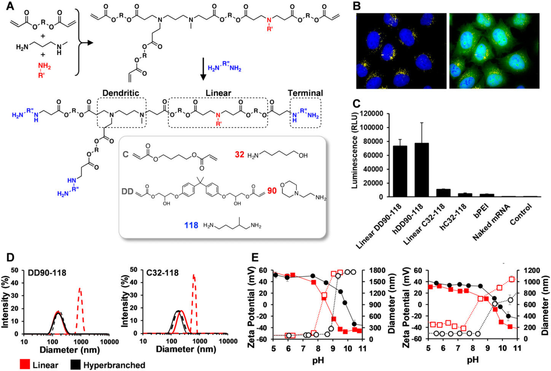
First we synthesized two chemically distinct PBAEs that have previously proved to be effective gene delivery vectors, C32–118[17] and DD90–118.[21,22] Linear PBAEs were synthesized via Michael addition of diacrylate (A2) with primary amine monomers (B2) and end-capped with terminal amines.[23] We synthesized hyperbranched PBAE polymers, using an A2-B2-BB′2 strategy.[24] To the A2-B2 reaction, we introduced a trifunctional amine, N-methyl 1,3 diaminopropane (BB′2) to generate hyperbranched hC32–118 and hDD90–118 (Figure 1A). This amine was selected due to its analogy with amine “103” (1,3 diaminopropane), known to mediate efficient gene transfection.[25] We tested transfec tion efficacy in vitro to see if these polymers were capable of functional mRNA delivery to A549 lung epithelial cells and confirmed that both linear and hyperbranched versions facilitated efficient translation of IVT-mRNA encoding for firefly luciferase, with DD90–118-based PBAEs performing significantly better than C32–118 (Figure 1C).
Figure 1.

A) hDD90–118 and hC32–118 hyperbranched PBAEs were synthesized via addition of a tri-functional amine, N-methyl 1,3 diaminopropane. B) PBAE polymers were complexed with Cy-5 tagged mRNA encoding for GFP to confirm particle uptake (yellow) and translation to GFP (overlaid image on the right, green) in A549 lung epithelial cells (nuclei, blue), hDD90–118 polyplexes shown. C) Comparison of in vitro transfection efficiency in A549 cells using hyperbranched and linear polymers delivering 0.003 mg mL−1 of luciferase mRNA (n = 3, +SD). D) Polyplex stability at varying mRNA concentration; at pH 7.4, linear PBAEs formulated with mRNA at 0.5 mg mL−1 (red dashed line) become unstable and aggregate into large particles whereas hyperbranched analogues (black dashed line) remain stable nanoparticles below 200 nm. Both linear and branched PBAEs at 0.003 mg mL−1 mRNA remain stable (red and black solid lines, respectively). E) Particle stability at varying pH (DD90, left; C32, right); zeta potential of hyperbranched PBAE polyplexes display higher isoelectric points (black closed circles) compared to linear (red closed squares). A reduction in surface zeta potential occurs at increasing pH which correlates with growth in particle size at pHs above 7.5 for linear PBAEs (red open squares) compared to hyperbranced polymers which remain stable below 200 nm up to pH 8.5 (black open circles).
For inhaled delivery of drugs, the vibrating mesh nebulizer is one of the most efficient devices for aerosol generation but requires highly stable formulations that can withstand shearing forces. It has been estimated that ≈0.2–0.5% of the total nebulized dose is deposited in the lung using whole body chambers[3,26] and this requires development of delivery vectors that can be concentrated. We aimed to deliver IVT-mRNA encoding for firefly luciferase at a concentration of 0.5 mg mL−1, 150 times that used in vitro. At this concentration, linear DD90–118 and C32–118 formed visible aggregates and particle diameters were measured to be 1192 and 962 nm, respectively, with large polydispersity values approaching 1.0 (Table S1, Supporting Information). In contrast, hyperbranched PBAE nanoparticles remained stable at the higher mRNA concentration, below 200 nm (Figure 1D) with low polydispersities of 0.1 (Table S1, Supporting Information). H1 NMR indicated that the incorporation of dendritic units correlates with an increase in terminal end-cap amine groups in hyperbranched polymer (Figure S2, Supporting Information). These end-cap amines consequently increase the density of primary and secondary amines in the PBAE which may influence pKa and charge density. Complexation assays using varying ratios of PBAE to mRNA demonstrated that the hyperbranched polymer could retard the movement of mRNA during gel electrophoresis at a ratio of 5 to 1 whereas linear polymer required a ratio of 10 to 1, this correlated with surface zeta potential measurements which only became positive at these respective ratio’s (Figure S3, Supporting Information). Furthermore, characterization of nanoparticle zeta potential across varying pH shows an increase in the isoelectric point of hyperbranched PBAEs compared to their linear counterpart regardless of the chemistry of the PBAE (Figure 1E). This means that compared to their linear versions, hDD90–118 and hC32118 are able to maintain a greater positive surface charge closer to physiological pH which is important for maintaining particle stability (Figure 1E) in the absence of steric stabilization such as that afforded by poly(ethylene glycol) (PEG) coatings.[27] PEGylation has been used to stabilize nanoparticles for improved systemic delivery of mRNA by intravenous injection.[22] To ascertain whether PEGylation could also improve nebulized delivery, we synthesized linear DD90–118 with an alkyl amine “C12” to enable coformulation with PEG-lipid as described previously.[21] However, there was no improvement compared to the nonPEGylated formulation (Figure S5, Supporting Information) and therefore was not taken forward as a strategy to stabilize the PBAE formulations.
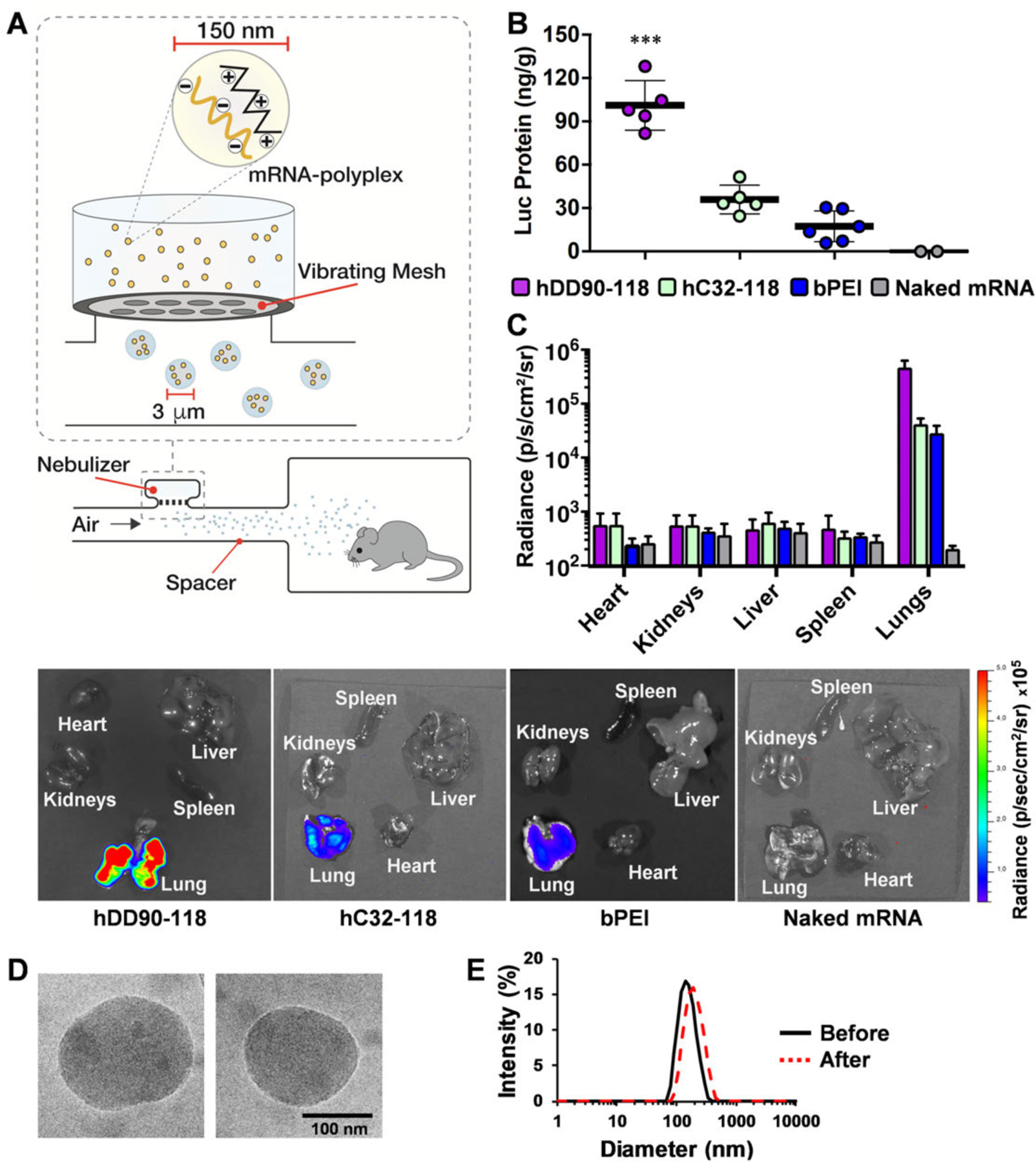
We then nebulized the stable hyperbranched PBAE and bPEI vectors to mice at a concentration of 0.5 mg mL−1 mRNA encoding for firefly luciferase using a vibrating mesh nebulizer connected to a whole-body chamber (Figure 2A). Electron microscopy confirmed that hDD90–118 polyplexes remained stable before (137 nm ± 21) and after (146 nm ± 40) nebulization (Figure S6, Supporting Information). Organs were harvested 24 h after nebulization and bioluminescence was observed to be localized to the lung only unlike intravenous delivery where translation may also be observed in the spleen and liver.[14,22] Luminescent radiance was significantly higher in lungs transfected with hDD90–118 polyplexes (4.8 · 105 p s−1 cm−2 sr−1) compared to hC32–118 (4.3 · 104 p s−1 cm−2 sr−1) and bPEI (2.9 · 104 p s−1 cm−2 sr−1) (Figure 2C). Quantification of luciferase protein in lung tissue homogenates again demonstrated significantly higher translation of luciferase encoding mRNA with hDD90–118 polyplexes producing 101.2 ng g−1 (±15.3) of luciferase protein relative to total protein compared to hC32–118 (35.9 ng g−1, ± 8.9) and bPEI (17.4 ng g−1, ± 9.7) (Figure 2B). Our data confirms that the chemical composition of PBAE has significant influence on protein production from IVT-mRNA. Screening of chemically diverse PBAE libraries have previously highlighted important structure-function relationships between the chemical composition of the vector and transfection efficiency.[17] Therefore, strategies that can improve nanoformulation without changing chemistry, such as hyperbranching could be critical for the development of effective yet stable formulations.
Figure 2. Inhalation of mRNA polyplexes.

A) A vibrating mesh nebulizer connected to a whole-body chamber was used to deliver IVT-mRNA encoding for firefly luciferase to mice. The nebulizer generates micrometer sized droplets optimal for lung deposition, containing nanoparticles for intracellular delivery. B) Quantification of luciferase protein/total protein in the lung 24 h after nebulized delivery of 1.0 mg mRNA (p < 0.001, ±S.D, n = 5 – 6). C) Bioluminescence 24 h after inhalation of polyplexes, hDD90–118 vectors produced significantly higher radiance localized to the lung, compared to hC32–118 and bPEI (p < 0.001, +S.D, n = 4). Statistical analysis using one-way ANOVA with post-hoc Tukey test. D) Electron microscopy of hDD90–118 particles before (left) and after (right) nebulization, particles had an average size of 137 nm (±21) and 146 nm (±40), respectively (±S.D, n = 13 – 15, additional images in Figure S6, Supporting Information). E) Particles have narrow size distribution with polydispersity indices of 0.10 before (black) and 0.11 after (red, dashed) nebulization.
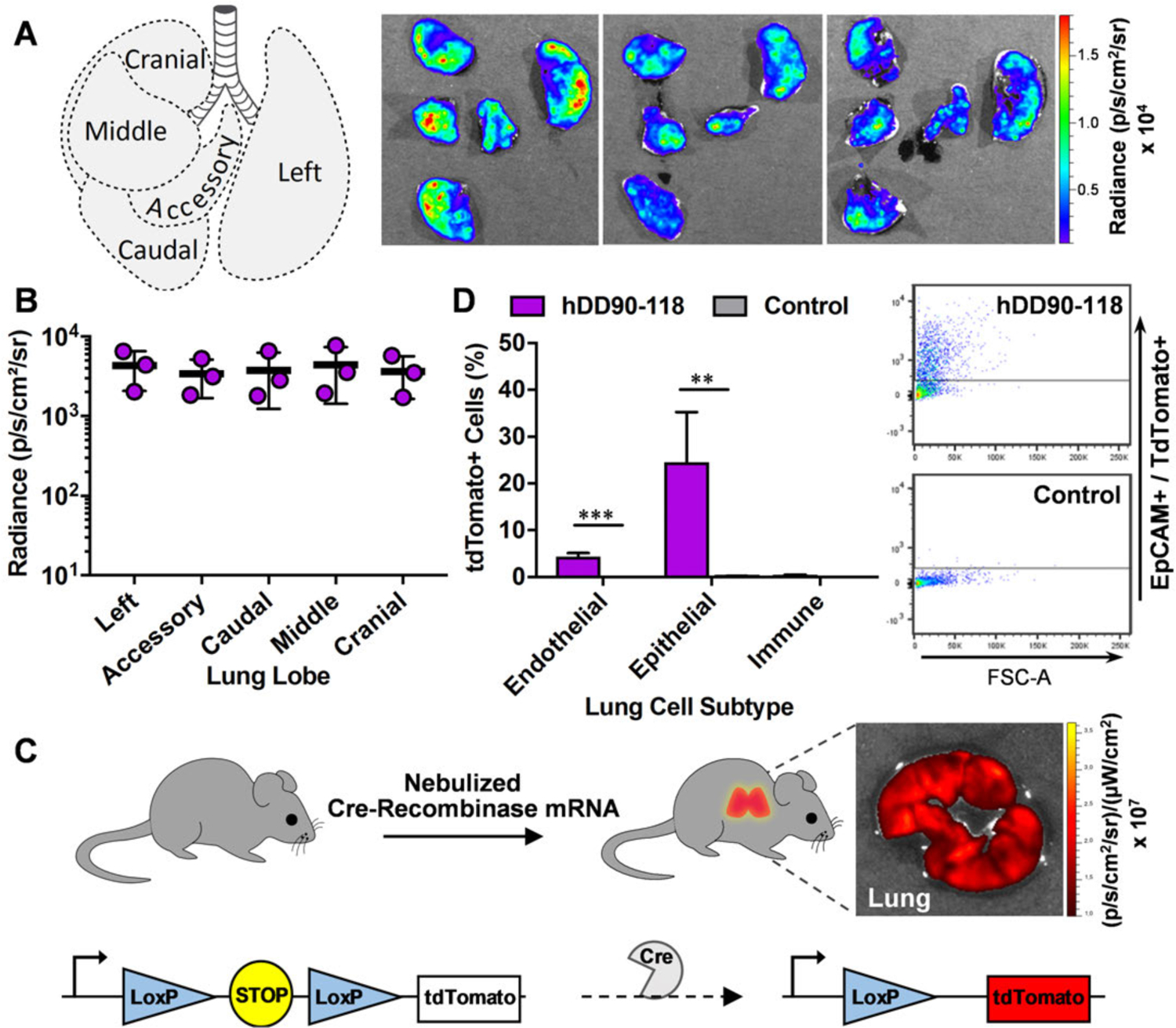
To determine the extent of distribution throughout the lung using our highly effective hDD90–118 polyplexes, the lungs were dissected into their five constituent lobes[28] 24 h postnebulization (Figure 3A) and revealed that uniform bioluminescence was achieved in all lobes of the lung (Figure 3B). To further identify which lung cell subtype was specifically transfected by hDD90–118 polyplexes, we adopted a sensitive single cell specific gene expression approach using Ai14 tdTomato reporter mice. These mice harbor a loxP-flanked stop cassette that controls gene expression of the fluorescent tdTomato protein which is only produced in the presence of Cre-recombinase[29,30] (Figure 3C). We nebulized hDD90–118 nanoparticles containing IVT-mRNA encoding for Cre-recombinase to mice and analyzed lung cells by flow cytometry using markers for endothelial (CD31), epithelial (EpCAM) or immune (CD45) cells (Figure S7, Supporting Information) and found that lung epithelial cells were the majority sub-type transfected with 24.6% (±8.6) of the total epithelial population expressing tdTomato, followed by 4.4% (±0.6) endothelial and 0.4% (±0.1) immune cell expression (Figure 3D). The extent of epithelial targeting required for phenotypic disease correction will depend on the disorder being treated but it has been suggested, for example, that 6–10% of CFTR gene correction in lung epithelial sheets[31] could alleviate symptoms of cystic fibrosis, with in vivo studies suggesting that 17–28% of epithelial transduction may be required for a 50% restoration of CFTR current.[32]
Figure 3.

Distribution of protein expression in the lung and cell sub-type transfected with hDD90–118 polyplexes. A) Dissection of mouse lung 24 h after nebulization of hDD90–118-luciferase mRNA polyplexes, bioluminescence is observed throughout all 5 lobes of the lung. B) Uniform radiance is quantified within each lobe (n = 3, ± SD). C) Quantitative assessment of lung cell subtype transfected by hDD90–118 polyplexes was determined using a cre-loxP mouse model designed to express tdTomato only in cells that translate cre-recombinase mRNA. D) Lungs expressing tdTomato fluorescence were analyzed by flow cytometry using markers for endothelial (CD31), epithelial (EpCAM) and immune (CD45) cells (unpaired T-test, **p < 0.01,***p < 0.005, n = 3, + SD).
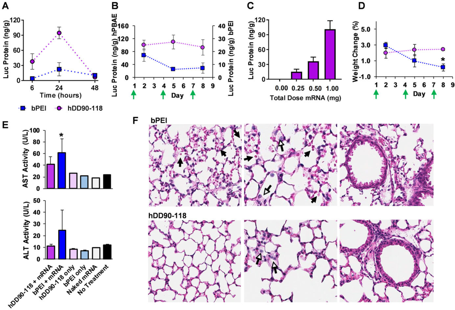
Characterization of the kinetics of protein expression after inhalation of aerosolized hDD90–118 vectors was investigated by quantifying luciferase protein in mouse lung at 6, 24, and 48 h postnebulization. Transient expression of luciferase protein was observed which is maximal at 24 h postnebulization and drops significantly at 48 h (Figure 4A). IVT-mRNA is promising for the treatment of lung conditions ranging from pulmonary fibrosis to protein deficiencies such as α1-antitrypsin but treatment of these conditions with mRNA may require persistent dosing. To determine if the degradable hDD90–118 vectors could be administered repeatedly, we nebulized 1 mg of luciferase mRNA every 72 h a total of three times and quantified luciferase protein in the lung 24 h after each dose. A dosing interval of 72 h was guided by our kinetics data to prevent accumulation of protein from the previous dose which may mask a reduction in protein expression over time. We observed that both hDD90–118 and bPEI could mediate repeat reporter protein production in the lung, with hDD90–118 producing significantly higher levels of protein after the third dose (93.6 ng g−1 ± 24.2) compared to bPEI (7.4 ng g−1 ± 4.0) (Figure 4B).
Figure 4.

Pharmacokinetics of firefly luciferase mRNA translation in mouse lung and toxicity. A) Quantification of luciferase protein at 6, 24, and 48 h post nebulization of hDD90–118 and bPEI polyplexes (n = 3, ± SD). B) Repeat dosing of 1.0 mg of mRNA every 3 d (green arrow) (n = 3–5, ± SD). C) Luciferase protein expression 24 h after inhalation of hDD90–118 polyplexes formulated with 0.25, 0.50, or 1.0 mg of mRNA (n = 3–5, ± SD). D) Weight change in mouse 24 h after nebulization (green arrows). Mice treated repeatedly with bPEI underwent a reduction in weight gain after the third dose (p < 0.05, n = 4–10, ±SD, one-way ANOVA with Tukeys test). E) Serum levels of liver enzymes, aspartate aminotransferase (AST) and alanine aminotransferase (ALT) after three doses of mRNA polyplexes or polymer only (p < 0.05, n = 4, ±SD, one-way ANOVA with Tukeys test). F) Histology of lungs after three inhaled doses (day 8) of bPEI or hDD90–118 polyplexes. Alveolar architecture is maintained in both samples, lungs exposed to bPEI display some occurrence of red blood cells but is not considered abnormal (black arrows). Normal alveolar macrophages are present in the lungs of both samples (white arrows). Bronchiolar architecture is maintained in both samples. H&E staining, 20· magnification.
To examine if the amount of protein expression could be controlled, we performed a dose-response study of inhaled mRNA-hDD90–118 vectors by quantifying the amount of reporter protein in the lung after delivering 0.25, 0.5, and 1.0 mg of mRNA encoding for luciferase. A corresponding increase in protein production was observed 24 h postnebulization, indicating that dose dependent control of endogenous protein expression is possible (Figure 4C). Repeated delivery of hDD90–118 vectors were well tolerated in mice as indicated by lack of weight loss, no increase in serum liver enzyme levels and normal lung histology (Figure 4D–F). This highlights a significant benefit of local mRNA delivery compared to systemic where gross toxicity characterized by weight loss may be observed after a single dose.
In order to increase the feasibility of its clinical use, the hydrolyzable hDD90–118 vectors must be formulated to improve shelf-life. We lyophilized nanoparticles containing luciferase mRNA with sucrose as a cryoprotectant[33] and stored them at −80 °C. The freeze-dried particles were reconstituted in water to a concentration of 0.5 mg mL−1 mRNA immediately prior to nebulization. After storage up to 90 d, the particles remained stable and luciferase-mRNA proved to be functional after nebulized delivery to mice (Figure S8, Supporting Information), adding substantial ease of preparation and shelf-life of the degradable vectors for noninvasive gene delivery to the lung.
In summary, we report materials capable of safe and effective IVT-mRNA delivery to lung epithelium which facilitates repeated functional protein production. Unlike proteins, where delivery systems must be tailored to each protein based on its charge and hydrophobicity, the chemical properties between different mRNA’s are often very similar. Therefore, materials such as those developed in this study that can: 1) condense mRNA, 2) facilitate intracellular uptake, followed by 3) cytosolic release to allow translation to the encoded protein are of significant interest as an enabling technology for the clinical application of IVT-mRNA therapeutics.
Experimental Section
Poly(beta amino ester) Synthesis:
Diacrylate and amine monomers were purchased from Sigma-Aldrich, Alfa Aesar, TCI America, and MonomerPolymer & Dajac Labs. Linear PBAEs were synthesized at a ratio of 1:0.95 acrylate:backbone amine. To synthesize hyperbranched C32 and DD90, acrylate:backbone amine:trifunctional amine monomers were reacted at a ratio of 1.2:0.3:0.4 and 1:0.5:0.2. Monomers were stirred in anhydrous dimethylformamide at a concentration of 150 mg mL−1 at 40 °C for 4 h then 90 °C for 48 h. The mixtures were allowed to cool to 30 °C and end cap amine was added at 1.5 molar equivalent relative to the excess acrylate and stirred for a further 24 h. The polymers were purified by dropwise precipitation into cold anhydrous diethyl ether spiked with glacial acetic acid, vortexed and centrifuged at 1250 G for 2 min to pellet the polymer. The supernatant was discarded and polymer washed twice more in fresh diethyl ether and dried under vacuum for 48 h. Polymers were stored at −20 °C.
Animal Studies:
C57BL/6 mice, 6–8 weeks old females (Charles River) were cared for in the USDA-inspected MIT Animal Facility under federal, state, local, and NIH guidelines for animal care.
Nebulized Delivery:
An AeroNeb vibrating mesh nebulizer (Aerogen Inc.) was connected to a whole-body nebulization chamber via a spacer half filled with silica (1–3 mm, Sigma). Nanoparticles were formulated at 0.5 mg mL−1 of IVT mRNA encoding for firefly luciferase (kindly provided by TranslateBio, MA, USA), with either 50 to 1 PBAE to mRNA by mass, in 100 · 10−3 m of sodium acetate buffer, pH 5.2 (Sigma) or bPEI 25 kDa (Sigma) at an N/P ratio of 10, titrated to pH 5 with hydrochloric acid. The nebulizer was loaded with the required volume of nanoparticles and an oxygen flow rate of 20 SCFH was used to direct the aerosol along the spacer into the chamber until no more aerosol could be observed. For IVIS imaging, mice were sacrificed 10 min after receiving an intraperitoneal injection of Luciferin, 0.2 mg g−1 (Xenolight, PerkinElmer), organs harvested and bioluminescence imaged (Xenogen IVIS Spectrum Imager). For luciferase protein quantification and serum collection, mice were euthanized followed by cardiac puncture and blood collection in BD SST microtainers, lungs were removed en bloc, washed in PBS and flash frozen in liquid nitrogen. Tissues were homogenized in a pestle and mortar on dry ice. Homogenates were lysed in reporter lysis buffer (Promega), centrifuged at 20 000 G for 5 min. Supernatant was assayed for luciferase content (BrightGlo and QuantiLum, Promega) and for total protein content (Pierce BCA kit, Invitrogen). Serum was analyzed using ALT and AST activity assay kits (Sigma).
Flow Cytometry Studies with Ai14 Cre Reporter Mice:
B6.CgGt(ROSA)26Sortm14(CAG-tdTomato)Hze/J mice (Jackson Laboratory, Bar Harbor, ME, USA) were nebulized with hDD90–118 nanoparticles loaded with 1 mg of mRNA encoding for Cre Recombinase (Trilink, NLS-Cre, 5meC, ¬). Nanoparticles were prepared according to the protocol described earlier. Control C57BL/6 mice were nebulized with buffer only. Mice were sacrificed 7 d postnebulization, lungs were minced and incubated for 1 h at 37 °C in PBS buffer (Gibco) containing 0.92 M HEPES (Gibco), 201.3 units mL−1 collagenase I (Sigma), 566.1 units mL−1 collagenase XI (Sigma), and 50.3 units mL−1 DNase I (Sigma). Digested tissue was filtered through a 70 · 10−6 m nylon cell strainer and treated with red blood cell (RBC) lysis buffer for 5 min. The suspension centrifuged at 400 G, pellet resuspended in PBS containing 0.5% bovine serum albumin and filtered through a 40· 10−6 m cell strainer. The cell suspension was centrifuged again, pellet resuspended, and then incubated for 30 min at 4 °C with antibodies against epithelial (EPCAM-APC), endothelial (CD31-AF488), and immune (CD45-BV421) cell markers at a 1:300 dilution (all antibodies from BioLegend, San Diego, CA, USA). The cells were then analyzed using an LSR HTS-II flow cytometer (BD Biosciences).
Supplementary Material
Acknowledgements
D.G.A. would like to acknowledge funding from TranslateBio, Lexington, MA. A.K.P. gratefully acknowledges the engineering and physical sciences research council (EPSRC) for the engineering, tissue engineering and regenerative medicine (E-TERM) award (EP/I017801/1). This work was supported in part by the Koch Institute Support (core) Grant P30-CA14051 from the National Cancer Institute. The authors thank Roderick Bronson, Dong Soo Yun, and Eliza Vasile at the Koch Institute Swanson Core for their assistance with histology, electron, and confocal microscopy, respectively. The authors thank Dr. Kevin Daniel and Umberto Palmiero for assistance with H1 NMR acquisition, Dr. Adam Behrens for assistance with DLS instrumentation, and Dr. Padmini Pillai for guidance on lung fixation procedures.
Footnotes
Supporting Information
Supporting Information is available online.
Conflict of Interest
A patent for the materials developed in this manuscript has been filed by A.K.P., J.C.K., K.J.K., and D.G.A.
Contributor Information
Asha Kumari Patel, Koch Institute for Integrative Cancer Research Massachusetts Institute of Technology Cambridge, MA 02139, USA; Division of Cancer and Stem Cells School of Medicine, and Division of Advanced Materials and Healthcare Technologies, School of Pharmacy University of Nottingham, NG7 2RD, UK.
James C. Kaczmarek, Koch Institute for Integrative Cancer Research Massachusetts Institute of Technology Cambridge, MA 02139, USA Department of Chemical Engineering, Massachusetts Institute of Technology, Cambridge, MA 02139, USA.
Suman Bose, Koch Institute for Integrative Cancer Research Massachusetts Institute of Technology Cambridge, MA 02139, USA; Department of Chemical Engineering, Massachusetts Institute of Technology, Cambridge, MA 02139, USA.
Kevin J. Kauffman, Koch Institute for Integrative Cancer Research Massachusetts Institute of Technology Cambridge, MA 02139, USA Department of Chemical Engineering, Massachusetts Institute of Technology, Cambridge, MA 02139, USA.
Faryal Mir, Koch Institute for Integrative Cancer Research Massachusetts Institute of Technology Cambridge, MA 02139, USA.
Michael W. Heartlein, TranslateBio, Lexington, MA 02421, USA
Frank DeRosa, TranslateBio, Lexington, MA 02421, USA.
Robert Langer, Koch Institute for Integrative Cancer Research Massachusetts Institute of Technology Cambridge, MA 02139, USA; Department of Chemical Engineering, Massachusetts Institute of Technology, Cambridge, MA 02139, USA; Harvard-MIT Division of Health Sciences and Technology Institute for Medical Engineering and Science Massachusetts Institute of Technology, Cambridge, MA 02139, USA; Institute for Medical Engineering and Science Massachusetts Institute of Technology Cambridge, MA 02139, USA.
Daniel G. Anderson, Koch Institute for Integrative Cancer Research Massachusetts Institute of Technology Cambridge, MA 02139, USA Institute for Medical Engineering and Science Massachusetts Institute of Technology Cambridge, MA 02139, USA; Department of Chemical Engineering, Massachusetts Institute of Technology, Cambridge, MA 02139, USA; Harvard-MIT Division of Health Sciences and Technology Institute for Medical Engineering and Science Massachusetts Institute of Technology, Cambridge, MA 02139, USA.
References
- [1].Sahin U, Karikó K, Türeci Ö, Nat. Rev. Drug Discovery 2014, 13, 759. [DOI] [PubMed] [Google Scholar]
- [2].Kormann MSD, Hasenpusch G, Aneja MK, Nica G, Flemmer AW, Herber-Jonat S, Huppmann M, Mays LE, Illenyi M, Schams A, Griese M, Bittmann I, Handgretinger R, Hartl D, Rosenecker J, Rudolph C, Nat. Biotechnol 2011, 29, 154. [DOI] [PubMed] [Google Scholar]
- [3].Beck SE, Laube BL, Barberena CI, Fischer AC, Adams RJ, Chesnut K, Flotte TR, Guggino WB, Mol. Ther 2002, 6, 546. [DOI] [PubMed] [Google Scholar]
- [4].Alton EWFW, Armstrong DK, Ashby D, Bayfield KJ, Bilton D, Bloomfield EV, Boyd AC, Brand J, Buchan R, Calcedo R, Carvelli P, Chan M, Cheng SH, Collie DDS, Cunningham S, Davidson HE, Davies G, Davies JC, Davies LA, Dewar MH, Doherty A, Donovan J, Dwyer NS, Elgmati HI, Featherstone RF, Gavino J, Gea-Sorli S, Geddes DM, Gibson JSR, Gill DR, Greening AP, Griesenbach U, Hansell DM, Harman K, Higgins TE, Hodges SL, Hyde SC, Hyndman L, Innes JA, Jacob J, Jones N, Keogh BF, Limberis MP, Lloyd-Evans P, Maclean AW, Manvell MC, McCormick D, McGovern M, McLachlan G, Meng C, Montero MA, Milligan H, Moyce LJ, Murray GD, Nicholson AG, Osadolor T, Parra-Leiton J, Porteous DJ, Pringle IA, Punch EK, Pytel KM, Quittner AL, Rivellini G, Saunders CJ, Scheule RK, Sheard S, Simmonds NJ, Smith K, Smith SN, Soussi N, Soussi S, Spearing EJ, Stevenson BJ, Sumner-Jones SG, Turkkila M, Ureta RP, Waller MD, Wasowicz MY, Wilson JM, Wolstenholme-Hogg P, Lancet Respir. Med 2015, 3, 684. [DOI] [PMC free article] [PubMed] [Google Scholar]
- [5].Griesenbach U, Pytel KM, Alton EWFW, Hum. Gene Ther 2015, 26, 266. [DOI] [PMC free article] [PubMed] [Google Scholar]
- [6].Arulmuthu ER, Williams DJ, Baldascini H, Versteeg HK, Hoare M, Biotechnol. Bioeng 2007, 98, 939. [DOI] [PubMed] [Google Scholar]
- [7].Johler SM, Rejman J, Guan S, Rosenecker J, PLoS One 2015, 10, e0137504. [DOI] [PMC free article] [PubMed] [Google Scholar]
- [8].Densmore CL, Orson FM, Xu B, Kinsey BM, Waldrep JC, Hua P, Bhogal B, Knight V, Mol. Ther 2000, 1, 180. [DOI] [PubMed] [Google Scholar]
- [9].Ernst N, Ulrichskötter S, Schmalix WA, Rädler J, Galneder R, Mayer E, Gersting S, Plank C, Reinhardt D, Rosenecker J, J. Gene Med 1999, 1, 331. [DOI] [PubMed] [Google Scholar]
- [10].McLachlan G, Davidson H, Holder E, Davies LA, Pringle IA, Sumner-Jones SG, Baker A, Tennant P, Gordon C, Vrettou C, Blundell R, Hyndman L, Stevenson B, Wilson A, Doherty A, Shaw DJ, Coles RL, Painter H, Cheng SH, Scheule RK, Davies JC, Innes JA, Hyde SC, Griesenbach U, Alton EWFW, Boyd AC, Porteous DJ, Gill DR, Collie DDS, Gene Ther. 2011, 18, 996. [DOI] [PubMed] [Google Scholar]
- [11].Rudolph C, Ortiz A, Schillinger U, Jauernig J, Plank C, Rosenecker J, J. Gene Med 2005, 7, 59. [DOI] [PubMed] [Google Scholar]
- [12].Godbey WT, Wu KK, Mikos AG, J. Biomed. Mater. Res 1999, 45, 268. [DOI] [PubMed] [Google Scholar]
- [13].Wightman L, Kircheis R, Rossler V, Carotta S, Ruzicka R, Kursa M, Wagner E, J. Gene Med 2001, 3, 362. [DOI] [PubMed] [Google Scholar]
- [14].Kowalski PS, Capasso Palmiero U, Huang Y, Rudra A, Langer R, Anderson DG, Adv. Mater 2018, 30, 1801151. [DOI] [PMC free article] [PubMed] [Google Scholar]
- [15].Park MR, Kim HW, Hwang CS, Han KO, Choi CY, Song SC, Myung HC, Cho CS, J. Gene Med 2008, 10, 198. [DOI] [PubMed] [Google Scholar]
- [16].Lynn DM, Langer R, J. Am. Chem. Soc 2000, 122, 10761. [Google Scholar]
- [17].Anderson DG, Akinc A, Hossain N, Langer R, Mol. Ther 2005, 11, 426. [DOI] [PubMed] [Google Scholar]
- [18].Wu Y. Liu, Jiang X, He L. Chen, Goh SH, Leong KW, Biomacromolecules 2005, 6, 3166. [DOI] [PubMed] [Google Scholar]
- [19].Cutlar L, Zhou D, Gao Y, Zhao T, Greiser U, Wang W, Wang W, Biomacromolecules 2015, 16, 2609. [DOI] [PubMed] [Google Scholar]
- [20].Gao C, Yan D, Prog. Polym. Sci 2004, 29, 183. [Google Scholar]
- [21].Eltoukhy AA, Chen D, Alabi CA, Langer R, Anderson DG, Adv. Mater 2013, 25, 1487. [DOI] [PMC free article] [PubMed] [Google Scholar]
- [22].Kaczmarek JC, Patel AK, Kauffman KJ, Fenton OS, Webber MJ, Heartlein MW, DeRosa F, Anderson DG, Angew. Chem., Int. Ed 2016, 55, 13808. [DOI] [PMC free article] [PubMed] [Google Scholar]
- [23].Zugates GT, Peng W, Zumbuehl A, Jhunjhunwala S, Huang Y-H, Langer R, Sawicki JA, Anderson DG, Mol. Ther 2007, 15, 1306. [DOI] [PubMed] [Google Scholar]
- [24].Gao C, Yan D, Macromolecules 2001, 34, 156. [Google Scholar]
- [25].Eltoukhy AA, Siegwart DJ, Alabi CA, Rajan JS, Langer R, Anderson DG, Biomaterials 2012, 33, 3594. [DOI] [PMC free article] [PubMed] [Google Scholar]
- [26].Koshkina NV, Agoulnik IY, Melton SL, Densmore CL, Knight V, Mol. Ther 2003, 8, 249. [DOI] [PubMed] [Google Scholar]
- [27].Verwey EJW, Overbeek JTG, Theory of the Stability of Lyophobic Colloids, Elsevier, Amsterdam, The Netherlands: 1948. [Google Scholar]
- [28].Metzger RJ, Klein OD, Martin GR, Krasnow MA, Nature 2008, 453, 745. [DOI] [PMC free article] [PubMed] [Google Scholar]
- [29].Madisen L, Zwingman TA, Sunkin SM, Oh SW, Zariwala HA, Gu H, Ng LL, Palmiter RD, Hawrylycz MJ, Jones AR, Lein ES, Zeng H, Nat. Neurosci 2010, 13, 133. [DOI] [PMC free article] [PubMed] [Google Scholar]
- [30].Kauffman KJ, Oberli MA, Dorkin JR, Hurtado JE, Kaczmarek JC, Bhadani S, Wyckoff J, Langer R, Jaklenec A, Anderson DG, Mol. Ther. – Nucleic Acids 2018, 10, 55. [DOI] [PMC free article] [PubMed] [Google Scholar]
- [31].Johnson LG, Olsen JC, Sarkadi B, Moore KL, Swanstrom R, Boucher RC, Nat. Genet 1992, 2, 21. [DOI] [PubMed] [Google Scholar]
- [32].Potash AE, Wallen TJ, Karp PH, Ernst S, Moninger TO, Gansemer ND, Stoltz DA, Zabner J, Chang EH, Mol. Ther 2013, 21, 947. [DOI] [PMC free article] [PubMed] [Google Scholar]
- [33].Tzeng SY, Guerrero-Cázares H, Martinez EE, Sunshine JC, Quiñones-Hinojosa A, Green JJ, Biomaterials 2011, 32, 5402. [DOI] [PMC free article] [PubMed] [Google Scholar]
Associated Data
This section collects any data citations, data availability statements, or supplementary materials included in this article.


