Abstract
Stress granules (SGs) are aggregates of translationally silenced messenger ribonucleoprotein (mRNP) complexes induced by oxidative, osmotic, hypoxic, thermal, viral, and genotoxic stresses. Over the past decade, extensive research has identified key components of SGs, their molecular interactions, and impact on stress‐induced reprogramming of protein expression and cell survival. However, studies defining the signaling pathways that modulate SG assembly have only been launched recently. These studies reveal that posttranslational modifications of selected SG proteins play important roles in the regulation of SG assembly and function. Here we provide an overview of the signaling pathways and posttranslational protein modifications that regulate the assembly and function of SGs. Copyright © 2010 John Wiley & Sons, Ltd.
This article is categorized under:
-
1
Translation > Translation Regulation
-
2
RNA Export and Localization > RNA Localization
-
3
RNA Turnover and Surveillance > Regulation of RNA Stability
Eukaryotic cells respond to environmental stress by reprogramming protein translation.1 The translation of mRNAs encoding ‘housekeeping’ proteins is turned off, allowing cells to conserve energy for the repair of stress‐induced molecular damage. At the same time, translation of mRNAs encoding proteins that re‐fold denatured proteins or repair oxidative damage is turned on, allowing cells to survive under adverse conditions. The mRNAs that are translationally repressed during stress accumulate at discrete cytoplasmic foci known as stress granules (SGs).2 The core components of SGs are translationally stalled non‐canonical 48S pre‐initiation complexes, but many other proteins are also concentrated at these foci.2
Processing bodies (PBs) are a related class of RNA granules at which untranslated mRNA accumulates while awaiting degradation.3, 4, 5 The core components of PBs are mRNA decay factors including deadenylases, decapping enzymes, and a 5′→3′ exonuclease. Unlike SGs, PBs are found in the cytoplasm of unstressed cells. Like SGs, the number of PBs increases when cells are exposed to adverse conditions. The messenger ribonucleoproteins (mRNPs) within SGs and PBs are in a dynamic equilibrium with polysomes.6,7 Consequently, the assembly of SGs and PBs is inhibited by drugs that freeze mRNAs within polysomes (e.g., emetine, cycloheximide). These observations led us to propose that SGs function as effectors of molecular triage whereby the composition of the mRNP determines whether individual transcripts are degraded, stored, or reinitiated.8
As recent reviews have described the dynamic properties of SGs and their components that are crucial for SG assembly,2,9,10 these topics will not be discussed here. In this review, we focus on the signaling pathways that produce posttranslational modifications that promote the assembly of SGs.
PHOSPHORYLATION
Phosphorylation and dephosphorylation play key roles in the assembly and disassembly of SGs. The reprogramming of protein translation observed in stressed cells is usually initiated by the phosphorylation of serine 51 on eIF2α.11 The stress‐activated signaling cascades responsible for this event are centered around a related family of serine/threonine kinases. This family includes: (1) PKR (protein kinase R), a double‐stranded RNA‐dependent kinase that is activated by viral infection, heat, and UV irradiation12,13; (2) PERK (PKR‐like endoplasmic reticulum kinase; also known as PEK, or pancreatic eIF2α kinase), a resident endoplasmic reticulum (ER) protein that is activated when unfolded proteins accumulate in the ER lumen14,15; (3) GCN2 (general control non‐repressible 2), a protein that is activated primarily by amino acid deprivation16; and (4) HRI (heme‐regulated inhibitor), a kinase that is activated under conditions of heme deprivation and arsenite‐induced oxidative stress.17,18 Stress‐induced phosphorylation of eIF2α converts eIF2 from a substrate to a competitive inhibitor of eIF2B, the guanine nucleotide exchange factor, depleting the level of the eIF2‐GTP‐methionyl tRNA required for cap‐dependent translation initiation, thereby inhibiting global translation and initiating SG assembly.2
Phosphorylation of eIF2α stalls translation initiation and allows ribosome runoff. Data from immunofluorescence microscopy suggest that stalled 48S pre‐initiation complexes aggregate into visible foci through the coordinate actions of multiple RNA‐binding proteins that contain self‐aggregation domains. One well‐characterized example of a self‐aggregation domain is the prion‐related glutamine‐rich domain in TIA‐1/TIAR, members of the RNA‐recognition motif (RRM) family of RNA‐binding proteins.11,19,20 Additional SG‐associated proteins possessing aggregation domains include G3BP, fragile X mental retardation protein (FMRP), and TTP.2
Phosphorylation of SG components also plays an important role in the life cycle of several different viruses. In cells infected with either semliki forest virus21 or herpes simplex virus type 1,22 activation of PKR results in the transient assembly of SGs that may contribute to the shut down of host protein synthesis. In contrast, during rotavirus infection, eIF2α is phosphorylated but SGs are not assembled.23 Relocalization of PABP to the nucleus has been proposed to contribute to the inhibition of SG assembly. It is possible that this virus facilitates the expression of its mRNAs by preventing the formation of SGs in the host cell. These studies suggest that manipulation of SG assembly may provide targets for the development of new anti‐viral therapies.
In cells infected with the coronavirus‐related virus that causes severe acute respiratory syndrome (SARS), the phosphorylation status of a nucleocapsid protein participating in viral RNA replication and transcription appears to modulate SG assembly.24 The phosphorylation of nucleocapsid protein within an arginine/serine‐rich (SR) motif somehow inhibits SG assembly during virus infection. Ectopic co‐expression of wild‐type nucleocapsid protein and SR protein kinase 1 prevents arsenite‐induced SG assembly. If inhibitors of SR protein kinase 1 allow SG assembly and inhibit viral replication, they may be candidates for a new class of anti‐viral agent.
Phosphorylation of TTP has been shown to regulate interactions between SGs and PBs. TTP is an adenine/uridine‐rich element (ARE) binding protein that promotes the decay of several transcripts encoding mediators of inflammation.25 This is accomplished by delivering ARE‐containing transcripts to the exosome, a multi‐subunit degradative enzyme, or the PB.4,26 These transcripts can be rescued from TTP‐mediated decay by lipopolysaccharide‐induced activation of MK2, a kinase that phosphorylates TTP on serine residues 52 and 178.27 This allows the binding of 14‐3‐3 proteins that effectively inactivate TTP.27 As these modifications also prevent TTP‐mediated interactions between SGs and PBs,8 it is possible that TTP allows selected transcripts to be presented to (or be transferred to) PBs for degradation.
Phosphorylation of G3BP, a protein originally characterized as a partner of the Ras‐GTPase‐activating protein,28 also regulates the assembly and disassembly of SGs. G3BP is a cytoplasmic protein that quantitatively moves to SGs in response to stress.29 Overexpression of G3BP is sufficient to induce SG assembly, and targeted knockdown of G3BP prevents SG assembly, indicating that it is a key regulator of this process. Phosphorylation of G3BP on serine residue 149 inhibits SG assembly, a possible consequence of reduced homotypic aggregation.29 Analysis of non‐phosphorylatable (S149A) and phosphomimetic (S149E) G3BP mutants confirmed the importance of this modification. Whereas both mutants are efficiently recruited to SGs in cells subjected to arsenite‐induced oxidative stress, overexpression of G3BP (S149A), but not G3BP (S149E), induces SG assembly. It remains to be determined how phosphorylation of G3BP regulates SG assembly.
Phosphorylation of growth factor receptor‐bound protein 7 (Grb7) by focal adhesion kinase (FAK) modulates the dynamics of SG assembly and disassembly during heat shock.30 Both Fak and Grb7 are integral SG components, and RNAi‐mediated depletion of Grb7 inhibits the appearance of SGs in cells cultured at supra‐ambient temperatures. Although phosphorylation of Grb7 does not inhibit heat‐stress‐induced SG assembly, it significantly enhances SG dissolution in cells allowed to recover from heat stress. Analysis of non‐phosphorylatable Grb7 mutants (Y483F/Y495F) confirmed the importance of phosphorylation in promoting the dissolution of SGs. Phosphorylation of Grb7 was found to prevent its interaction with the SG proteins HuR, TIA‐1, and Staufen in co‐immunoprecipitation assays. More importantly, this phosphorylation event partially restored the translation of selected transcripts (but not necessarily global translation) upon removal of heat stress.
More recently, CDK function has been shown to be required for SG formation in response to UV irradiation. In contrast, the main repair checkpoint pathways initiated by ATM or ATR do not modulate SG assembly.31 CDK does not regulate arsenite‐induced SG assembly, implying that UV‐damage‐induced SG assembly is selectively dependent upon CDK signaling. It will be of interest to determine the effects of downstream targets of CDK signaling on the assembly of SGs.
The sequestration of signaling components at SGs can also regulate the survival of stressed cells. RACK1 is a scaffold protein that is required for the activation of the MAP kinase cascade that culminates in the activation of p38 and JNK, potent effectors of stress‐induced apoptosis. In stressed cells, sequestration of RACK1 at SGs prevents stress‐induced apoptosis, allowing SGs to actively promote the survival of cells exposed to adverse conditions.32 Similarly, the recruitment of the RSK2 kinase to SGs and to the nucleus requires its binding to TIA‐1, which affects both its ability to induce cyclin D1 and promote survival of cells exposed to oxidative stress.33 It is possible that these kinases modulate SG assembly independent of their kinase activity. This could involve heterotypic interactions with specific SG components or disruption of interactions between SG components.
O‐GLCNACYLATION
We recently completed an siRNA‐mediated microscopic screen to identify genes that are required for the assembly of SGs.34 We found that knockdown of SORT1 (sortilin) strongly inhibits the assembly of SGs and PBs in cells subjected to arsenite‐induced oxidative stress. Consistent with this, ectopic overexpression of sortilin induced the assembly of SGs, confirming its importance as a regulator of SGs. As sortilin was not concentrated at SGs, we hypothesized that it may be a component of a signaling pathway that indirectly regulates SG assembly.
In 3T3‐L1 adipocytes, sortilin directs Glut4‐containing endosomes to the plasma membrane following insulin stimulation to facilitate glucose uptake.35 Internalized glucose can be multimerized to produce glycogen, metabolized to produce ATP and pyruvate, or modified to produce N‐acetylglucosamine, a substrate for the hexosamine biosynthetic pathway (HBP). The HBP reversibly adds the monosaccharide O‐linked N‐acetylglucosamine (O‐GlcNAc) to serine and threonine residues (O‐GlcNAcylation) to target proteins in a process that is similar to phosphorylation.36 The HBP modulates a wide array of cellular process, such as signal transduction,37 glucose sensing,38 the stress response,38,39 transcription,40 apoptosis, and41,42 proteosomal degradation.43 The first step in the conversion of glucose to GlcNAc is GFAT (L‐glutamine: D‐fructose‐6‐phosphate amidotransferase)‐mediated amidylation. The HBP culminates in the formation of uridine 5′‐diphosphate (UDP)‐GlcNAc, the unique monosccharide donor for the O‐GlcNAcylation of target proteins. The conjugation and deconjugation of O‐GlcNAc are mediated by two antagonistic enzymes, O‐GlcNAc transferase (OGT) and O‐GlcNAcase, respectively.
The fact that HBP is activated by various cellular stresses led us to further investigate the functional significance of this pathway for SG assembly.38,44 As expected, knockdown of genes encoding GFAT and OGT strongly inhibits arsenite‐induced SG assembly, whereas knockdown of O‐GlcNAcase, the enzyme that removes O‐GlcNAc from target protein, has no effect on SG assembly. GFAT has two isoforms, GFAT1 and GFAT2. In our assay, only GFAT2 knockdown inhibits SG assembly, suggesting that under stress conditions, the HBP is activated through GFAT2. Collectively, these data support a role for glycosylation in the regulation of SG assembly.
Immunofluorescence analysis using two different anti‐O‐GlcNAc antibodies detects strong O‐GlcNAc staining in arsenite‐induced SGs, indicating that O‐GlcNAcylated proteins are SG components.34 Moreover, we found that OGT is colocalized to arsenite‐induced SGs. To identify specific O‐GlcNAcylated proteins potentially recruited to SGs, we analyzed samples from polysome fractions from mock versus arsenite‐treated U2OS cells. Immunoblots probed with O‐GlcNAc antibodies revealed numerous O‐GlcNAcylated proteins sized 10∼40 KDa that co‐localize with translationally stalled 80S monosome fractions in sucrose gradients. Immuno‐purification of these O‐GlcNAcylated proteins revealed these modified proteins to be components of small and large ribosomal subunits, the ribosomal associated protein RACK1, glyceraldehyde‐3‐phosphate dehydrogenase (GAPDH), prohibitin‐2, and several other proteins.34 It was surprising to find large ribosomal subunit proteins as O‐GlcNAcylated targets because the large ribosomal subunit is excluded from SGs.45 As arsenite‐induced eIF2α phosphorylation and subsequent polysome disassembly are normal in sortilin and OGT knockdown cells, O‐GlcNAcylation appears to be required for the aggregation of untranslated mRNPs at SGs.34 It is possible that these sugars serve as a molecular glue that promotes the aggregation of untranslated mRNPs. Alternatively, O‐GlcNAc modifications may facilitate translational repression by interfering with ribosomal subunit association.
In the yeast, Saccharomyces cerevisiae, SG‐related foci termed EGP bodies induced upon glucose deprivation have been described. EGP bodies are composed of typical SG markers (e.g., eIF4E, eIF4G, and Pab1) but lack the 40S ribosomal subunits and eIF3.46, 47, 48 It is interesting to note that budding yeast apparently lacks O‐GlcNAcylation due to the absence of OGT, suggesting that modification of the translation machinery may account for differences between yeast EGP bodies and mammalian SGs.
HYPUSINATION
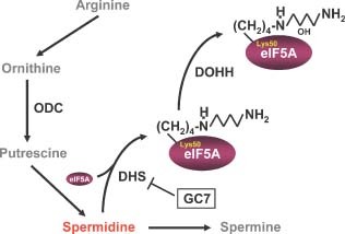
Hypusination is an unusual posttranslational modification mediated by the polyamine biosynthetic pathway that is involved in many cellular processes including cell proliferation and the stress response. Hypusine [Nε‐(4‐amino‐2‐hydroxybutyl)lysine], a derivative of spermidine, is covalently conjugated to eukaryotic translation initiation factor 5A (eIF5A) at a lysine residue (Lys50 in human) via two consecutive enzymatic reactions: first, the enzyme deoxyhypusine synthase (DHS) catalyzes the transfer of the aminobutyl moiety from spermidine to the lysine residue in eIF5A which in turn is hydroxylated by deoxyhypusine hydroxylase (DOHH)49 (Figure 1). eIF5A is the only known hypusinated protein in nature and this hypusination is absolutely required for its function.50
Figure 1.

Hypusination of eIF5A via the polyamine biosynthetic pathway. ODC, ornithine decarboxylase; DHS, deoxyhypusine synthase; DOHH, deoxyhypusine hydroxylase. N1‐Guanyl‐1,7‐diaminoheptane (GC7) is a competitive inhibitor of DHS.
The role of eIF5A in mRNA translation has been extensively studied but its actual function is still in question. eIF5A was first purified from rabbit reticulocytes as a ribosome‐associated translation initiation factor and shown to stimulate methionyl‐puromycin synthesis, suggestive of a role in the formation of the first peptide bond.51 However, analyses using eIF5A‐deficient yeast S. cerevisiae showed that global protein synthesis was inhibited by only 30%, suggesting that it has a non‐essential role in protein synthesis.52 Depletion of eIF5A in mammalian cells using siRNAs also displayed only a marginal effect on global translation rate (decrease by ∼10%).53 Thus, it was hypothesized that eIF5A may function in a different stage of translation or affect only a subset of mRNAs.
Recently, several studies along with ours implicated a role for hypusinated eIF5A in the translation elongation process.53, 54, 55, 56 Mass‐spectrometric analysis revealed that GST‐eIF5A fusion protein can be co‐purified with the translation elongation factor 2 (eEF2) in yeast, suggesting that eIF5A associates with translationally elongating ribosomes.54 Genetic evidence also showed that an eIF5A mutant tif51A‐3 interacts with the translation elongation mutant eft2H699K (eEF2). Moreover, polysome profiling analysis with temperature‐sensitive eIF5A mutants showed an increase in polysome peaks compared to isogenic wild‐type controls that is also observed in the eft2H699K mutant.55 The retention of polysomes under cyclohexamide (CHX)‐free conditions was also observed in the yeast tif51a‐td, a temperature‐sensitive eIF5A degron mutant cultured at the restrictive temperature. Most importantly, eIF5A but not a non‐hypusinatable mutant [eIF5A(K51R)] can stimulate translation elongation and termination in an in vitro system.56
Our RNAi screen revealed that ornithine decarboxylase (ODC), an enzyme required for the synthesis of polyamines, is essential for arsenite‐induced assembly of SGs.53 Moreover, siRNA‐mediated depletion of eIF5A, a downstream target of the polyamine pathway, dramatically reduces SG formation in cells exposed to oxidative stress. This is further supported by the finding that knockdown (using siRNAs) or inhibition (using GC7) of DHS, an enzyme that covalently joins spermidine to eIF5A,57 inhibits SG assembly. We concluded that hypusination of eIF5A via polyamine synthesis pathway is crucial for SG formation.53
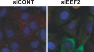
Because stalled translation elongation induced by cycloheximide or emetine inhibits SG assembly, eIF5A may be required to maintain translation elongation in stressed cells. Several lines of evidence support this hypothesis. First, temperature‐sensitive eIF5A mutants are hypersensitive to the peptidyltransferase inhibitors sparsomycin and anisomycin.54 Second, translation elongation factor 2 (eEF2) and 80S ribosomal subunits co‐purify with eIF5A.54 Third, ribosomal transit time is abnormal upon eIF5A inactivation and this phenomenon mimics effects of the eEF2 inhibitor sordarin.55,56 Finally, we have found that eIF5A knockdown delays ribosome runoff in U2OS cells subjected to arsenite‐induced oxidative stress.53 Similar results were observed following ODC1 knockdown which significantly impairs eIF5A hypusination.53 Thus, hypusinated eIF5A may play an important role in promoting translation elongation in stressed compared to unstressed cells. Depletion of eEF2 also has a strong inhibitory effect on SG formation34 (Figure 2). As eEF2 is phosphorylated in response to stress, this modification may modulate interactions with eIF5A. It is tempting to speculate that eIF5A may physically interact with phospho‐eEF2 to maintain ribosome elongation in cells subjected to adverse conditions.
Figure 2.

Depletion of eukaryotic translation elongation factor 2 (eEF2) inhibits SG assembly. RDG3 (GFP‐G3BP, SG marker; RFP‐Dcp1a, PB marker) double stable cells were transfected with control (siCONT) or eEF2‐specific (siEEF2) siRNAs, then SGs were induced by treating with sodium arsenite (0.5 mM) for 30 min.
METHYLATION
Methylation of lysine and arginine residues regulates many different cellular processes including the DNA damage response, chromatin remodeling, and RNA metabolism.58, 59, 60 S‐adenosyl‐L‐methionine is an essential co‐factor used by different types of methyltransferases.61 Arginine methylation mediated by lysine and peptidylarginine methyltransferses transfers methyl groups from S‐adenosyl‐L‐methionine to one or more terminal nitrogens on lysine or arginine residues of target proteins.61, 62, 63 Arginine can be mono‐ or di‐methylated in a symmetric (one methyl group on each nitrogen) or asymmetric (both methyl groups on one terminal nitrogen) manner.64 Protein arginine methylation typically occurs within RGG domains of RNA‐binding proteins such as the FMRP and heterogeneous ribonucleoproteins.60,65
Methylation of the RGG domain of FMRP has been implicated in SG assembly.66 FMRP, which exhibits a punctate cytoplasmic distribution under non‐stressed conditions, quantitatively relocalizes to SGs in response to heat or arsenite‐induced oxidative stress.66,67 The methylation inhibitor adenosine‐2′, 3′‐dialdehyde (AdOx) increases the assembly of FMRP‐containing granules at baseline, inhibits arsenite‐induced SG assembly, and prevents the recruitment of FMRP to SGs. Thus, methylation of FMRP appears to be required for both recruitment to SGs and optimal SG assembly.
Methylation of the cold‐inducible RNA‐binding protein (CIRP) is also required for the assembly of SG.68 CIRP contains an RRM in its amino‐terminus and an RGG motif in its carboxyl‐terminus, both of which are required for RNA binding and SG formation. In contrast to FMRP, methylation of RGG promotes the relocalization of CIRP from the nucleus to the cytoplasm of stressed cells. Unlike FMRP, methylation inhibitors (e.g., AdOx) do not promote the cytoplasmic aggregation of CIRP, suggesting that methylated RGG domains may not be sufficient to induce SG‐independent protein aggregation.
OTHER MODIFICATIONS
Additional posttranslational modifiers identified in our RNAi screen as modulators of SG assembly include EP300, USP10, and UBE2M.34 EP300 is a histone acetyltransferase that modulates transcription by remodeling chromatin.69 Its role in SG assembly may be an indirect consequence of depleting the mRNA pool, but it is possible that SG assembly is enhanced by acetylation of specific SG components. USP10 is a deubiquinating enzyme that has been implicated in various cellular functions.70 As ubiquitination has been implicated in SG assembly,71,72 it is possible that the balance between ubiquitination and deubiquitination modulates this process. UBE2M is an NEDD8‐conjugating E2 enzyme that is required for neddylation of target proteins.73 An understanding of the diverse regulatory pathways involved in SG assembly will require additional investigation.
CONCLUSIONS
During the past decade, significant progress has been made in understanding the role of SGs in maintaining the survival of cells exposed to adverse conditions. SGs help to silence the expression of housekeeping genes and promote the expression of molecular chaperones that repair stress‐induced damage.74 At the same time, sequestration of selected proteins within SGs (e.g., RACK1, RSK2) may determine whether stressed cells live and repair stress‐induced damage, or die by apoptosis. In this sense, SGs may play a key role in modulating life or death decisions in cells exposed to adverse conditions. The finding that SG assembly and disassembly are regulated by myriad posttranslational modifications of SG components indicates that this process is subject to complex regulation. It is likely that the identification of new types of modifications, additional modified target proteins, and upstream signaling pathways will be essential to fully understand the functions of SGs. Drugs that target these signaling cascades may prove useful in the treatment of infectious diseases and cancer.
RELATED WIREs ARTICLE
REFERENCES
- 1. Yamasaki S, Anderson P. Reprogramming mRNA translation during stress. Curr Opin Cell Biol 2008, 20: 222–226. [DOI] [PMC free article] [PubMed] [Google Scholar]
- 2. Anderson P, Kedersha N. Stress granules: the Tao of RNA triage. Trends Biochem Sci 2008, 33: 141–150. [DOI] [PubMed] [Google Scholar]
- 3. Parker R, Sheth U. P bodies and the control of mRNA translation and degradation. Mol Cell 2007, 25: 635–646. [DOI] [PubMed] [Google Scholar]
- 4. Franks T, Lykke‐Andersen J. The control of mRNA decapping and P‐body formation. Mol Cell 2008, 32: 605–615. [DOI] [PMC free article] [PubMed] [Google Scholar]
- 5. Eulalio A, Behm‐Ansmant I, Izaurralde E. P bodies: at the crossroads of posttranscriptional pathways. Nat Rev 2007, 8: 9–22. [DOI] [PubMed] [Google Scholar]
- 6. Kedersha N, Cho MR, Li W, Yacono P, Chen S, Gilks N, Golan DE, Anderson P. Dynamic shuttling of TIA‐1 accompanies the recruitment of mRNA to mammalian stress granules. J Cell Biol 2000, 151: 1257–1268. [DOI] [PMC free article] [PubMed] [Google Scholar]
- 7. Kedersha N, Cho M, Li W, Yacono P, Chen S, Golan D, Anderson P. Cold Spring Harbor Press, Cold Spring Harbor Mammalian Stress Granules: Highly Dynamic Sites of mRNA Triage during Stress Induced Translational Arrest. Cold Spring Harbor Press; 2000. [Google Scholar]
- 8. Kedersha N, Stoecklin G, Ayodele M, Yacono P, Lykke‐Andersen J, Fitzler M, Scheuner D, Kaufman R, Golan DE, Anderson P. Stress granules and processing bodies are dynamically liked sites of mRNP remodeling. J Cell Biol 2005, 169: 871–884. [DOI] [PMC free article] [PubMed] [Google Scholar]
- 9. Anderson P, Kedersha N. RNA granules: posttranscriptional and epigenetic modulators of gene expression. Nat Rev 2009, 10: 430–436. [DOI] [PubMed] [Google Scholar]
- 10. Balagopal V, Parker R. P bodies and stress granules: states and fates of eukaryotic mRNAs. Curr Opin Cell Biol 2009, 21: 403–408. [DOI] [PMC free article] [PubMed] [Google Scholar]
- 11. Kedersha NL, Gupta M, Li W, Miller I, Anderson P. RNA‐binding proteins TIA‐1 and TIAR link the phosphorylation of eIF‐2α to the assembly of mammalian stress granules. J Cell Biol 1999, 147: 1431–1441. [DOI] [PMC free article] [PubMed] [Google Scholar]
- 12. Srivastava SP, Kumar KU, Kaufman RJ. Phosphorylation of eukaryotic translation initiation factor 2 mediates apoptosis in response to activation of the double‐stranded RNA‐ dependent protein kinase. J Biol Chem 1998, 273: 2416–2423. [DOI] [PubMed] [Google Scholar]
- 13. Williams BR. Signal integration via PKR. Sci STKE 2001, 2001: re2. [DOI] [PubMed] [Google Scholar]
- 14. Harding HP, Zhang Y, Bertolotti A, Zeng H, Ron D. Perk is essential for translational regulation and cell survival during the unfolded protein response. Mol Cell 2000, 5: 897–904. [DOI] [PubMed] [Google Scholar]
- 15. Wek RC, Cavener DR. Translational control and the unfolded protein response. Antioxid Redox Signal 2007, 9: 2357–2371. [DOI] [PubMed] [Google Scholar]
- 16. Wek SA, Zhu S, Wek RC. The histidyl‐tRNA synthetase‐related sequence in the eIF‐2 alpha protein kinase GCN2 interacts with tRNA and is required for activation in response to starvation for different amino acids. Mol Cell Biol 1995, 15: 4497–4506. [DOI] [PMC free article] [PubMed] [Google Scholar]
- 17. McEwen E, Kedersha N, Song B, Scheuner D, Gilks N, Han A, Chen JJ, Anderson P, Kaufman RJ. Heme‐regulated inhibitor (HRI) kinase‐mediated phosphorylation of eukaryotic translation initiation factor 2 (eIF2) inhibits translation, induces stress granule formation, and mediates survival upon arsenite exposure. J Biol Chem 2005, 280: 16925–16933. [DOI] [PubMed] [Google Scholar]
- 18. Lu L, Han AP, Chen JJ. Translation initiation control by heme‐regulated eukaryotic initiation factor 2alpha kinase in erythroid cells under cytoplasmic stresses. Mol Cell Biol 2001, 21: 7971–7980. [DOI] [PMC free article] [PubMed] [Google Scholar]
- 19. Tian Q, Streuli M, Saito H, Schlossman S, Anderson P. A polyadenylate binding protein localized to the granules of cytolytic lymphocytes induces DNA fragmentation in target cells. Cell 1991, 67: 629–639. [DOI] [PubMed] [Google Scholar]
- 20. Gilks N, Kedersha N, Ayodele M, Shen L, Stoecklin G, Dember LM, Anderson P. Stress granule assembly is mediated by prion‐like aggregation of TIA‐1. Mol Biol Cell 2004, 15: 5383–5398. [DOI] [PMC free article] [PubMed] [Google Scholar]
- 21. McInerney GM, Kedersha NL, Kaufman RJ, Anderson P, Liljestrom P. Importance of eIF2alpha phosphorylation and stress granule assembly in alphavirus translation regulation. Mol Biol Cell 2005, 16: 3753–3763. [DOI] [PMC free article] [PubMed] [Google Scholar]
- 22. Esclatine A, Taddeo B, Roizman B. Herpes simplex virus 1 induces cytoplasmic accumulation of TIA‐1/TIAR and both synthesis and cytoplasmic accumulation of tristetraprolin, two cellular proteins that bind and destabilize AU‐rich RNAs. J Virol 2004, 78: 8582–8592. [DOI] [PMC free article] [PubMed] [Google Scholar]
- 23. Montero H, Rojas M, Arias CF, Lopez S. Rotavirus infection induces the phosphorylation of eIF2alpha but prevents the formation of stress granules. J Virol 2008, 82: 1496–1504. [DOI] [PMC free article] [PubMed] [Google Scholar]
- 24. Peng TY, Lee KR, Tarn WY. Phosphorylation of the arginine/serine dipeptide‐rich motif of the severe acute respiratory syndrome coronavirus nucleocapsid protein modulates its multimerization, translation inhibitory activity and cellular localization. FEBS J 2008, 275: 4152–4163. [DOI] [PMC free article] [PubMed] [Google Scholar]
- 25. Taylor GA, Carballo E, Lee DM, Lai WS, Thompson MJ, Patel DD, Schenkman DI, Gilkeson GS, Broxmeyer HE, Haynes BF, Blackshear PJ. A pathogenetic role for TNF alpha in the syndrome of cachexia, arthritis, and autoimmunity resulting from tristetraprolin (TTP) deficiency. Immunity 1996, 4: 445–454. [DOI] [PubMed] [Google Scholar]
- 26. Franks TM, Lykke‐Andersen J. TTP and BRF proteins nucleate processing body formation to silence mRNAs with AU‐rich elements. Genes Dev 2007, 21: 719–735. [DOI] [PMC free article] [PubMed] [Google Scholar]
- 27. Stoecklin G, Stubbs T, Kedersha N, Blackwell TK, Anderson P. MK2‐induced tristetraprolin:14‐3‐3 complexes prevent stress granule association and ARE‐mRNA decay. EMBO J 2004, 23: 1313–1324. [DOI] [PMC free article] [PubMed] [Google Scholar]
- 28. Parker F, Maurier F, Delumeau I, Duchesne M, Faucher D, Debussche L, Dugue A, Schweighoffer F, Tocque B. A Ras‐GTPase‐activating protein SH3‐domain‐binding protein. Mol Cellular Biol 1996, 16: 2561–2569. [DOI] [PMC free article] [PubMed] [Google Scholar]
- 29. Tourriere H, Chebli K, Zekri L, Courselaud B, Blanchard JM, Bertrand E, Tazi J. The RasGAP‐associated endoribonuclease G3BP assembles stress granules. J Cell Biol 2003, 160: 823–831. [DOI] [PMC free article] [PubMed] [Google Scholar] [Retracted]
- 30. Tsai NP, Ho PC, Wei LN. Regulation of stress granule dynamics by Grb7 and FAK signalling pathway. EMBO J 2008, 27: 715–726. [DOI] [PMC free article] [PubMed] [Google Scholar]
- 31. Pothof J, Verkaik NS, van Kuijk IW, Wiemer EA, Ta VT, van der Horst GT, Jaspers NG, van Gent DC, Hoeijmakers JH, Persengiev SP. MicroRNA‐mediated gene silencing modulates the UV‐induced DNA‐damage response. EMBO J 2009, 28: 2090–2099. [DOI] [PMC free article] [PubMed] [Google Scholar]
- 32. Arimoto K, Fukuda H, Imajoh‐Ohmi S, Saito H, Takekawa M. Formation of stress granules inhibits apoptosis by suppressing stress‐responsive MAPK pathways. Nat Cell Biol 2008, 10: 1324–1332. [DOI] [PubMed] [Google Scholar]
- 33. Eisinger‐Mathason TS, Andrade J, Groehler AL, Clark DE, Muratore‐Schroeder TL, Pasic L, Smith JA, Shabanowitz J, Hunt DF, Macara IG, et al. Codependent functions of RSK2 and the apoptosis‐promoting factor TIA‐1 in stress granule assembly and cell survival. Mol Cell 2008, 31: 722–736. [DOI] [PMC free article] [PubMed] [Google Scholar]
- 34. Ohn T, Kedersha N, Hickman T, Tisdale S, Anderson P. A functional RNAi screen links O‐GlcNAc modification of ribosomal proteins to stress granule and processing body assembly. Nat Cell Biol 2008, 10: 1224–1231. [DOI] [PMC free article] [PubMed] [Google Scholar]
- 35. Shi J, Kandror KV. Sortilin is essential and sufficient for the formation of Glut4 storage vesicles in 3T3‐L1 adipocytes. Dev Cell 2005, 9: 99–108. [DOI] [PubMed] [Google Scholar]
- 36. Slawson C, Housley MP, Hart GW. O‐GlcNAc cycling: how a single sugar posttranslational modification is changing the way we think about signaling networks. J Cell Biochem 2006, 97: 71–83. [DOI] [PubMed] [Google Scholar]
- 37. Slawson C, Hart GW. Dynamic interplay between O‐GlcNAc and O‐phosphate: the sweet side of protein regulation. Curr Opin Struct Biol 2003, 13: 631–636. [DOI] [PubMed] [Google Scholar]
- 38. Zachara NE, Hart GW. O‐GlcNAc a sensor of cellular state: the role of nucleocytoplasmic glycosylation in modulating cellular function in response to nutrition and stress. Biochim Biophys Acta 2004, 1673: 13–28. [DOI] [PubMed] [Google Scholar]
- 39. Fulop N, Zhang Z, Marchase RB, Chatham JC. Glucosamine cardioprotection in perfused rat hearts associated with increased O‐linked N‐acetylglucosamine protein modification and altered p38 activation. Am J Physiol 2007, 292: H2227–H2236. [DOI] [PMC free article] [PubMed] [Google Scholar]
- 40. Love DC, Hanover JA. The hexosamine signaling pathway: deciphering the “O‐GlcNAc code”. Sci STKE 2005, 2005: re13. [DOI] [PubMed] [Google Scholar]
- 41. Wells L, Hart GW. O‐GlcNAc turns twenty: functional implications for posttranslational modification of nuclear and cytosolic proteins with a sugar. FEBS Lett 2003, 546: 154–158. [DOI] [PubMed] [Google Scholar]
- 42. Wells L, Vosseller K, Hart GW. A role for N‐acetylglucosamine as a nutrient sensor and mediator of insulin resistance. Cell Mol Life Sci 2003, 60: 222–228. [DOI] [PMC free article] [PubMed] [Google Scholar]
- 43. Zhang F, Su K, Yang X, Bowe DB, Paterson AJ, Kudlow JE. O‐GlcNAc modification is an endogenous inhibitor of the proteasome. Cell 2003, 115: 715–725. [DOI] [PubMed] [Google Scholar]
- 44. Zachara NE, O'Donnell N, Cheung WD, Mercer JJ, Marth JD, Hart GW. Dynamic O‐GlcNAc modification of nucleocytoplasmic proteins in response to stress. A survival response of mammalian cells. J Biol Chem 2004, 279: 30133–30142. [DOI] [PubMed] [Google Scholar]
- 45. Kedersha N, Chen S, Gilks N, Li W, Miller IJ, Stahl J, Anderson P. Evidence that ternary complex (eIF2‐GTP‐tRNA(i)(Met))‐deficient preinitiation complexes are core constituents of mammalian stress granules. Mol Biol Cell 2002, 13: 195–210. [DOI] [PMC free article] [PubMed] [Google Scholar]
- 46. Brengues M, Parker R. Accumulation of polyadenylated mRNA, Pab1p, eIF4E, and eIF4G with P‐bodies in Saccharomyces cerevisiae. Mol Biol Cell 2007, 18: 2592–2602. [DOI] [PMC free article] [PubMed] [Google Scholar]
- 47. Buchan JR, Muhlrad D, Parker R. P bodies promote stress granule assembly in Saccharomyces cerevisiae. J Cell Biol 2008, 183: 441–455. [DOI] [PMC free article] [PubMed] [Google Scholar]
- 48. Hoyle NP, Castelli LM, Campbell SG, Holmes LE, Ashe MP. Stress‐dependent relocalization of translationally primed mRNPs to cytoplasmic granules that are kinetically and spatially distinct from P‐bodies. J Cell Biol 2007, 179: 65–74. [DOI] [PMC free article] [PubMed] [Google Scholar]
- 49. Murphey RJ, Gerner EW. Hypusine formation in protein by a two‐step process in cell lysates. J Biol Chem 1987, 262: 15033–15036. [PubMed] [Google Scholar]
- 50. Park MH. The posttranslational synthesis of a polyamine‐derived amino acid, hypusine, in the eukaryotic translation initiation factor 5A (eIF5A). J Biochem 2006, 139: 161–169. [DOI] [PMC free article] [PubMed] [Google Scholar]
- 51. Benne R, Hershey JW. The mechanism of action of protein synthesis initiation factors from rabbit reticulocytes. J Biol Chem 1978, 253: 3078–3087. [PubMed] [Google Scholar]
- 52. Kang H, Hershey JW. Effect of initiation factor eiF5A depletion on protein synthesis and proliferation of Saccharomyces cerevisiae. J Biol Chem 1994, 269: 3934–3940. [PubMed] [Google Scholar]
- 53. Li C, Ohn T, Ivanov P, Tisdale S, Anderson P. eIF5A promotes translation elongation, polysome disassembly and stress granule assembly. PLoS ONE 2010, e9942. [DOI] [PMC free article] [PubMed] [Google Scholar]
- 54. Zanelli CF, Maragno AL, Gregio AP, Komili S, Pandolfi JR, Mestriner CA, Lustri WR, Valentini SR. eIF5A binds to translational machinery components and affects translation in yeast. Biochem Biophys Res Commun 2006, 348: 1358–1366. [DOI] [PubMed] [Google Scholar]
- 55. Gregio AP, Cano VP, Avaca JS, Valentini SR, Zanelli CF. eIF5A has a function in the elongation step of translation in yeast. Biochem Biophys Res Commun 2009, 380: 785–790. [DOI] [PubMed] [Google Scholar]
- 56. Saini P, Eyler DE, Green R, Dever TE. Hypusine‐containing protein eIF5A promotes translation elongation. Nature 2009, 459: 118–121. [DOI] [PMC free article] [PubMed] [Google Scholar]
- 57. Joe YA, Wolff EC, Park MH. Cloning and expression of human deoxyhypusine synthase cDNA. Structure‐function studies with the recombinant enzyme and mutant proteins. J Biol Chem 1995, 270: 22386–22392. [DOI] [PubMed] [Google Scholar]
- 58. Boisvert FM, Dery U, Masson JY, Richard S. Arginine methylation of MRE11 by PRMT1 is required for DNA damage checkpoint control. Genes Dev 2005, 19: 671–676. [DOI] [PMC free article] [PubMed] [Google Scholar]
- 59. Chen D, Ma H, Hong H, Koh SS, Huang SM, Schurter BT, Aswad DW, Stallcup MR. Regulation of transcription by a protein methyltransferase. Science 1999, 284: 2174–2177. [DOI] [PubMed] [Google Scholar]
- 60. Shen EC, Henry MF, Weiss VH, Valentini SR, Silver PA, Lee MS. Arginine methylation facilitates the nuclear export of hnRNP proteins. Genes Dev 1998, 12: 679–691. [DOI] [PMC free article] [PubMed] [Google Scholar]
- 61. Schubert HL, Blumenthal RM, Cheng X. Many paths to methyltransfer: a chronicle of convergence. Trends Biochem Sci 2003, 28: 329–335. [DOI] [PMC free article] [PubMed] [Google Scholar]
- 62. Najbauer J, Johnson BA, Young AL, Aswad DW. Peptides with sequences similar to glycine, arginine‐rich motifs in proteins interacting with RNA are efficiently recognized by methyltransferase(s) modifying arginine in numerous proteins. J Biol Chem 1993, 268: 10501–10509. [PubMed] [Google Scholar]
- 63. Paik W, Kim S. Natural occurence of various methylated amino acid derivatives In: Meister A, ed. Protein Methylation. New York: John Wiley & Sons; 1980, 8–25. [Google Scholar]
- 64. Bedford MT, Clarke SG. Protein arginine methylation in mammals: who, what, and why. Mol Cell 2009, 33: 1–13. [DOI] [PMC free article] [PubMed] [Google Scholar]
- 65. Denman RB, Dolzhanskaya N, Sung YJ. Regulating a translational regulator: mechanisms cells use to control the activity of the fragile X mental retardation protein. Cell Mol Life Sci 2004, 61: 1714–1728. [DOI] [PMC free article] [PubMed] [Google Scholar]
- 66. Dolzhanskaya N, Merz G, Aletta JM, Denman RB. Methylation regulates the intracellular protein‐protein and protein‐RNA interactions of FMRP. J Cell Sci 2006, 119: 1933–1946. [DOI] [PubMed] [Google Scholar]
- 67. Mazroui R, Huot ME, Tremblay S, Filion C, Labelle Y, Khandjian EW. Trapping of messenger RNA by Fragile X Mental Retardation protein into cytoplasmic granules induces translation repression. Human Mol Genet 2002, 11: 3007–3017. [DOI] [PubMed] [Google Scholar]
- 68. De Leeuw F, Zhang T, Wauquier C, Huez G, Kruys V, Gueydan C. The cold‐inducible RNA‐binding protein migrates from the nucleus to cytoplasmic stress granules by a methylation‐dependent mechanism and acts as a translational repressor. Exp Cell Res 2007, 313: 4130–4144. [DOI] [PubMed] [Google Scholar]
- 69. Dekker FJ, Haisma HJ. Histone acetyl transferases as emerging drug targets. Drug Discovery Today 2009, 14: 942–948. [DOI] [PubMed] [Google Scholar]
- 70. Jochemsen AG, Shiloh Y. USP10: friend and foe. Cell 2010, 140: 308–310. [DOI] [PubMed] [Google Scholar]
- 71. Kwon S, Zhang Y, Matthias P. The deacetylase HDAC6 is a novel critical component of stress granules involved in the stress response. Genes Dev 2007, 21: 3381–3394. [DOI] [PMC free article] [PubMed] [Google Scholar]
- 72. Mazroui R, Di Marco S, Kaufman RJ, Gallouzi IE. Inhibition of the ubiquitin‐proteasome system induces stress granule formation. Mol Biol Cell 2007, 18: 2603–2618. [DOI] [PMC free article] [PubMed] [Google Scholar]
- 73. Xirodimas DP. Novel substrates and functions for the ubiquitin‐like molecule NEDD8. Biochem Soc Trans 2008, 36: 802–806. [DOI] [PubMed] [Google Scholar]
- 74. Kedersha N, Anderson P. Stress granules: sites of mRNA triage that regulate mRNA stability and translatability. Biochem Soc Trans 2002, 30: 963–969. [DOI] [PubMed] [Google Scholar]


