Abstract
Ovarian cancer is a highly heterogeneous disease and its formation is affected by many epidemiological factors. It has typical lack of early signs and symptoms, and almost 70% of ovarian cancers are diagnosed in advanced stages. Robust, early and non-invasive ovarian cancer diagnosis will certainly be beneficial. Herein we analysed the regulatory sequence methylation profiles of the RASSF1, PTEN, CDH1 and PAX1 tumour suppressor genes by pyrosequencing in healthy, benign and malignant ovarian tissues, and corresponding plasma samples. We recorded statistically significant higher methylation levels (p < 0.05) in the CDH1 and PAX1 genes in malignant tissues than in controls (39.06 ± 18.78 versus 24.22 ± 6.93; 13.55 ± 10.65 versus 5.73 ± 2.19). Higher values in the CDH1 gene were also found in plasma samples (22.25 ± 14.13 versus 46.42 ± 20.91). A similar methylation pattern with positive correlation between plasma and benign lesions was noted in the CDH1 gene (r = 0.886, p = 0.019) and malignant lesions in the PAX1 gene (r = 0.771, p < 0.001). The random forest algorithm combining methylation indices of all four genes and age determined 0.932 AUC (area under the receiver operating characteristic (ROC) curve) prediction power in the model classifying malignant lesions and controls. Our study results indicate the effects of methylation changes in ovarian cancer development and suggest that the CDH1 gene is a potential candidate for non-invasive diagnosis of ovarian cancer.
Keywords: liquid biopsy, pyrosequencing, ovarian cancer, CDH1, PTEN, PAX1, RASSF1, cfDNA
1. Introduction
Ovarian cancer (OC) is one of the most lethal gynaecological cancerous diseases and the fifth leading cause of cancer-related death in women. Although it is globally diagnosed in almost 240,000 women annually and responsible for over 150,000 deaths each year [1], the incidence varies regionally and it is generally higher in both developed countries and caucasian women. The following OC rates in 100,000 women have been established: (1) Eastern Europe has 11.4 women diagnosed with OC in every 100,000, (2) the Central Europe and North America have over 8, (3) Central and South America have up to 6 and (4) the lowest rates are observed in Asia and Africa, at less than 3, usually [1,2]. It is also important that OC incidence has been slightly decreasing in developed countries since 1990 but increasing in developing countries [3,4,5]. OC is also very heterogeneous, with a variety of benign, borderline and malignant variants, almost all of which arise from transformation of epithelial, stromal and germ cell types [6]. Of these, the most common malignant neoplasms have their origin in the epithelium in up to 90% OC while the germ cell type are very rare with only 2–3% [6].
Ovarian tumours (OT) were traditionally divided into five groups: Serous, mucinous, endometrioid, clear cell and transitional [7]. However, each of those also comprised variable forms of benign, borderline and malignant lesions, with a total of over 40 tumour types. This classification primarily focused on the ovarian mesothelial surface as the point of origin for epithelial OT [7], but the more recent 2014 WHO classification, established in parallel with new FIGO staging implementation, excluded transitional cell tumours, but added the new sero-mucinous tumour group [7]. This latest classification is considered more consistent and it also comprises only 28 tumour types. While Meinhold-Heerlein et al. [7] have compared the traditional and new classification in detail many analyses have been based on older classification, and the results from those studies quoted herein are in their original form. However, it is essential to remember that OC is a very variable group of cancerous diseases and their formation is affected by many epidemiological factors [2]. It is especially typical in its lack of early signs and symptoms, and almost 70% of these cancers are diagnosed in advanced stages [8].
Diagnosed OC is primarily treated surgically with subsequent application of platine and taxane based adjuvant chemotherapy [9], but the efficacy of surgical treatment followed by chemotherapy rapidly decreases with the identified stage of the tumour [8]. The statistics for variable OC histotypes highlight that only 20% of women in advanced stages have five-year survival after diagnosis, while 89% diagnosed in stage I and 71% in stage II survive five years [8]. Moreover, OC metastases spread very quickly using two strategies: (1) Transcoelomic passive dissemination of tumour cell spheroids in the peritoneal cavity through ascites and (2) haematogenous metastasis of OC cells in the systemic circulation followed by the preferred seeding of the omentum [10,11,12]. It is therefore essential to use the robust, sensitive and non-invasive liquid biopsy (LB) approach for early OC diagnosis and differentiation. This methodology overcomes the typical limitations of solid tumour biopsies, such as invasiveness connected with unexpected complications during surgical procedures, difficulties in operating on organs that lie deep within the body, false negativity from sampling bias and the impossibility of repeating the same intervention [13]. Most importantly, solid biopsies provide no possibility for early tumour diagnosis and differentiation; they give tumour information only at particular points in time and they preclude monitoring the response to medication during treatment and follow up. In contrast, these are all possible with the LB approaches focused on detection and characterisation of abnormalities in circulating tumour cells (CTC), circulating tumour DNA (ctDNA), circulating cell-free microRNAs (cfmiRNA) and released exosome vesicles [10].
This cross-sectional study focuses on epigenetic changes, particularly alterations in methylation levels in primary tumours and the paired plasma samples. It then analyses the model’s predictive power in distinguishing between diagnoses so that this can be used in clinically predicting the risk of OC. For this purpose, four tumour-suppressor genes: Ras Association Domain Family Member 1 (RASSF1), Paired Box 1 (PAX1), Cadherin 1 (CDH1) and Phosphatase and Tensin Homolog (PTEN) were analysed.
RASSF1 inactivation is connected with the development and progression of many cancer types, and while this inactivation can be caused by deletions and point mutations, it most often arises from methylation changes [14,15]. RASSF1 encodes a protein similar to the RAS effector proteins [16] and this product regulates cell-cycle progression and apoptosis pathways, especially the Ras/PI3K/AKT, Ras/RAF/MEK/ERK and Hippo apoptosis pathways [16]. It also regulates Bax-mediated cell death [17], interacts with the XPA DNA repair gene [18] and inhibits accumulation of cyclin D1 [19]. In addition, it inhibits anaphase-promoting complex (APC) activity and mitotic progression following interaction with CDC20 [20]. PTEN is a multifunctional tumour-suppressor gene encoding phosphatidylinositol-3,4,5-trisphosphate 3-phosphatase, which de-phosphorylates phosphoinositide substrates, and it is also an important regulator of insulin signalling, glucose metabolism and the PI3K/AKT/mTOR pathway [21]. The CDH1 gene encodes the cadherin–calcium dependent cell adhesion protein which regulates the mobility and proliferation of epithelial cells [22], and abnormalities in this gene are connected with the development of colorectal [22], breast [23], gastric [24] and most likely also OC [25]. The PAX1 gene is a member of the paired-box transcriptional factor gene family which regulates proper tissue development and cellular differentiation in embryos [26]. Although its role in cancer development and progression has not been sufficiently established [27], this gene is aberrantly hyper-methylated and down-regulated in different types of cancer [28], and is suggested to inhibit the phosphorylation of multiple kinases, especially after challenges with the EGF and IL6 oncogenic growth factors [28]. In addition, PAX1 has the ability to activate variable phosphatases, including DUSP-1, 5, and 6, and to inhibit EGF/MAPK signaling. Its interaction with SET1B increases histone H3K4 methylation and the DNA demethylation of numerous phosphatase-encoding genes [28].
Finally, changes in DNA methylation were repeatedly noted in OC and these affected the activity of variable genes, and therefore contributed to the tumorigenesis of variable histotypes. Furthermore, methylation changes in OC can be prognostic for shorter progression-free survival (PFS) and chemotherapy resistance [29,30,31]. Importantly for this study, all four selected genes have previously been observed to be abnormally methylated in primary OC tumours [25,32,33,34,35,36,37,38,39,40]. Moreover, lower mRNA expression in at least one OC histotype in the RASSF1 and PTEN genes [41,42,43] and lower CDH1 gene protein expression [44] in OC have been noted. Decreased CDH1 gene expression also correlated with the methylation levels and all these genes can, therefore, be suitable candidates for methylation analysis of LB samples from women with OC.
2. Material and Methods
Our sample collection consists of 128 variable types of ovarian tissues and paired plasma samples from central European caucasian women who underwent surgical excision as part of their treatment. This includes healthy (mean age 63.83 ± 11.23 years), benign (64.57 ± 16.40 years) and malignant samples (57.20 ± 11.12 years), and also a few ovarian cancers subsequent to breast cancer (BC-OC) (59.25 ± 12.74 years) (Table 1). The histological characteristics of the OC samples are summarised by FIGO staging and subtype in Table 2 and Table 3. Personal and gynaecological anamnesis information was obtained during medical examination and the study was approved by the ethical committee of Jessenius Faculty of Medicine in Martin Slovakia under number 1933/2016, and the research was performed in compliance with the Declaration of Helsinki.
Table 1.
Number of samples according to the diagnosis.
| Tissue | Diagnosis | Plasma | Paired Samples | ||
|---|---|---|---|---|---|
| n | % | n | % | n | |
| 17 | 13.3 | Control | 9 | 7.0 | 7 |
| 8 | 6.3 | Benign | 5 | 3.9 | 5 |
| 49 | 38.3 | OC | 33 | 25.8 | 33 |
| 4 | 3.1 | BC-OC | 3 | 2.3 | 3 |
Note: OC—ovarian cancer; BC-OC—ovarian cancer subsequent to breast cancer.
Table 2.
FIGO classification of OC samples.
| Stage | n | % |
|---|---|---|
| 1A | 5 | 10.2 |
| 1C | 3 | 6.1 |
| 1C2 | 1 | 2.0 |
| 3b | 1 | 2.0 |
| 3B | 3 | 6.1 |
| 3C | 17 | 34.7 |
| 4 | 7 | 14.3 |
| 4B | 5 | 10.2 |
| No data | 7 | 14.3 |
Table 3.
Histopathological subtypes of OC samples.
| Subtype | n | % |
|---|---|---|
| Clear cell | 2 | 4.1 |
| Endometrioid | 2 | 4.1 |
| Mucinous | 4 | 8.2 |
| Serous | 16 | 32.7 |
| Serous papillary | 25 | 51.0 |
2.1. DNA Isolation and Bisulfite Conversion
Immediately after section, the OT and healthy control tissues were stabilised in RNAlater solution and kept at −20 °C. DNA from tissue and plasma was isolated by DNeasy Blood and Tissue Kit (Qiagen GmbH, Hilden, Germany) and QIAamp® DSP Virus Kit (Qiagen GmbH, Hilden, Germany). The concentration was measured by NanodropTM 2000 (Thermo Fisher Inc, Wilmington, DE, USA) and QuBit (Thermo Fisher Inc, Wilmington, DE, USA). Finally, subsequent bisulfite DNA conversion was performed by the Epitect Bisulfite Kit (Qiagen Inc., Valencia, CA, USA) and the converted DNA was stored at −20 °C.
2.2. Methylation Analyses
Methylation of bisulfite modified DNA from control tissues, primary tumours and plasma samples were analysed by pyrosequencing on Pyromark Q96ID (Qiagen GmbH, Hilden, Germany). This is a very practical and reliable method and it provides quantitave methylation levels relatively quickly.
Sequencing was preceded by PCR amplification by Pyromark PCR Kit® (Qiagen GmbH). The total reaction volume was 25 µL and comprised 21 µL Pyromark PCR Kit mixture (12.5 µL MasterMix, 2.5 µL CoralLoad Concetrate, 1µL MgCl2, 5µL Q-Solution), 2.5 µL primer pairs, 0.5 µL RNAse free water and 1 µL bisulfite modified DNA.
The PCR reaction steps were as follows: Activation of polymerase (95 °C, 15 min), 45 cycles (RASSF1) or 50 cycles (PTEN, CDH1, PAX1) of denaturation (94 °C, 15 s), annealing (56 °C (RASSF1, PAX1)/62 °C (PTEN, CDH1) for 30 s), extension (72 °C, 30 s) and final extension at 72 °C for 10 min. The qualitative and quantitative amplicon parameters were subsequently assessed by 1.5% agarose gel electrophoresis. The amplification process was the same for both DNA from tumorous tissue and circulating DNA. The PCR amplification primers were modified so that the reverse primer had a biotinylated 5′ end. This enabled separation of the strand for pyrosequencing analysis.
The PCR product was subsequently used for pyrosequencing. This was first mixed with streptavidin-coated sepharose beads (GE Healthcare Life Sciences, Chalfont, UK), binding buffer (Qiagen GmbH) and nuclease free water in a total volume of 80 µL in a Pyromark plate and shaken for 10 min at 2,500 rpm. The 5′-biotinylated strand for sequencing was immobilised by Pyromark working station, transferred to a mixture of 0.4 M sequencing primer and binding buffer (Qiagen GmbH) and incubated for 2 min at 80 °C. Finally, the pyrosequencing results were interpreted by Pyromark Q96 software version 2.5.8 (Qiagen GmbH). The commercially available CpG methylation assays® (Qiagen GmbH) were used herein to analyse methylation levels in the following sequences (Table 4):
Table 4.
Analysed sequences of genes RASSF1, PTEN, CDH1 and PAX, and their location in human genome.
| Gene | Sequence to Analyse | Location |
|---|---|---|
| RASSF1 | 5′-GGTAGCGCAGTCGCCGCGGGTCAAGTCGCGGC-3′ | 3:50,377,907–50,377,938 |
| PTEN | 5′-CGCGAGGCGAGGATAACGAGCTAAGCCTCGGC-3′ | 10:89,622,923–89,622,954 |
| CDH1 | 5′-CGGCAGCGCGCCCTCACCTCTGCCCAGGACGCGGC-3′ | 16:68,772,300–68,772,331 |
| PAX1 | 5′-CGGAATCTGCTAGCTTCGTCGGGCGCGA-3′ | 20:21,684,296–21,684,323 |
2.3. Biostatistical Analyses
Exploratory data analysis was performed by standard summary statistics; the methylation data was visualised by boxplot overlain with swarmplot; and the heat map provided methylation level mean values. Data normality was assessed by a quantile-quantile plot with 95% bootstrap confidence band, and robust ANOVA tested the hypothesis of equality of population methylation medians in the disease levels. Null hypothesis rejection was followed by the Tukey HSD post-hoc test. The discriminative ability of the methylation indices and age for various pairs of disease level was then assessed by the random forest learning algorithm. This quantified the importance of the individual predictors by minimal depth. The trained random forest discriminative ability was visualised by the ROC curve based on “out-of-bag” data and the AUC area under the ROC curve and the 95% AUC confidence interval quantified the discriminative ability of methylation indices and age. Finally, we determined sample size and performed power analysis for the one-way ANOVA test, assuming balanced design. All data analysis was performed in R, version 3.5.2 [45].
3. Results
3.1. CpG Sites Methylation Status of the RASSF1, PTEN, CDH1 and PAX1 Gene Regulatory Regions
DNA methylation profiling of 21 CpG sites in four genes, comprising six CpG sites in the RASSF1 gene and five CpG sites in each of the PTEN, CDH1 and PAX1 genes was performed by pyrosequencing on the four different tissue types—healthy ovarian tissues, benign OT, OC and BC-OC, and also on the corresponding plasma samples. The mean value, median and standard deviation of each CpG site is provided in Supplementary Table S1 for tissues and Table S2 for plasma.
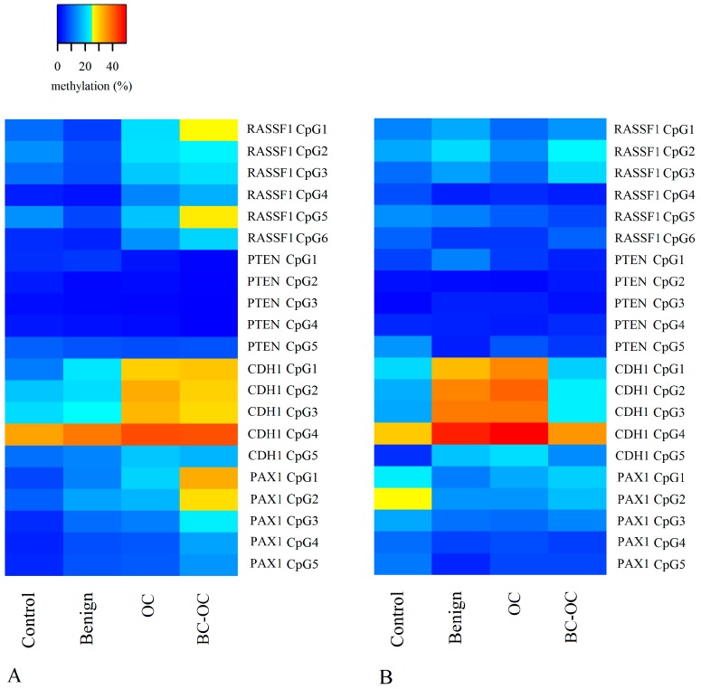
Figure 1 shows the methylation value of each CpG site in the tissue (A) and plasma samples (B) according to diagnosis. The general overview provides information on the hypomethylation of PTEN CpG sites for all diagnoses and tissue and plasma samples. Greater variability is obvious in the RASSF1 and PAX1 CpG sites, and the highest methylation was detected in the CDH1 CpG sites. The comparison of tissue samples (Figure 1A) shows the increasing methylation trend from the control sample group to cancer samples in the RASSF1, CDH1 and PAX1 genes. The statistical comparison of means revealed the most significant differences between controls and OC samples in two CDH1 and three PAX1 gene CpG sites and between controls and BC-OC in three PAX1 gene CpG sites. The increasing mean values for RASSF1 were not statistically significant because of the wide variance in values (detailed statistical data is contained in Table S1). Figure 1B depicts variability in the plasma group methylation values, with no visible linear trends detected between the diagnostic groups. Finally, the statistically significant higher methylation levels were detected in OCs in four CDH1 gene CpG sites compared to controls, and all detailed statistical data is listed in Table S2.
Figure 1.
Heat map of each CpG site methylation values in tissue (A) and plasma samples (B) according to diagnosis. OC—ovarian cancer; BC-OC—ovarian cancer subsequent to breast cancer.
3.2. DNA Methylation Indices of the RASSF1, PTEN, CDH1 and PAX1 Gene Regulatory Regions
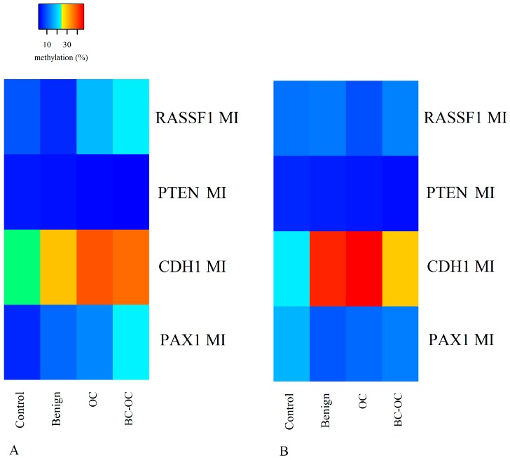
The methylation index (MI) is the mean percentage of methylation across all CpG sites analysed per gene, and that was calculated for each gene and diagnostic group in the tissue and plasma samples. The MI values of each diagnostic group are depicted by the heat maps in Figure 2A for tissue and 2B for plasma. Although PTEN gene methylation indices were very low, they were statistically significantly different between benign tumours (3.90 ± 4.10) and malignant OC (3.00 ± 2.60) in the tissue samples. No statistically significant differences were determined in RASSF1 MI between diagnostic groups in either the tissue or plasma samples. In the tissue samples, the CDH1 and PAX1 gene methylation indices had increasing tendency toward OC, with statistically significant difference between controls (24.22 ± 6.93 for CDH1 and 5.73 ± 2.19 for PAX1) and OCs (39.06 ± 18.78 and 13.55 ± 10.65) in both genes, and also between controls (5.73 ± 2.19) and BC-OC samples (22.90 ± 13.56) in the PAX1 gene. In the plasma samples, there was only one statistically significant difference between controls (22.25 ± 14.13) and OC samples (46.42 ± 20.91), in the CDH1 gene. Detailed data is provided in Supplementary Table S3.
Figure 2.
Heat map of MI values in tissue (A) and plasma (B) samples according to diagnosis. MI—methylation index, OC—ovarian cancer; BC-OC—ovarian cancer subsequent to breast cancer.
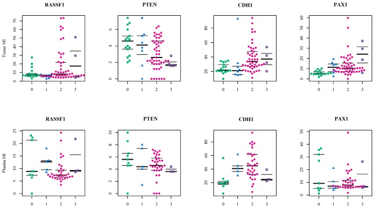
Analysed sample distribution is depicted in Figure 3 according to the sample type and diagnosis for all four studied genes. Wider variance in the mean values is obvious in the PTEN methylation index, and that explains the small differences between groups.
Figure 3.
The swarmplots of methylation indices of RASSF1, PTEN, CDH1 and PAX1 genes, showing the mean values of all observations. MI—methylation index; 0—control tissues (green squares); 1—benign tumours (blue triangles); 2—ovarian cancers (pink circles); 3—ovarian cancers subsequent to breast cancer (purple crossed circles).
3.3. Methylation Pattern by FIGO Staging and Histological Type
The methylation levels of all CpG sites revealed little statistically significant differences between the compared FIGO stages (data not shown) and the MI data pattern is depicted in Table 5. Two following trends were observed: The RASSF1 and PAX1 methylation indices decreased with disease severity increase, and the CDH1 and PTEN values had increasing tendency, thus followiing the stages. Statistical comparison of the mean values revealed significant differences only in the PTEN gene and between stages I and II, and stages I and IV in the PTEN gene.
Table 5.
MI averages of CpG sites in regulatory regions of the RASSF1, PTEN, CDH1 and PAX1 genes in tissue samples according to FIGO stage.
| Stage I (n = 9) | Stage II (n = 21) | Stage IV (n = 12) | p-Value | |||||||
|---|---|---|---|---|---|---|---|---|---|---|
| Mean | Median | SD | Mean | Median | SD | Mean | Median | SD | ||
| MI_RASSF1 | 19.24 | 5.33 | 20.46 | 17.24 | 7.50 | 19.80 | 8.01 | 6.83 | 3.95 | ns |
| MI_PTEN | 2.09 | 2.00 | 1.37 | 3.47 | 3.60 | 1.49 | 3.63 | 3.60 | 1.37 | a, b |
| MI_CDH1 | 32.89 | 29.00 | 16.81 | 39.98 | 30.50 | 20.72 | 48.38 | 44.25 | 17.76 | ns |
| MI_PAX1 | 17.11 | 13.60 | 17.36 | 11.56 | 10.40 | 5.42 | 10.98 | 8.50 | 6.28 | ns |
Note: Due to number of cases in each category, we created only three groups, based on the FIGO stage numbers (for example 1A, 1B and 1C were merged into stage I); ns—non significant difference between groups; a—statistically significant difference (p < 0.05) between FIGO I and II; b—between FIGO I and IV.
Comparison of the methylation levels and indices between histological subtypes revealed only sporadic differences and indicated possible uniform methylation of the studied genes in this OC group.
3.4. Comparison of Methylation Indices in Tissue and Plasma Paired Samples
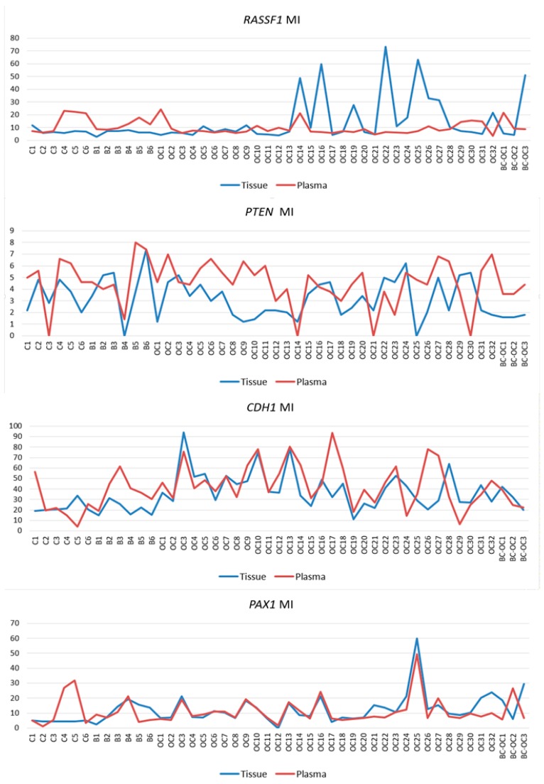
The MI values of paired tissue and plasma samples were compared to assess if the plasma methylation status reflects those observed in the primary tumours. Figure 4 shows similarities and differences in the methylation indices of all RASSF1, PTEN, CDH1 and PAX1 gene paired samples, and statistical analysis determined significant plasma and tissue correlation only in the CDH1 and PAX1 genes. The correlation coefficient of CDH1 MI was negative in controls (r = −0.812, p = 0.05), but positive in benign samples (r = 0.886, p = 0.019) and OC samples (r = 0.428, p = 0.015). However, strong correlation was established between plasma and tissue in all PAX1 CpG sites and between PAX1 methylation indices, but only in the OC pairs (r = 0.771, p < 0.001).
Figure 4.
Methylation indices of RASSF1, PTEN, CDH1 and PAX1 genes in paired tissue and plasma samples. MI—methylation index; C—control samples; B—benign; OC—ovarian cancer; BC-OC—ovarian cancer subsequent to breast cancer.
The results for sample size determination, including the effect size and the number of observations in each group needed to maintain 80% power level, are included in Supplementary Table S4 for tissue samples and Table S5 for plasma samples.
3.5. Evaluation of the Diagnostic Predictive Model Using ROC Analyses
Finally, we used the random forest algorithm with ROC analysis to evaluate if the studied methylation indices combined with age could significantly distinguish between diagnoses. Table 6 provides the AUC values with 95% confidence intervals for tandem discriminations. The “Important variable” column denotes the most relevant variables based on importance measured by minimal graph depth. The best AUC values were obtained in the model which discriminate between controls and OC samples based on both the MI of the four genes in tissues (AUC = 0.932) and in plasma (AUC = 0.822). The first “tissue” model had perfect sensitivity (98%), but specificity was weak (34%). The same was observed in the “plasma” model, with 91% sensitivity and 56% specificity. Based on the AUC values and 95% CI, these models indicate strong predictive power, and can, therefore, be considered for diagnostic discrimination clinically.
Table 6.
Predictive performance of the model distinguishing diagnostic groups.
| AUC | 95% CI | Overall Error Rate (%) | Important Variable | ||
|---|---|---|---|---|---|
| Tissue | Control vs. Benign | 0.738 | 0.391–1.000 | 15.38 | PAX1 |
| Control vs. OC | 0.932 | 0.823–1.000 | 9.09 | PAX1 | |
| Benign vs. OC | 0.499 | 0.195–0.802 | 12.5 | CDH1 | |
| Plasma | Control vs. Benign | 0.778 | 0.512–1.000 | 21.43 | CDH1 |
| Control vs. OC | 0.822 | 0.667–0.976 | 16.67 | CDH1 | |
| Benign vs. OC | 0.630 | 0.309–0.951 | 13.16 | PTEN | |
Note: OC—ovarian cancer, AUC—area under the curve, CI—confidence interval.
4. Discussion
OC is one of the most lethal cancerous diseases, and despite the progress in surgical techniques, better management, advanced chemotherapy and targeted therapy, the OC patient survival rate is still very low and almost 70% of all patients are diagnosed in advanced stages. This is mainly due to the lack of early symptoms in this disease [8]. Early OC screening approaches have been traditionally based on serum CA-125 concentration and trans-vaginal ultrasound, but both these methods lack sufficient sensitivity and specificity [46,47]. Therefore, non-invasive biomarkers able to predict early OC presence and monitor response to treatment and cancer progression are imperative. However, those with sufficient predictive power are still lacking and most possible candidates require further investigation [46,48,49]. Moreover, further use of the tumour markers is crucial in all gynaecological cancerous diseases [50,51,52]. For example, CTC, ctDNA, cfmiRNAs and released exosome vesicles can be analysed in the assessment of molecular markers. This assessment can be performed by a variety of methods, from basic PCR modifications to advanced multi-parallel sequencing approaches [10].
Herein, we focused on the epigenetic impact on OC formation by analysing the methylation levels of the RASSF1, PTEN, CDH1 and PAX1 tumour suppressor genes’ regulatory sequences. We compared the methylation status of normal tissue, benign, OC and BC-OC samples. We then assessed the methylation levels in these genes in the corresponding plasma samples in order to estimate their potential for LB utilisation. Finally, we compared the methylation patterns in the tissue samples and the paired plasma samples to assess if these patterns were markedly different.
Previously published articles reported the importance of some genes’ plasma methylation status in OC management. For example, the C2 CD4 D, WNT6 and COL23 A1 genes’ methylation status is not only significantly altered in OC patients’ ctDNA, but different responses to platinum-based therapy can also be associated with their methylation levels [53]. Moreover, Giannopolou et al. [54] discovered altered ESR1 gene methylation levels in the ctDNA samples of high-grade serous OC patients, and these abnormalities are very similar to those in primary tumours [54].
The most notable methylation changes in solid OT are in the BRCA1 [29,55], OPCML [56], HOXA9 [33] and P16 INK4α genes [57]. BRCA1 is methylated in up to 30% [29,55] of these tumours and OPCML is methylated in over 80% of OT [56]. Interestingly, RASSF1 A is the major RASSF1 gene isoform and this has been repeatedly analysed for methylation changes in both OC tissues and plasma samples. For example, de Caceres et al. [32] noted methylation of the RASSF1 A gene in 50% of primary OT and detected an identical pattern of gene hyper-methylation in 41 of 50 (82%) matched plasma DNA. In addition, hyper-methylation of this gene was recorded in all OC histological types, grades and stages. Wu et al. [33] reported very similar results in solid tissues, with hyper-methylation in 49% of patients with various OC types and grades. In contrast, Agathanggelou et al. [58] found hyper-methylation in only 10% of solid OT.
Methylation changes in the RASSF1 A promoter have already been analysed for utilization in non-invasive differentiation of epithelial ovarian carcinoma from healthy controls. For example, 90% sensitivity and 86% specificity for cancer detection was determined when methylation levels of the RASSF1 A, EP300 and CALCA genes were used in combination [59]. In addition, the multiplex methylation specific PCR (MSP) assay of seven different genes, including RASSF1 A, achieved 85.3% sensitivity and 90.5% specificity in differentiating epithelial OC from healthy controls in serum samples [60]. Variable RASSF1 A methylation has also been noted in tissue samples of serous and non-serous types and benign tumours [61].
Herein, the RASSF1 A MI differed in all four tissue types. It was higher in primary malignant tumours (17.98 ± 19.73) than in healthy tissue (9.50 ± 6.21), but the opposite was found in plasma ctDNA. Here, the controls have higher MI (11.74 ± 8.34) compared to malignant samples (8.93 ± 4.54). However, no statistically significant differences were established between particular CpGs or mean methylation indices. Importantly, although these differences proved insignificant, they can indicate wide methylation changes in RASSF1 gene regulating sequences and the important role of methylation in silencing this gene in OC development as is generally suggested [29].
Although the methylation status of the PTEN CpG sites and methylation indices was generally low and homogenous, there were statistically significant differences between benign and malignant lesions. The decreasing tendency in methylation indices was observed in both tissue and plasma samples, but comparison between FIGO stages revealed a statistically significant increase in methylation indices only in the higher FIGO stages. This theoretically indicates its important role in cancer formation and spread. Herein, however, we compared only the main FIGO categories where subtypes were merged in one main category, because of the small sample size. In addition, the differences between the FIGO stages were not only low, but the methylation indices themselves also had low values. This leads to debate if such small increases in methylation levels can contribute to tumour progression from stage I to stage IV. This is especially pertinent when previous very low PTEN methylation levels have occurred without differences in OC FIGO stages [34].
Yang et al. [34] and Zuberi et al. [35] recorded relatively low methylation values in the PTEN gene with higher methylation in only 16% and 8.2% of the samples, respectively. Schondorf et al. [62] suggested that PTEN methylation should have only a subordinate role in OC progression and only the one study by Qi et al. [36] reported relatively higher methylation levels of this gene in OC. Overall, the low and homogeneous PTEN methylation concord with the most of previous studies, and the small increase in methylation should not reflect progression from stage I to stage IV, though statistical significance is established between the FIGO stages. Our results also indicate the potential effect of PTEN methylation changes in benign lesion formation. That suggests that further analysis of the methylation changes will be beneficial, because there is no rule that benign lesions must be less methylated than malignant lesions, and different mechanisms can lead to abnormal methylation in benign and malignant tumours [63].
In comparison to RASSF1 and PTEN, the PAX1 gene had greater variation in methylation levels with statistically significant differences between controls, OC and BC-OC. There was also wide variability in particular CpG islands, especially in the control and malignant samples. This gene’s methylation provides a potential biomarker for cervical cancer screening [64,65,66], but important methylation changes in this gene were also noted in OC. In particular, Su et al. [37] reported 50% PAX1 methylation rates in OC, but only 14.3% in borderline tumours and 4% in benign, and Hassan et al. [38] found hyper-methylation in 50%, 50%, 46.6% and 78% of patients depending on the FIGO stage of epithelial OC. In addition, the authors associated methylation levels with the presence of HPV16/18 infection. Neither of these studies can be quantified in the same manner as our quantitative pyrosequencing results because they used MSP in their assessment, but the significant differences we achieved between healthy tissues and cancer samples can be interpreted quite similarly—that dynamic methylation changes indicating OC’s presence occur in the aforementioned gene’s regulation sequences. Although our results established no statistical significance, they can certainly help move the debate on how this gene’s methylation changes affect the formation of benign lesion because the methylation levels were as high they are in cancerous tissues.
Finally, the CDH1 gene had statistically significant variability in methylation levels between control and malignant tissue samples, and this agrees with previous analyses [25,39,40]. Importantly, this gene also had statistically significant plasma samples differences in both mean methylation levels and those of particular CpG dinucleotides in both healthy and cancerous groups. Therefore, the CDH1 gene could be considered a very promising candidate for non-invasive LB-based diagnosis of malignant OC, but its potential for non-invasive investigation of benign tumour presence requires further analysis.
It is important to realize that even non-significant, but observable differences in plasma samples methylation levels were revealed also in the RASSF1, PTEN and PAX1 genes, and this could inspire additional analyses of these genes. The large deviation in the final methylation levels of these genes was very likely the main obstacle to achieving statistical significance. However, larger sample collection could help to solve this problem.
One of the issues in the analysis of plasma samples is the lack of definition and accepted consensus if changes, including those in ctDNA methylation, remain exactly the same as in the primary tumours that released them. While some authors consider that the aberrant patterns remain identical to those in the primary tumour [53,67,68,69,70,71,72], others support diverse patterns [73,74,75]. Herein, we conform to similarity because of the strong correlations between tissue and plasma samples in the CDH1 and PAX1 genes, and also in several RASSF1 and PTEN gene samples. Discrepancies in tissue and plasma sample methylation levels could have also occurred herein because circulating cfDNA of cancer patients contains a mixture of DNA from both normal and cancerous cells. The ratio of ctDNA in the entire cfDNA fraction can vary because this depends on several tumour parameters [76]. Our methodology cannot estimate how plasma sample results are affected by the presence of non-tumorous cfDNA elements, and we cannot exclude that this is responsible for the variability obtained in the results. Although the ctDNA ratio is generally quite low it can vary widely, and some estimations of ctDNA abundance reach 90% [76,77]. Importantly, it has been demonstrated that the concentration of all circulating free molecules, and not only ctDNA, is associated with a tumour volume which gives shorter overall survival (OS) for OC patients [78].
Previously performed LB-based methylation analyses assessing the methylation pattern in OCs using non-NGS approaches have provided very interesting results. Those include (1) Teschendorff et al. [79], when they used a methylation-array based study and observed significant differences in the blood of healthy and cancerous patients, and (2) Flanagan et al. [80], who utilised the pyrosequencing approach and determined significant correlation of SFN gene methylation with PFS. In addition, their subsequent study [81] noted specific CpGs’ alterations in blood DNA following relapse from platinum-based chemotherapy, and their results proved an independent significant association with survival. Moreover, both pyrosequencing and MSP have previously been used to assess methylation abnormalities in plasma samples from different cancer types, and some of these achieved significant results assessing the profiles of all circulating DNAs, not only ctDNA [82,83,84,85,86]. Therefore, even methodology which is unable to distinguish ctDNAs in the whole cfDNA fraction can be useful in assessing disease characteristics from LB samples. We also incline toward the consensus that changes in primary tumours should remain preserved in ctDNA elements and the changes noted in primary tumours could then be used as inspiration for initiating further experiments utilising LB samples. Therefore, known abnormalities in primary tumours can be very helpful because these can be searched and analysed in LB samples.
OC is widely recognised as a very heterogeneous group and it should, therefore, be important to analyse and interpret the results of each histotype. However, the rarity of some morphological subtypes causes study limitations, and the small numbers in some histotypes herein prevented performance of statistical analysis. Only the serous and serous papillary histotypes were sufficient for analysis, and these recorded no significant differences in their methylation levels and were even almost identical. While studies have confirmed that some differences are typical for all OC histotypes [25,32,33,34,56], it cannot be ignored that unintentional variance in our results is due to the heterogeneity of our collection and also the impossibility of separating and analysing particular histotypes.
The effect size and the number of observations in each group required to maintain 80% power were assessed by power analysis, and these are listed in supplementary Tables S4 and S5. Herein, the number of malignant tissues was mostly sufficient, but we occasionally noted a lack in the number of healthy controls and the number of benign lesions was quite often lower than proposed. Therefore, although the power analysis results may limit our study through having a sample size insufficient to assess methylation levels in all CpGs with sufficient statistical power, it can help design the same or similar experiments. The highest number of required observations, excluding three PTEN gene CpG sites in plasma samples, was 64, and it is quite possible to collect at least that number of samples for future analysis.
It has been noted that there are generally higher cfDNA levels in malignant OC patients than in healthy patients and in those with benign lesions [87,88]. The cfDNA levels are also lower in FIGO I–II stages than in the III–IV stages [78]. Although we assessed only four genes’ methylation indices and not the entire cfDNA levels, and therefore cannot directly associate these parameters, analysis of the relationship between methylation changes, tumour stage and general cfDNA levels could most likely provide clinical implications. It would be beneficial, therefore, to design further studies which assess these relationships.
The cfDNA characteristics can also vary depending on age [89] and the presence of concomitant disease [90]. Furthermore, it is presumed that length of cfDNA fragments correlates with shorter PFS, and also OS [91]. Unfortunately, we can neither confirm nor preclude that our results are affected by other clinical attributes, such as the presence or absence of other patient disease. However, further studies should establish associations between a wide variety of clinical parameters, methylation and also cfDNA levels, as previously suggested.
Herein we evaluated the predictive performance of the combined model of DNA methylation signature (MI of the genes) and the age (in years) using ROC analyses computed by the random forest algorithm. We ran six models separated for sample type. The model which discriminated between control and OC samples had the highest predictive power—at 0.932 AUC in tissue samples and 0.822 in plasma. This initially appeared a perfect model with high sensitivity and clinical application potential, but 34% specificity for tissue samples and 56% for plasma samples means that too many healthy women would be incorrectly classified as cancer patients. The most likely reason for these results was the small number of controls, and this could have affected statistical analyses and hindered efforts to ensure a strong statistical model. Adequate AUC values were obtained in the model discriminating control samples and benign samples (tissue: AUC = 0.729, 85.7% sensitivity and 83.3% specificity; plasma: AUC = 0.778, 100% sensitivity and 66.7% specificity). Our last discrimination model of benign and OC samples had insufficient classifiers. In these models, PAX1 MI and CDH1 MI were selected as the most important variables for tissue and plasma respectively, and this indicates the previously described results about tissue and plasma non-conformity. Although these analyses promised a high-quality classifier, other possibilities should now be also considered. The most important would be the inclusion of additional control samples, as this would provide more accurate specificity. It is also worth considering the inclusion of more variables in the model, such as gynaecological anamnesis, life style risk factors and also CA-125 or other novel biomarkers with the potential to detect serous OC at earlier stages and predict patient survival prognosis [92,93,94]. Although this is beyond the scope of this pilot project, it should provide inspiration for further analyses focused on specific variables identified by a multinomial approach and offer personalised tailored therapy. Because of the great variability in this disease, the management of OC should be individualized, and performance status of the patient should be considered. This is best reflected in necessity for different treatment option for younger and older women [95,96]. In addition, future individual OC treatment should also be based on different patient physiological and health parameters.
In conclusion, our study confirmed higher methylation levels in several CpG sites and also higher methylation index in RASSF1 A, PAX1 and CDH1 genes in OC tissues compared to control tissues. However, no statistical significance was noted in the RASSF1 gene because of high variance. Statistically significant higher MI and CpG sites methylation levels were detected in plasma samples only in the CDH1 gene. PTEN gene was slightly and homogenously methylated. In addition, we recorded similar methylation patterns with strong correlations in the CDH1 and PAX1 genes in OC plasma and tissue, and the results suggest that the CDH1 gene is a prospective candidate for non-invasive, LB-based differentiation in OC. Our diagnostic discrimination model combined the methylation indices of all four genes with age, and this revealed the highest AUC 0.932 predictive power in the model, comparing OC subjects with controls. Finally, it will prove highly beneficial to employ as many LB-based approaches as possible in cancer detection and treatment, especially in OC, which has such high risk and generally delayed diagnosis. LB is capable of providing early OC diagnosis which can help decrease the global expense of fighting this disease, but most importantly it can save lives, or at least prolong them with greater life quality for the thousands of women affected by this lethal cancer.
Abbreviations
| AUC | Area under the curve |
| BC-OC | Ovarian cancer subsequent to breast cancer |
| CDH1 | Cadherin 1 |
| cfmiRNA | Circulating cell-free microRNAs |
| CTC | Circulating tumour cells |
| ctDNA | Circulating tumour DNA |
| LB | Liquid biopsy |
| MI | Methylation index |
| MSP | Methylation specific PCR |
| OC | Ovarian cancer |
| OS | Overall survival |
| OT | Ovarian tumours |
| PAX1 | Paired Box 1 |
| PFS | Progression-free survival |
| PTEN | Phosphatase and Tensin Homolog |
| RASSF1 | Ras Association Domain Family Member 1 |
| ROC | Receiver operating characteristic |
Supplementary Materials
Supplementary Materials can be found at https://www.mdpi.com/1422-0067/20/17/4119/s1.
Author Contributions
Conceptualization: B.N., B.S. and Z.D.; data curation: M.G.; funding acquisition: P.Z. and Z.D.; investigation: D.D. and D.B.; methodology: Z.D., D.D., D.B., M.J. and K.Z.; project administration: P.Z., Z.L., and Z.D.; software: M.G. and Z.D.; supervision: B.N., P.Z. and Z.L.; writing—original draft: D.D., D.B. and Z.D.; writing—reviewing and editing: B.N., R.P. and Z.D.
Funding
This study was supported by Slovak Research and Development Agency Grants APVV-14-0815, VEGA Grant 1/0199/17 of the Scientific Grant Agency of the Ministry of Education of the Slovak Republic and by implementation of project: Molecular diagnosis of cervical cancer, ITMS: 26220220113 supported by the Operational Programme Research and Innovation funded by the ERDF.
Conflicts of Interest
The authors declare no conflict of interest.
References
- 1.Ferlay J., Soerjomataram I., Ervik M., Dikshit R., Eser S., Mathers C., Rebelo M., Parkin D.M., Forman D., Bray F. GLOBOCAN 2012 v1.0, Cancer Incidence and Mortality Worldwide: IARC CancerBase No. 11. Lyon, France: International Agency for Research on Cancer. [(accessed on 28 February 2014)];2013 Available online: http://globocan.iarc.fr.
- 2.Brett M. R., Jennifer B. P., Thomas A. S., Brett M. R., Jennifer B. P., Thomas A. S. Epidemiology of ovarian cancer: A reviewepidemiology of ovarian cancer: A review. Cancer Biol. Med. 2017;14:9–32. doi: 10.20892/j.issn.2095-3941.2016.0084. [DOI] [PMC free article] [PubMed] [Google Scholar]
- 3.Bray F., Loos A.H., Tognazzo S., La Vecchia C. Ovarian cancer in europe: Cross-sectional trends in incidence and mortality in 28 countries, 1953–2000. Int. J. Cancer. 2005;113:977–990. doi: 10.1002/ijc.20649. [DOI] [PubMed] [Google Scholar]
- 4.Malvezzi M., Carioli G., Rodriguez T., Negri E., La Vecchia C. Global trends and predictions in ovarian cancer mortality. Ann. Oncol. 2016;27:2017–2025. doi: 10.1093/annonc/mdw306. [DOI] [PubMed] [Google Scholar]
- 5.Teng Z., Han R., Huang X., Zhou J., Yang J., Luo P., Wu M. Increase of incidence and mortality of ovarian cancer during 2003–2012 in Jiangsu province, China. Front. Public Heal. 2016;4:146. doi: 10.3389/fpubh.2016.00146. [DOI] [PMC free article] [PubMed] [Google Scholar]
- 6.Sankaranarayanan R., Ferlay J. Worldwide Burden of gynaecological cancer: The size of the problem. Best Pract. Res. Clin. Obstet. Gynaecol. 2006;20:207–225. doi: 10.1016/j.bpobgyn.2005.10.007. [DOI] [PubMed] [Google Scholar]
- 7.Meinhold-Heerlein I., Fotopoulou C., Harter P., Kurzeder C., Mustea A., Wimberger P., Hauptmann S., Sehouli J. The new WHO classification of ovarian, fallopian tube, and primary peritoneal cancer and its clinical implications. Arch. Gynecol. Obstet. 2016;293:695–700. doi: 10.1007/s00404-016-4035-8. [DOI] [PubMed] [Google Scholar]
- 8.Torre L.A., Trabert B., DeSantis C.E., Miller K.D., Samimi G., Runowicz C.D., Gaudet M.M., Jemal A., Siegel R.L. Ovarian cancer statistics, 2018. CA. Cancer J. Clin. 2018;68:284–296. doi: 10.3322/caac.21456. [DOI] [PMC free article] [PubMed] [Google Scholar]
- 9.Wimberger P., Wehling M., Lehmann N., Kimmig R., Schmalfeldt B., Burges A., Harter P., Pfisterer J., du Bois A. Influence of residual tumor on outcome in ovarian cancer patients with FIGO stage IV disease. Ann. Surg. Oncol. 2010;17:1642–1648. doi: 10.1245/s10434-010-0964-9. [DOI] [PubMed] [Google Scholar]
- 10.Giannopoulou L., Zavridou M., Kasimir-Bauer S., Lianidou E.S. Liquid biopsy in ovarian cancer: The potential of circulating MiRNAs and exosomes. Transl. Res. 2019;205:77–91. doi: 10.1016/j.trsl.2018.10.003. [DOI] [PubMed] [Google Scholar]
- 11.Tan D.S., Agarwal R., Kaye S.B. Mechanisms of transcoelomic metastasis in ovarian cancer. Lancet Oncol. 2006;7:925–934. doi: 10.1016/S1470-2045(06)70939-1. [DOI] [PubMed] [Google Scholar]
- 12.Yeung T.L., Leung C.S., Yip K.P., Au Yeung C.L., Wong S.T.C., Mok S.C. Cellular and molecular processes in ovarian cancer metastasis. A review in the theme: Cell and molecular processes in cancer metastasis. Am. J. Physiol. Physiol. 2015;309:C444–C456. doi: 10.1152/ajpcell.00188.2015. [DOI] [PMC free article] [PubMed] [Google Scholar]
- 13.Bedard P.L., Hansen A.R., Ratain M.J., Siu L.L. Tumour heterogeneity in the clinic. Nature. 2013;501:355–364. doi: 10.1038/nature12627. [DOI] [PMC free article] [PubMed] [Google Scholar]
- 14.Donninger H., Vos M.D., Clark G.J. The RASSF1 a tumor suppressor. J. Cell Sci. 2007;120:3163–3172. doi: 10.1242/jcs.010389. [DOI] [PubMed] [Google Scholar]
- 15.Grawenda A.M., O’Neill E. Clinical utility of RASSF1 a methylation in human malignancies. Br. J. Cancer. 2015;113:372–381. doi: 10.1038/bjc.2015.221. [DOI] [PMC free article] [PubMed] [Google Scholar]
- 16.Van der Weyden L., Adams D.J. The ras-association domain family (RASSF) members and their role in human tumourigenesis. Biochim. Biophys. Acta Rev. Cancer. 2007;1776:58–85. doi: 10.1016/j.bbcan.2007.06.003. [DOI] [PMC free article] [PubMed] [Google Scholar]
- 17.Law J., Yu V.C., Baksh S. Modulator of apoptosis 1: A highly regulated RASSF1 a-interacting BH3-like protein. Mol. Biol. Int. 2012;2012:536802. doi: 10.1155/2012/536802. [DOI] [PMC free article] [PubMed] [Google Scholar]
- 18.Donninger H., Clark J., Rinaldo F., Nelson N., Barnoud T., Schmidt M.L., Hobbing K.R., Vos M.D., Sils B., Clark G.J. The RASSF1 a tumor suppressor regulates XPA-mediated DNA repair. Mol. Cell. Biol. 2015;35:277–287. doi: 10.1128/MCB.00202-14. [DOI] [PMC free article] [PubMed] [Google Scholar]
- 19.Shivakumar L., Minna J., Sakamaki T., Pestell R., White M.A. The RASSF1 a tumor suppressor blocks cell cycle progression and inhibits cyclin D1 accumulation. Mol. Cell. Biol. 2002;22:4309–4318. doi: 10.1128/MCB.22.12.4309-4318.2002. [DOI] [PMC free article] [PubMed] [Google Scholar]
- 20.Song S.J., Song M.S., Kim S.J., Kim S.Y., Kwon S.H., Kim J.G., Calvisi D.F., Kang D., Lim D.S. Aurora a regulates prometaphase progression by inhibiting the ability of RASSF1 A to suppress APC-Cdc20 activity. Cancer Res. 2009;69:2314–2323. doi: 10.1158/0008-5472.CAN-08-3984. [DOI] [PubMed] [Google Scholar]
- 21.Yin Y., Shen W.H. PTEN: A new guardian of the genome. Oncogene. 2008;27:5443–5453. doi: 10.1038/onc.2008.241. [DOI] [PubMed] [Google Scholar]
- 22.Tsanou E., Peschos D., Batistatou A., Charalabopoulos A., Charalabopoulos K. The E-cadherin adhesion molecule and colorectal cancer. A global literature approach. Anticancer Res. 2008;28:3815–3826. [PubMed] [Google Scholar]
- 23.Corso G., Intra M., Trentin C., Veronesi P., Galimberti V. CDH1 Germline mutations and hereditary lobular breast cancer. Fam. Cancer. 2016;15:215–219. doi: 10.1007/s10689-016-9869-5. [DOI] [PubMed] [Google Scholar]
- 24.Luo W., Fedda F., Lynch P., Tan D. CDH1 gene and hereditary diffuse gastric cancer syndrome: Molecular and histological alterations and implications for diagnosis and treatment. Front. Pharmacol. 2018;9:1421. doi: 10.3389/fphar.2018.01421. [DOI] [PMC free article] [PubMed] [Google Scholar]
- 25.Wang Q., Wang B., Zhang Y.M., Wang W. The association between CDH1 promoter methylation and patients with ovarian cancer: A systematic meta-analysis. J. Ovarian Res. 2016;9:23. doi: 10.1186/s13048-016-0231-1. [DOI] [PMC free article] [PubMed] [Google Scholar]
- 26.Robson E.J.D., He S.J., Eccles M.R. A PANorama of PAX genes in cancer and development. Nat. Rev. Cancer. 2006;6:52–62. doi: 10.1038/nrc1778. [DOI] [PubMed] [Google Scholar]
- 27.Li C.G., Eccles M.R. PAX genes in cancer; friends or foes? Front. Genet. 2012;3:1–6. doi: 10.3389/fgene.2012.00006. [DOI] [PMC free article] [PubMed] [Google Scholar]
- 28.Su P.H., Lai H.C., Huang R.L., Chen L.Y., Wang Y.C., Wu T.I., Chan M.W.Y., Liao C.C., Chen C.W., Lin W.Y., et al. Paired box-1 (PAX1) activates multiple phosphatases and inhibits kinase cascades in cervical cancer. Sci. Rep. 2019;9:9195. doi: 10.1038/s41598-019-45477-5. [DOI] [PMC free article] [PubMed] [Google Scholar]
- 29.Hentze J., Høgdall C., Høgdall E. Methylation and ovarian cancer: Can DNA methylation be of diagnostic use? Mol. Clin. Oncol. 2019;10:323–330. doi: 10.3892/mco.2019.1800. [DOI] [PMC free article] [PubMed] [Google Scholar]
- 30.Losi L., Fonda S., Saponaro S., Chelbi S.T., Lancellotti C., Gozzi G., Alberti L., Fabbiani L., Botticelli L., Benhattar J. Distinct DNA methylation profiles in ovarian tumors: Opportunities for novel biomarkers. Int. J. Mol. Sci. 2018;19:1559. doi: 10.3390/ijms19061559. [DOI] [PMC free article] [PubMed] [Google Scholar]
- 31.Lund R.J., Huhtinen K., Salmi J., Rantala J., Nguyen E.V., Moulder R., Goodlett D.R., Lahesmaa R., Carpén O. DNA methylation and transcriptome changes associated with cisplatin resistance in ovarian cancer. Sci. Rep. 2017;7:1469. doi: 10.1038/s41598-017-01624-4. [DOI] [PMC free article] [PubMed] [Google Scholar]
- 32.De Caceres I.I., Battagli C., Esteller M., Herman J.G., Dulaimi E., Edelson M.I., Bergman C., Ehya H., Eisenberg B.L., Cairns P. Tumor cell-specific BRCA1 and RASSF1A hypermethylation in serum, plasma, and peritoneal fluid from ovarian cancer patients. Cancer Res. 2004;64:6476–6481. doi: 10.1158/0008-5472.CAN-04-1529. [DOI] [PubMed] [Google Scholar]
- 33.Wu Q., Lothe R.A., Ahlquist T., Silins I., Tropé C.G., Micci F., Nesland J.M., Suo Z., Lind G.E. DNA methylation profiling of ovarian carcinomas and their in vitro models identifies HOXA9, HOXB5, SCGB3 A1, and CRABP1 as novel targets. Mol. Cancer. 2007;6:45. doi: 10.1186/1476-4598-6-45. [DOI] [PMC free article] [PubMed] [Google Scholar]
- 34.Yang H.J., Liu V.W.S., Wang Y., Tsang P.C.K., Ngan H.Y.S. Differential DNA methylation profiles in gynecological cancers and correlation with clinico-pathological data. BMC Cancer. 2006;6:212. doi: 10.1186/1471-2407-6-212. [DOI] [PMC free article] [PubMed] [Google Scholar]
- 35.Zuberi M., Mir R., Dholariya S., Najar I., Yadav P., Javid J., Guru S., Mirza M., Gandhi G., Khurana N., et al. RASSF1 and PTEN promoter hypermethylation influences the outcome in epithelial ovarian cancer. Clin. Ovarian other Gynecol. Cancer. 2014;7:33–39. doi: 10.1016/j.cogc.2014.12.002. [DOI] [Google Scholar]
- 36.Qi Q., Ling Y., Zhu M., Zhou L., Wan M., Bao Y., Liu Y. Promoter region methylation and loss of protein expression of PTEN and significance in cervical cancer. Biomed. Rep. 2014;2:653–658. doi: 10.3892/br.2014.298. [DOI] [PMC free article] [PubMed] [Google Scholar]
- 37.Su H.Y., Lai H.C., Lin Y.W., Chou Y.C., Liu C.Y., Yu M.H. An epigenetic marker panel for screening and prognostic prediction of ovarian cancer. Int. J. Cancer. 2009;124:387–393. doi: 10.1002/ijc.23957. [DOI] [PubMed] [Google Scholar]
- 38.Hassan Z.K., Zekri A.R.N., Hafez M.M., Kamel M.M. Human papillomavirus genotypes and methylation of CADM1, PAX1, MAL and ADCYAP1 genes in epithelial ovarian cancer patients. Asian Pac. J. Cancer Prev. 2017;18:169–176. doi: 10.22034/APJCP.2017.18.1.169. [DOI] [PMC free article] [PubMed] [Google Scholar]
- 39.Lin H.W., Fu C.F., Chang M.C., Lu T.P., Lin H.P., Chiang Y.C., Chen C.A., Cheng W.F. CDH1, DLEC1 and SFRP5 methylation panel as a prognostic marker for advanced epithelial ovarian cancer. Epigenomics. 2018;10:1397–1413. doi: 10.2217/epi-2018-0035. [DOI] [PubMed] [Google Scholar]
- 40.Koukoura O., Spandidos D.A., Daponte A., Sifakis S. DNA methylation profiles in ovarian cancer: Implication in diagnosis and therapy. Mol. Med. Rep. 2014;10:3–9. doi: 10.3892/mmr.2014.2221. [DOI] [PMC free article] [PubMed] [Google Scholar]
- 41.Ma L., Zhang J., Liu F., Zhang X. Expression of RASSF1 A and RASSF1C transcripts in human primary ovarian cancers. Chinese J. Pathol. 2005;34:150–153. [PubMed] [Google Scholar]
- 42.Chen Y., Zheng H., Yang X., Sun L., Xin Y. Effects of mutation and expression of PTEN gene MRNA on tumorigenesis and progression of epithelial ovarian cancer. Chinese Med. Sci. J. 2004;19:25–30. [PubMed] [Google Scholar]
- 43.Chen Y., Zhao Y.J., Zheng H.C., Yang X.F., Wang G.L., Xin Y. MRNA expression of PTEN and VEGF genes in epithelial ovarian cancer. Chinese J. Cancer Res. 2003;15:252–256. doi: 10.1007/BF02974887. [DOI] [Google Scholar]
- 44.Wu X., Zhuang Y.X., Hong C.Q., Chen J.Y., You Y.J., Zhang F., Huang P., Wu M.Y. Clinical importance and therapeutic implication of E-cadherin gene methylation in human ovarian cancer. Med. Oncol. 2014;31:100. doi: 10.1007/s12032-014-0100-y. [DOI] [PubMed] [Google Scholar]
- 45.R Core Team R: A Language and Environment for Statistical Computing R Foundation for Statistical Computing, Vienna, Austria. [(accessed on 10 July 2019)]; Available online: https://www.R-project.org/
- 46.Chang L., Ni J., Zhu Y., Pang B., Graham P., Zhang H., Li Y. Liquid biopsy in ovarian cancer: Recent advances in circulating extracellular vesicle detection for early diagnosis and monitoring progression. Theranostics. 2019;9:4130–4140. doi: 10.7150/thno.34692. [DOI] [PMC free article] [PubMed] [Google Scholar]
- 47.Henderson J.T., Webber E.M., Sawaya G.F. Screening for ovarian cancer: Updated evidence report and systematic review for the US preventive services task force. JAMA. 2018;319:595–606. doi: 10.1001/jama.2017.21421. [DOI] [PubMed] [Google Scholar]
- 48.Dong X., Men X., Zhang W., Lei P. Advances in Tumor Markers of Ovarian Cancer for Early Diagnosis. Indian J. Cancer. 2014;51:e72–e76. doi: 10.4103/0019-509X.154049. [DOI] [PubMed] [Google Scholar]
- 49.Giannopoulou L., Kasimir-Bauer S., Lianidou E.S. Liquid biopsy in ovarian cancer: Recent advances on circulating tumor cells and circulating tumor DNA. Clin. Chem. Lab. Med. 2018;56:186–197. doi: 10.1515/cclm-2017-0019. [DOI] [PubMed] [Google Scholar]
- 50.Valenti G., Vitale S.G., Tropea A., Biondi A., Laganà A.S. Tumor markers of uterine cervical cancer: A new scenario to guide surgical practice? Updates Surg. 2017;69:441–449. doi: 10.1007/s13304-017-0491-3. [DOI] [PubMed] [Google Scholar]
- 51.Muinelo-Romay L., Casas-Arozamena C., Abal M. Liquid biopsy in endometrial cancer: New opportunities for personalized oncology. Int. J. Mol. Sci. 2018;19:2311. doi: 10.3390/ijms19082311. [DOI] [PMC free article] [PubMed] [Google Scholar]
- 52.Cheung T.H., Yim S.F., Yu M.Y., Worley M.J., Fiascone S.J., Chiu R.W.K., Lo K.W.K., Siu N.S.S., Wong M.C.S., Yeung A.C.M., et al. Liquid biopsy of HPV DNA in cervical cancer. J. Clin. Virol. 2019;114:32–36. doi: 10.1016/j.jcv.2019.03.005. [DOI] [PubMed] [Google Scholar]
- 53.Widschwendter M., Zikan M., Wahl B., Lempiäinen H., Paprotka T., Evans I., Jones A., Ghazali S., Reisel D., Eichner J., et al. The potential of circulating tumor DNA methylation analysis for the early detection and management of ovarian cancer. Genome Med. 2017;9:116. doi: 10.1186/s13073-017-0500-7. [DOI] [PMC free article] [PubMed] [Google Scholar]
- 54.Giannopoulou L., Mastoraki S., Buderath P., Strati A., Pavlakis K., Kasimir-Bauer S., Lianidou E.S. ESR1 methylation in primary tumors and paired circulating tumor DNA of patients with high-grade serous ovarian cancer. Gynecol. Oncol. 2018;150:355–360. doi: 10.1016/j.ygyno.2018.05.026. [DOI] [PubMed] [Google Scholar]
- 55.Wang C., Horiuchi A., Imai T., Ohira S., Itoh K., Nikaido T., Katsuyama Y., Konishi I. Expression of BRCA1 protein in benign, borderline, and malignant epithelial ovarian neoplasms and its relationship to methylation and allelic loss of the BRCA1 gene. J. Pathol. 2004;202:215–223. doi: 10.1002/path.1507. [DOI] [PubMed] [Google Scholar]
- 56.Czekierdowski A., Czekierdowska S., Szymanski M., Wielgos M., Kaminski P., Kotarski J. Opioid-binding protein/cell adhesion molecule-like (OPCML) gene and promoter methylation status in women with ovarian cancer. Neuroendocrinol. Lett. 2006;27:609–613. [PubMed] [Google Scholar]
- 57.Xiao X., Cai F., Niu X., Shi H., Zhong Y. Association between P16 INK4 a promoter methylation and ovarian cancer: A meta-analysis of 12 published studies. PLoS ONE. 2016;11:e0163257. doi: 10.1371/journal.pone.0163257. [DOI] [PMC free article] [PubMed] [Google Scholar]
- 58.Agathanggelou A., Honorio S., Macartney D.P., Martinez A., Dallol A., Rader J., Fullwood P., Chauhan A., Walker R., Shaw J.A., et al. Methylation associated inactivation of RASSF1A from region 3p21.3 in lung, breast and ovarian tumours. Oncogene. 2001;20:1509–1518. doi: 10.1038/sj.onc.1204175. [DOI] [PubMed] [Google Scholar]
- 59.Liggett T.E., Melnikov A., Yi Q., Replogle C., Hu W., Rotmensch J., Kamat A., Sood A.K., Levenson V. Distinctive DNA methylation patterns of cell-free plasma DNA in women with malignant ovarian tumors. Gynecol. Oncol. 2011;120:113–120. doi: 10.1016/j.ygyno.2010.09.019. [DOI] [PMC free article] [PubMed] [Google Scholar]
- 60.Zhang Q., Hu G., Yang Q., Dong R., Xie X., Ma D., Shen K., Kong B. A multiplex methylation-specific PCR assay for the detection of early-stage ovarian cancer using cell-free serum DNA. Gynecol. Oncol. 2013;130:132–139. doi: 10.1016/j.ygyno.2013.04.048. [DOI] [PubMed] [Google Scholar]
- 61.Rattanapan Y., Korkiatsakul V., Kongruang A., Chareonsirisuthigul T., Rerkamnuaychoke B., Wongkularb A., Wilailak S. EGFL7 and RASSF1 promoter hypermethylation in epithelial ovarian cancer. Cancer Genet. 2018;224:37–40. doi: 10.1016/j.cancergen.2018.04.117. [DOI] [PubMed] [Google Scholar]
- 62.Schondorf T., Ebert M.P., Hoffmann J., Becker M., Moser N., Pur S., Göhring U.J., Weisshaar M.P. Hypermethylation of the PTEN gene in ovarian cancer cell lines. Cancer Lett. 2004;207:215–220. doi: 10.1016/j.canlet.2003.10.028. [DOI] [PubMed] [Google Scholar]
- 63.Pfeifer G.P. Defining driver DNA methylation changes in human cancer. Int. J. Mol Sci. 2018;19:1166. doi: 10.3390/ijms19041166. [DOI] [PMC free article] [PubMed] [Google Scholar]
- 64.Fang C., Wang S.Y., Liou Y.L., Chen M.H., Ouyang W., Duan K.M. The promising role of PAX1 (Aliases: HUP48, OFC2) gene methylation in cancer screening. Mol. Genet. Genomic Med. 2019;7:e506. doi: 10.1002/mgg3.506. [DOI] [PMC free article] [PubMed] [Google Scholar]
- 65.Kan Y.Y., Liou Y.L., Wang H.J., Chen C.Y., Sung L.C., Chang C.F., Liao C.I. ATL. Int. J. Gynecol. Cancer. 2014;24:928–934. doi: 10.1097/IGC.0000000000000155. [DOI] [PubMed] [Google Scholar]
- 66.Xu J., Xu L., Yang B., Wang L., Lin X., Tu H. Assessing methylation status of PAX1 in cervical scrapings, as a novel diagnostic and predictive biomarker, was closely related to screen cervical cancer. Int. J. Clin. Exp. Pathol. 2015;8:1674–1681. [PMC free article] [PubMed] [Google Scholar]
- 67.Warton K., Mahon K.L., Samimi G. Methylated circulating tumor DNA in blood: Power in cancer prognosis and response. Endocr.-Relat. Cancer. 2016;23:R157–R171. doi: 10.1530/ERC-15-0369. [DOI] [PMC free article] [PubMed] [Google Scholar]
- 68.Xu R., Wei W., Krawczyk M., Wang W., Luo H., Flagg K., Yi S., Shi W., Quan Q., Li K., et al. Circulating tumour DNA methylation markers for diagnosis and prognosis of hepatocellular carcinoma. Nat. Mater. 2017;16:1155–1161. doi: 10.1038/nmat4997. [DOI] [PubMed] [Google Scholar]
- 69.Gorgannezhad L., Umer M., Islam M.N., Nguyen N.-T., Shiddiky M.J.A. Circulating Tumor DNA and Liquid Biopsy: Opportunities, Challenges, and Recent Advances in Detection Technologies. Lab Chip. 2018;18:1174–1196. doi: 10.1039/C8LC00100F. [DOI] [PubMed] [Google Scholar]
- 70.Wan J.C.M., Massie C., Garcia-Corbacho J., Mouliere F., Brenton J.D., Caldas C., Pacey S., Baird R., Rosenfeld N. Liquid biopsies come of age: Towards implementation of circulating tumour DNA. Nat. Rev. Cancer. 2017;17:223–238. doi: 10.1038/nrc.2017.7. [DOI] [PubMed] [Google Scholar]
- 71.Ng S.B., Chua C., Ng M., Gan A., Poon P.S., Teo M., Fu C., Leow W.Q., Lim K.H., Chung A., et al. Individualised multiplexed circulating tumour DNA assays for monitoring of tumour presence in patients after colorectal cancer surgery. Sci. Rep. 2017;7:40737. doi: 10.1038/srep40737. [DOI] [PMC free article] [PubMed] [Google Scholar]
- 72.Yang N., Li Y., Liu Z., Qin H., Du D., Cao X., Cao X., Li J., Li D., Jiang B., et al. The characteristics of CtDNA reveal the high complexity in matching the corresponding tumor tissues. BMC Cancer. 2018;18:319. doi: 10.1186/s12885-018-4199-7. [DOI] [PMC free article] [PubMed] [Google Scholar]
- 73.Guo Q., Wang J., Xiao J., Wang L., Hu X., Yu W., Song G., Lou J., Chen J. Heterogeneous mutation pattern in tumor tissue and circulating tumor DNA warrants parallel NGS panel testing. Mol. Cancer. 2018;17:131. doi: 10.1186/s12943-018-0875-0. [DOI] [PMC free article] [PubMed] [Google Scholar]
- 74.Kammesheidt A., Tonozzi T.R., Lim S.W., Braunstein G.D. Mutation Detection using plasma circulating tumor DNA (CtDNA) in a cohort of asymptomatic adults at increased risk for cancer. Int. J. Mol. Epidemiol. Genet. 2018;9:1–12. [PMC free article] [PubMed] [Google Scholar]
- 75.Toor O.M., Ahmed Z., Bahaj W., Boda U., Cummings L.S., McNally M.E., Kennedy K.F., Pluard T.J., Hussain A., Subramanian J., et al. Correlation of somatic genomic alterations between tissue genomics and CtDNA employing next-generation sequencing: Analysis of lung and gastrointestinal cancers. Mol. Cancer Ther. 2018;17:1123–1132. doi: 10.1158/1535-7163.MCT-17-1015. [DOI] [PubMed] [Google Scholar]
- 76.Qin Z., Ljubimov V.A., Zhou C., Tong Y., Liang J. Cell-free circulating tumor DNA in cancer. Chinese J. Cancer. 2016;35:36. doi: 10.1186/s40880-016-0092-4. [DOI] [PMC free article] [PubMed] [Google Scholar]
- 77.Elazezy M., Joosse S.A. Techniques of using circulating tumor DNA as a liquid biopsy component in cancer management. Comput. Struct. Biotechnol. J. 2018;16:370–378. doi: 10.1016/j.csbj.2018.10.002. [DOI] [PMC free article] [PubMed] [Google Scholar]
- 78.Shao X., He Y., Ji M., Chen X., Qi J., Shi W., Hao T., Ju S. Quantitative analysis of cell-free DNA in ovarian cancer. Oncol. Lett. 2015;10:3478–3482. doi: 10.3892/ol.2015.3771. [DOI] [PMC free article] [PubMed] [Google Scholar]
- 79.Teschendorff A.E., Menon U., Gentry-Maharaj A., Ramus S.J., Gayther S.A., Apostolidou S., Jones A., Lechner M., Beck S., Jacobs I.J., et al. An epigenetic signature in peripheral blood predicts active ovarian cancer. PLoS ONE. 2009;4:e8274. doi: 10.1371/journal.pone.0008274. [DOI] [PMC free article] [PubMed] [Google Scholar]
- 80.Flanagan J.M., Wilhelm-Benartzi C.S., Metcalf M., Kaye S.B., Brown R. Association of somatic DNA methylation variability with progression-free survival and toxicity in ovarian cancer patients. Ann. Oncol. Off. J. Eur. Soc. Med. Oncol. 2013;24:2813–2818. doi: 10.1093/annonc/mdt370. [DOI] [PubMed] [Google Scholar]
- 81.Flanagan J.M., Wilson A., Koo C., Masrour N., Gallon J., Loomis E., Flower K., Wilhelm-Benartzi C., Hergovich A., Cunnea P., et al. Platinum-based chemotherapy induces methylation changes in blood DNA associated with overall survival in patients with ovarian cancer. Clin. Cancer Res. 2017;23:2213–2222. doi: 10.1158/1078-0432.CCR-16-1754. [DOI] [PubMed] [Google Scholar]
- 82.Lu Y., Li S., Zhu S., Gong Y., Shi J., Xu L.L. Methylated DNA/RNA in Body Fluids as Biomarkers for Lung Cancer. Biol. Proced. Online. 2017;19:2. doi: 10.1186/s12575-017-0051-8. [DOI] [PMC free article] [PubMed] [Google Scholar]
- 83.Lissa D., Robles A.I. Methylation analyses in liquid biopsy. Transl. Lung Cancer Res. 2016;5:492–504. doi: 10.21037/tlcr.2016.10.03. [DOI] [PMC free article] [PubMed] [Google Scholar]
- 84.Balgkouranidou I., Chimonidou M., Milaki G., Tsarouxa E.G., Kakolyris S., Welch D.R., Georgoulias V., Lianidou E.S. Breast cancer metastasis suppressor-1 promoter methylation in cell-free DNA provides prognostic information in non-small cell lung cancer. Br. J. Cancer. 2014;110:2054–2062. doi: 10.1038/bjc.2014.104. [DOI] [PMC free article] [PubMed] [Google Scholar]
- 85.Kristensen L.S., Hansen J.W., Kristensen S.S., Tholstrup D., Harsløf L.B.S., Pedersen O.B., De Nully Brown P., Grønbæk K. Aberrant methylation of cell-free circulating DNA in plasma predicts poor outcome in diffuse large B cell lymphoma. Clin. Epigenetics. 2016;8:95. doi: 10.1186/s13148-016-0261-y. [DOI] [PMC free article] [PubMed] [Google Scholar]
- 86.Majchrzak-Celińska A., Paluszczak J., Kleszcz R., Magiera M., Barciszewska A.M., Nowak S., Baer-Dubowska W. Detection of MGMT, RASSF1A, P15INK4B, and P14ARF promoter methylation in circulating tumor-derived DNA of central nervous system cancer patients. J. Appl. Genet. 2013;54:335–344. doi: 10.1007/s13353-013-0149-x. [DOI] [PMC free article] [PubMed] [Google Scholar]
- 87.Zhou Q., Li W., Leng B., Zheng W., He Z., Zuo M., Chen A. Circulating cell free DNA as the diagnostic marker for ovarian cancer: A systematic review and meta-analysis. PLoS ONE. 2016;11:e0155495. doi: 10.1371/journal.pone.0155495. [DOI] [PMC free article] [PubMed] [Google Scholar]
- 88.Kamat A.A., Baldwin M., Urbauer D., Dang D., Han L.Y., Godwin A., Karlan B.Y., Simpson J.L., Gershenson D.M., Coleman R.L., et al. Plasma cell-free DNA in ovarian cancer: An independent prognostic biomarker. Cancer. 2010;116:1918–1925. doi: 10.1002/cncr.24997. [DOI] [PMC free article] [PubMed] [Google Scholar]
- 89.Teo Y.V., Capri M., Morsiani C., Pizza G., Faria A.M.C., Franceschi C., Neretti N. Cell-free DNA as a biomarker of aging. Aging Cell. 2019;18:e12890. doi: 10.1111/acel.12890. [DOI] [PMC free article] [PubMed] [Google Scholar]
- 90.Moss J., Magenheim J., Neiman D., Zemmour H., Loyfer N., Korach A., Samet Y., Maoz M., Druid H., Arner P., et al. Comprehensive human cell-type methylation atlas reveals origins of circulating cell-free DNA in health and disease. Nat. Commun. 2018;9:5068. doi: 10.1038/s41467-018-07466-6. [DOI] [PMC free article] [PubMed] [Google Scholar]
- 91.Lapin M., Oltedal S., Tjensvoll K., Buhl T., Smaaland R., Garresori H., Javle M., Glenjen N.I., Abelseth B.K., Gilje B., et al. Fragment size and level of cell-free DNA provide prognostic information in patients with advanced pancreatic cancer. J. Transl. Med. 2018;16 doi: 10.1186/s12967-018-1677-2. [DOI] [PMC free article] [PubMed] [Google Scholar]
- 92.Han C., Bellone S., Siegel E.R., Altwerger G., Menderes G., Bonazzoli E., Egawa-Takata T., Pettinella F., Bianchi A., Riccio F., et al. A novel multiple biomarker panel for the early detection of high-grade serous ovarian carcinoma. Gynecol. Oncol. 2018;149:585–591. doi: 10.1016/j.ygyno.2018.03.050. [DOI] [PMC free article] [PubMed] [Google Scholar]
- 93.Pisanic T.R., Cope L.M., Lin S.F., Yen T.T., Athamanolap P., Asaka R., Nakayama K., Fader A.N., Wang T.H., Shih I.M., et al. Methylomic analysis of ovarian cancers identifies tumor-specific alterations readily detectable in early precursor lesions. Clin. Cancer Res. 2018;24:6536–6547. doi: 10.1158/1078-0432.CCR-18-1199. [DOI] [PMC free article] [PubMed] [Google Scholar]
- 94.Guo W., Zhu L., Yu M., Zhu R., Chen Q., Wang Q. A Five-DNA methylation signature act as a novel prognostic biomarker in patients with ovarian serous cystadenocarcinoma. Clin. Epigenetics. 2018;10:142. doi: 10.1186/s13148-018-0574-0. [DOI] [PMC free article] [PubMed] [Google Scholar]
- 95.Vitale S.G., Capriglione S., Zito G., Lopez S., Gulino F.A., Di Guardo F., Vitagliano A., Noventa M., La Rosa V.L., Sapia F., et al. Management of endometrial, ovarian and cervical cancer in the elderly: current approach to a challenging condition. Arch. Gynecol. Obstet. 2019;299:299–315. doi: 10.1007/s00404-018-5006-z. [DOI] [PubMed] [Google Scholar]
- 96.Schuurman M.S., Kruitwagen R.F.P.M., Portielje J.E.A., Roes E.M., Lemmens V.E.P.P., Van der Aa M.A. Treatment and outcome of elderly patients with advanced stage ovarian cancer: A nationwide analysis. Gynecol. Oncol. 2018;149:270–274. doi: 10.1016/j.ygyno.2018.02.017. [DOI] [PubMed] [Google Scholar]
Associated Data
This section collects any data citations, data availability statements, or supplementary materials included in this article.


