Abstract
Osteoarthritic chondrocytes secrete matrix metalloproteinase-13 (MMP-13) in response to interleukin-1 (IL-1), causing digestion of type II collagen in cartilage. Using chondrocytic cells, we previously determined that IL-1 induced a strong MMP-13 transcriptional response that requires p38 MAPK, JNK and the transcription factor NF-κB. Now, we have studied the tissue-specific transcriptional regulation of MMP-13. Constitutive expression of the transcription factor Runx-2 correlated with the ability of a cell type to express MMP-13 and was required for IL-1 induction; moreover, Runx-2 enhanced IL-1 induction of MMP-13 transcription by synergizing with the p38 MAPK signaling pathway. Transiently transfected MMP-13 promoters were not IL-1 inducible. However, –405 bp of stably integrated promoter was sufficient for 5- to 6-fold IL-1 induction of reporter activity and this integrated reporter required the same p38 MAPK pathway as the endogenous gene. Finally, mutation of the proximal Runx binding site and the proximal AP-1 site blunted the transcriptional response to IL-1, and double mutation synergistically decreased reporter activity. In summary, our data suggest that the transcriptional MMP-13 response to IL-1 is controlled by the p38 pathway interacting at the MMP-13 promoter through the tissue-specific transcription factor Runx-2 and the ubiquitous AP-1 transcription factor.
INTRODUCTION
The matrix metalloproteinases (MMPs) are a family of enzymes that collectively degrade the components of the extracellular matrix. MMPs have crucial roles in normal physiological processes such as development and wound healing, where expression of these enzymes is typically low. In contrast, aberrant MMP expression occurs in several disease states, including atherosclerosis, tumor invasion and arthritic diseases (1,2). MMPs mediate irreversible matrix degradation and subsequent joint destruction in rheumatoid arthritis and osteoarthritis (OA). Irreversible degradation of type II collagen is a critical early event in the pathogenesis of OA (3) and MMP expression is restricted to active OA cartilage lesions, where destruction occurs. A subset of MMPs, the collagenases (MMP-1, MMP-8, MMP-13 and MMP-14), are the only enzymes that perform the initial cleavage of triple helical collagen and, thus, they are the rate-limiting step for collagen degradation in OA.
OA chondrocytes express MMP-13 and this MMP cleaves type II collagen more efficiently than MMP-1 and MMP-8 (4). Moreover, MMP-13 expression co-localizes with type II collagen degradation in active OA lesions, implying that the enzyme plays a pivotal role in cartilage degradation in this disease (4). Additionally, inhibition of MMP-13 in cartilage explant models greatly reduced type II collagen destruction and overexpression of MMP-13 by articular chondrocytes in a transgenic mouse model resulted in cartilage destruction that strongly resembled OA (5). MMP-13 is, therefore, believed to mediate type II collagen degradation in OA.
The expression of MMP-13 is more restricted than the other secreted collagenase, MMP-1. Normally, MMP-13 expression by chondrocytes appears to be limited to development; chondrocytes produce MMP-13 to remove type II collagen at the growth plate during bone formation (6,7). In OA, autocrine secretion of the inflammatory cytokine interleukin-1 (IL-1) by chondrocytes stimulates MMP-13 expression and cartilage degradation in the absence of inflammatory cells (3,4,8). We recently demonstrated that IL-1 induction of MMP-13 gene expression in chondrocytes required the transcription factor NF-κB as well as p38 MAPK and JNK, but not ERK MAPK (9). JNK was recently shown to be required for IL-1 induction of MMP-13 expression in mouse fibroblasts and a rheumatoid arthritis model (10). IL-1-activated MAPK/JNK pathways affect numerous regulatory processes, including the synthesis and activity of the AP-1 transcription factor composed of Fos/Jun heterodimers and Jun/Jun homodimers (11). A proximal AP-1 site is found in the MMP-13 promoter and in the promoter of many inducible genes, including numerous MMPs (12). Coupled with our previous work studying signaling pathways, this observation suggests that AP-1 is involved in the regulation of MMP-13 transcription by IL-1, but that it alone is not responsible for tissue-specific expression of MMP-13.
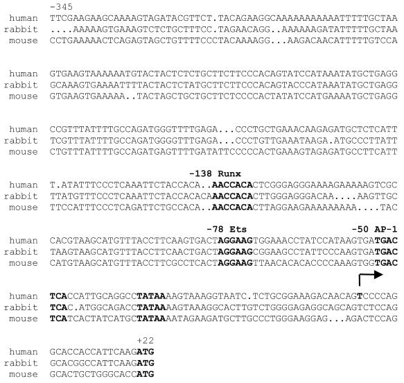
First, to explore the tissue-specific expression of MMP-13, we examined the functional role of a potential Runx protein-binding site 138 bp upstream of the transcriptional start site (Fig. 1) that is not found in other MMP promoters. The Runx site binds proteins from the Runx family, including Runx-2 (Cbfa1), an essential transcription factor for osteoblast and chondrocyte development (13–16). Runx-2 forms a heterodimeric complex with its partner Cbf-β; Runx-2 binds DNA directly while Cbf-β modulates the affinity of the complex for DNA (17). We found that constitutive Runx-2 expression correlated with the ability of a cell to produce MMP-13 in response to IL-1. Moreover, we determined that Runx-2 in cooperation with AP-1 was required for IL-1 induction of MMP-13 and that Runx-2 activity was modulated by IL-1-induced p38 MAPK activation. Second, we found that stable integration of the proximal 405 bp of the MMP-13 promoter was necessary for IL-1 induction of transcription, implying that chromatin structure may be important for transcriptional regulation.
Figure 1.
Homology alignment of the proximal MMP-13 promoters from mouse, rabbit and human sequence. The numbering is based on the start of transcription mapped in IL-1-treated OA chondrocytes (36). Conserved AP-1, Runx and Ets sites are in bold; the arrow denotes the human transcriptional start site. The translational start site is highlighted in bold from nucleotide +22 to +24. Alignment was made using GeneInspector (Textco Inc.)
MATERIALS AND METHODS
Cell culture
SW-1353 cells were obtained from the American Type Culture Collection and cells were grown in 150 mm diameter culture dishes in DMEM containing 10% fetal calf serum (FCS) and penicillin/streptomycin (37°C in 5% CO2) (Life Technologies, Rockville, MD). Cells were passaged 1:4 with 0.25% trypsin. For most experiments, cells were grown to confluence in 35 mm 6-well plates, washed with Hanks’ balanced salt solution (HBSS) to remove traces of serum and placed in 2 ml of serum-free DMEM with 0.2% lactalbumin hydrosylate (DMEM/LH), with or without 10 ng/ml IL-1β (92 U/ng; Promega, Madison, WI).
Western blots
One milliliter of serum-free medium from each well of 6-well cultures was TCA precipitated and analyzed by SDS–PAGE and western immunoblotting as described previously (18). Briefly, 10% SDS–PAGE gels were run and proteins were transferred to Immobilon PVDF membranes (Millipore, Bedford, MA). Supernatants from transfection experiments were assayed for MMP-1 and MMP-13 protein. MMP-13 antibody was graciously provided by Peter Mitchell (Pfizer) (18). Anti-MMP-1 polyclonal antibody was purchased from Chemicon (Temecula, CA). Anti-Runx2 antibody was graciously provided by Gerard Karsenty (Baylor School of Medicine) (15). Peroxidase-conjugated secondary antibodies and enhanced chemiluminesence (Amersham, Piscataway, NJ) were used to visualize bound primary antibodies. NIH Image software was used to compare band intensities.
Northern blots
Total RNA was isolated using the Trizol protocol (Life Technologies) and was subjected to northern blot analysis. Briefly, 10 µg RNA was loaded on a 1% agarose–formaldehyde–formamide gel, electrophoresed and blotted to GeneScreen membrane (NEN, Boston, MA). RNA was then UV crosslinked and probed with a 1017 bp SstI–EcoRI fragment of mouse Runx2 cDNA that was random hexamer labeled with [α-32P]dCTP and visualized by autoradiography.
Reporter constructs
All MMP-13 promoter constructs were cloned into the luciferase reporter vector pGL3 Basic (Promega). The 3.4 and 1.6 kb constructs were obtained by rapid amplification of genomic DNA ends; smaller constructs were created by PCR cloning utilizing the SstI and KpnI sites of pGL3 basic. A segment of 8.0 kb of 5′-flanking DNA was provided by Karen Hasty (University of Tennessee Medical School). Mutations to the proximal AP-1- and Runx-binding sites were made using overlapping PCR (19). The proximal AP-1 site was changed from TGACTCA to ACTCTCA. The proximal Runx-binding site was changed from AACCACA to ACTAACA. The sequence of all promoter constructs was verified by DNA sequencing. The runt domain of Runx-2 was cloned into pCMVTag5A (Stratagene, La Jolla, CA) using PCR. An expression plasmid containing the mouse Runx-2 isoform starting with the amino acids MASN was provided by Gerard Karsenty (Baylor University Medical School) (15). The MKK3 plasmid was provided by Roger Davis (University of Massachusetts Medical School). The ΔMEKK1 plasmid was obtained from Stratagene. The pyridinyl imidazole p38 inhibitor SB203580 and the MEK inhibitor PD98059 were purchased from CalBiochem (San Diego, CA).
Transient transfections
All experiments were performed in triplicate. SW-1353 cells were plated at a density of 2 × 10⁵ per well in 6-well tissue culture plates. The following morning 1 µg total DNA plus 10 µl of Geneporter-1 (Gene Therapy Systems, San Diego, CA) reagent in 1 ml of serum-free medium were added for 4 h. One milliliter of 20% FCS DMEM was then added for overnight incubation. The next day, the experimental treatments were added for the times indicated. Cells were then washed three times with ice-cold phosphate-buffered saline and lysed in 25 mM glycylglycine, 4 mM EGTA, 15 mM MgSO4, 1% Triton X-100 and 1 mM DTT. Luciferase activity was measured in a Dynatech ML2250 luminometer and reported as relative light units (RLU).
For analysis of expression of the endogenous MMP-13 gene, SW-1353 cells were plated at 3 × 10⁵ cells/well in 6-well plates and cultured overnight in 10% FCS DMEM. The following day, 1 µg total DNA plus 10 µl of Geneporter-1 per well was incubated with the cells for 5 h in 1 ml of serum-free medium (LH), followed by addition of 1 ml of DMEM containing 20% FCS for overnight incubation at 37°C. The following day cells were washed three times with HBSS, and then 2 ml of DMEM/LH with or without 10 ng/ml IL-1β was added. For western blot analysis, after 24 h 1 ml of supernatant was TCA precipitated and analyzed by SDS–PAGE and immunoblotting.
Stable cell lines
For each construct, a 90 mm plate of SW-1353 cells that were 70% confluent was co-transfected with 2.3 µg of the neomycin-expressing vector pCMV-Tag5A (Stratagene) and 2.3 µg of the corresponding pGL3-promoter construct using 46.7 µl/plate Geneporter. Two days after transfection, cells were passaged into a 150 mm plate containing 800 µg/ml G418 (Stratagene) in 10% FCS. Cells were passaged once a week for 3 weeks, when single colonies were isolated by serial dilution in 96-well plates. Cells were selected for 8 weeks in G418 before experiments were begun. Stable cell lines were maintained by selection in 800 µg/ml G418. To create pooled stable lines, cells were transfected as above and plates containing several thousand colonies were passaged once a week in 800 µg/ml G418 for 4 weeks before analysis.
RESULTS
Constitutive Runx-2 expression correlates with MMP-13 production
Previously, we determined that the SW-1353 human chondrosarcoma cell line provides an appropriate model for studying the signaling pathways used by primary chondrocytes to produce MMP-13 transcript in response to IL-1 (9). We demonstrated that IL-1 strongly induces MMP-13 transcription in the SW-1353 line by 2 h via the p38 MAPK, JNK and NF-κB inflammatory signaling pathways (9). Because the MMP-13 promoter contains a Runx-binding site (Fig. 1), and because Runx-2 is involved in cartilage and bone development (13–16), we hypothesized that the transcription factor Runx-2 is involved in IL-1 induction of MMP-13. Therefore, the expression of Runx-2 mRNA was compared among human foreskin fibroblasts (HFFs), rabbit synovial fibroblasts (RFs) and the chondrosarcoma cell line SW-1353. A time course experiment showed that Runx-2 was constitutively present in SW-1353 cells treated with serum-free medium up to 24 h (Fig. 2), but that Runx-2 was not expressed in HFFs or RFs, cells that make no to low levels of MMP-13 in response to IL-1, respectively (18). Further, IL-1 slightly increased Runx-2 message in SW-1353 cells up to 2-fold at 4 and 24 h. Western blot analysis of HFF and SW-1353 nuclear extracts verified the results of northern blots (data not shown). These experiments demonstrate that Runx-2 expression correlates with MMP-13 production and that mRNA and protein levels of Runx-2 are constitutively high in the nucleus of SW-1353 cells.
Figure 2.
Runx-2 expression correlates with IL-1 inducibility of MMP-13. Total RNA was harvested from SW-1353 cells, rabbit fibroblasts or human foreskin fibroblasts after treatment for the times indicated. Aliquots of 10 µg were analyzed by northern blot analysis using a 32P-labled probe from the 3′-end of the mouse Runx-2 cDNA. rRNA is shown as a loading control. Band intensities were compared with NIH Image software.
IL-1 induction of endogenous MMP-13 requires Runx-2
The Runx proteins bind to DNA via the runt domain, a conserved domain found in the middle of the polypeptide. Expression of the Runx-2 runt domain acts as an inhibitor of transactivation (20,21), since the overexpressed runt domain binds to its binding site more tightly than the wild-type protein and is transactivation deficient (20). In order to test the hypothesis that Runx-2 contributes to IL-1 induction of MMP-13, SW-1353 cells were transfected with CMV-driven expression plasmids containing either wild-type Runx-2 or the runt domain. Twenty-four hours after transfection, cells were placed in serum-free medium with or without IL-1 for 24 h and supernatants were analyzed by western blotting. As expected, untreated cells produced undetectable levels of MMP-13 and MMP-1 and treatment with IL-1 resulted in substantial production of both proteins (9; Fig. 3). We also found that overexpression of the runt domain inhibited IL-1 induction of MMP-13 by ∼50%, which parallels the transfection efficiency (see below). In contrast, transfection with wild-type Runx-2 increased MMP-13 induction by ∼2-fold. To analyze the specificity of the effect of Runx-2, MMP-1 levels were assayed from the same experiment and they were unaffected by transfection of either construct (Fig. 3). Transfection efficiency was estimated to be ∼50% by transfection of a GFP expression plasmid (9; data not shown). These experiments demonstrate that Runx-2 contributes to expression of the endogenous MMP-13 gene by IL-1, but not to induction of MMP-1. Further, the contribution of Runx-2 to gene expression correlates with the presence of a Runx-2 site in the MMP-13 promoter.
Figure 3.
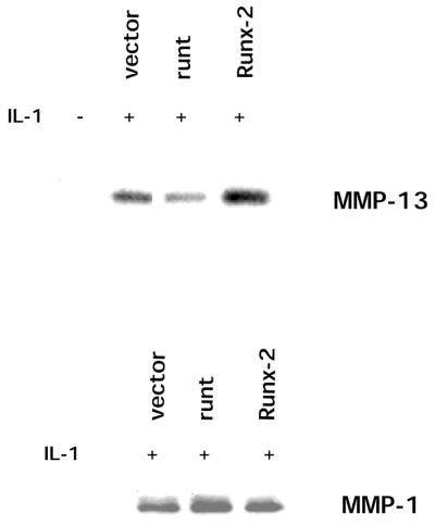
Runt domain expression specifically inhibits IL-1 induction of endogenous MMP-13. Samples of 3 × 10⁵ SW-1353 cells were transfected with 1 µg empty pCMV-Tag5A expression plasmid, a CMV driven Runx-2 expression plasmid or pCMV-Tag5A-runt using 10 µl/well Geneporter reagent. After overnight recovery, cells were treated for 24 h with 2 ml of serum-free DMEM/LH or LH and 10 ng/ml IL-1β. Next, 1 ml of supernatant was TCA precipitated and analyzed by SDS–PAGE and western blotting. Polyclonal antibodies to either MMP-13 or MMP-1 were used to visualize protein. Band intensities were compared with NIH Image. A transfection efficiency of 50% was measured by transfection of the green fluorescent protein-expressing vector pEGFP and fluorescence microscopy (9).
ΔMEKK1 and MKK3 synergize with Runx-2 to activate 405 bp of MMP-13 promoter
DNA sequence analysis of the proximal promoter demonstrated that the first 370 bp of MMP-13 5′-flanking DNA are highly conserved among mouse, rabbit and human (Fig. 1). This sequence contains a putative AP-1 site, an ETS-binding site and a Runx-binding site (Fig. 1). Initially, we used gel mobility shift assays (22) to determine whether IL-1 induced changes in the pattern of proteins binding to the AP-1 and Runx-2 sites. We found high constitutive binding and no significant changes after IL-1 treatment (data not shown). Therefore, we hypothesized that the AP-1 and Runx-2 proteins are constitutively bound and the effect of IL-1 on MMP-13 gene expression may occur through kinase-mediated phosphorylation of these target transcription factors, rather than through an increase in their synthesis. A recent study demonstrated that ERK MAPK can phosphorylate Runx-2 and that phosphorylation increases the ability of the protein to transactivate the osteocalcin promoter (23). Since we have demonstrated that IL-1 activation of p38 MAPK and JNK was necessary for induction of MMP-13 (9), we postulated that p38 MAPK and/or JNK may phosphorylate Runx-2 and that phosphorylation is involved in transcriptional activation of the MMP-13 promoter.
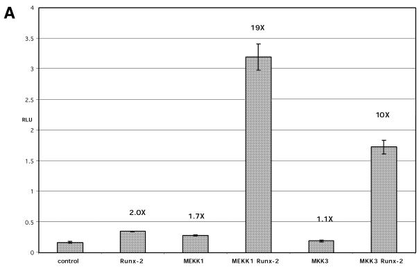
Previous studies have shown that when MMP-13 promoter constructs and Runx-2 are co-transfected into HeLa cells (cells that do not express Runx-2 or MMP-13), Runx-2 could drive MMP-13 reporter activity (24). Therefore, we transiently co-transfected SW-1353 cells with 405 bp of MMP-13 promoter in the pGL3 luciferase reporter along with expression constructs of Runx-2 and upstream activators of MAPK pathways, such as ΔMEKK1, a constitutively active MAPK kinase that activates JNK, p38 and ERK (25). Transfection of Runx-2 alone induced reporter activity 2-fold, while ΔMEKK1 alone induced the promoter–reporter to a similar extent (1.7-fold) (Fig. 4A). However, when ΔMEKK1 and Runx-2 were co-transfected with the reporter gene, a synergistic 19-fold increase in reporter activity occurred. Another upstream MAPK activator is MKK3, a MAPK kinase that phosphorylates and activates p38 MAPK, which is a kinase required for MMP-13 transcription (26). Transfection of constitutively active MKK3 alone did not increase reporter activity. Combined with Runx-2, MKK3 synergistically induced transcription 10-fold. Transfection of the empty pGL3 vector resulted in extremely low reporter expression that was not affected by Runx-2 co-transfection (data not shown). This synergism between Runx-2 and the p38 MAPK activator suggests direct integration between MAPK signaling and Runx-2 binding at the promoter of MMP-13.
Figure 4.
(Opposite) ΔMEKK1 and MKK3 synergistically activate 405 bp of MMP-13 reporter through Runx-2. Samples of 2 × 10⁵ SW-1353 cells were transfected with 500 ng of the proximal 405 bp of MMP-13 (A), of 5′-flanking DNA (B) or 4.3 kb of MMP-1 promoter (C), linked to the luciferase reporter gene in pGL3. Cells were co-transfected with 500 ng total of expression plasmid containing combinations of cDNAs for Runx-2, constitutively active ΔMEKK1 (Stratagene) or constitutively active MKK3 (26) driven by a CMV promoter. Total DNA was kept constant by addition of the empty pCMVTag5A expression plasmid. Aliquots of 10 µl of Geneporter (Gene Therapy Systems) were used to transfect the cells. Twenty-four hours after transfection, cells were placed in serum-free medium for 24 h or in 10 µM SB203580, cell extracts were made and assayed for luciferase activity. Units shown are relative light units (RLU). Individual treatments were performed in triplicate; error bars represent standard deviations. Values indicate fold induction over basal activity of the 405 bp construct.
SB203580 inhibits MKK3 Runx-2 synergism
Because MKK3 activates p38 MAPK, the synergism seen with Runx-2 should be sensitive to treatment with the p38 inhibitor SB203580, an inhibitor that blocks IL-1 induction of the endogenous MMP-13 gene (9). Therefore, cells were transiently co-transfected with 405 bp of MMP-13 promoter along with expression constructs for Runx-2 and MKK3 (Fig. 4B). As in the previous experiment, Runx-2 and MKK3 alone gave a modest increase in transcription, while together the increase was synergistic. Addition of the p38 inhibitor reduced the synergistic induction mediated by MKK3 and Runx-2 from 6.4- to 3.4-fold over the control. In summary, MKK3–Runx-2 synergism through 405 bp of the MMP-13 promoter is inhibited by a p38 MAPK inhibitor, demonstrating that, like the endogenous MMP-13 gene, active p38 MAPK is required for the effect.
Runx-2 activation and synergism is specific for MMP-13
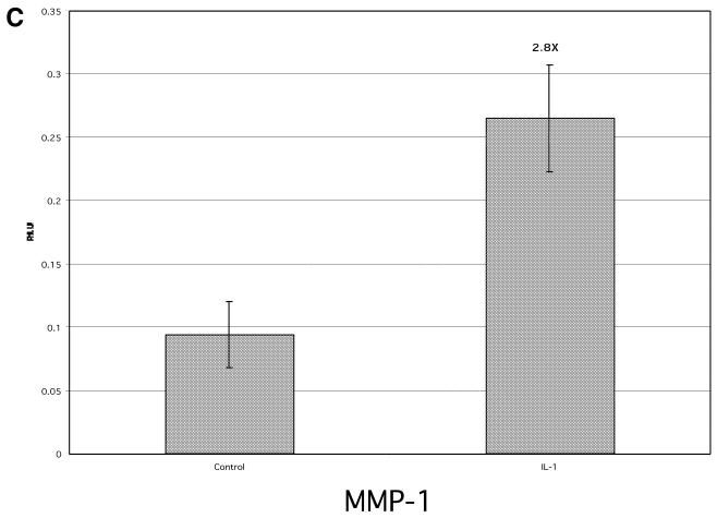
Because MMP-1 does not contain a Runx-2-binding sequence in its promoter, but does contain AP-1 sites that respond to the MAPK/JNK pathways (27), as a control we investigated the ability of Runx-2 to synergistically transactivate the MMP-1 promoter with expression constructs for MAPK and JNK activators (Fig. 4C). When a 4.3 kb piece of MMP-1 promoter DNA that is responsive to IL-1 (28) was transiently transfected into SW-1353 cells along with Runx-2, there was no effect on reporter activity. In contrast to the MMP-13 promoter, MKK3 and ΔMEKK1 each activated transcription of the MMP-1 reporter, but co-transfection of these expression plasmids and Runx-2 did not increase transcription further. This experiment demonstrates the specificity of p38 MAPK–Runx-2 synergism for the MMP-13 promoter and suggests that the Runx-binding site is needed for IL-1 induction of MMP-13.
IL-1 does not stimulate transiently transfected MMP-13 promoter–reporter constructs
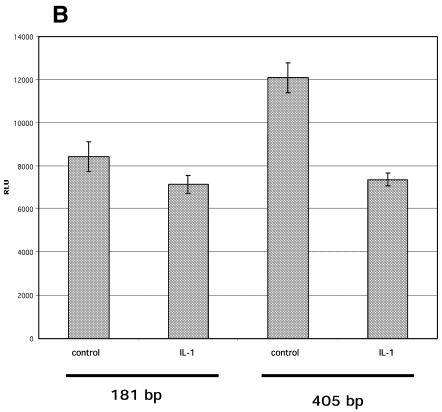
Next, we focused on defining the cis-acting sequences required for regulation of MMP-13 transcription by IL-1 by generating a series of MMP-13 5′-flanking constructs in the luciferase reporter vector pGL3 basic (Fig. 5A). When constructs from –181 bp to –8 kb were transiently transfected into SW-1353 cells, treatment with IL-1 resulted either in no effect or repression of luciferase activity, in contrast to the endogenous gene, which shows a strong transcriptional induction (9; Fig. 5B and data not shown). We saw the same results when we used a β-galactosidase reporter vector or when other cell lines and primary cells that produce MMP-13 were transfected. In contrast, a pGL3-MMP-1 promoter construct (28) was IL-1 inducible (Fig. 5C) and a NF-κB site-driven luciferase reporter was 4.8-fold increased after IL-1 treatment (data not shown). In summary, we were unable to detect IL-1 regulatory regions in 8.0 kb of MMP-13 5′-flanking DNA and 1.6 kb of 3′-flanking DNA by transient transfections. This suggests that additional cis-acting sequences are required or that integration of promoter DNA into native chromatin is needed to observe IL-1 regulation of MMP-13 (see below).
Figure 5.
(A) pGL3 MMP-13 reporter constructs. The numbering of constructs is based on the start of translation. 5′-Flanking DNA was cloned into pGL3 basic by PCR (181 and 405 bp constructs) and rapid amplification of genomic DNA ends (RAGE) (1.6 and 3.4 kb constructs). The 8.0 kb 5′-flanking region was subcloned from a pBetaGal construct graciously provided by Karen Hasty (University of Tennessee). (B) Transient transfection of 181 and 405 bp of MMP-13 5′-flanking DNA into SW-1353 cells. Samples of 2 × 10⁵ SW-1353 cells were transfected with 1 µg pGL3 luciferase reporter construct containing MMP-13 5′-flanking DNA using 10 µl of Geneporter. (C) Transient transfection of 4.3 kb of MMP-1 5′-flanking DNA. Samples of 2 × 10⁵ SW-1353 cells were transfected with 1 µg pGL3 luciferase reporter construct containing 4.3 kb of MMP-1 5′-flanking DNA (37). Cells were treated with 10 ng/ml IL-1β for 24 h. In all three experiments, luciferase activity was assayed in a luminometer and expressed as RLU. Error bars denote standard deviations.
IL-1 stimulates MMP-13 promoter–reporter activity in stable transfectants
To test the hypothesis that integration of the reporter plasmid into the chromosome was required for appropriate regulation of the promoter by IL-1, stable transfectants were created by co-transfecting pGL3 reporter constructs containing either 8.0 kb or 405 bp of MMP-13 5′-flanking DNA with pCMVTag5, a neomycin expression vector (see Materials and Methods). Three of five clones containing 405 bp and one of five clones containing 8.0 kb expressed the luciferase reporter and were IL-1 inducible (Fig. 6A and data not shown). All luciferase-positive clones demonstrated a similar fold induction after IL-1 treatment, suggesting that proximal sequences control transcription and that the sequence between –8.0 kb and –405 bp is not necessary. Additionally, control lines created by stable transfection with the empty pGL3 vector did not basally express luciferase and were not IL-1 inducible, demonstrating that the IL-1-responsive sequences are not found in the vector DNA (data not shown).
Figure 6.
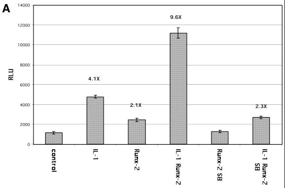
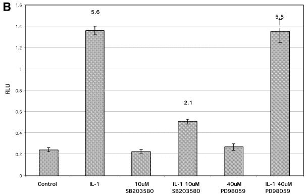
Stable transfectants of MMP-13 5′-flanking DNA reporter constructs respond to IL-1β. Stably transfected SW-1353 cells containing 405 bp of MMP-13 5′-flanking DNA were created by co-transfecting cells with a reporter construct and the pCMV-Tag5A vector expressing the neomycin resistance gene, followed by 2 months of selection in G418. To assay the integrated reporter constructs, 2 × 10⁵ cells derived from single cell clones were plated and treated with 10 ng/ml IL-1β for 24 h. Luciferase activity was assayed and plotted as RLU. (A) The 405 bp stably integrated reporter constructs are inhibited by the same MAPK inhibitors as the endogenous MMP-13 gene. A 405 bp clone was treated with 10 ng/ml IL-1β for 24 h with and without 40 µM MEK inhibitor PD98059 or 10 µM p38 inhibitor SB203580. Luciferase activity was assayed and plotted as RLU. (B) IL-1 synergizes with Runx-2 in stable transfectants. Samples of 2 × 10⁵ SW-1353 cells stably transfected with 405 bp of MMP-13 promoter DNA were transiently transfected with 1 µg pCMVTag5A or pCMV Runx-2 using 10 µl of Geneporter. Twenty-four hours after transfection, cells were treated with 10 ng/ml IL-1 for 24 h and/or 10 µM SB203580 in serum-free medium. All treatments were done in triplicate; cells were harvested and assayed for luciferase activity. Luciferase activity is shown as RLU; error bars represent standard deviations. Values indicate fold induction over control.
Next, we investigated whether the stably integrated promoter could be inhibited by the p38 inhibitor SB203580, which inhibits IL-1 induction of the endogenous gene (9). As noted above, treatment of the 405(3) clone with IL-1 induced the luciferase reporter 5.6-fold (Fig. 6A) and concomitant treatment of the cell line with 10 µM SB203580 reduced IL-1 induction to 2.1-fold, with no effect on basal activity. Treatment with 40 µM PD98059 had no effect on basal activity or IL-1 induction, also consistent with the endogenous gene (9). These data demonstrate that integration of the MMP-13 promoter results in appropriate reporter gene induction and suggest that integration is required for IL-1 induction of MMP-13. Moreover, the stably integrated promoter–reporter constructs require p38 MAPK and do not require the MEK/ERK pathway, exactly like the endogenous MMP-13 gene (9). Thus, these stable cell lines provide a model system for elucidating the specific cis-acting sequences required for IL-1 induction of MMP-13.
IL-1 and Runx-2 synergize to activate the MMP-13 promoter in stable transfectants
Next, we tested the ability of a stably transfected cell line to respond to Runx-2 and IL-1. The cells were transiently transfected with an empty vector or with the same vector containing a Runx-2 construct, with and without IL-1 treatment. In cells transfected with empty vector, IL-1 stimulated induction 4.1-fold over the control. Runx-2 transfection increased basal levels 2.1-fold and, together, IL-1 and Runx-2 synergistically increased reporter expression 9.6-fold (Fig. 6B). Treatment with 10 µM SB203580 blocked synergism between Runx-2 and IL-1, demonstrating that p38 MAPK is required for this effect. These experiments demonstrate that IL-1-induced p38 MAPK signaling synergizes with Runx-2 to activate MMP-13 transcription.
Mutational analysis of the stably transfected MMP-13 promoter
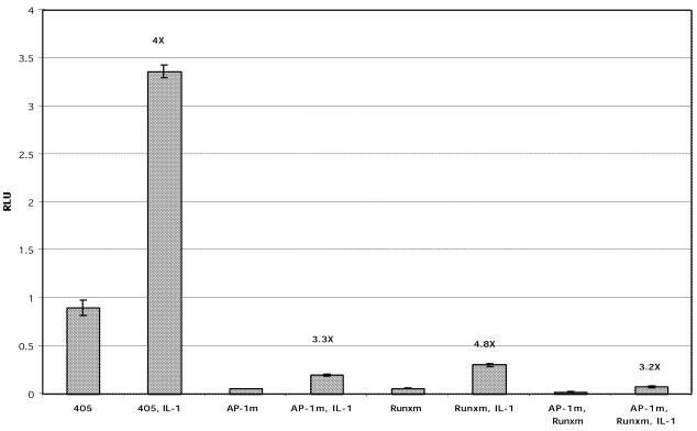
Finally, to further define the cis-acting sequences required for IL-1 induction of MMP-13 transcription, we created single mutants of the proximal AP-1 site and the Runx site and a double mutant in the context of 405 bp of MMP-13 5′-flanking DNA in pGL3. Pooled stable clones were created by co-transfection followed by selection in G418 for 1 month. IL-1 treatment of cells stably transfected with 405 bp of promoter resulted in 4-fold IL-1 induction (Fig. 7). Clones containing a mutation in the AP-1 site exhibited an 18-fold reduction in both basal expression and IL-1 induction, while clones containing a mutated Runx site showed 12-fold reduced basal and IL-1 induction. Mutation of both sites caused a 46-fold reduction in basal and IL-1-induced reporter expression. In summary, mutation of each site dramatically reduced both basal and IL-1-induced expression without substantially altering the fold induction over control. Double mutation caused a synergistic decrease in basal and IL-1-induced reporter activity, implying that the AP-1 and Runx proteins interact at the promoter. However, the double mutant remained 3.2-fold inducible in response to IL-1, suggesting that other IL-1-responsive sites remain and that these sites can drive lower levels of transcription.
Figure 7.
Mutational analysis of the MMP-13 promoter using stable integrants. Pooled stable cell lines were created by transfection with the reporter constructs indicated followed by 1 month of weekly passages and selection in G418. Cells were plated at 2 × 10⁵ per well in 6-well plates and treated with 10 ng/ml IL-1 in serum-free medium for 24 h. All treatments were done in triplicate; cells were harvested and assayed for luciferase activity. Luciferase activity is shown as RLU; error bars represent standard deviations. Values indicate fold IL-1 induction over basal expression for each construct.
DISCUSSION
We determined that Runx-2 participates in IL-1 induction of MMP-13 in chondrocytic cells. We found that Runx-2 mRNA and protein were high in untreated and IL-1-treated SW-1353 cells, which produce MMP-13, and much lower in RFs and HFFs, which produce little or no MMP-13, respectively. Therefore, the ability of a cell type to produce MMP-13 in response to IL-1 correlated with Runx-2 expression, implying that Runx-2 may participate in MMP-13 regulation in a tissue-specific manner (Fig. 8). Indeed, transfection of a plasmid expressing the full-length Runx-2 protein augmented IL-1 induction of the endogenous MMP-13 gene while transfection of the transactivation-deficient runt domain specifically blocked IL-1 induction of the endogenous MMP-13 gene, showing that this protein is required for gene expression.
Figure 8.
IL-1 signaling to AP-1 and Runx-2 at the MMP-13 promoter. The model depicts the expression levels and roles of Runx and AP-1 proteins at the MMP-13 promoter in SW-1353 human chondrosarcoma cells and HFFs human foreskin fibroblasts. Arrows denote activation of signaling pathways or transcriptional activation at the MMP-13 promoter. The question mark refers to unidentified IL-1-responsive elements.
Recent evidence suggesting that Runx-2 is a target of the ERK MAPK pathway (23) and our data demonstrating the importance of MAPK pathways for MMP-13 regulation prompted us to hypothesize that IL-1 treatment resulted in p38 MAPK and/or JNK modulation of Runx-2 activity. Additional data showing that protein kinase A (PKA) was required for parathyroid hormone induction of rat MMP-13 and that PKA could phosphorylate Runx-2 in vitro also demonstrate that Runx-2 is a possible target for a kinase (29,30). We indirectly tested this model by measuring the ability of transiently transfected upstream MAPK activators to activate MMP-13 transcription through Runx-2. Transfection of both ΔMEKK1 (an activator of p38 MAPK, ERK and JNK) and MKK3 (an activator of p38 MAPK) synergistically activated the MMP-13 promoter when co-transfected with Runx-2. This effect was not seen with the MMP-1 promoter, demonstrating the specificity of Runx-2 for MMP-13 over MMP-1. These results parallel analysis of parathyroid hormone regulation of the rat MMP-13 promoter using the UMR rat osteosarcoma cell line (30) and the demonstration that transfection of rat osteosarcoma cells with Runx-2 augmented TGF-β induction of the endogenous MMP-13 gene (24). Additionally, expression of Runx-2 in cell lines stably transfected with the MMP-13 promoter synergistically activated reporter activity with IL-1 treatment, illustrating that IL-1 can signal through Runx-2 to activate gene expression. Finally, mutation of the Runx site decreased overall transcription from an integrated reporter construct, demonstrating that this site participates in the regulation of MMP-13 gene expression. Mutation of both the AP-1 site and the Runx-2 site caused a synergistic reduction in transcription, suggesting that transcription factors bound to these two sites may interact (Fig. 8). Perhaps IL-1-induced p38 and JNK directly phosphorylate Runx-2, leading to coactivator recruitment and an increase in transcription. Alternatively, Runx-2 may interact with AP-1 proteins directly or indirectly through other factors after the phosphorylation of AP-1 by these kinases. Indeed, direct interaction between Runx-2 and the AP-1 proteins c-Fos and c-Jun was recently demonstrated both in vivo and in vitro (31).
We determined that transient transfections of MMP-13 5′-flanking DNA linked to a luciferase reporter gene did not recapitulate the transcriptional induction of the endogenous MMP-13 gene in response to IL-1. Other reporter constructs like MMP-1–pGL3 and NF-κB–luciferase were IL-1 responsive. In contrast, 405 bp or 8 kb of stably transfected promoter was IL-1 inducible, suggesting that 405 bp is sufficient for IL-1 induction. The 405 bp construct contains a continuous region of high homology among mouse, human and rabbit promoters with conserved AP-1, Ets and Runx transcription factor-binding sites (Fig. 1). When compared to the endogenous gene, the same pattern of reporter gene inhibition by small molecule MAPK inhibitors was seen with the 405 bp stable transfectants, verifying that the reporter gene requires the same pathways as the endogenous gene for IL-1 induction (9). Mutational analysis revealed that both the AP-1 site and the Runx-binding site are required for basal and IL-1-induced MMP-13 transcription.
The failure of transiently transfected DNA to respond to IL-1 has at least two possible explanations. The first is that limiting amounts of transcription factors or other proteins involved in IL-1 signaling are available in the nucleus. In a transiently transfected cell, the number of reporter gene copies may exceed the available pool of transcription factors and the endogenous gene may be preferentially bound or targeted by these factors after IL-1 treatment. In contrast, a stable line may contain fewer copies of the promoter–reporter construct, thus the available pool of factors is not exceeded. This model could explain the inhibition of reporter activity after IL-1 treatment of transiently transfected cells. Although we attempted to rescue IL-1 induction in transient transfections by co-transfecting an expression plasmid for Runx-2, this approach did not restore IL-1 induction (data not shown). A second explanation is that chromatin structure is required for regulation of MMP-13. Chromosomally integrated DNA may organize histones and other chromatin-associated proteins, thereby repressing transcription in the absence of a stimulus. If this were the case, transient transfection may result in high basal reporter expression because unintegrated plasmid cannot assume a native repressed chromatin structure and cannot respond appropriately to IL-1.
The structure of transiently transfected DNA has been analyzed with conflicting results. Nucleosomal ladders of transiently transfected DNA were found by one group to have repeat lengths similar to cellular chromatin (187 ± 5 bp), while others have demonstrated spacings of 199 or 280 bp or a complete lack of regular nucleosomes, respectively (reviewed in 32). The latter results demonstrate that transiently transfected DNA may be more unwound and, therefore, more accessible to transcription factors and less appropriately regulated compared to stably integrated reporter constructs.
There may be a direct link between regulation of chromatin structure and the activities of Runx-2 protein. Recent evidence indicates that Runx-2 protein is targeted to and interacts with the nuclear matrix and that this ability is required for proper functioning of the protein (33,34). Additionally, another member of the Runx family, Runx-1, interacts with coactivators like the histone acetylase p300 through domains conserved in Runx-2 protein (35). These putative Runx-2 properties may be involved in IL-1 regulation of MMP-13 transcription.
When compared to other IL-1-inducible MMPs, MMP-13 has a more restricted expression pattern and requires a unique set of signaling pathways and transcription factors that lead to tissue-specific expression in chondrocytes. Our studies suggest that both the AP-1 and the tissue-restricted Runx-2 transcription factors are important targets of IL-1-induced p38 MAPK leading to MMP-13 transcription. Future studies directed towards defining the direct downstream targets of the p38 MAPK and JNK pathways and studies identifying additional proteins that activate polymerase II transcription of MMP-13 are needed to understand the molecular mechanism driving gene expression. Understanding the mechanism of gene regulation may uncover new targets for the inhibition of MMP-13 expression in arthritis.
Acknowledgments
ACKNOWLEDGEMENTS
The authors thank Charlie Coon for technical assistance and advice, Karen Hasty for graciously providing 8 kb of 5′-flanking DNA, Peter Mitchell for providing the MMP-13 antibody, Gerard Karsenty for providing the anti-Runx-2 antibody and the Runx-2 expression construct and, finally, Roger Davis for providing the MKK3 expression plasmid. This work was supported by grants from the NIH (AR 26599 and CA 77267) and grants from the RGK Foundation (Austin, TX), the Susan G.Komen Foundation and DoD BC991121 to C.E.B., the NIH (AR-02024 and AR 46977) to M.P.V. and the NIH (T32 AR07576 and T32 AI07363-09) and a scholarship from the American Federation for Aging Research to J.A.M.
References
- 1.Libby P. and Aikawa,M. (1998) New insights into plaque stabilisation by lipid lowering. Drugs, 56, 9–13; Discussion, 33. [DOI] [PubMed] [Google Scholar]
- 2.McCawley L.J. and Matrisian,L.M. (2000) Matrix metalloproteinases: multifunctional contributors to tumor progression. Mol. Med. Today, 6, 149–156. [DOI] [PubMed] [Google Scholar]
- 3.Billinghurst R.C., Dahlberg,L., Ionescu,M., Reiner,A., Bourne,R., Rorabeck,C., Mitchell,P., Hambor,J., Diekmann,O., Tschesche,H. et al. (1997) Enhanced cleavage of type II collagen by collagenases in osteoarthritic articular cartilage. J. Clin. Invest., 99, 1534–1545. [DOI] [PMC free article] [PubMed] [Google Scholar]
- 4.Mitchell P.G., Magna,H.A., Reeves,L.M., Lopresti-Morrow,L.L., Yocum,S.A., Rosner,P.J., Geoghegan,K.F. and Hambor,J.E. (1996) Cloning, expression and type II collagenolytic activity of matrix metalloproteinase-13 from human osteoarthritic cartilage. J. Clin. Invest., 97, 761–768. [DOI] [PMC free article] [PubMed] [Google Scholar]
- 5.Neuhold L.A., Killar,L., Zhao,W., Sung,M.L., Warner,L., Kulik,J., Turner,J., Wu,W., Billinghurst,C., Meijers,T. et al. (2001) Postnatal expression in hyaline cartilage of constitutively active human collagenase-3 (MMP-13) induces osteoarthritis in mice. J. Clin. Invest., 107, 35–44. [DOI] [PMC free article] [PubMed] [Google Scholar]
- 6.Johansson N., Saarialho-Kere,U., Airola,K., Herva,R., Nissinen,L., Westermarck,J., Vuorio,E., Heino,J. and Kahari,V.M. (1997) Collagenase-3 (MMP-13) is expressed by hypertrophic chondrocytes, periosteal cells and osteoblasts during human fetal bone development. Dev. Dyn., 208, 387–397. [DOI] [PubMed] [Google Scholar]
- 7.Stahle-Backdahl M., Sandstedt,B., Bruce,K., Lindahl,A., Jimenez,M.G., Vega,J.A. and Lopez-Otin,C. (1997) Collagenase-3 (MMP-13) is expressed during human fetal ossification and re-expressed in postnatal bone remodeling and in rheumatoid arthritis. Lab. Invest., 76, 717–728. [PubMed] [Google Scholar]
- 8.Shlopov B.V., Lie,W.R., Mainardi,C.L., Cole,A.A., Chubinskaya,S. and Hasty,K.A. (1997) Osteoarthritic lesions: involvement of three different collagenases. Arthritis Rheum., 40, 2065–2074. [DOI] [PubMed] [Google Scholar]
- 9.Mengshol J.A., Vincenti,M.P., Coon,C.I., Barchowsky,A. and Brinckerhoff,C.E. (2000) Interleukin-1 induction of collagenase 3 (matrix metalloproteinase 13) gene expression in chondrocytes requires p38, c-Jun N-terminal kinase and nuclear factor kappaB: differential regulation of collagenase 1 and collagenase 3. Arthritis Rheum., 43, 801–811. [DOI] [PubMed] [Google Scholar]
- 10.Han Z., Boyle,D.L., Chang,L., Bennett,B., Karin,M., Yang,L., Manning,A.M. and Firestein,G.S. (2001) c-Jun N-terminal kinase is required for metalloproteinase expression and joint destruction in inflammatory arthritis. J. Clin. Invest., 108, 73–81. [DOI] [PMC free article] [PubMed] [Google Scholar]
- 11.Karin M., Liu,Z. and Zandi,E. (1997) AP-1 function and regulation. Curr. Opin. Cell Biol., 9, 240–246. [DOI] [PubMed] [Google Scholar]
- 12.Vincenti M.P., White,L.A., Schroen,D.J., Benbow,U. and Brinckerhoff,C.E. (1996) Regulating expression of the gene for matrix metalloproteinase-1 (collagenase): mechanisms that control enzyme activity, transcription and mRNA stability. Crit. Rev. Eukaryot. Gene Expr., 6, 391–411. [DOI] [PubMed] [Google Scholar]
- 13.Inada M., Yasui,T., Nomura,S., Miyake,S., Deguchi,K., Himeno,M., Sato,M., Yamagiwa,H., Kimura,T., Yasui,N. et al. (1999) Maturational disturbance of chondrocytes in Cbfa1-deficient mice. Dev. Dyn., 214, 279–290. [DOI] [PubMed] [Google Scholar]
- 14.Komori T., Yagi,H., Nomura,S., Yamaguchi,A., Sasaki,K., Deguchi,K., Shimizu,Y., Bronson,R.T., Gao,Y.H., Inada,M. et al. (1997) Targeted disruption of Cbfa1 results in a complete lack of bone formation owing to maturational arrest of osteoblasts. Cell, 89, 755–764. [DOI] [PubMed] [Google Scholar]
- 15.Ducy P., Zhang,R., Geoffroy,V., Ridall,A.L. and Karsenty,G. (1997) Osf2/Cbfa1: a transcriptional activator of osteoblast differentiation. Cell, 89, 747–754. [DOI] [PubMed] [Google Scholar]
- 16.Enomoto H., Enomoto-Iwamoto,M., Iwamoto,M., Nomura,S., Himeno,M., Kitamura,Y., Kishimoto,T. and Komori,T. (2000) Cbfa1 is a positive regulatory factor in chondrocyte maturation. J. Biol. Chem., 275, 8695–8702. [DOI] [PubMed] [Google Scholar]
- 17.Tahirov T.H., Inoue-Bungo,T., Morii,H., Fujikawa,A., Sasaki,M., Kimura,K., Shiina,M., Sato,K., Kumasaka,T., Yamamoto,M. et al. (2001) Structural analyses of DNA recognition by the AML1/Runx-1 Runt domain and its allosteric control by CBFbeta. Cell, 104, 755–767. [DOI] [PubMed] [Google Scholar]
- 18.Vincenti M.P., Coon,C.I., Mengshol,J.A., Yocum,S., Mitchell,P. and Brinckerhoff,C.E. (1998) Cloning of the gene for interstitial collagenase-3 (matrix metalloproteinase-13) from rabbit synovial fibroblasts: differential expression with collagenase-1 (matrix metalloproteinase-1). Biochem. J., 331, 341–346. [DOI] [PMC free article] [PubMed] [Google Scholar]
- 19.Ho S.N., Hunt,H.D., Horton,R.M., Pullen,J.K. and Pease,L.R. (1989) Site-directed mutagenesis by overlap extension using the polymerase chain reaction. Gene, 77, 51–59. [DOI] [PubMed] [Google Scholar]
- 20.Ducy P., Starbuck,M., Priemel,M., Shen,J., Pinero,G., Geoffroy,V., Amling,M. and Karsenty,G. (1999) A Cbfa1-dependent genetic pathway controls bone formation beyond embryonic development. Genes Dev., 13, 1025–1036. [DOI] [PMC free article] [PubMed] [Google Scholar]
- 21.Ueta C., Iwamoto,M., Kanatani,N., Yoshida,C., Liu,Y., Enomoto-Iwamoto,M., Ohmori,T., Enomoto,H., Nakata,K., Takada,K. et al. (2001) Skeletal malformations caused by overexpression of Cbfa1 or its dominant negative form in chondrocytes. J. Cell Biol., 153, 87–100. [DOI] [PMC free article] [PubMed] [Google Scholar]
- 22.Vincenti M.P., Coon,C.I. and Brinckerhoff,C.E. (1998) Nuclear factor kappaB/p50 activates an element in the distal matrix metalloproteinase 1 promoter in interleukin-1beta-stimulated synovial fibroblasts. Arthritis Rheum., 41, 1987–1994. [DOI] [PubMed] [Google Scholar]
- 23.Xiao G., Jiang,D., Thomas,P., Benson,M.D., Guan,K., Karsenty,G. and Franceschi,R.T. (2000) MAPK pathways activate and phosphorylate the osteoblast-specific transcription factor, Cbfa1. J. Biol. Chem., 275, 4453–4459. [DOI] [PubMed] [Google Scholar]
- 24.Jimenez M.J., Balbin,M., Lopez,J.M., Alvarez,J., Komori,T. and Lopez-Otin,C. (1999) Collagenase 3 is a target of Cbfa1, a transcription factor of the runt gene family involved in bone formation. Mol. Cell. Biol., 19, 4431–4442. [DOI] [PMC free article] [PubMed] [Google Scholar]
- 25.Schlesinger T.K., Fanger,G.R., Yujiri,T. and Johnson,G.L. (1998) The TAO of MEKK. Front. Biosci., 3, D1181–D1186. [DOI] [PubMed] [Google Scholar]
- 26.Raingeaud J., Whitmarsh,A.J., Barrett,T., Derijard,B. and Davis,R.J. (1996) MKK3- and MKK6-regulated gene expression is mediated by the p38 mitogen-activated protein kinase signal transduction pathway. Mol. Cell. Biol., 16, 1247–1255. [DOI] [PMC free article] [PubMed] [Google Scholar]
- 27.Barchowsky A., Frleta,D. and Vincenti,M.P. (2000) Integration of the NF-kappaB and mitogen-activated protein kinase/AP-1 pathways at the Collagenase-1 promoter: divergence of IL-1 and TNF-dependent signal transduction in rabbit primary synovial fibroblasts. Cytokine, 12, 1469–1479. [DOI] [PubMed] [Google Scholar]
- 28.Rutter J.L., Benbow,U., Coon,C.I. and Brinckerhoff,C.E. (1997) Cell-type specific regulation of human interstitial collagenase-1 gene expression by interleukin-1 beta (IL-1 beta) in human fibroblasts and BC-8701 breast cancer cells. J. Cell. Biochem., 66, 322–336. [PubMed] [Google Scholar]
- 29.Selvamurugan N., Pulumati,M.R., Tyson,D.R. and Partridge,N.C. (2000) Parathyroid hormone regulation of the rat collagenase-3 promoter by protein kinase A-dependent transactivation of core binding factor alpha1. J. Biol. Chem., 275, 5037–5042. [DOI] [PubMed] [Google Scholar]
- 30.Selvamurugan N., Chou,W.Y., Pearman,A.T., Pulumati,M.R. and Partridge,N.C. (1998) Parathyroid hormone regulates the rat collagenase-3 promoter in osteoblastic cells through the cooperative interaction of the activator protein-1 site and the runt domain binding sequence. J. Biol. Chem., 273, 10647–10657. [DOI] [PubMed] [Google Scholar]
- 31.Hess J., Porte,D., Munz,C. and Angel,P. (2001) AP-1 and Cbfa/runt physically interact and regulate parathyroid hormone-dependent MMP13 expression in osteoblasts through a new osteoblast-specific element 2/AP-1 composite element. J. Biol. Chem., 276, 20029–20038. [DOI] [PubMed] [Google Scholar]
- 32.Smith C.L. and Hager,G.L. (1997) Transcriptional regulation of mammalian genes in vivo. A tale of two templates. J. Biol. Chem., 272, 27493–27496. [DOI] [PubMed] [Google Scholar]
- 33.Choi J.Y., Pratap,J., Javed,A., Zaidi,S.K., Xing,L., Balint,E., Dalamangas,S., Boyce,B., van Wijnen,A.J., Lian,J.B. et al. (2001) Subnuclear targeting of Runx/Cbfa/AML factors is essential for tissue-specific differentiation during embryonic development. Proc. Natl Acad. Sci. USA, 98, 8650–8655. [DOI] [PMC free article] [PubMed] [Google Scholar]
- 34.Lian J.B., Stein,J.L., Stein,G.S., Montecino,M., van Wijnen,A.J., Javed,A. and Gutierrez,S. (2001) Contributions of nuclear architecture and chromatin to vitamin D-dependent transcriptional control of the rat osteocalcin gene. Steroids, 66, 159–170. [DOI] [PubMed] [Google Scholar]
- 35.Kitabayashi I., Yokoyama,A., Shimizu,K. and Ohki,M. (1998) Interaction and functional cooperation of the leukemia-associated factors AML1 and p300 in myeloid cell differentiation. EMBO J., 17, 2994–3004. [DOI] [PMC free article] [PubMed] [Google Scholar]
- 36.Tardif G., Pelletier,J.P., Dupuis,M., Hambor,J.E. and Martel-Pelletier,J. (1997) Cloning, sequencing and characterization of the 5′-flanking region of the human collagenase-3 gene. Biochem. J., 323, 13–16. [DOI] [PMC free article] [PubMed] [Google Scholar]
- 37.Rutter J.L., Mitchell,T.I., Buttice,G., Meyers,J., Gusella,J.F., Ozelius,L.J. and Brinckerhoff,C.E. (1998) A single nucleotide polymorphism in the matrix metalloproteinase-1 promoter creates an Ets binding site and augments transcription. Cancer Res., 58, 5321–5325. [PubMed] [Google Scholar]


