Abstract
One of the most promising approaches to improve recovery after spinal cord injury (SCI) is the augmentation of spontaneously occurring plasticity in uninjured neural pathways. Acute intermittent hypoxia (AIH, brief exposures to reduced O2 levels alternating with normal O2 levels) initiates plasticity in respiratory systems and has been shown to improve recovery in respiratory and non-respiratory spinal systems after SCI in experimental animals and humans. Although the mechanism by which AIH elicits its effects after SCI are not well understood, AIH is known to alter protein expression in spinal neurons in uninjured animals. Here, we examine hypoxia- and plasticity-related protein expression using immunofluorescence in spinal neurons in SCI rats that were treated with AIH combined with motor training, a protocol which has been demonstrated to improve recovery of forelimb function in this lesion model. Specifically, we assessed protein expression in spinal neurons from animals with incomplete cervical SCI which were exposed to AIH treatment + motor training either for 1 or 7 days. AIH treatment consisted of 10 episodes of AIH: (5 min 11% O2: 5 min 21% O2) for 7 days beginning at 4 weeks post-SCI. Both 1 or 7 days of AIH treatment + motor training resulted in significantly increased expression of the transcription factor hypoxia-inducible factor-1α (HIF-1α) relative to normoxia-treated controls, in neurons both proximal (cervical) and remote (lumbar) to the SCI. All other markers examined were significantly elevated in the 7 day AIH + motor training group only, at both cervical and lumbar levels. These markers included vascular endothelial growth factor (VEGF), brain-derived neurotrophic factor (BDNF), and phosphorylated and nonphosphorylated forms of the BDNF receptor tropomyosin-related kinase B (TrkB). In summary, AIH induces plasticity at the cellular level after SCI by altering the expression of major plasticity- and hypoxia-related proteins at spinal regions proximal and remote to the SCI. These changes occur under the same AIH protocol which resulted in recovery of limb function in this animal model. Thus AIH, which induces plasticity in spinal circuitry, could also be an effective therapy to restore motor function after nervous system injury.
Introduction
Spinal cord injury (SCI) damages axonal pathways, interrupting synaptic transmission between the brain and spinal cord and subsequently altering motor, sensory and autonomic functions below the level of injury. Most SCIs are incomplete, and the sparing of undamaged pathways contributes to spontaneous recovery of some limb and respiratory function following SCI. Nevertheless, this recovery is frequently inadequate to restore normal function. A variety of approaches have been used to enhance functional recovery in animal models of SCI, including methods to facilitate plasticity in uninjured neural pathways throughout the brain and spinal cord [1].
Acute intermittent hypoxia (AIH) is one approach known to induce plasticity in multiple physiological systems [2]. Intermittent hypoxia involves exposure of persons or animals to short periods of low oxygen levels. Beneficial effects of acute or low-dose exposure to intermittent hypoxia has been most thoroughly studied in the respiratory system, where brief (<5 min) exposures to reduced oxygen levels (~10.5% inspired O2), alternating with exposure to normal levels (20% O2), results in a sustained increase in the output of phrenic motoneurons for several hours after the stimulus has ended [2, 3]. This increase is known as long-term facilitation (LTF) and is potentially a manifestation of a compensatory mechanism which stabilizes respiratory motor output following hypoxia exposure [1, 3–5].
The mechanism of action of AIH is complex, but it has been well established that multiple convergent intracellular pathways are responsible for AIH-induced LTF within spinal neurons (for review, see [1]). These pathways are mainly described as “Q” and “S” pathways, based on the type of G protein (Gq- and Gs) with which the response-initiating metabotropic receptors are coupled. Moderate AIH elicits LTF through the Q pathway, which requires spinal serotonin type 2 receptor (5-HT2) activation, protein kinase (PK) C-mediated increase in brain-derived neurotrophic factor (BDNF) synthesis and activation of its high-affinity receptor, tropomyosin-related kinase B (TrkB), followed by ERK/MAP kinase activity [1]. However, the S pathway signaling is mostly activated in response to more severe AIH conditions and is characterized by spinal adenosine 2A and 5-HT7 receptor activation, formation of cyclic adenosine monophosphate (cAMP) and synthesis of an immature TrkB isoform (BDNF-independent) and downstream signaling via the phosphatidylinositol 3-kinase/protein kinase B (PI3K/Akt) pathway [3, 6]. As an alternate pathway, it has been shown that vascular endothelial growth factor (VEGF) is also able to elicit LTF by acting through its high affinity VEGF type 2 receptor (VEGFR2), and by the activation of both ERK/MAP kinase and Akt signaling (for review, see [1]). Both VEGF and VEGFR2 are expressed in motor neurons and their expression is regulated by hypoxia-inducible factor-1α (HIF-1α), a transcriptional activator which mediates a range of systemic and cellular responses to hypoxia [7].
While these pathways have been shown to induce LTF in anesthetized, ventilated rodent preparations, it is not yet clear whether the same mechanisms are responsible for the positive behavioural effects of AIH administered in animals or humans with SCI. AIH protocols administered in animal SCI models and in persons with SCI have been shown to have beneficial effects for both respiratory and non-respiratory motor systems. For example, repetitive exposure to AIH (daily for 7 days) improves respiratory tidal volume in rats with cervical hemisection [8]. Indeed, we have shown that the same 7 day AIH exposure in cervical spinal-injured rats will also improve performance on a horizontal ladder task. Rats with a unilateral transection of the dorsolateral funiculus at the second cervical spinal segment (C2) are able to cross a horizontal ladder but will consistently make foot-slip errors with the forepaw ipsilateral to the injury [8, 9]. When these same rats are subject to 7 days of AIH treatment starting 4 weeks post-injury, AIH-treated animals make fewer foot-slip errors compared to control-treated animals [8]. We have more recently shown that this AIH-induced improvement in ladder performance requires that animals receive motor rehabilitative training concurrently with AIH treatment [9]. Remarkably, AIH exposure also improves motor function in persons with SCI. In people with incomplete chronic spinal cord injuries, a single AIH exposure increases ankle strength; furthermore, daily exposure to AIH for 5 days enhances walking speed and endurance when combined with walking training [10, 11].
AIH is therefore a promising therapy which has the potential to improve motor recovery after SCI. Nevertheless, the mechanisms by which AIH exerts functional effects in spinal-injured animals and humans are as yet unknown. As a first step, it was previously demonstrated that several key proteins involved in the intracellular pathways described above, i.e. those pathways underlying AIH-induced respiratory plasticity in the form of LTF, are altered by AIH exposure [12, 13]. In awake uninjured rats, AIH exposure 3 times per week for 10 weeks resulted in increases in spinal serotonin (type 2A) receptor, BDNF and its high affinity receptor TrkB, vascular endothelial growth factor A (VEGF-A) and its high-affinity receptor (VEGFR-2), and HIF-1α [12, 13]. Interestingly, AIH altered protein expression in neurons situated bilaterally and in multiple locations along the spinal cord. The effects of AIH are not restricted to the spinal cord–in a separate study using mice, AIH increased HIF-1α and VEGF levels in sensory neurons in dorsal root ganglia, and promoted axonal regeneration in a HIF-1α -dependent manner [14].
The purpose of this experiment was to investigate whether our AIH protocol (a combination of 7 days AIH + rehabilitative training, which we previously showed to improve forelimb function in cervically injured rats [8, 9]) is also associated with altered plasticity- and hypoxia- related protein expression in spinal neurons. Here, we used a sham treatment-controlled, randomized study to examine the effect of AIH exposure on the response of spinal neurons in multiple locations along the spinal cord. Our results show a rapid significant early increase in expression of the hypoxia-associated transcription factor in AIH-treated rats relative to normoxia controls in spinal neurons both proximal and remote to the incomplete cervical SCI. This increase was still evident following 7 days of therapy, with increased plasticity-related protein expression only observed after 7 days of treatment. These findings revealed an early impact of AIH in regulation of hypoxia-related transcription factor expression that was then followed by a more protracted regulation of plasticity-associated gene expression.
Materials and methods
Animals and experimental design
Twenty-four male Lewis rats (225-250g, Charles River Laboratories, Quebec) were housed 3 rats/cage upon arrival to the facility and allowed to acclimate to the colony room for 5 days prior to handling. Temperature of the room was maintained at 20°C and lights were on an automated cycle of 12hL: 12hD at the Animal Care Facility, Western College of Veterinary Medicine, University of Saskatchewan. Cages (51 x 28cm) contained wood chip bedding, PVC tubes for hiding and sleeping in, and wood blocks for chewing. Rats were fed rodent chow ad libitum until an approximate weight of 320g was reached, at which time they were restricted to 4 pellets/rat/day for duration of the study. Rats had ad libitum access to water throughout the study. Prior to initiation of behavioural training, rats were handled gently 10 min/day for approximately 3 days or until deemed comfortable with the handler. Individual animals were handled by the same person for all procedures. All animal procedures were approved by the University of Saskatchewan Committee on Animal Care and Supply and carried out in accordance with standards set out by the Canadian Council on Animal Care.
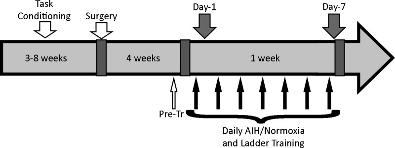
The experimental timeline is shown in Fig 1. All rats were initially conditioned on the ladder task prior to surgery for unilateral transection of the dorsolateral funiculus at the C2 level. The location and size of spinal transection injury was chosen because it produces a consistent foot-slip deficit of the ipsilateral forepaw during the horizontal ladder task [8, 9, 15]. Four weeks after surgery, performance on the ladder task was evaluated and animals were grouped based on their ladder task deficit such that the deficits were matched between the groups. The groups were randomly assigned to receive either AIH or control (normoxia) treatment (n = 12 per treatment group). This ensured equivalence of lesion severity between AIH and normoxia treatment groups. Within each treatment group, animals were randomly assigned to receive either 1 day or 7 days of treatment plus ladder training. The experimental groups were therefore 1 day normoxia; 7 day normoxia; 1 day AIH; or 7 day AIH (n = 6 per experimental group). During the treatment week, rats underwent ladder training 1 hour after each treatment.
Fig 1. Experimental design.
All animals were initially conditioned on the ladder task for 3–8 weeks, underwent spinal surgery and were assessed on the ladder task 4 weeks after surgery but before treatment (Pre-tr). Animals then received AIH or normoxia treatment plus ladder training for one day (Day 1) or every day for 7 days (Day 7). Spinal cord tissue was taken from half of the animals (n = 6/treatment group), which were euthanized and perfused after 1 day of treatment and training (Day 1), and from the remaining animals (n = 6/treatment group) after 7 days of treatment and training (Day 7).
Ladder-walking task
Rats were trained to cross a horizontal runway, consisting of a ladder apparatus 120 cm in length, with 20 cm opaque plexiglass platforms at either end allowing the rats to turn around. The central ladder portion (80 cm in length) consisted of 2 mm diameter wire rungs spaced 2 cm apart. The ladder was positioned above a 45° angled mirror so both lateral and ventral aspects of the rat movements were visible in the digital video camera (EOS Rebel, T2i EOS 550D Canon) positioned perpendicular to the runway.
Ladder conditioning and training took place at the same time each day for each group of animals. Animals were initially conditioned to walk on the runway for 3–8 weeks, after which time they would repeatedly cross the ladder consistently and quickly with very few long pauses or hesitations. Training on the ladder-task was carried out at the following time points: pre-surgery, pre-treatment, and each of the days of treatment. All training sessions involved 12 complete crossings.
Surgery
Animals were placed under isoflurane anaesthesia and administered an antibiotic (trimethoprim and sulfadoxine (TMS), Trivetrin, Schering Canada Inc., QC, Canada, 30mg/kg sub-cutaneous (SC)) and pre-emptive analgesia (buprenorphine, Buprenex; Reckitt Benckiser Pharmaceuticals Inc., Richmond, VA, USA, 0.05mg/kg SC). The surgical site over the dorsum of the neck was prepared by removing fur with clippers and cleaning the skin with stanhexidine and 70% isopropyl alcohol. Under an operating microscope, the spinal laminae of the 2nd and 3rd cervical vertebrae were exposed using sterile technique, and a laminectomy and durotomy performed to expose the 2nd cervical spinal segment. The dorsolateral funiculus was transected unilaterally on the left side using a modified 25-gauge bevel tipped needle. The muscle was closed with a simple continuous suture, and the skin was closed with a subcuticular suture technique. The entire time spent under anaesthetic was approximately 20–30 min. Following surgery, rats were administered 3 ml warm sterile saline (SC) and housed individually in cages equipped with wood chip bedding, a plastic tube and an extraneous heat source. Post-operative analgesia (buprenorphine 0.05mg/kg) and antibiotic (TMS, 30mg/kg) were administered for 48h post-surgery, and longer if necessary. Rats were monitored several times daily for 5 days post-surgery and assessed for change in weight, presence and severity of porphyrin, hydration, healing of the incision site, mobility, and general behavior. At day 3 post-surgery, rats were rehoused with their original cage mates.
Intermittent hypoxia treatment
Rats were acclimated to the treatment apparatus by placing them for 30 min into custom-made Plexiglas chambers (1 rat per chamber; 30cm × 17cm × 12cm) under normoxia (21% inspired O2), 1 day prior to the first treatment day as previously described [8, 9]. Subsequently, on each day of treatment, the rats were placed into the Plexiglas chamber and then exposed to AIH, consisting of ten 5 min hypoxic episodes (11% inspired O2), alternating with 5 min normoxic intervals. Alteration in normoxic and hypoxic conditions were established by automatically switching the incoming air between premixed O2 and N2 gas (FlO2 = 0.11) and medical air (FlO2 = 0.21). Animals receiving control treatment (normoxia) were placed in adjacent chambers for the same total duration of time under continuous normoxic conditions (FlO2 = 0.21). The oxygen levels in the chambers were continuously monitored using an oxygen analyzer (AX300-1 Portable Oxygen Analyzer; Teledyne Analytical Instruments).
Tissue preparation
Beginning one hour after the training portion of the treatment on either day 1 or day 7, animals were deeply anaesthetized with isoflurane and perfused trans-cardially with heparinized phosphate-buffered saline followed by cold 4% paraformaldehyde in 0.01M phosphate buffer. The vertebral column was removed and post-fixed overnight in 4% paraformaldehyde. The spinal cord was dissected from the vertebral column and segments C6-7 (containing forelimb motoneurons) and L4-5 (containing hindlimb motoneurons) were postfixed for 1–1.5h in 4% paraformaldehyde followed by cryoprotection in 10% and then 20% sucrose overnight at 4°C, respectively. Subsequently, segments of spinal cord from each of the experimental groups were embedded in the same cryomold covered in OCT compound (Tissue Tek; Miles Laboratories, Elkhart, IN), and frozen in isopentane cooled in a slurry of dry ice and acetone. Thus, each block contained 8 pieces of spinal cord, representing each of the 4 experimental conditions (1 day normoxia; 7 day normoxia; 1 day AIH; 7 day AIH) from each of the spinal cord regions (C6-7 and L4-5). Blocks were then stored at -80°C until sectioning. Tissues were sectioned at 10μm on a Microm cryostat, thaw-mounted onto silanized slides (VWR Superfrost Plus) and stored at -80°C. The presence of representative spinal cord sections from all experimental groups on each slide allowed processing under identical conditions and intergroup analysis within the same slide.
Immunofluorescence
Slides were brought to room temperature, then washed 3×10 min in 0.01M PBS, pH 7.4. For BNDF and TrkB only, citrate antigen retrieval was performed prior to blocking and incubation with primary antibodies. For this extra step, slides were placed in 0.01M citrate buffer (10% 0.1M sodium citrate buffer in ddH2O, pH 6) at 50°C and then warmed to 90°C over 45min. Slides were then allowed to cool for 20min. For all antibodies, slides were incubated with blocking solution containing undiluted Sea Block buffer (Abcam) for 1hr at room temperature. Primary antibodies were diluted with 10% Sea Block in primary diluent (0.1% Triton X-100 in 0.01M PBS) to the following concentrations: mouse anti- HIF-1α (NB 100–105, Novus Biological) 1:200, rabbit anti-VEGF (sc-152, Santa Cruz Biotech, Inc) 1:200, chicken anti-BDNF (Promega) 1:200, rabbit anti-TrkB (sc-12, Santa Cruz Biotech, Inc) 1:100, rabbit anti phospho-TrkB (rat phospho Y705, Y706, ab111545, Abcam) 1:400. After 24hr incubation with the primary at 4°C in a humidified chamber, slides were washed for 3× 10min in 0.01M PBS. The following secondary antibodies were used: goat-anti-rabbit Alexa Fluor 488, donkey-anti-mouse Alexa Fluor 488 (Jackson Immuno Research Laboratories, Inc) at 1:2000, goat-anti-chicken (F-1005, aves labs, Inc) 1:1000 diluted in 0.01M PBS. After 1hr incubation in the dark at room temperature, slides were washed for 3× 10min in 0.01M PBS, and then coverslipped using ProLong Gold Antifade Reagent (P36931, Molecular Probes, Invitrogen).
For double staining for choline acetyl-transferase (ChAT) and HIF-1α, sections were brought to room temperature, underwent antigen retrieval with citrate buffer as described above and blocked in undiluted Sea Block buffer for 1h. Anti-ChAT (AB144P, Millipore) and anti-HIF-1α (GTX127309, GeneTex) antibodies were diluted at 1:100 and 1:200 respectively in 0.01M PBS with 10% Sea Block and 0.1% Triton X-100 and were applied simultaneously on the sections. Sections were incubated with these antibodies for 24hr at 4°C in a humidified chamber. The slides were subsequently washed 3× 10min in 0.01M PBS before simultaneous application of the secondary antibodies, Alexa Fluor 594 (A-11058, Invitrogen) and Alexa Fluor 488 (A-21206, Invitrogen), diluted 1:500 in 0.01M PBS. Sections were incubated with the secondary antibodies for 1hr in the dark at room temperature. The secondary solution was removed with 3× 10min washes in 0.01M PBS and the nuclei were stained with DAPI (D9542, Sigma-Aldrich) for 10min at room temperature using a 300 nM DAPI staining solution in 0.01M PBS. Finally, the slides were again washed 3 × 10min in 0.01M PBS before coverslipping with a 1:1 glycerol/0.01M PBS solution.
For all markers specificity of the secondary antibodies employed in this study was confirmed in experiments where the primary antibodies were omitted, revealing an absence of nonspecific staining.
Image analysis / quantification
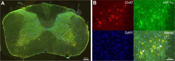
Immunofluorescence-processed sections were examined and imaged using a Zeiss Axioskop (20× objective) with all spinal cord ventral cord regions of the different experimental groups imaged under identical conditions. Initially, spinal neurons located on both sides of the cord were analyzed separately and the data were pooled after we determined that there were no differences between the levels of protein expression from side to side. All slides were first qualitatively examined and alterations in immunofluorescence noted. Immunofluorescence signal intensity was quantified for 3 out of 6 animals in each experimental group using Northern Eclipse (Empix Imaging) and FIJI (ImageJ). Briefly, the cell bodies of neurons in ventral grey matter on both sides of the spinal cord were identified by their location and size. Only cells for which the cytoplasmic area visible was equal to or greater than the area of the nucleus were included. Immunofluorescence labeling for ChAT confirmed that these identified neurons were motoneurons (Fig 2). All neurons meeting these criteria were analyzed from 2 separate slides for each grouping (n = 3) of all 8 experimental conditions mounted on the same slide (1 day normoxia C6-7 & L4-5; 7 day normoxia C6-7 & L4-5; 1 day AIH C6-7 & L4-5; 7 day AIH C6-7 & L4-5), representing a total of 60–85 neurons analysed/time point/experimental condition. Quantitative image analysis was performed by an individual blinded to experimental condition of the image being analysed. Neurons were circumscribed manually and the mean gray value for the marker being examined was obtained for the cytoplasm of each neuron. To determine whether neuronal size, and consequently the mean gray value, would be affected by treatment, we compared the mean cell areas (micron2) for each experimental condition and found no significant differences between groups. The area of the nuclei was excluded from analysis. Mean gray background values were determined over regions of the neuropil devoid of positive signal and subtracted from the mean gray value for each neuron analyzed in that spinal cord section and then the net mean gray value determined for that grouping of neurons. The mean gray value for each slide was averaged to get the mean gray value for each animal within each experimental group. The mean gray values for each of 3 animals in each of 4 experimental groups (1 day normoxia; 7 day normoxia; 1 day AIH; 7 day AIH) were then averaged to obtain mean gray values for each group at both the cervical and lumbar levels.
Fig 2. Spinal neurons identified as motoneurons with choline acetyl-transferase (ChAT).
Photomicrograph of the spinal cord in C6-7 region (A) processed for ChAT, HIF-1α and DAPI immunofluorescence from a 7 day AIH-treated spinal-injured rat. Scale bar = 250 μm. White box shows location of representative images in (B) processed for each of ChAT, HIF-1α and DAPI. Merged image shows HIF-1α co-localization with ChAT and DAPI in spinal neurons (e.g. arrows). Scale bar = 100 μm.
Bar graphs were made in Graphpad prism v6.02 (La Jolla, CA) with composite figures compiled using Adobe Photoshop CC software (Adobe System, San Jose, CA).
Statistical analysis
Statistical analysis was performed with IBM SPSS Statistics v20 for Windows software. Differences between the mean gray values for each experimental group at C6-7 or L4-5 spinal segments for each of BDNF, TrkB, phospho-TrkB, HIF-1α and VEGF were examined by using one-way analysis of variance (ANOVA) and Tukey HSD test was used for post hoc analysis. Differences were considered significant if p<0.05.
Results
AIH and motor training results in early and sustained increases in HIF-1α protein expression in spinal neurons at cervical and lumbar spinal segments
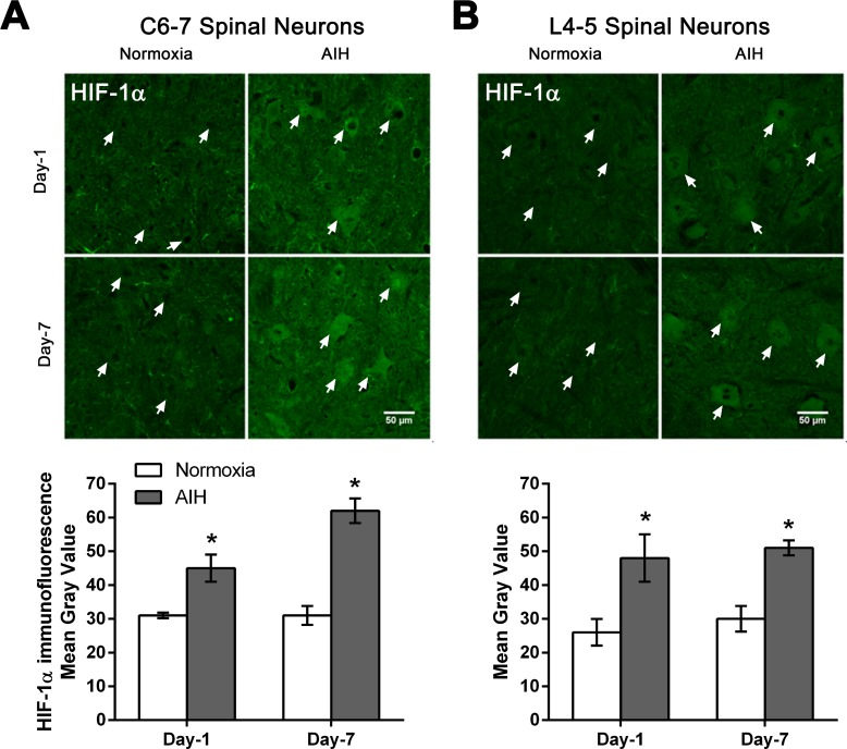
HIF-1α is a transcriptional regulator of genes controlling a number of adaptive responses to low oxygen tension in order to maintain oxygen homeostasis in mammalian cells and more recently linked to neurorepair [16, 17]. AIH treatment and motor training for 1 or 7 days increased HIF-1α protein expression in spinal motoneurons at C6-7 and L4-5 segments of spinal cord (Fig 3). Photomicrographs of ventral grey matter region of spinal cord processed for HIF-1α immunofluorescence showed increased immunoreactivity in neurons in AIH-treated + trained SCI rats compared to normoxia-treated + trained SCI rats after 1 day (Day-1). A similar change occurred in response to 7 days (Day-7) of treatment in putative motoneurons (large neurons in ventral horn) of both C6-7 (Fig 3A) and L4-5 (Fig 3B) spinal segments. Quantitative analysis confirmed that AIH treatment plus motor training for either 1 day or 7 days significantly increased HIF-1α protein levels in the neurons of ventral grey matter of C6-7 and L4-5 spinal segments in AIH-treated rats relative to normoxia-treated control rats (p < 0.05) (Fig 3A–3B and S1 Table).
Fig 3. AIH treatment for either 1 or 7 days increased HIF-1α protein levels in multiple spinal segments.
Representative photomicrographs of the ventral grey matter in C6-7 (A) or L4-5 (B) spinal segments sections processed for HIF-1α immunofluorescence from 1 day (Day-1) or 7 day (Day-7) in normoxia-treated versus AIH-treated spinal injured rats. Arrows indicate representative neurons. Scale bar = 50 μm. Histograms summarize the mean immunofluorescence signal intensity detected ± SEM as measured in gray values over individual neurons within the ventral horn from normoxia- and AIH-treated spinal injured rats [n = 3 rats per experimental group analysed; 60–65 neurons analysed/time point/experimental condition at C6-7 (A); 80–85 neurons analysed/time point/experimental condition at L4-5 (B)]. Asterisks indicate significant differences between experimental groups; * p < 0.05.
VEGF protein expression in spinal neurons is increased in response to AIH and motor training for 7 days
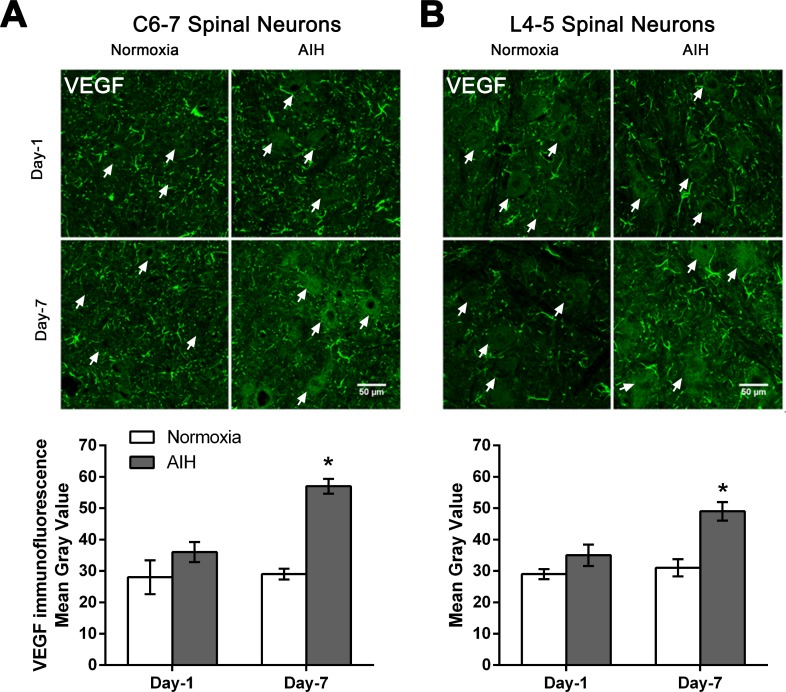
We next investigated a target gene of HIF-1α, vascular endothelial growth factor (VEGF; [18]), a dimeric glycoprotein and fundamental regulator of angiogenesis that also appears to play neurotrophic and neuroprotective roles in spinal cord and brain injury [19–22]. AIH treatment + motor training for 7 days increased the expression of VEGF protein in spinal neurons at C6-7 and L4-5 segments of spinal cord, with no marked change in VEGF immunoreactivity following 1 day of treatment + training (Fig 4). Photomicrographs of spinal ventral grey matter processed for VEGF protein immunofluorescence show increased neuronal VEGF expression in AIH-treated spinal-injured rats relative to normoxia-treated spinal-injured rats after Day-7 of treatment in C6-7 (Fig 4A) and L4-5 (Fig 4B). Quantitative analysis confirmed that AIH treatment plus motor training for 7 days significantly increased protein expression of VEGF levels in neurons of ventral grey matter of spinal segments C6-7 and L4-5 in AIH-treated rats versus normoxia-treated rats (p < 0.05) (Fig 4A–4B and S2 Table).
Fig 4. AIH treatment for 7 days increased neuronal VEGF protein levels in multiple spinal segments.
Representative photomicrographs of the ventral grey matter in C6-7 (A) or L4-5 (B) spinal segments sections processed for VEGF immunofluorescence from 1 day (Day-1) or 7 day (Day-7) in normoxia-treated versus AIH-treated spinal injured rats. Arrows indicate representative neurons. Scale bar = 50 μm. Histograms summarize the mean immunofluorescence signal intensity detected ± SEM as measured in gray values over individual neurons within the ventral horn from normoxia- and AIH-treated spinal injured rats [n = 3 rats per experimental group analysed; 60–65 neurons analysed/time point/experimental condition at C6-7 (A); 80–85 neurons analysed/time point/experimental condition at L4-5 (B)]. Asterisks indicate significant differences between experimental groups; * p < 0.05.
AIH treatment and motor training for 7 days resulted in higher neuronal expression of BDNF in cervical and lumbar neurons
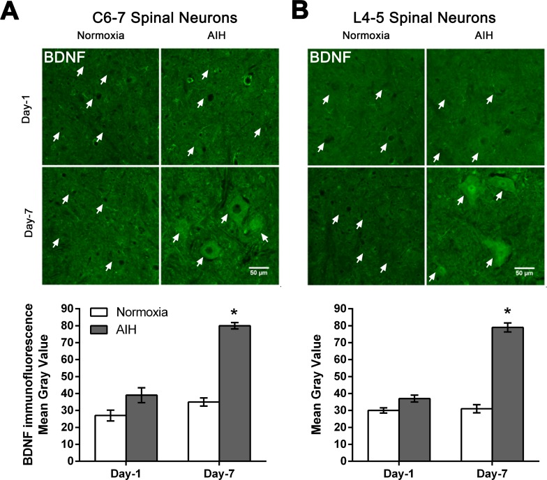
BDNF was also examined due to its diverse role in modulating neural plasticity and enhancing functional recovery following SCI [23–29]. AIH treatment and motor training for 7 days increased the expression of BDNF protein in spinal neurons in both C6-7 and L4-5 segments of spinal cord relative to the normoxia controls and as detected using immunofluorescence (Fig 5). However, there was no discernible change in BDNF immunofluorescence over neurons at either cervical or lumbar spinal cord levels examined following 1 day of treatment and training (Fig 5). Quantitative analysis confirmed that the increase in BDNF immunofluorescence signal observed in neurons of C6-7 and L4-5 spinal segments in SCI rats in response to 7 day AIH and motor training was significantly higher than those detected in the normoxia plus motor training-treated SCI rats (p < 0.05) (Fig 5A–5B and S3 Table).
Fig 5. AIH treatment for 7 days increased BDNF protein levels in multiple spinal segments.
Representative photomicrographs of the ventral grey matter in C6-7 (A) or L4-5 (B) spinal segments sections processed for BDNF immunofluorescence from 1 day (Day-1) or 7 day (Day-7) in normoxia-treated versus AIH-treated spinal injured rats. Arrows indicate representative neurons. Scale bar = 50 μm. Histograms summarize the mean immunofluorescence signal intensity detected ± SEM as measured in gray values over individual neurons within the ventral horn from normoxia- and AIH-treated spinal injured rats [n = 3 rats per experimental group analysed; 60–65 neurons analysed/time point/experimental condition at C6-7 (A); 80–85 neurons analysed/time point/experimental condition at L4-5 (B)]. Asterisks indicate significant differences between experimental groups; * p < 0.05.
AIH treatment and motor training for 7 days resulted in increased expression and activation of TrkB protein expression in ventral grey matter
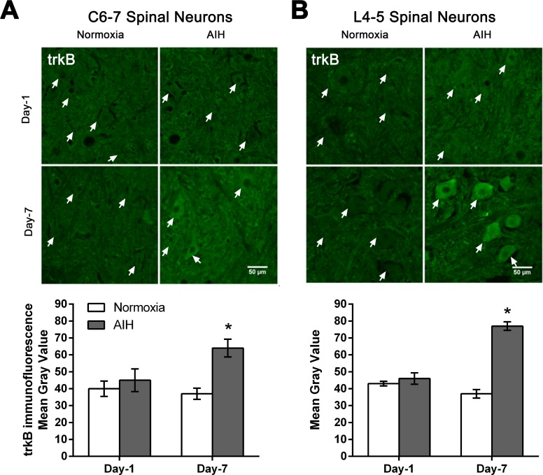
BDNF signaling through its cognate receptor, tropomyosin-related kinase B (TrkB) [30] is a key pathway involved in activity-dependent processes driving neural plasticity. Thus, we next examined whether the elevations in neuron BDNF expression induced by AIH and motor training were also associated with increased expression of its receptor TrkB in this population of neurons. Immunofluorescence analysis of tissue sections at C6-7 and L4-5 spinal segments revealed that AIH treatment and motor training for 7 days effects increased the expression of TrkB protein in spinal neurons, with little change in expression noted after only one day of treatment (Fig 6). Quantitative analysis confirmed that these changes in TrkB expression in response to AIH treatment plus motor training for 7 days were significant (Fig 6 and S4 Table).
Fig 6. AIH treatment for 7 days increased TrkB protein levels in multiple spinal segments.
Representative photomicrographs of the ventral grey matter in C6-7 (A) or L4-5 (B) spinal segments sections processed for TrkB immunofluorescence from 1 day (Day-1) or 7 day (Day-7) in normoxia-treated versus AIH-treated spinal injured rats. Arrows indicate representative neurons. Scale bar = 50 μm. Histograms summarize the mean immunofluorescence signal intensity detected ± SEM as measured in gray values over individual neurons within the ventral horn from normoxia- and AIH-treated spinal injured rats [n = 3 rats per experimental group analysed; 60–65 neurons analysed/time point/experimental condition at C6-7 (A); 80–85 neurons analysed/time point/experimental condition at L4-5 (B)]. Asterisks indicate significant differences between experimental groups; * p < 0.05.
BDNF induces rapid phosphorylation of TrkB receptors [31–33], which is necessary for activation of many downstream actions. To assess whether the increased BDNF and TrkB expression effected by AIH and motor training was also associated with increased BDNF signaling in cervical and lumbar neurons, we processed additional tissue sections for immunofluorescence to detect alterations in the levels of tyrosine phosphorylated/activated TrkB.
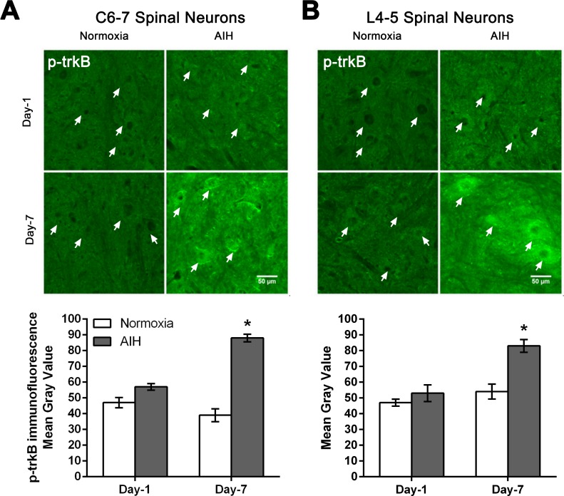
AIH treatment and motor training for 7 days in the SCI rats resulted in an increased phosphorylation and activation of TrkB protein in spinal neurons at C6-7 and L4-5 segments of spinal cord relative to normoxia and motor training treated control SCI rats (Fig 7). This increased phospho-TrkB immunofluorescence was already evident, albeit at low levels following one day of AIH treatment, with greatly increased levels observed after 7 days of AIH treatment at C6-7 (Fig 7A) and L4-5 (Fig 7B) spinal segment levels. The punctate pattern of staining observed at the level of the cell membrane and in the neuronal cytosol is consistent with activation at the membrane level, followed by internalization of the signaling endosome. Quantitative analysis confirmed that AIH treatment plus motor training resulted in significantly increased levels of phospho-TrkB detected in the C6-7 and L4-5 neurons from 7 day treated animals relative to normoxia-treated rats, but failed to reach significance in the one day AIH treated animals (Fig 7 and S5 Table).
Fig 7. AIH treatment for 7 days increases phospho-TrkB (p-TrkB) protein levels in multiple spinal segments.
Representative photomicrographs of the ventral grey matter in C6-7 (A) or L4-5 (B) spinal segments sections processed for p-TrkB immunofluorescence from 1 day (Day-1) or 7 day (Day-7) in normoxia-treated versus AIH-treated spinal injured rats. Arrows indicate representative neurons. Scale bar = 50 μm. Histograms summarize the mean immunofluorescence signal intensity detected ± SEM as measured in gray values over individual neurons within the ventral horn from normoxia- and AIH-treated spinal injured rats [n = 3 rats per experimental group analysed; 60–65 neurons analysed/time point/experimental condition at C6-7 (A); 80–85 neurons analysed/time point/experimental condition at L4–5 (B) ]. Asterisks indicate significant differences between experimental groups; * p < 0.05.
Discussion
In the present study, we assessed the combined effect of AIH and rehabilitative training on expression of hypoxia-associated (HIF-1α, VEGF) and plasticity-associated proteins (BDNF, TrkB, phospho-TrkB) in the spinal neurons of cervical SCI rats. AIH plus training administered for 7 days starting 4 weeks post-SCI significantly increased the expression of HIF-1α, VEGF, BDNF and TrkB molecules and enhanced TrkB activation in spinal neurons of cervical SCI rats at spinal segments C6-7 and L4-5. Unlike other proteins examined, the expression of the oxygen-sensitive transcription factor HIF-1α was also increased in spinal neurons after only 1 day of AIH treatment and motor training in SCI rats.
AIH and motor training increased hypoxia-associated protein expression
The HIF-1 protein is a heterodimer, composed of two subunits: the constitutively expressed HIF-1β and HIF-1α expressed under hypoxia conditions [19, 34–36]. HIF-1 regulates the transcription of several dozen hypoxia-inducible target genes including VEGF, erythropoietin, inducible nitric oxide synthase, heme oxygenase-1, glucose transporter-1 and the glycolytic enzymes [35, 37–44]. Hypoxia induces the expression of HIF-1α in numerous cell types of the CNS including neurons, astrocytes, oligodendrocytes and microglia [45, 46].
The current finding that HIF-1α expression increased in spinal neurons after only one session of AIH treatment is consistent with the findings that exposure to low oxygen for only minutes to hours is sufficient to enhance HIF-1α levels. One hour of systemic hypoxia (6% O2) is sufficient to increase HIF-1α protein expression in brain tissues of mice, especially in the neurons of cerebral cortex and the granular layer of dentate gyrus in the hippocampus [47]. Only a few minutes of hypoxia induces the expression of HIF-1α proteins in the human epithelial carcinoma cell line HeLaS3, and after 1 hour of anoxia, HIF-1α protein expression reached its maximum level, and this maximum level was maintained for 4 hours [48]. Moreover, exposure to low oxygen (6% to 7% O2) for 30 minutes increased the expression of HIF-1α protein, but not its mRNA, 7 fold in rat retina, followed by a further increase after 3 hours of hypoxia to 15 fold compared to control animals [49]. Mice exposed to AIH for 120 minutes, comparable to the present study, showed increased HIF-1α expression in dorsal root ganglion neurons[14].
HIF-1α regulates the expression of VEGF, a 45kDa dimeric glycoprotein which is a fundamental regulator of pathological and physiological angiogenesis [19] but also plays neurotrophic and neuroprotective roles in the central and peripheral nervous systems [19, 50–54]. VEGF and its high affinity receptor VEGFR-2 are expressed in spinal neurons [55]. Repetitive AIH exposure (3×/week AIH: 10, 5-min episodes of 10.5% O2; 5-min normoxic intervals) upregulated the expression of VEGF and VEGFR-2 in respiratory and non-respiratory neurons in the spinal cord [13, 55, 56]. Consistent with these previous findings, the current study shows that AIH treatment for 7 days also significantly enhanced the expression of HIF-1α and VEGF at both C6-7 and L4-5 levels in the spinal cord (Figs 3 and 4). While AIH exposure for 1 day increased expression of HIF-1α, it was not sufficient to discernibly alter the expression of VEGF in spinal neurons. It is possible that a more sustained elevation in HIF-1α levels beyond that produced by 1 day of AIH treatment is required to induce downstream increases in VEGF expression.
Whether increased VEGF expression is related to improvements in ladder task performance seen in SCI rats after AIH + motor training is not yet known. In mice, 2 hours of in vivo AIH exposure results a HIF-1α -dependent improvement in axonal regeneration, along with hypoxia-induced JNK phosphorylation and upregulation of VEGF in dorsal root ganglion neurons[14]. In respiratory systems, exogenous application of VEGF in vivo elicits phrenic motor plasticity via the ERK/Akt intracellular pathways [1, 55, 56]. Collectively, these findings suggest that VEGF signalling might contribute to improved function in AIH-treated animals with nervous system injury [8, 9].
AIH and motor training enhance the spinal expression of BDNF
Growth factors and trophic factors, including BDNF, play important roles in multiple forms of neural plasticity. BDNF mediates these effects through its high affinity receptor TrkB [57, 58]. Exogenous application of BDNF promotes neuroprotection, axonal regeneration, survival of neurons and axonal sprouting at the site of injury in SCI animals and has been reported to improve functional recovery after SCI [57–62]. Nevertheless, detrimental functional effects of exogenous BDNF have also been reported, including increased neuropathic pain and spasticity in SCI rats [63, 64]. Additionally, exogenous administration of BDNF must overcome difficulties related to access across the blood brain barrier, down-regulation of BDNF receptors, and triggering of immune responses [65, 66].
Increases in endogenous levels of BDNF might be more effective for facilitating functional recovery. Motor training following SCI enhances BDNF protein expression and this increase in BDNF expression is associated with locomotor functional recovery following SCI in animals [67–70]. Locomotor training, including voluntary wheel running and forced treadmill training, enhances the expression of BDNF protein and mRNA in spinal neurons and in skeletal muscles in spinal-injured rats [67, 71–76]. Furthermore, weight-supported treadmill training, either 100 or 1000 steps/training session daily for 4 weeks, has been shown to increase the BDNF expression in ventral grey matter of SCI rats [67]. Of particular interest to the current study, 4 weeks of treadmill training initiated 1 month after injury increased BDNF expression in the lumbar spinal neurons caudal to a complete spinal transection in rats [73]. In the current study, 1 week of ladder training initiated 4 weeks after spinal injury was not sufficient to increase BDNF expression in cervical or lumbar neurons (Fig 5, Day 7-Normoxia). Instead, 1 week of AIH administered in addition to motor training increased BDNF levels in spinal neurons (Fig 5, Day 7-AIH).
Consistent with our findings, several different protocols of AIH (daily for 7 days or 3×/week for 10 weeks) have shown increased expression of BDNF and TrkB as well as enhanced activation of TrkB (phospho-TrkB) in spinal motor nuclei [8, 13, 56, 77]. Importantly, brief episodes of hypoxia in intact rats initiated the synthesis of new BDNF in the cervical cord, and this BDNF was sufficient and necessary to induce respiratory plasticity [77, 78]. The current study extends these previous findings by confirming that BDNF expression also increases at multiple spinal sites after 7 days of treatment with AIH treatment plus motor training.
AIH and motor training enhance the spinal expression of TrkB and its activation
The BDNF receptor TrkB is a transmembrane proteins of the Trk family of neurotrophin receptors [30]. TrkB receptors are activated by BDNF-induced formation of receptor dimers, whereupon the dimerized receptors rapidly phosphorylate each other and activate downstream signaling pathways [79–81]. In the current study, the increased spinal expression of TrkB and its subsequent phosphorylation/activation after 7 days of AIH and motor training in SCI rats strongly suggests that the effects of this combined treatment are at least in part mediated by the activation of TrkB signalling pathways. This is consistent with increased BDNF expression described above, as well as with previous findings of increased TrkB expression and its activation after repetitive AIH treatment in intact rats, and AIH-induced activation of TrkB receptors in spinal neurons resulting in increased output of the phrenic nerve [8, 56, 77, 78, 82].
Conclusion
In the present study, we have shown that a low-dose protocol of AIH treatment + motor training in SCI rats produced temporal and spatial differential expression of hypoxia- and plasticity-related proteins in spinal neurons. The pattern of expression of these proteins occurs under identical experimental conditions which result in recovery of skilled forelimb use in SCI animals. Together with the capacity of AIH and motor training to improve walking abilities in persons with chronic incomplete SCI, these findings suggest that AIH has potential as an effective therapy to restore motor function after nervous system injury.
Supporting information
(XLSX)
(XLSX)
(XLS)
(XLSX)
(XLSX)
Acknowledgments
The authors acknowledge the advice and support of Gordon Mitchell, and the invaluable assistance of Erin Prosser-Loose, Aphroditi Antonopoulos, Joelle Nadeau and Jayne Johnston.
Data Availability
All relevant data are within the paper and its Supporting Information files.
Funding Statement
This work was supported by the US Department of Defense SC090355P1 (GDM), Saskatchewan Health Research Fund 2668 (VMV, GDM), Saskatchewan Health Research Fund 3695 (GDM, VMV), and Saskatchewan Health Research Fund 6362 (GDM, VMV). The funders had no role in study design, data collection and analysis, decision to publish, or preparation of the manuscript.
References
- 1.Dale EA, Ben Mabrouk F, Mitchell GS. Unexpected benefits of intermittent hypoxia: enhanced respiratory and nonrespiratory motor function. Physiology. 2014;29(1):39–48. doi: 10.1152/physiol.00012.2013 . [DOI] [PMC free article] [PubMed] [Google Scholar]
- 2.Navarrete-Opazo A, Mitchell GS. Therapeutic potential of intermittent hypoxia: a matter of dose. American journal of physiology Regulatory, integrative and comparative physiology. 2014;307(10):R1181–97. doi: 10.1152/ajpregu.00208.2014 ; PubMed Central PMCID: PMC4315448. [DOI] [PMC free article] [PubMed] [Google Scholar]
- 3.Devinney MJ, Huxtable AG, Nichols NL, Mitchell GS. Hypoxia-induced phrenic long-term facilitation: emergent properties. Annals of the New York Academy of Sciences. 2013;1279:143–53. doi: 10.1111/nyas.12085 ; PubMed Central PMCID: PMC3880582. [DOI] [PMC free article] [PubMed] [Google Scholar]
- 4.Golder FJ, Mitchell GS. Spinal synaptic enhancement with acute intermittent hypoxia improves respiratory function after chronic cervical spinal cord injury. The Journal of neuroscience: the official journal of the Society for Neuroscience. 2005;25(11):2925–32. doi: 10.1523/JNEUROSCI.0148-05.2005 . [DOI] [PMC free article] [PubMed] [Google Scholar]
- 5.Fuller DD, Johnson SM, Olson EB Jr., Mitchell GS. Synaptic pathways to phrenic motoneurons are enhanced by chronic intermittent hypoxia after cervical spinal cord injury. The Journal of neuroscience: the official journal of the Society for Neuroscience. 2003;23(7):2993–3000. . [DOI] [PMC free article] [PubMed] [Google Scholar]
- 6.Dougherty BJ, Fields DP, Mitchell GS. Mammalian target of rapamycin is required for phrenic long-term facilitation following severe but not moderate acute intermittent hypoxia. Journal of neurophysiology. 2015;114(3):1784–91. doi: 10.1152/jn.00539.2015 ; PubMed Central PMCID: PMC4571765. [DOI] [PMC free article] [PubMed] [Google Scholar]
- 7.Forsythe JA, Jiang BH, Iyer NV, Agani F, Leung SW, Koos RD, et al. Activation of vascular endothelial growth factor gene transcription by hypoxia-inducible factor 1. Molecular and cellular biology. 1996;16(9):4604–13. ; PubMed Central PMCID: PMC231459. [DOI] [PMC free article] [PubMed] [Google Scholar]
- 8.Lovett-Barr MR, Satriotomo I, Muir GD, Wilkerson JE, Hoffman MS, Vinit S, et al. Repetitive intermittent hypoxia induces respiratory and somatic motor recovery after chronic cervical spinal injury. The Journal of neuroscience: the official journal of the Society for Neuroscience. 2012;32(11):3591–600. doi: 10.1523/JNEUROSCI.2908-11.2012 ; PubMed Central PMCID: PMC3349282. [DOI] [PMC free article] [PubMed] [Google Scholar]
- 9.Prosser-Loose EJ, Hassan A, Mitchell GS, Muir GD. Delayed Intervention with Intermittent Hypoxia and Task Training Improves Forelimb Function in a Rat Model of Cervical Spinal Injury. Journal of neurotrauma. 2015;32(18):1403–12. doi: 10.1089/neu.2014.3789 . [DOI] [PubMed] [Google Scholar]
- 10.Trumbower RD, Jayaraman A, Mitchell GS, Rymer WZ. Exposure to acute intermittent hypoxia augments somatic motor function in humans with incomplete spinal cord injury. Neurorehabilitation and neural repair. 2012;26(2):163–72. doi: 10.1177/1545968311412055 . [DOI] [PubMed] [Google Scholar]
- 11.Hayes HB, Jayaraman A, Herrmann M, Mitchell GS, Rymer WZ, Trumbower RD. Daily intermittent hypoxia enhances walking after chronic spinal cord injury: A randomized trial. Neurology. 2014;82(2):104–13. doi: 10.1212/01.WNL.0000437416.34298.43 . [DOI] [PMC free article] [PubMed] [Google Scholar]
- 12.Dale EA, Satriotomo I, Mitchell GS. Cervical spinal erythropoietin induces phrenic motor facilitation via extracellular signal-regulated protein kinase and Akt signaling. The Journal of neuroscience: the official journal of the Society for Neuroscience. 2012;32(17):5973–83. doi: 10.1523/JNEUROSCI.3873-11.2012 ; PubMed Central PMCID: PMC3505998. [DOI] [PMC free article] [PubMed] [Google Scholar]
- 13.Satriotomo I, Nichols NL, Dale EA, Emery AT, Dahlberg JM, Mitchell GS. Repetitive acute intermittent hypoxia increases growth/neurotrophic factor expression in non-respiratory motor neurons. Neuroscience. 2016;322:479–88. doi: 10.1016/j.neuroscience.2016.02.060 . [DOI] [PMC free article] [PubMed] [Google Scholar]
- 14.Cho Y, Shin JE, Ewan EE, Oh YM, Pita-Thomas W, Cavalli V. Activating Injury-Responsive Genes with Hypoxia Enhances Axon Regeneration through Neuronal HIF-1alpha. Neuron. 2015;88(4):720–34. doi: 10.1016/j.neuron.2015.09.050 ; PubMed Central PMCID: PMC4655162. [DOI] [PMC free article] [PubMed] [Google Scholar]
- 15.Kanagal SG, Muir GD. Task-dependent compensation after pyramidal tract and dorsolateral spinal lesions in rats. Experimental neurology. 2009;216(1):193–206. doi: 10.1016/j.expneurol.2008.11.028 . [DOI] [PubMed] [Google Scholar]
- 16.Calvani M, Comito G, Giannoni E, Chiarugi P. Time-dependent stabilization of hypoxia inducible factor-1alpha by different intracellular sources of reactive oxygen species. PloS one. 2012;7(10):e38388 doi: 10.1371/journal.pone.0038388 ; PubMed Central PMCID: PMC3483303. [DOI] [PMC free article] [PubMed] [Google Scholar]
- 17.Khan M, Dhammu TS, Matsuda F, Baarine M, Dhindsa TS, Singh I, et al. Promoting endothelial function by S-nitrosoglutathione through the HIF-1alpha/VEGF pathway stimulates neurorepair and functional recovery following experimental stroke in rats. Drug design, development and therapy. 2015;9:2233–47. doi: 10.2147/DDDT.S77115 ; PubMed Central PMCID: PMC4408969. [DOI] [PMC free article] [PubMed] [Google Scholar]
- 18.Zhang B, Tanaka J, Yang L, Yang L, Sakanaka M, Hata R, et al. Protective effect of vitamin E against focal brain ischemia and neuronal death through induction of target genes of hypoxia-inducible factor-1. Neuroscience. 2004;126(2):433–40. doi: 10.1016/j.neuroscience.2004.03.057 . [DOI] [PubMed] [Google Scholar]
- 19.Rosenstein JM, Krum JM. New roles for VEGF in nervous tissue—beyond blood vessels. Experimental neurology. 2004;187(2):246–53. doi: 10.1016/j.expneurol.2004.01.022 . [DOI] [PubMed] [Google Scholar]
- 20.Svensson B, Peters M, Konig HG, Poppe M, Levkau B, Rothermundt M, et al. Vascular endothelial growth factor protects cultured rat hippocampal neurons against hypoxic injury via an antiexcitotoxic, caspase-independent mechanism. Journal of cerebral blood flow and metabolism: official journal of the International Society of Cerebral Blood Flow and Metabolism. 2002;22(10):1170–5. doi: 10.1097/00004647-200210000-00003 . [DOI] [PubMed] [Google Scholar]
- 21.Krum JM, Rosenstein JM. VEGF mRNA and its receptor flt-1 are expressed in reactive astrocytes following neural grafting and tumor cell implantation in the adult CNS. Experimental neurology. 1998;154(1):57–65. doi: 10.1006/exnr.1998.6930 . [DOI] [PubMed] [Google Scholar]
- 22.Facchiano F, Fernandez E, Mancarella S, Maira G, Miscusi M, D'Arcangelo D, et al. Promotion of regeneration of corticospinal tract axons in rats with recombinant vascular endothelial growth factor alone and combined with adenovirus coding for this factor. Journal of neurosurgery. 2002;97(1):161–8. doi: 10.3171/jns.2002.97.1.0161 . [DOI] [PubMed] [Google Scholar]
- 23.Ma L, Wang DD, Zhang TY, Yu H, Wang Y, Huang SH, et al. Region-specific involvement of BDNF secretion and synthesis in conditioned taste aversion memory formation. The Journal of neuroscience: the official journal of the Society for Neuroscience. 2011;31(6):2079–90. doi: 10.1523/JNEUROSCI.5348-10.2011 ; PubMed Central PMCID: PMC3044502. [DOI] [PMC free article] [PubMed] [Google Scholar]
- 24.Nagahara AH, Tuszynski MH. Potential therapeutic uses of BDNF in neurological and psychiatric disorders. Nature reviews Drug discovery. 2011;10(3):209–19. doi: 10.1038/nrd3366 . [DOI] [PubMed] [Google Scholar]
- 25.Vavrek R, Girgis J, Tetzlaff W, Hiebert GW, Fouad K. BDNF promotes connections of corticospinal neurons onto spared descending interneurons in spinal cord injured rats. Brain: a journal of neurology. 2006;129(Pt 6):1534–45. doi: 10.1093/brain/awl087 . [DOI] [PubMed] [Google Scholar]
- 26.Hiebert GW, Khodarahmi K, McGraw J, Steeves JD, Tetzlaff W. Brain-derived neurotrophic factor applied to the motor cortex promotes sprouting of corticospinal fibers but not regeneration into a peripheral nerve transplant. Journal of neuroscience research. 2002;69(2):160–8. doi: 10.1002/jnr.10275 . [DOI] [PubMed] [Google Scholar]
- 27.Liao GY, Bouyer K, Kamitakahara A, Sahibzada N, Wang CH, Rutlin M, et al. Brain-derived neurotrophic factor is required for axonal growth of selective groups of neurons in the arcuate nucleus. Molecular metabolism. 2015;4(6):471–82. doi: 10.1016/j.molmet.2015.03.003 ; PubMed Central PMCID: PMC4443292. [DOI] [PMC free article] [PubMed] [Google Scholar]
- 28.Streijger F, Plunet WT, Lee JH, Liu J, Lam CK, Park S, et al. Ketogenic diet improves forelimb motor function after spinal cord injury in rodents. PloS one. 2013;8(11):e78765 doi: 10.1371/journal.pone.0078765 ; PubMed Central PMCID: PMC3817084. [DOI] [PMC free article] [PubMed] [Google Scholar]
- 29.Waterhouse EG, Xu B. New insights into the role of brain-derived neurotrophic factor in synaptic plasticity. Molecular and cellular neurosciences. 2009;42(2):81–9. doi: 10.1016/j.mcn.2009.06.009 ; PubMed Central PMCID: PMC2748315. [DOI] [PMC free article] [PubMed] [Google Scholar]
- 30.Spencer-Segal JL, Waters EM, Bath KG, Chao MV, McEwen BS, Milner TA. Distribution of phosphorylated TrkB receptor in the mouse hippocampal formation depends on sex and estrous cycle stage. The Journal of neuroscience: the official journal of the Society for Neuroscience. 2011;31(18):6780–90. doi: 10.1523/JNEUROSCI.0910-11.2011 ; PubMed Central PMCID: PMC3108038. [DOI] [PMC free article] [PubMed] [Google Scholar]
- 31.Klein R, Nanduri V, Jing SA, Lamballe F, Tapley P, Bryant S, et al. The trkB tyrosine protein kinase is a receptor for brain-derived neurotrophic factor and neurotrophin-3. Cell. 1991;66(2):395–403. ; PubMed Central PMCID: PMC2710095. [DOI] [PMC free article] [PubMed] [Google Scholar]
- 32.Guo W, Ji Y, Wang S, Sun Y, Lu B. Neuronal activity alters BDNF-TrkB signaling kinetics and downstream functions. Journal of cell science. 2014;127(Pt 10):2249–60. doi: 10.1242/jcs.139964 . [DOI] [PubMed] [Google Scholar]
- 33.Jia JM, Chen Q, Zhou Y, Miao S, Zheng J, Zhang C, et al. Brain-derived neurotrophic factor-tropomyosin-related kinase B signaling contributes to activity-dependent changes in synaptic proteins. The Journal of biological chemistry. 2008;283(30):21242–50. doi: 10.1074/jbc.M800282200 ; PubMed Central PMCID: PMC3258936. [DOI] [PMC free article] [PubMed] [Google Scholar]
- 34.Wang GL, Jiang BH, Rue EA, Semenza GL. Hypoxia-inducible factor 1 is a basic-helix-loop-helix-PAS heterodimer regulated by cellular O2 tension. Proceedings of the National Academy of Sciences of the United States of America. 1995;92(12):5510–4. ; PubMed Central PMCID: PMC41725. [DOI] [PMC free article] [PubMed] [Google Scholar]
- 35.Xiaowei H, Ninghui Z, Wei X, Yiping T, Linfeng X. The experimental study of hypoxia-inducible factor-1alpha and its target genes in spinal cord injury. Spinal cord. 2006;44(1):35–43. doi: 10.1038/sj.sc.3101813 . [DOI] [PubMed] [Google Scholar]
- 36.Lando D, Peet DJ, Whelan DA, Gorman JJ, Whitelaw ML. Asparagine hydroxylation of the HIF transactivation domain a hypoxic switch. Science. 2002;295(5556):858–61. doi: 10.1126/science.1068592 . [DOI] [PubMed] [Google Scholar]
- 37.Kimura H, Weisz A, Kurashima Y, Hashimoto K, Ogura T, D'Acquisto F, et al. Hypoxia response element of the human vascular endothelial growth factor gene mediates transcriptional regulation by nitric oxide: control of hypoxia-inducible factor-1 activity by nitric oxide. Blood. 2000;95(1):189–97. . [PubMed] [Google Scholar]
- 38.Ballermann M, Tse AD, Misiaszek JE, Fouad K. Adaptations in the walking pattern of spinal cord injured rats. Journal of neurotrauma. 2006;23(6):897–907. doi: 10.1089/neu.2006.23.897 . [DOI] [PubMed] [Google Scholar]
- 39.Ke Q, Costa M. Hypoxia-inducible factor-1 (HIF-1). Molecular pharmacology. 2006;70(5):1469–80. doi: 10.1124/mol.106.027029 . [DOI] [PubMed] [Google Scholar]
- 40.Xiong Y, Mahmood A, Meng Y, Zhang Y, Qu C, Schallert T, et al. Delayed administration of erythropoietin reducing hippocampal cell loss, enhancing angiogenesis and neurogenesis, and improving functional outcome following traumatic brain injury in rats: comparison of treatment with single and triple dose. Journal of neurosurgery. 2010;113(3):598–608. doi: 10.3171/2009.9.JNS09844 ; PubMed Central PMCID: PMC2898921. [DOI] [PMC free article] [PubMed] [Google Scholar]
- 41.Semenza GL, Nejfelt MK, Chi SM, Antonarakis SE. Hypoxia-inducible nuclear factors bind to an enhancer element located 3' to the human erythropoietin gene. Proceedings of the National Academy of Sciences of the United States of America. 1991;88(13):5680–4. ; PubMed Central PMCID: PMC51941. [DOI] [PMC free article] [PubMed] [Google Scholar]
- 42.Melillo G, Musso T, Sica A, Taylor LS, Cox GW, Varesio L. A hypoxia-responsive element mediates a novel pathway of activation of the inducible nitric oxide synthase promoter. The Journal of experimental medicine. 1995;182(6):1683–93. ; PubMed Central PMCID: PMC2192245. [DOI] [PMC free article] [PubMed] [Google Scholar]
- 43.Lee PJ, Jiang BH, Chin BY, Iyer NV, Alam J, Semenza GL, et al. Hypoxia-inducible factor-1 mediates transcriptional activation of the heme oxygenase-1 gene in response to hypoxia. The Journal of biological chemistry. 1997;272(9):5375–81. . [PubMed] [Google Scholar]
- 44.Stein I, Neeman M, Shweiki D, Itin A, Keshet E. Stabilization of vascular endothelial growth factor mRNA by hypoxia and hypoglycemia and coregulation with other ischemia-induced genes. Molecular and cellular biology. 1995;15(10):5363–8. ; PubMed Central PMCID: PMC230785. [DOI] [PMC free article] [PubMed] [Google Scholar]
- 45.Bergeron M, Yu AY, Solway KE, Semenza GL, Sharp FR. Induction of hypoxia-inducible factor-1 (HIF-1) and its target genes following focal ischaemia in rat brain. The European journal of neuroscience. 1999;11(12):4159–70. . [DOI] [PubMed] [Google Scholar]
- 46.Chavez JC, LaManna JC. Activation of hypoxia-inducible factor-1 in the rat cerebral cortex after transient global ischemia: potential role of insulin-like growth factor-1. The Journal of neuroscience: the official journal of the Society for Neuroscience. 2002;22(20):8922–31. . [DOI] [PMC free article] [PubMed] [Google Scholar]
- 47.Stroka DM, Burkhardt T, Desbaillets I, Wenger RH, Neil DA, Bauer C, et al. HIF-1 is expressed in normoxic tissue and displays an organ-specific regulation under systemic hypoxia. FASEB journal: official publication of the Federation of American Societies for Experimental Biology. 2001;15(13):2445–53. doi: 10.1096/fj.01-0125com . [DOI] [PubMed] [Google Scholar]
- 48.Jewell UR, Kvietikova I, Scheid A, Bauer C, Wenger RH, Gassmann M. Induction of HIF-1alpha in response to hypoxia is instantaneous. FASEB journal: official publication of the Federation of American Societies for Experimental Biology. 2001;15(7):1312–4. . [PubMed] [Google Scholar]
- 49.Crosson LA, Kroes RA, Moskal JR, Linsenmeier RA. Gene expression patterns in hypoxic and post-hypoxic adult rat retina with special reference to the NMDA receptor and its interactome. Molecular vision. 2009;15:296–311. ; PubMed Central PMCID: PMC2635851. [PMC free article] [PubMed] [Google Scholar]
- 50.Connolly DT, Heuvelman DM, Nelson R, Olander JV, Eppley BL, Delfino JJ, et al. Tumor vascular permeability factor stimulates endothelial cell growth and angiogenesis. The Journal of clinical investigation. 1989;84(5):1470–8. doi: 10.1172/JCI114322 ; PubMed Central PMCID: PMC304011. [DOI] [PMC free article] [PubMed] [Google Scholar]
- 51.Senger DR, Perruzzi CA, Feder J, Dvorak HF. A highly conserved vascular permeability factor secreted by a variety of human and rodent tumor cell lines. Cancer research. 1986;46(11):5629–32. . [PubMed] [Google Scholar]
- 52.Mackenzie F, Ruhrberg C. Diverse roles for VEGF-A in the nervous system. Development. 2012;139(8):1371–80. doi: 10.1242/dev.072348 . [DOI] [PubMed] [Google Scholar]
- 53.Nowacka MM, Obuchowicz E. Vascular endothelial growth factor (VEGF) and its role in the central nervous system: a new element in the neurotrophic hypothesis of antidepressant drug action. Neuropeptides. 2012;46(1):1–10. doi: 10.1016/j.npep.2011.05.005 . [DOI] [PubMed] [Google Scholar]
- 54.Zachary I. Neuroprotective role of vascular endothelial growth factor: signalling mechanisms, biological function, and therapeutic potential. Neuro-Signals. 2005;14(5):207–21. doi: 10.1159/000088637 . [DOI] [PubMed] [Google Scholar]
- 55.Dale-Nagle EA, Satriotomo I, Mitchell GS. Spinal vascular endothelial growth factor induces phrenic motor facilitation via extracellular signal-regulated kinase and Akt signaling. The Journal of neuroscience: the official journal of the Society for Neuroscience. 2011;31(21):7682–90. doi: 10.1523/JNEUROSCI.0239-11.2011 ; PubMed Central PMCID: PMC3172810. [DOI] [PMC free article] [PubMed] [Google Scholar]
- 56.Satriotomo I, Dale EA, Dahlberg JM, Mitchell GS. Repetitive acute intermittent hypoxia increases expression of proteins associated with plasticity in the phrenic motor nucleus. Experimental neurology. 2012;237(1):103–15. doi: 10.1016/j.expneurol.2012.05.020 . [DOI] [PMC free article] [PubMed] [Google Scholar]
- 57.Mantilla CB, Gransee HM, Zhan WZ, Sieck GC. Motoneuron BDNF/TrkB signaling enhances functional recovery after cervical spinal cord injury. Experimental neurology. 2013;247:101–9. doi: 10.1016/j.expneurol.2013.04.002 ; PubMed Central PMCID: PMC3742616. [DOI] [PMC free article] [PubMed] [Google Scholar]
- 58.Weishaupt N, Blesch A, Fouad K. BDNF: the career of a multifaceted neurotrophin in spinal cord injury. Experimental neurology. 2012;238(2):254–64. doi: 10.1016/j.expneurol.2012.09.001 . [DOI] [PubMed] [Google Scholar]
- 59.Weishaupt N, Li S, Di Pardo A, Sipione S, Fouad K. Synergistic effects of BDNF and rehabilitative training on recovery after cervical spinal cord injury. Behavioural brain research. 2013;239:31–42. doi: 10.1016/j.bbr.2012.10.047 . [DOI] [PubMed] [Google Scholar]
- 60.Bregman BS, McAtee M, Dai HN, Kuhn PL. Neurotrophic factors increase axonal growth after spinal cord injury and transplantation in the adult rat. Experimental neurology. 1997;148(2):475–94. doi: 10.1006/exnr.1997.6705 . [DOI] [PubMed] [Google Scholar]
- 61.Novikova LN, Novikov LN, Kellerth JO. Differential effects of neurotrophins on neuronal survival and axonal regeneration after spinal cord injury in adult rats. The Journal of comparative neurology. 2002;452(3):255–63. doi: 10.1002/cne.10381 . [DOI] [PubMed] [Google Scholar]
- 62.Boyce VS, Park J, Gage FH, Mendell LM. Differential effects of brain-derived neurotrophic factor and neurotrophin-3 on hindlimb function in paraplegic rats. The European journal of neuroscience. 2012;35(2):221–32. doi: 10.1111/j.1460-9568.2011.07950.x ; PubMed Central PMCID: PMC3509221. [DOI] [PMC free article] [PubMed] [Google Scholar]
- 63.Fouad K, Bennett DJ, Vavrek R, Blesch A. Long-term viral brain-derived neurotrophic factor delivery promotes spasticity in rats with a cervical spinal cord hemisection. Frontiers in neurology. 2013;4:187 doi: 10.3389/fneur.2013.00187 ; PubMed Central PMCID: PMC3832889. [DOI] [PMC free article] [PubMed] [Google Scholar]
- 64.Garraway SM, Huie JR. Spinal Plasticity and Behavior: BDNF-Induced Neuromodulation in Uninjured and Injured Spinal Cord. Neural plasticity. 2016;2016:9857201 doi: 10.1155/2016/9857201 ; PubMed Central PMCID: PMC5046018. [DOI] [PMC free article] [PubMed] [Google Scholar]
- 65.Banks WA, Erickson MA. The blood-brain barrier and immune function and dysfunction. Neurobiology of disease. 2010;37(1):26–32. doi: 10.1016/j.nbd.2009.07.031 . [DOI] [PubMed] [Google Scholar]
- 66.Toth A, Veszelka S, Nakagawa S, Niwa M, Deli MA. Patented in vitro blood-brain barrier models in CNS drug discovery. Recent patents on CNS drug discovery. 2011;6(2):107–18. . [DOI] [PubMed] [Google Scholar]
- 67.de Leon RD, See PA, Chow CH. Differential effects of low versus high amounts of weight supported treadmill training in spinally transected rats. Journal of neurotrauma. 2011;28(6):1021–33. doi: 10.1089/neu.2010.1699 ; PubMed Central PMCID: PMC3113444. [DOI] [PMC free article] [PubMed] [Google Scholar]
- 68.Ying Z, Roy RR, Edgerton VR, Gomez-Pinilla F. Exercise restores levels of neurotrophins and synaptic plasticity following spinal cord injury. Experimental neurology. 2005;193(2):411–9. doi: 10.1016/j.expneurol.2005.01.015 . [DOI] [PubMed] [Google Scholar]
- 69.Skup M, Ziemlinska E, Gajewska-Wozniak O, Platek R, Maciejewska A, Czarkowska-Bauch J. The impact of training and neurotrophins on functional recovery after complete spinal cord transection: cellular and molecular mechanisms contributing to motor improvement. Acta neurobiologiae experimentalis. 2014;74(2):121–41. . [DOI] [PubMed] [Google Scholar]
- 70.Vaynman S, Gomez-Pinilla F. License to run: exercise impacts functional plasticity in the intact and injured central nervous system by using neurotrophins. Neurorehabilitation and neural repair. 2005;19(4):283–95. doi: 10.1177/1545968305280753 . [DOI] [PubMed] [Google Scholar]
- 71.Gomez-Pinilla F, Ying Z, Roy RR, Molteni R, Edgerton VR. Voluntary exercise induces a BDNF-mediated mechanism that promotes neuroplasticity. Journal of neurophysiology. 2002;88(5):2187–95. doi: 10.1152/jn.00152.2002 . [DOI] [PubMed] [Google Scholar]
- 72.Keeler BE, Liu G, Siegfried RN, Zhukareva V, Murray M, Houle JD. Acute and prolonged hindlimb exercise elicits different gene expression in motoneurons than sensory neurons after spinal cord injury. Brain research. 2012;1438:8–21. doi: 10.1016/j.brainres.2011.12.015 ; PubMed Central PMCID: PMC3273584. [DOI] [PMC free article] [PubMed] [Google Scholar]
- 73.Joseph MS, Tillakaratne NJ, de Leon RD. Treadmill training stimulates brain-derived neurotrophic factor mRNA expression in motor neurons of the lumbar spinal cord in spinally transected rats. Neuroscience. 2012;224:135–44. doi: 10.1016/j.neuroscience.2012.08.024 . [DOI] [PMC free article] [PubMed] [Google Scholar]
- 74.Hutchinson KJ, Gomez-Pinilla F, Crowe MJ, Ying Z, Basso DM. Three exercise paradigms differentially improve sensory recovery after spinal cord contusion in rats. Brain: a journal of neurology. 2004;127(Pt 6):1403–14. doi: 10.1093/brain/awh160 . [DOI] [PubMed] [Google Scholar]
- 75.Macias M, Nowicka D, Czupryn A, Sulejczak D, Skup M, Skangiel-Kramska J, et al. Exercise-induced motor improvement after complete spinal cord transection and its relation to expression of brain-derived neurotrophic factor and presynaptic markers. BMC neuroscience. 2009;10:144 doi: 10.1186/1471-2202-10-144 ; PubMed Central PMCID: PMC2802589. [DOI] [PMC free article] [PubMed] [Google Scholar]
- 76.Dupont-Versteegden EE, Houle JD, Dennis RA, Zhang J, Knox M, Wagoner G, et al. Exercise-induced gene expression in soleus muscle is dependent on time after spinal cord injury in rats. Muscle & nerve. 2004;29(1):73–81. doi: 10.1002/mus.10511 . [DOI] [PubMed] [Google Scholar]
- 77.Wilkerson JE, Mitchell GS. Daily intermittent hypoxia augments spinal BDNF levels, ERK phosphorylation and respiratory long-term facilitation. Experimental neurology. 2009;217(1):116–23. doi: 10.1016/j.expneurol.2009.01.017 ; PubMed Central PMCID: PMC2691872. [DOI] [PMC free article] [PubMed] [Google Scholar]
- 78.Baker-Herman TL, Fuller DD, Bavis RW, Zabka AG, Golder FJ, Doperalski NJ, et al. BDNF is necessary and sufficient for spinal respiratory plasticity following intermittent hypoxia. Nature neuroscience. 2004;7(1):48–55. doi: 10.1038/nn1166 . [DOI] [PubMed] [Google Scholar]
- 79.Schecterson LC, Bothwell M. Neurotrophin receptors: Old friends with new partners. Developmental neurobiology. 2010;70(5):332–8. doi: 10.1002/dneu.20767 . [DOI] [PubMed] [Google Scholar]
- 80.Ji Y, Pang PT, Feng L, Lu B. Cyclic AMP controls BDNF-induced TrkB phosphorylation and dendritic spine formation in mature hippocampal neurons. Nature neuroscience. 2005;8(2):164–72. doi: 10.1038/nn1381 . [DOI] [PubMed] [Google Scholar]
- 81.Samarajeewa A, Goldemann L, Vasefi MS, Ahmed N, Gondora N, Khanderia C, et al. 5-HT7 receptor activation promotes an increase in TrkB receptor expression and phosphorylation. Frontiers in behavioral neuroscience. 2014;8:391 doi: 10.3389/fnbeh.2014.00391 ; PubMed Central PMCID: PMC4224134. [DOI] [PMC free article] [PubMed] [Google Scholar]
- 82.Hoffman MS, Mitchell GS. Spinal 5-HT7 receptor activation induces long-lasting phrenic motor facilitation. The Journal of physiology. 2011;589(Pt 6):1397–407. doi: 10.1113/jphysiol.2010.201657 ; PubMed Central PMCID: PMC3082099. [DOI] [PMC free article] [PubMed] [Google Scholar]
Associated Data
This section collects any data citations, data availability statements, or supplementary materials included in this article.
Supplementary Materials
(XLSX)
(XLSX)
(XLS)
(XLSX)
(XLSX)
Data Availability Statement
All relevant data are within the paper and its Supporting Information files.


