Abstract
Herpesviruses have evolved exquisite virus-host interactions that co-opt or evade a number of host pathways to persist. Persistence of human cytomegalovirus (CMV or HCMV), the prototypical β-herpesvirus, is particularly complex in the host organism. Depending on host physiology and the cell types infected, CMV persistence is comprised of latent, chronic, and productive states that may occur concurrently. Viral latency is a central strategy by which herpesviruses ensure their life-long persistence. While much remains to be defined about the virus-host interactions important to CMV latency, it is clear that checkpoints comprised of viral and cellular factors exist to either maintain a latent state or initiate productive replication in response to host cues. CMV offers a rich platform for defining the virus-host interactions and understanding the host biology important to viral latency. This review describes current understanding of the virus-host interactions that contribute to viral latency and reactivation.
Keywords: Herpesvirus, Cytomegalovirus, Latency, EGFR, Signaling, ULb’
INTRODUCTION
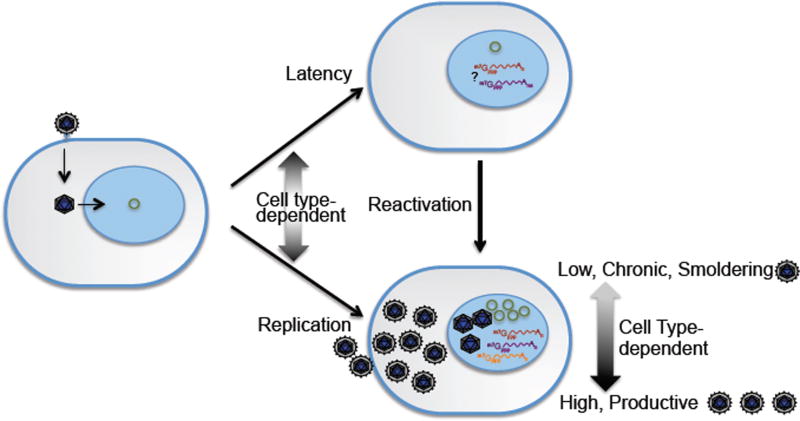
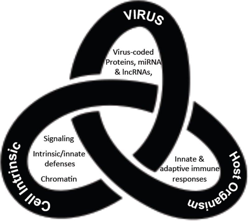
The aphorism “herpes is forever”, reflects the long-appreciated fact that herpesvirus infections cannot yet be cured. Herpesviruses are ancient viruses that persist by establishing life-long latent infections: the maintenance of viral genomes in the absence of active replication and progeny virus production (Fig.1). Critical to survival of the virus is the capacity to reactivate replication from latency in response to changes in the host. The virus-host interactions governing the entry into, maintenance of and exit from latency have yet to be fully defined. However, it is clear that the control of latency is multifaceted, involving the intrinsic responses, innate and adaptive immune response, cellular signaling, chromatin remodeling, and viral factors regulating viral replication or host responses to infection. As such, latency resembles a Gordian knot, presenting what has proved to be a highly intertwined and nuanced puzzle (Fig. 2). This review will discuss our current understanding of viral and cellular mechanisms controlling CMV latency in the broader context of herpesvirus latency and viral persistence.
Figure 1. Schematic of CMV latency.
Following entry into the cell and delivery of the genome (green circle) to the nucleus, cell type-specific i defenses, the activity of viral tegument proteins, and as yet undefined virus-host interactions begin to define the pattern of infection: productive, chronic, or latent. Cellular differentiation and other host cues can reactivate virus replication. In productive states of replication, all three classes of viral transcripts are expressed. The transcriptome associated with viral latency is more poorly defined.
Figure 2. The Gordian knot.
CMV latency is a complex and poorly understood process that is controlled by (i) viral factors, (ii) cell type-specific factors in the infected cell, and (iii) innate and adaptive host responses. These three aspects of CMV persistence are inextricably linked. Untangling the knot will require an integrated inquiry into these facets of latency.
HCMV is the prototypical β-herpesvirus, a subfamily of herpesviruses characterized by slow replication and strict tropism for host species but broad tropism for the cell types infected within the host. HCMV is the largest of the human herpesviruses, with a ~230,000 base-pair, linear double-stranded DNA genome encoding at least 170 proteins (1, 2) and possibly as many as 750 proteins (3), as well as many small and long non-coding RNAs. The genome complexity of CMV provides a rich molecular platform, manipulating a multitude of diverse host processes in its capacity to modulate latent, chronic, and productive patterns of infections.
CMV asymptomatically infects a large majority of the world’s population and typically causes disease only in the absence of adequate cellular immunity. The prevalence of CMV ranges from 40–99% of the world’s population, with higher seroprevalence in developing countries (4). Asymptomatic long-term virus shedding in urine, saliva, and genital secretions usually marks the primary infection in healthy individuals (5). CMV establishes life-long persistence during which it replicates at low subclinical levels or reactivates from a latent state sporadically and likely frequently in response to normal biological processes, including the differentiation of monocytes into macrophages (6, 7) or lactation (8). These reactivation events are typically controlled by the immune system and rarely result clinical presentation, although they likely contribute to transmission of the virus. However, infection or reactivation from latency in a host with inadequate or compromised T cell immunity poses a serious disease risk. Indeed, CMV contributes significantly to viral disease following solid organ or stem cell transplantation (9–11). Consequences of CMV disease in the immunocompromised include interstitial pneumonia, gastroenteritis, retinitis, hepatitis, graft failure, and death. CMV further contributes to graft versus host disease and myelosuppression. Antivirals available to control CMV can exacerbate myelosuppression, resulting in leukopenias and increased risk of fungal or bacterial infections following transplantation. Due to these risks, CMV infection is an important factor in the long-term outcome for solid organ and stem cell transplants (9, 12).
CMV also is the leading cause of infectious disease-related congenital infection (13, 14). One in 150 children are born with CMV infection in the U.S., making it more prevalent than Down’s syndrome, spina bifida, fetal alcohol syndrome, or pediatric AIDS. According to the CDC, 1 in 5 children born with a congenital CMV infection will develop a permanent disability. Congenital infections resulting from primary infection of the mother during pregnancy are associated with the greatest risk for severe pathologies, although it is estimated that 75% of congenital CMV infections in the U.S. are due to reactivation or reinfection of women who were seropositive prior to pregnancy (15–17). While CMV may be asymptomatic at birth, congenital CMV may result in mild to severe hearing loss, cognitive impairment, microcephaly, and cerebral palsy. Utah and Connecticut have recently passed laws requiring the education of pregnant women about the risks of CMV and mandating CMV testing in children who fail two newborn hearing tests.
Until recently, CMV persistence in healthy individuals was considered without health consequences. While CMV infection may actually benefit young, healthy individuals by enhancing the immune response to vaccination or infection (18), important long-term costs of viral persistence are beginning to emerge. CMV infection is implicated in heightened inflammatory states and an increased risk of vascular disease, including atherosclerosis (19, 20) and cardiovascular mortality, particularly in older women (>70 years) (21, 22). CMV-specific T cell responses account for 10% of both CD4+ and CD8+ memory T cells in the peripheral circulation of infected individuals (23) and may expand up to 30% in the elderly (24). Effector memory CD4+ and CD8+ T cells specific to CMV epitopes expand with age as naïve cells decrease in numbers (25, 26), yet there is no clear functional consequence for this expansion. The expansion of CMV-specific T cells likely reflects the broad repertoire of responding T cells (23), low-level, repeated antigen exposure from frequent viral reactivation, and superinfection (27). In contrast to long-term persistence of HIV or hepatitis C virus, life-long CMV persistence does not result in T-cell exhaustion (28), perhaps because CMV persistence has periods of latency where antigens are not produced.
CMV remains an intractable health risk because the immune response is not clearing, there is no vaccine, and available antivirals target only cells actively replicating the virus and cannot be used long-term because of toxicity. Developing strategies to target latently infected cells in the absence of disease is critical to controlling or eliminating CMV disease—a goal that requires a fundamental understanding of the molecular basis of latency.
A COMPLEX PERSISTENCE
During primary infection, HCMV infects numerous cell types resulting in a variety of infection states in the host (29). While some cell types support robust viral replication from which the virus eventually may be cleared, other cell types such as endothelial and epithelial cells support a chronic or “smoldering” infection that may result in low-level virus shedding for months to years (Fig. 1). Hematopoietic progenitor cells (HPCs) and cells of the myeloid lineage (e.g., CD14+ monocytes) harbor viral genomes in the absence of active replication, providing latent reservoirs for the virus (30, 31). Our understanding of how cell type affects viral gene expression and establishment of distinct patterns of infection (e.g., replicative, chronic, latent) is limited. Given the diversity of cell types infected, persistence of the virus in the host organism is complex, reflecting an aggregate of infection states—which may occur concurrently and subclinically in the immune competent host. Latency in the healthy host is overlayed by contained bouts of replication to allow transmission. The dependence of infection states on cell type reflects a requirement for specific host environments and factors to support latent, chronic, or replicative states. While some cell type-specific host factors have been identified, much remains to be defined about factors underlying restriction or permissivity to replicative or latent infection states.
LATENT RESERVOIRS
In infected persons, CMV genomes have been detected as far back as CD34+ HPCs in the hierarchy of hematopoietic differentiation (30–32). Reactivation of virus replication occurs in hematopoietic cells in response to allogeneic stimulation and differentiation cues (7, 33, 34). Hematopoietic cells have been a primary focus of latency studies; however, there has been no comprehensive study to identify cellular reservoirs of latency in the host, and other latent reservoirs cannot be ruled out. Defining cellular reservoirs for latent HCMV and understanding the molecular program of HCMV latency is challenging for a number of reasons. First, latent genomes are maintained in few cells and at low copy numbers—estimated at 1 genome in every 104 to 105 mononuclear cells from healthy G-CSF stimulated donors (35). Second, viral genes are expressed at low levels (if at all) in latently infected cells. Third, given the nature of the hematopoietic reservoir for HCMV latency, HCMV latency may not be represented by a single, static state. HCMV latency may resemble latency of γ-herpesviruses such as EBV, which establishes multiple programs of latency characterized by distinct viral protein and RNA expression profiles in different cell populations (36). As such, model systems are needed to dissect CMV infection states in specific cell types.
In vitro model systems for studying latency
The species restriction of HCMV to humans limits the majority of latency studies to in vitro models. Primary human hematopoietic cell models have been the gold standard, but have a high potential for aberrant ex vivo differentiation and proliferation in culture. A further complication to understanding the latent program has been the multitude of culture systems used to study latency. One broadly used yet nonstandardized approach with regards to media and cytokine composition is stromal-free culture systems with exogenously supplied cytokine cocktails designed to either maintain CD34+ phenotypes or promote differentiation down a particular lineage (37–41). While the ease of this approach is appealing, cytokine concentrations and combinations can result in aberrant differentiation or expansion of cells that have little physiological relevance to function in vivo. In an attempt to closely mimic HPC maintenance and differentiation in vivo, clonal stromal cell lines selected for their ability to recapitulate a physiologically relevant hematopoietic microenvironment in vitro have been adopted for CMV latency studies (42–44). Cultured stromal cell clones secrete a physiologically balanced milieu of cytokines to support HPC differentiation and stem cell self-renewal that can subsequently support serial, long-term hematopoietic reconstitution in an animal following xenotransplantation (45, 46). HPC co-culture with stromal cells support HCMV latency (42, 47–51), and importantly, studies using this system are recapitulated in humanized mice (49, 52). This system has been used to determine the frequency of cells reactivating, providing a means to quantitatively compare latency and reactivation between virus strains (47), viruses containing specific gene disruptions (47, 50, 53, 54), or different hematopoietic subpopulations (42).
Primary cells used for studying latency have largely consisted of CD34+ HPCs (42, 48, 55–57), granulocyte-macrophage progenitor cells (GMPs) (58–60), and CD14+ monocytes (33, 40, 61–63). While primary cells may be the most relevant system for studying latency, the availability, cost, and donor variability presents a significant challenge. Cell line models including the THP-1 cells (64–66), the CD34+ Kasumi 3 leukemic cell line (67) and embryonic stem cell lines (68) have been demonstrated to harbor latent genomes and can be stimulated to reactivate viral gene expression. While cell lines offer the attractive advantage of homogeneity and availability, their application is since cell lines infected prior to differentiation reactivate inefficiently.
Infection of primary human HPCs is highly variable (ranging between 5 and 50% of the cells depending on the donor, cell population composition, and experiment). Therefore, using pure populations of infected cells or at a minimum normalizing for input genomes in mixed populations of infected and uninfected cells is critical when making comparisons between different virus strains, mutant viruses, or when analyzing host responses. Expression of a fluorescent protein from the viral genome is a useful strategy to mark infection, allowing for isolation of pure populations of infected cells by fluorescent-activated cell sorting. Marker expression (e.g. GFP) from an SV40 early promoter driven cassette engineered into an intergenic region is silenced between 3–5 days following infection and will be expressed again following reactivation. This is a bona fide marker for infection since all HPCs receiving virus (positive for tegument protein) will express GFP whereas the GFP/tegument-negative population does not contain viral genomes or support reactivation (48).
In vivo models systems for studying latency
A major barrier to understanding CMV latency has been the lack of an animal model. Due to the species restriction of CMV, humanized mice represent the only approach for studying the human virus in vivo. NOD-scidIL2Rγc null (huNSG) mice engrafted with human CD34+ HPCs and infused with CMV-infected fibroblasts support reactivation from latency in response to stem cell mobilization by granulocyte-colony stimulating factor (G-CSF) (69). HuNSG mice disseminate infected cells to multiple tissues and recapitulate observations made in bone marrow transplant patients receiving G-CSF-mobilized CD34+ HPCs from CMV-infected donors (70). Humanized mice provide a method for investigating HCMV latency and reactivation in the context of an organism with aspects of the immune response in tact.
LATENT PROGRAMS of VIRAL GENE EXPRESSION
Viral gene expression during the replicative cycle of herpesviruses, including CMV, is described as an orderly cascade beginning with the activation of immediate early (IE) genes with subsequent activation of early and late genes depending on the successful completion of the previous phase (71). IE genes (e.g., IE1 and IE2) are activated by cellular factors and viral tegument components in the absence of de novo synthesis of viral proteins. IE gene products influence the cellular environment for replication and transactivate early genes, which are expressed independently of viral DNA synthesis. Early-late and late genes are characterized by their increasing dependence on viral genome synthesis for their expression. This cascade of gene expression has been defined for productive infection in fibroblasts; it is less clear how the viral transcriptome is regulated in other cell types and during other patterns of infection.
Viral gene expression following infection of cells that will ultimately establish a latent infection is controversial. It is possible that the viral genome is epigenetically silenced immediately following nuclear entry of a cell permissive for latency and no viral genes are expressed. However, studies of primary cells infected in vitro as well as the detection of viral transcripts in cells derived from seropositive individuals suggests that limited viral gene expression occurs in latency (40, 42, 48, 72). In vitro studies have demonstrated broad expression of a number of IE and early genes, including a number of genes that have either no known function or that are dispensable for replication in fibroblasts. It is not clear how these genes contribute to the establishment of latency or if sustained gene expression is required for the maintenance of latency. Since studies to date have been carried out in batch using heterogeneous populations of hematopoietic cells, it is not yet clear if there is one or several different viral transcriptomes associated with latency. The conspicuous absence of “lytic” cycle gene transcripts, such as those encoding structural proteins or envelope glycoproteins, in latent transcriptomes is reassuring that the latent transcriptomes are not skewed by a minority of productively infected cells (40, 42, 48, 72).
CMV latency is likely achieved from the collusion of replication-suppressive viral factors (73) and a non-permissive cellular environment that promotes epigenetic silencing of the viral genome (41, 64, 74) to inhibit “lytic” viral gene expression (38). Latently infected CD14+ or CD34+ cells from healthy seropositive individuals express genes from the UL133-UL138 locus (47, 49, 54), UL144 (75), the latent unique nuclear antigen (LUNA) (76–78), the US28 viral G-coupled receptor (79, 80), the latency-associated viral homolog of IL-10 (LAcmvIL-10) encoded by UL111A (81, 82), a small form of the UL123-encoded IE1 protein (IE1×4) (83), a latency-associated transcript originating from a distal promoter in the MIE region (58, 84), and 2.7-kb and 4.9-kb long non-coding (lnc) RNAs (40). While these genes are expressed during both latent and lytic infection, during the latent state they are expressed in the absence of broader lytic viral gene expression.
The transcriptome associated with reactivation is equally as challenging, again due to the heterogeneity of the cell populations harboring virus and the low frequency at which latently infected cells reactivate (42, 47). Expression of the IE1 or IE2 genes has been used as a marker of reactivation, and it is presumed that the classic cascade of gene expression and progeny virus production will follow. However, the latent state is more dynamic than originally appreciated, and MIE expression alone does not guarantee full reactivation and production of progeny virus. Latently infected cells from healthy CMV carriers sporadically express IE genes but fail to produce progeny virus when stimulated ex vivo (34) and “blips” of IE gene expression are observed in mice latently infected in the absence of reactivation and production of progeny virus (85). Furthermore, ectopic expression of the IE1–72kDa and IE2–86kDa proteins is not sufficient to drive viral genome synthesis or infectious progeny production in infected THP-1 cells (66). Similar to CMV, broad gene expression has been detected in neurons latently infected with HSV or VZV in the absence of replication (86–89). Disruption of a neuron-specific miRNA targeting ICP0 of HSV-1 increases ICP0 expression in neurons without inducing reactivation (90). Further, IE, early and late viral gene expression is induced by host signaling (e.g., phosphoinositide 3-kinase, PI3K, and cJun N-terminal kinase, JNK) in initial phase of HSV-1reactivation in neurons (91–93). Yet, this first phase of gene expression in reactivation is not sufficient for reactivation and histone deymethylation is required to drive HSV-1 gene expression beyond a threshold to support productive reactivation (93, 94). Therefore, limited measures of viral gene expression, IE or otherwise, cannot serve as a proxy for authentic viral reactivation. Certainly, if the replicative cycle was induced each time MIE genes are expressed, latency would be difficult if not impossible to maintain. While the expression of replicative cycle transactivators (e.g., IE2) may sporadically occur during latency, their expression may have to be sustained beyond a biologically meaningful threshold in order to drive infection towards reactivation and the production of virus progeny.
VIRAL GENOMES
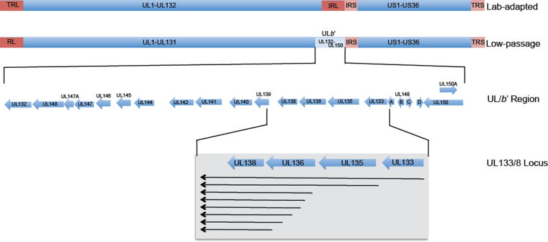
The propagation of viruses in cultured cells results in selection of more highly replicative and cytopathic viruses. As such, laboratory-adapted virus strains are variants that do not reflect the viruses circulating in the human population and in many cases have lost replication-suppressive functions (41, 47, 57, 95). Indeed, laboratory-adapted strains of CMV, such as AD169 and Towne, contain a number of genomic rearrangements due to passage in cultured fibroblasts and replicate with increased kinetics and produce higher yields compared with clinical isolates. The most notable rearrangement is the loss of the ULb’ region (Fig. 2), whose existence was first appreciated in 1990 when the first low-passage strain was sequenced (96). The ULb’ region of the genome is comprised of approximately 15-kilobase pairs encompassing UL132-UL150 that is repeatedly lost upon serial passage of the virus in fibroblasts to acquire laboratory-adapted strains of the virus (Fig. 3) (1, 96). That ULb’ sequences are dispensable for virus replication in cultured fibroblasts suggests that these genes are required for infection in other cell types or persistence in the host. Indeed, enhanced replication and tropism is documented for low-passage strains containing the ULb’ region relative to lab-adapted strains (97–101). Furthermore, the individual gene functions described for ULb’ genes include functions important in tropism or immune evasion. UL146 and UL147 encode putative αCXC chemokines that enhance infection of polymononuclear cells (99, 102). UL141 prevents the killing of infected cells by downregulating natural kill cell-activating ligands CD155 (103), CD112 (in cooperation with US2) (104), and Trail death receptors (105) by sequestering receptors in the lumen of the endoplasmic reticulum. UL144 is related to the tumor necrosis factor receptor (TNFR) superfamily and activates NFκB, partially overcoming an IE2–86kDa-mediated NFκB block (75, 106). UL135 and UL136 are required for post-entry tropism in endothelial cells (52, 107, 108). Finally, the UL128-UL131A gene locus adjoins the ULb’ region and, while present, it is typically mutated in laboratory-adapted strains. Mutations in UL128-UL131A result in inefficient entry into monocytes, endothelial, and epithelial cells (109–111). Thus, genes spanning UL128-UL150 encode a number of functions to mediate dissemination and survival of specialized cells in the infected host.
Figure 3. The CMV genome, UL/b’, and UL133/8 locus.
Low-passage strains retain a ~15-kb region of the genome termed UL/b’ that is lost during the serial passage of the virus in fibroblasts. The UL/b’ region encodes 19–20 ORFs, many of which have no defined function. The UL133/8 locus coordinates the expression of 4 genes from a series of polycistronic transcripts. The UL133/8 proteins have roles in latency and reactivation in CD34+ HPCs, as well as for replication in endothelial cells.
The ULb’ region also encodes functions important to latency (41, 47, 49, 112, 113). Low-passage strains (Toledo, FIX/VR1814 or TB40/E) are restricted in their gene expression and replication in CD34+ HPCs in the absence of a reactivation stimulus (47). In contrast, laboratory-adapted strains (AD169 and Towne) express viral genes and produce progeny virus in the absence of a reactivation stimulus, suggesting a failure to support a latent infection in CD34+ HPCs. The capacity of laboratory strains to establish latency is controversial. Previous reports (60, 72, 81) and well as more recent publications (37, 41, 68, 114) have carried out latency studies using laboratory-adapted strain (AD169 or TownvarRIT3) infection in HPCs (CD34+ or CD33+ GMPs). While few side-by-side comparisons have been made, the baseline replication of a laboratory-adapted strain (in the absence of a reactivation stimulus) is 10- to 100-fold greater than that of low-passage strains in THP-1 (68) and CD34+ HPCs (47), respectively. Thus, laboratory-adapted strains replicate with increased efficiency in cell-culture models of latency. Further, THP-1 or CD34+ HPCs infected low-passage strains are more refractory to induction of IE gene expression by an inhibitor of histone deacetylation than AD169 laboratory-adapted strains (41, 113). This restriction is attributed in part to UL138 (113). Further, disruption of ULb’ genes results in higher levels of replication in CD34+ HPCs (47, 49, 53, 54, 115, 116). Together these findings support a role for ULb’ genes in regulating viral replication and latency.
ANATOMY of a LOCUS –VIRAL DETERMINANTS of LATENCY
The UL133-UL138 gene locus (herein referred to as UL133/8) located within the ULb’ region of the HCMV genome encodes four genes regulating latency (47, 112) (Fig. 3). UL138, the gene at the 3’-most end of this locus, was first appreciated for the capacity to promote viral latency or suppress viral replication as its disruption results in increased yields in fibroblasts and in CD34+ HPCs in the absence of a reactivation stimulus (loss of latency phenotype) (47). UL138 is encoded on a series of unspliced, 3’ co-terminal RNAs that initiate as far upstream as UL133 (53, 54, 117). Therefore, the locus includes UL133, UL135, UL136, and UL138. UL134 and UL137 are also encoded within this region but on the opposite strand of the genome and it is not clear yet if they are bona fide open reading frames. The UL133/8 genes all encode cytosolic, membrane-associated proteins that distribute within the secretory pathway. UL135 and UL136 encode multiple protein isoforms, some of which are not membrane-associated (50, 52). The coordinated expression of the UL133/8 genes (50, 117), the physical interaction between proteins (115), and the collective roles of the proteins in latency (49, 53, 54, 115) suggest that the function of each UL133/8 gene must be considered in the context of the entire locus.
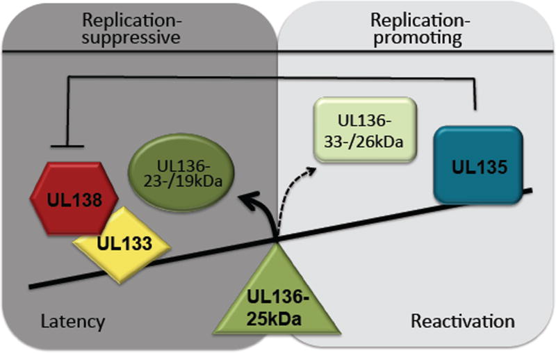
While the UL133/8 locus as a whole is suppressive to replication in CD34+ HPCs (49), a more complex role in latency and reactivation emerges when considering the disruption of individual UL133/8 locus genes. Like UL138, UL133 is suppressive to replication (115); disruption of either results in a loss of latency in CD34+ HPCs and a modest increase in yields in fibroblasts. By contrast, disruption of UL135 alone results in virus that is defective for reconstitution of infection from the transfection of infectious genomes into fibroblasts (50), indicating a role for UL135 in overcoming suppression imposed by other genes in the locus (Fig. 4). Indeed, the defect associated with disruption of UL135 is largely overcome by disruption of UL138 but not UL133 or UL136. Further, because the UL135-mutant defect is largely ameliorated in infection from a virus stock, UL135 may be required to initiate replication from a small number of genomes in the absence of virion proteins, a situation analogous to reactivation from latency. In CD34+ HPCs, the UL135-mutant virus is defective in reactivation even at multiplicities where the virus exhibits no defect for replication in fibroblasts and even when UL138 is disrupted. These findings indicate a greater requirement for UL135 for replication in CD34+ HPCs relative to fibroblasts and a role for UL135 in reactivation beyond overcoming UL138-mediated suppression. The suppressive affect of other UL133/8 genes may impose greater restrictions in CD34+ HPCs than are apparent in fibroblasts in the absence of UL135. The antagonism between UL135 (replication-activating) and UL133/8 replication-suppressive genes suggests that UL133/8 proteins comprise a molecular switch balancing states of latency and replication (Fig. 4).
Figure 4. A model for synergistic and antagonistic interactions between UL133/8 proteins in regulating latency.
UL133 and UL138 promote viral latency. UL138-mediated suppression of virus replication is overcome in part by replication-promoting activity of UL135 for reactivation. UL136 isoforms synergize and antagonize one another in regulating the level of virus replication in CD34+ HPCs. The UL136 33- and 26-kDa isoforms promote replication and are required for replication, whereas the 23- and 19-kDa isoforms suppress replication. The 25-kDa isoform has context-dependent roles. The 25-kDa isoform promotes the maintenance of latency in CD34+ HPCs, but is also required for reactivation or dissemination in humanized mice.
Both replication-promoting and -suppressing roles have emerged for UL136. UL136 expresses five protein isoforms from alternate transcription initiation and possibly alternative translation initiation (53). The isoforms are designated based on predicted molecular weights, 33-, 26-, 25-, 23-, and 19-kDa, and differ in their amino-terminal ends. The UL136 protein isoforms are largely dispensable for replication in fibroblasts, but they have unique, antagonistic and synergistic functions in endothelial cells, CD34+ HPCs, and huNSG mice (52, 53). The two large, membrane-associated isoforms (33- and 26-kDa) are required for replication and reactivation, whereas the two small, non-membrane-associated isoforms (23- and 19-kDa) suppress replication in CD34+ HPCs or huNSG mice. Accordingly, disruption of these pairs of isoforms reduce reactivation and replication in the case of the 33-/26-kDa isoforms and enhance replication in the case of the 23-/19-kDa isoforms. These findings suggest antagonistic functions for 33-/26-kDa and 23-/19-kDa isoforms, similar to the phenotypes associated with UL135 and UL138 (Fig. 4). The roles of the UL136 isoforms may be less absolute, since mutant viruses lacking 33-kDa, 26-kDa, or both isoforms are not as restricted in the capacity to reconstitute replication from infectious genomes as UL135-mutant viruses.
Functions of the UL136 25-kDa isoform of UL136 are dependent on infection context (116). Like the other isoforms, it is dispensable for replication in fibroblasts; however, it suppresses reactivation in CD34+ HPCs to maintain latency. In contrast, in huNSG mice, the 25-kDa isoform is required to either stimulate reactivation or disseminate reactivated cells to tissues. A third phenotype appears in endothelial cells; the 25-kDa isoform enhances the replication-promoting properties of the 33- and 25-kDa isoforms. This context-dependent property of the 25-kDa isoform may allow the conserved network of replication-promoting (33- and 26-kDa) and -suppressing (23-/19-kDa) isoforms to be dynamic and respond to changes in the host state. UL136 may constitute a locus within the UL133/8 locus, which functions to tip the balance of infection between replicative (UL135-dominant) and latent (UL138-dominant) states (Fig. 4). The antagonistic or synergistic roles of protein isoforms are poorly understood in biology but represent an important aspect of post-translational control.
VIRUS-HOST INTERACTIONS for LATENCY
Virus-host interactions important for the entrance into, maintenance of, and exit from latency include those that affect cell survival, cell differentiation, chromatin remodeling, host homeostasis and signaling, as well as the immune response to infection (Fig. 6). Herpesviruses contribute to viral latency by preventing cell death or immune clearance of latently infected cells and by altering host pathways to induce an environment with the capacity to support latency and to silence viral gene expression. Roles for CMV UL111A, US28, IE1×4, lncRNAs, and UL133/8 in latency have been defined genetically using in vitro models of latency and from this virus-host interactions important for CMV latency are beginning to emerge (Fig. 6). Given that only some cell types support viral latency, virus-host interactions important to latency are expected to be context-dependent.
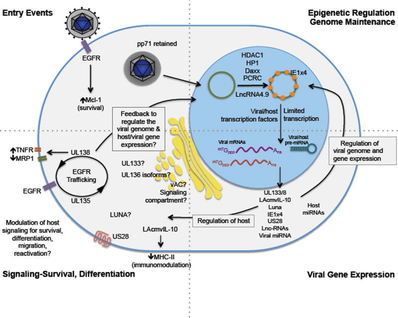
Figure 6. A model of virus-host interactions underlying latency.
CMV latency requires coordinated regulation of viral gene expression, maintenance of the viral genome, and modulation of host processes and responses. Virus entry stimulates EGFR and induces Mcl-1 in monocytes—events that enhance cellular motility and survival. The viral genome is chromatinized upon delivery to the nucleus. Viral transcription is restricted by host epigenetic modulators and virus factors, but the mechanisms are not completely defined. While the latent transcriptome is not well understood, expression of a number of viral genes has been detected and their roles in regulating viral gene expression or host responses are beginning to be defined. Thematically, many CMV latency determinants regulate host signaling as a means by which to control differentiation, survival, and detection by the immune response. The mechanisms by which viral factors regulate host signaling and the effect of these interactions on patterns of infection in various cell types remains to be fully understood.
Regulating viral genomes
Immediately following entry, the pp71 viral tegument protein traffics to the nucleus and targets the host repressor, Daxx, for degradation to prevent silencing of IE gene expression in fibroblasts infected with laboratory-adapted strains (118, 119). However, in THP-1 and CD34+ HPCs, pp71 is retained in the cytoplasm, and Daxx-mediated deacetylation silences the viral genome (41, 64). These findings indicate that early entry events differ in a cell type-dependent manner and impact the establishment of replicative or latent states. While the loss of Daxx in undifferentiated THP-1 cells is not sufficient to break the latent-like state, its loss increases IE2 gene expression following differentiation (120). This finding suggests that additional suppressive forces contribute to the establishment of latency and that gene silencing by Daxx contributes to preventing reactivation. In addition to Daxx, several other epigenetic regulatory mechanisms involving promyelocytic leukemia protein (PML) and Sp100 (localizing with Daxx at nuclear domain 10 or ND10 structures) (120, 121), heterochromatin protein 1 (HP1) (74), histone deacetylase 1 (HDAC1) (122), and the facilitates chromatin transcription (FACT) complex favor the establishment or maintenance of latency and must be overcome for reactivation (51). Furthermore, low-passage strains of CMV encode dominant-replication suppressive functions (41, 47), attributed in part the ULb’ UL138-mediated prevention histone demethylation to maintain viral genome silencing (113).
Long non-coding RNAs have diverse roles in cell biology and virology, including modulating chromatin remodeling. Four long non-coding RNAs of 1.2-, 2.7-, 4.9- and 5.0-kilobases are abundantly expressed during lytic infection (123). LncRNA2.7 and 4.9 have been detected in infected monocytes and CD34+ HPCs (40). The lncRNA2.7 (also referred to as β2.7) protects cells from apoptosis by stabilizing mitochondrial membrane potential (124), but this role of lncRNA2.7 remains to be explored in latency. LncRNA4.9 associates with the MIE promoter and interacts with the polycomb repressive complex (PRC) to enrich repressive chromatin marks in CD14+ monocytes (40). PRC2 increases repressive histone methylation marks on the viral genome in the THP-1 model of latency and may contribute to the establishment or maintenance of latency (125).
Viral microRNAs (miRNAs) function to limit immune detection or to modulate viral gene expression (126). While none of the 24 miRNAs encoded by CMV have demonstrated roles in CMV latency, they have replication-suppressive functions that might aide latent infection, as has been demonstrated for miRNAs derived from the latency-associated transcript of HSV-1 that target ICP0 and ICP4 (127). miR-UL112 targets the CMV MIE transcripts to reduce levels of the IE1–72kDa protein and viral genome synthesis (128, 129), but its role in latency has not been explored.
Host miRNAs expressed in a cell-type specific manner suppress viral gene transcription to maintain latent infection in cell types supporting latency. Three of 5 miRNAs in the hsa-miR-200 cluster target the 3’ untranslated region (UTR) of UL122 encoding the IE2 major transactivator (38). Disruption of the seed sequence in the 3’ UTR of UL122 increases IE2 gene expression and virus replication in Kasumi 3 and CD34+ HPC models of latency. Consistent with a role for these miRNAs in establishing latency, the has-miR-200 cluster is highly expressed in undifferentiated hematopoietic cells that support latency, but these are expressed at lower levels in differentiated cells permissive for virus replication. Similarly, the hsa-miR200 family of miRNAs also reduces the expression of the EBV ZEB1 and ZEB2 host proteins, which repress expression of the EBV Zta protein critical for reactivation (130, 131). Further, neurons express high levels of host miR-138, which targets the 3’ UTR of transcripts encoding the HSV-1 IE protein, ICP0 (90). Collectively, these studies indicate a critical role for cell type-specific miRNAs in suppressing viral gene expression for latency.
In addition to the regulation of viral gene expression, latency requires the maintenance of an episomal viral genome. The mechanisms by which the viral genome is replicated and partitioned to daughter cells during division of latently infected cells are largely open questions. IE1–72kDa interacts with host chromatin and may provide a mechanism by which the genome is tethered to host chromosome for maintenance (132). A small variant of IE1 initiating at the second ATG in exon 4 of IE1 (IE1x4) has been reported to bind terminal repeat sequences to replicate and maintain the genome in CD34+ HPCs through interaction with Sp-1 and topoisomerase IIβ (83).
Modulation of cytokines and antigen presentation
US28 is one of the four HCMV G-protein-coupled receptors that binds multiple host chemokines (133), altering adhesion and migration of monocytes to promote dissemination and viral latency (43, 134). Further, UL111A encodes a homologue of human IL-10 (cmvIL-10) and a latency-associated form of cmvIL-10 (LAcmvIL-10) that is derived from alternative splicing (81). While cmvIL-10 mimics many functional properties of IL-10, LAcmvIL-10 has reduced immunomodulatory capabilities relative to cmvIL-10 and primarily downregulates MHC-II on monocytes and GM-Ps (135). Further, LAcmvIL-10 enhances cell survival and increases cellular IL-10 and CCL8 cytokine secretion by suppressing host miRNA, hsa-miR92a (82). Although a direct role of LAcmvIL-10 in latency has not been demonstrated, the functions described are consistent with a role for LAcmvIL-10 in modulating the immune response to preserve latently infected cells. CMV encodes several other genes that suppress antigen presentation and evade NK recognition (136). Some of these genes have been shown to be important in rhesus models of infection for superinfection, which would also be important evading the immune response early stages of reactivation (27).
Host cell signaling
Entry into or exit from latency presents a challenge in that the virus must “sense” and “respond” to changes in the host to maintain latency or progress towards reactivation. Therefore, it is not surprising that cellular signaling is a linchpin of herpesvirus latency and reactivation. Myeloid progenitor cell differentiation is inextricably linked to reactivation (7, 33, 34, 122). The viral genome, and particularly the MIE region, is regulated (positively or negatively) by a multitude of host transcription factors, which undoubtedly couple viral gene expression to changes in differentiation or activation state in a cell type-specific manner (137). These include Elk-1 (138), NFκB (139–141), YY1 (142), ERF (143), and GATA-2 (144). Through these and other host transcription factor binding sites across the viral genome, the virus can “sense” and “respond” to changes in host signaling to regulate latent and replicative states (75, 144, 145). As an example, infection-mediated changes in host miRNAs in CD34+ HPCs increases expression of GATA-2 and IL-10, which in turn regulates the viral genes, UL144 and LUNA (75, 144).
Epidermal growth factor receptor (EGFR) and its downstream signaling pathways (e.g., PI3K and ERK/MAPK) are homeostatic regulators of cell survival, differentiation, and proliferation. EGFR is also an important regulators of herpesvirus latency and replication . PI3K signaling is stimulated by gB binding to EGFR and viral entry into normal fibroblasts (146, 147) and monocytes (148). These early events in viral replication might set the stage for later replication events and influence the outcome of infection. Activation of EGFR/PI3K during viral entry promotes monocyte motility (148, 149), which may aid the dissemination of latently infected cells. Furthermore, EGFR/PI3K/ERK pathways stimulate expression of myeloid cell leukemia-1 protein (Mcl-1) during entry into myeloid progenitors and monocytes to promote survival of latently infected cells by inhibiting caspase 3 activation (150–152). Following CMV entry into fibroblasts, EGFR is transcriptionally repressed during replication by the induction of Wilm’s tumor factor 1 (WT1) by a yet unidentified early viral gene product (153, 154), suggesting that high levels of EGFR activity impede post entry steps in the replicative cycle. Suppression of EGFR or PI3K with chemical inhibitors (following viral entry) enhances virus replication and reactivation (155), indicating a role for sustained EGFR/PI3K signaling in latency. The importance of EGFR/PI3K signaling for latency extends beyond CMV. Sustained PI3K signaling triggered through the analogous nerve growth factor pathway is required to maintain HSV-1 latency (92). Further, latency membrane protein (LMP)-1 of EBV upregulates EGFR and PI3K pathways through distinct NFκB pathways (156–159) and LMP-2A sustains PI3K/Akt signaling for B-cell survival (160).
During replication in fibroblasts, EGFR is sequestered in an active form in the viral assembly compartment (vAC) (155), a juxtanuclear coalescence of host secretory membranes and the microtubule organizing center contributes to virion maturation (161–163). Transferrin (162, 164, 165) and activated mammalian target of rapamycin (mTOR) (166) also localize to the vAC during infection. Intriguingly, activation of EGFR and mTOR at the vAC is refractory to starvation or stress in fibroblasts (155, 166) that would normally inactivate both kinases. Sequestration of activated signaling molecules at the vAC may insulate signaling from external regulation as a means to control host signaling, maintaining precise protein levels and activities optimal for infection.
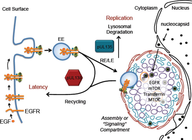
The UL138 latency determinant and its antagonist, UL135, interact with EGFR and oppose each other in regulating surface levels of EGFR (155) (Fig. 5). UL138 enhances surface expression of EGFR, while UL135 targets EGFR for internalization and degradation. The opposing regulation of EGFR suggests a mechanism to explain the antagonism between UL135 and UL138 in regulating viral latency. During latency, UL138 expression sustains levels of EGFR signaling, while UL135 expression during reactivation suppresses signaling to promote replication. While the precise mechanisms by which UL135 and UL138 regulate EGFR are not known, the effects of UL135 and UL138 on surface levels of EGFR suggest that UL135 and UL138 regulate vesicular trafficking, consistent with their association with secretory membranes (49, 54).
Figure 5. UL135 and UL138 antagonistically regulate EGFR surface levels and trafficking.
During CMV infection in fibroblasts, EGFR is sequestered in an activated form at the vAC. UL138 enhances recycling of EGFR to sustain cell surface levels, while UL135 promotes EGFR turnover. EGFR, together with UL135 and UL138, constitute a molecular switch controlling latency. EGFR and downstream PI3K signaling sustain latency. The mechanisms by which UL135 and UL138 regulate EGFR trafficking are not defined.
UL138 affects levels of two other cell-surface receptors, tumor necrosis factor receptor 1 (TNFR1) (167, 168) and multidrug resistance-associated protein 1 (MRP-1) (169). TNFR signaling is an important mediator of proliferation, differentiation, survival, and inflammation, at least in part through the activation of NFκB. Similar to the interaction with EGFR, UL138 increases TNFR1 surface expression (167, 168) and extends its half-life (167) perhaps to potentiate TNFR signaling. While TNF-α signaling stimulates reactivation in CMV latency models (170–172), TNF blockade increases the risk of reactivation of several herpesviruses (173). Regulation of signaling molecules such as TNFR and EGRF likely contributes to the fine-tuning of cellular events required in balancing latency and reactivation. In the case of EGFR, complete downregulation might threaten cell survival or inhibit downstream pathways important for replication, such as mTOR (166, 174–176). In contrast to EGFR and TNFR1, UL138 reduces surface levels of MRP-1, resulting in increased susceptibility of latently-infected cells to killing by vincristine (169). While this work suggests a means by which latently infected cells could be targeted, it is not clear how UL138-mediated regulation of MRP-1 might contribute to latency. The mechanism by which UL138 differentially regulates the surface expression of multiple receptors is not known.
Despite clear importance of host signaling to latency, the key functions precipitated by host signaling in establishment, maintenance, or reactivation from latency are far from clear. UL138 suppresses MIE gene expression by preventing histone demethylation and preventing accumulation of the Golgi-localized CtBP1 protein on the MIEP (113). It is possible that signaling events emanating from UL138-mediated regulation of EGFR or TNFR precipitate changes in epigenetic silencing. Consistent with this idea, JNK stress signaling (stimulated by a decrease in PI3K activity) is essential for the first phase of HSV-1 gene expression during reactivation (93). Further linking signaling and epigenetic changes critical to latency and reactivation, mTOR-mediated phosphorylation of Kruppel-associated box domain-associated protein-1 (KAP1) reduces HP-1, SETDB1, and repressive methylation modification of histones on CMV genomes for reactivation (177). KAP1 plays a similar role in the regulation of KSHV latency and reactivation (178, 179).
SUMMARY and FUTURE ISSUES
CMV persistence has been fine-tuned over millions of years of co-evolution with the human host resulting in a complex Gordian knot that will require an integrated and in-depth inquiry into the viral, cellular, and host components to fully untangle (Fig. 2). The infected host cell contributes a number of context-dependent suppressive activities to silence the genome and assist the establishment of latency. However, latency is not an inevitable default imposed by the cell. It is equally clear that the virus encodes factors to manipulate host pathways to direct both its entry into and exit from latency. A model summarizing known host and viral mechanisms is shown in Figure 6. Like the productive cycle of replication, latency is likely governed by an elaborate system of checks and balances—gate keepers that allow progression into or successful exit from latency to proceed only when specific thresholds of viral or cellular activity are met. Understanding these virus-host interactions and how they regulate transition into and out of latency is critical to ultimately controlling or eliminating latent infections. As we move forward, a number of fundamental questions about the nature of HCMV persistence and mechanisms controlling the latent state remain.
Do other reservoirs of latent CMV exist outside of hematopoietic compartment?
What are the host signaling pathways and viral regulators of signaling important for latency and reactivation?
How do the many distinct mechanisms regulating CMV latency (entry events, epigenetic regulation, viral factors, host signaling) interface (and to what extent) to regulate states of latency and reactivation?
How does expression of the UL133/8 genes and other viral determinants control the transition between latent and replicative states?
What is the latent transcriptome and how is it regulated?
DEFINITION BOXES.
Gordian Knot: a legend of Phrygian Gordium associated with Alexander the Great; a metaphor for an intractable problem that will be solved by a careful approach and thinking outside the box.
Persistence: is long-term maintenance of viral infection, which in the case of herpesviruses is incurable. CMV persistence is comprised of latent, chronic, and replicative states of infection, which need not be mutually exclusive in time.
Latency: refers to a quiescent state of infection during which viral genomes are maintained in the absence of productive virus replication. The extent to which the viral genome is transcribed during latency and the requirement for gene products in the establishment or maintenance of latency is controversial.
Acknowledgments
Research in the author’s laboratory was funded by the National Institutes of Health. Many thanks to James Alwine, Jason Buehler, Katie Caviness, John Purdy, Mike Rak, and Sebastian Zeltzer for helpful discussion and critical feedback. I acknowledge Nat Moorman and Jeremy Kamil for helpful discussions. I regret that page and citation limitations precluded the citation of much work important to latency.
Footnotes
DISCLOSURE STATEMENT
The author is not aware of any affiliations, memberships, funding, or financial holdings that might be perceived as affecting objectivity of this review.
LITERATURE CITED
- 1.Dolan A, Cunningham C, Hector RD, Hassan-Walker AF, Lee L, et al. Genetic content of wild-type human cytomegalovirus. J Gen Virol. 2004;85:1301–12. doi: 10.1099/vir.0.79888-0. [DOI] [PubMed] [Google Scholar]
- 2.Murphy E, Rigoutsos I, Shibuya T, Shenk TE. Reevaluation of human cytomegalovirus coding potential. Proc Natl Acad Sci U S A. 2003;100:13585–90. doi: 10.1073/pnas.1735466100. [DOI] [PMC free article] [PubMed] [Google Scholar]
- 3.Stern-Ginossar N, Weisburd B, Michalski A, Le VT, Hein MY, et al. Decoding human cytomegalovirus. Science. 2012;338:1088–93. doi: 10.1126/science.1227919. [DOI] [PMC free article] [PubMed] [Google Scholar]
- 4.Cannon MJ, Schmid DS, Hyde TB. Review of cytomegalovirus seroprevalence and demographic characteristics associated with infection. Rev Med Virol. 2010;20:202–13. doi: 10.1002/rmv.655. [DOI] [PubMed] [Google Scholar]
- 5.Britt W. Manifestations of human cytomegalovirus infection: proposed mechanisms of acute and chronic disease. Curr Top Microbiol Immunol. 2008;325:417–70. doi: 10.1007/978-3-540-77349-8_23. [DOI] [PubMed] [Google Scholar]
- 6.Smith MS, Goldman DC, Bailey AS, Pfaffle DL, Kreklywich CN, et al. Granulocyte-colony stimulating factor reactivates human cytomegalovirus in a latently infected humanized mouse model. Cell Host Microbe. 2010;8:284–9. doi: 10.1016/j.chom.2010.08.001. [DOI] [PMC free article] [PubMed] [Google Scholar]
- 7.Soderberg-Naucler C, Fish KN, Nelson JA. Reactivation of latent human cytomegalovirus by allogeneic stimulation of blood cells from healthy donors. Cell. 1997;91:119–26. doi: 10.1016/s0092-8674(01)80014-3. [DOI] [PubMed] [Google Scholar]
- 8.Lanzieri TM, Dollard SC, Josephson CD, Schmid DS, Bialek SR. Breast milk-acquired cytomegalovirus infection and disease in VLBW and premature infants. Pediatrics. 2013;131:e1937–45. doi: 10.1542/peds.2013-0076. [DOI] [PMC free article] [PubMed] [Google Scholar]
- 9.Boeckh M, Geballe AP. Cytomegalovirus: pathogen, paradigm, and puzzle. J Clin Invest. 2011;121:1673–80. doi: 10.1172/JCI45449. [DOI] [PMC free article] [PubMed] [Google Scholar]
- 10.Razonable RR, Humar A Practice ASTIDCo. Cytomegalovirus in solid organ transplantation. Am J Transplant. 2013;4(13 Suppl):93–106. doi: 10.1111/ajt.12103. [DOI] [PubMed] [Google Scholar]
- 11.Ljungman P, Hakki M, Boeckh M. Cytomegalovirus in hematopoietic stem cell transplant recipients. Hematol Oncol Clin North Am. 2011;25:151–69. doi: 10.1016/j.hoc.2010.11.011. [DOI] [PMC free article] [PubMed] [Google Scholar]
- 12.Potena L, Valantine HA. Cytomegalovirus-associated allograft rejection in heart transplant patients. Curr Opin Infect Dis. 2007;20:425–31. doi: 10.1097/QCO.0b013e328259c33b. [DOI] [PubMed] [Google Scholar]
- 13.Adler SP, Nigro G, Pereira L. Recent advances in the prevention and treatment of congenital cytomegalovirus infections. Semin Perinatol. 2007;31:10–8. doi: 10.1053/j.semperi.2007.01.002. [DOI] [PubMed] [Google Scholar]
- 14.Cannon MJ. Congenital cytomegalovirus (CMV) epidemiology and awareness. J Clin Virol. 2009;46(46 Suppl):S6–10. doi: 10.1016/j.jcv.2009.09.002. [DOI] [PubMed] [Google Scholar]
- 15.Boppana SB, Rivera LB, Fowler KB, Mach M, Britt WJ. Intrauterine transmission of cytomegalovirus to infants of women with preconceptional immunity. N Engl J Med. 2001;344:1366–71. doi: 10.1056/NEJM200105033441804. [DOI] [PubMed] [Google Scholar]
- 16.Colugnati FA, Staras SA, Dollard SC, Cannon MJ. Incidence of cytomegalovirus infection among the general population and pregnant women in the United States. BMC Infect Dis. 2007;7:71. doi: 10.1186/1471-2334-7-71. [DOI] [PMC free article] [PubMed] [Google Scholar]
- 17.Read JS, Cannon MJ, Stanberry LR, Schuval S. Prevention of mother-to-child transmission of viral infections. Curr Probl Pediatr Adolesc Health Care. 2008;38:274–97. doi: 10.1016/j.cppeds.2008.08.001. [DOI] [PubMed] [Google Scholar]
- 18.Furman D, Jojic V, Sharma S, Shen-Orr SS, CJ LA, et al. Cytomegalovirus infection enhances the immune response to influenza. Sci Transl Med. 2015;7 doi: 10.1126/scitranslmed.aaa2293. 281ra43. [DOI] [PMC free article] [PubMed] [Google Scholar]
- 19.Bentz GL, Yurochko AD. Human CMV infection of endothelial cells induces an angiogenic response through viral binding to EGF receptor and beta1 and beta3 integrins. Proc Natl Acad Sci U S A. 2008;105:5531–6. doi: 10.1073/pnas.0800037105. [DOI] [PMC free article] [PubMed] [Google Scholar]
- 20.Botto S, Streblow DN, DeFilippis V, White L, Kreklywich CN, et al. IL-6 in human cytomegalovirus secretome promotes angiogenesis and survival of endothelial cells through the stimulation of survivin. Blood. 2011;117:352–61. doi: 10.1182/blood-2010-06-291245. [DOI] [PMC free article] [PubMed] [Google Scholar]
- 21.Wang GC, Kao WH, Murakami P, Xue QL, Chiou RB, et al. Cytomegalovirus infection and the risk of mortality and frailty in older women: a prospective observational cohort study. Am J Epidemiol. 2010;171:1144–52. doi: 10.1093/aje/kwq062. [DOI] [PMC free article] [PubMed] [Google Scholar]
- 22.Simanek AM, Dowd JB, Pawelec G, Melzer D, Dutta A, Aiello AE. Seropositivity to cytomegalovirus, inflammation, all-cause and cardiovascular disease-related mortality in the United States. PLoS One. 2011;6:e16103. doi: 10.1371/journal.pone.0016103. [DOI] [PMC free article] [PubMed] [Google Scholar]
- 23.Sylwester AW, Mitchell BL, Edgar JB, Taormina C, Pelte C, et al. Broadly targeted human cytomegalovirus-specific CD4+ and CD8+ T cells dominate the memory compartments of exposed subjects. J Exp Med. 2005;202:673–85. doi: 10.1084/jem.20050882. [DOI] [PMC free article] [PubMed] [Google Scholar]
- 24.Hadrup SR, Strindhall J, Kollgaard T, Seremet T, Johansson B, et al. Longitudinal studies of clonally expanded CD8 T cells reveal a repertoire shrinkage predicting mortality and an increased number of dysfunctional cytomegalovirus-specific T cells in the very elderly. J Immunol. 2006;176:2645–53. doi: 10.4049/jimmunol.176.4.2645. [DOI] [PubMed] [Google Scholar]
- 25.Chidrawar S, Khan N, Wei W, McLarnon A, Smith N, et al. Cytomegalovirus-seropositivity has a profound influence on the magnitude of major lymphoid subsets within healthy individuals. Clin Exp Immunol. 2009;155:423–32. doi: 10.1111/j.1365-2249.2008.03785.x. [DOI] [PMC free article] [PubMed] [Google Scholar]
- 26.Wertheimer AM, Bennett MS, Park B, Uhrlaub JL, Martinez C, et al. Aging and cytomegalovirus infection differentially and jointly affect distinct circulating T cell subsets in humans. J Immunol. 2014;192:2143–55. doi: 10.4049/jimmunol.1301721. [DOI] [PMC free article] [PubMed] [Google Scholar]
- 27.Hansen SG, Powers CJ, Richards R, Ventura AB, Ford JC, et al. Evasion of CD8+ T cells is critical for superinfection by cytomegalovirus. Science. 2010;328:102–6. doi: 10.1126/science.1185350. [DOI] [PMC free article] [PubMed] [Google Scholar]
- 28.Nikolich-Zugich J. Ageing and life-long maintenance of T-cell subsets in the face of latent persistent infections. Nat Rev Immunol. 2008;8:512–22. doi: 10.1038/nri2318. [DOI] [PMC free article] [PubMed] [Google Scholar]
- 29.Hendrix RM, Wagenaar M, Slobbe RL, Bruggeman CA. Widespread presence of cytomegalovirus DNA in tissues of healthy trauma victims. J Clin Pathol. 1997;50:59–63. doi: 10.1136/jcp.50.1.59. [DOI] [PMC free article] [PubMed] [Google Scholar]
- 30.Mendelson M, Monard S, Sissons P, Sinclair J. Detection of endogenous human cytomegalovirus in CD34+ bone marrow progenitors. J Gen Virol. 1996;77:3099–102. doi: 10.1099/0022-1317-77-12-3099. [DOI] [PubMed] [Google Scholar]
- 31.von Laer D, Meyer-Koenig U, Serr A, Finke J, Kanz L, et al. Detection of cytomegalovirus DNA in CD34+ cells from blood and bone marrow. Blood. 1995;86:4086–90. [PubMed] [Google Scholar]
- 32.Maciejewski JP, Bruening EE, Donahue RE, Mocarski ES, Young NS, St Jeor SC. Infection of hematopoietic progenitor cells by human cytomegalovirus. Blood. 1992;80:170–8. [PubMed] [Google Scholar]
- 33.Soderberg-Naucler C, Streblow DN, Fish KN, Allan-Yorke J, Smith PP, Nelson JA. Reactivation of latent human cytomegalovirus in CD14(+) monocytes is differentiation dependent. J Virol. 2001;75:7543–54. doi: 10.1128/JVI.75.16.7543-7554.2001. [DOI] [PMC free article] [PubMed] [Google Scholar]
- 34.Taylor-Wiedeman J, Sissons P, Sinclair J. Induction of endogenous human cytomegalovirus gene expression after differentiation of monocytes from healthy carriers. J Virol. 1994;68:1597–604. doi: 10.1128/jvi.68.3.1597-1604.1994. [DOI] [PMC free article] [PubMed] [Google Scholar]
- 35.Slobedman B, Mocarski ES. Quantitative analysis of latent human cytomegalovirus. J Virol. 1999;73:4806–12. doi: 10.1128/jvi.73.6.4806-4812.1999. [DOI] [PMC free article] [PubMed] [Google Scholar]
- 36.Price AM, Luftig MA. To be or not IIb: a multi-step process for Epstein-Barr virus latency establishment and consequences for B cell tumorigenesis. PLoS Pathog. 2015;11:e1004656. doi: 10.1371/journal.ppat.1004656. [DOI] [PMC free article] [PubMed] [Google Scholar]
- 37.Coronel R, Takayama S, Juwono T, Hertel L. Dynamics of Human Cytomegalovirus Infection in CD34+ Hematopoietic Cells and Derived Langerhans-Type Dendritic Cells. J Virol. 2015;89:5615–32. doi: 10.1128/JVI.00305-15. [DOI] [PMC free article] [PubMed] [Google Scholar]
- 38.O'Connor CM, Vanicek J, Murphy EA. Host microRNA regulation of human cytomegalovirus immediate early protein translation promotes viral latency. J Virol. 2014;88:5524–32. doi: 10.1128/JVI.00481-14. [DOI] [PMC free article] [PubMed] [Google Scholar]
- 39.Poole E, Reeves M, Sinclair JH. The use of primary human cells (fibroblasts, monocytes, and others) to assess human cytomegalovirus function. Methods Mol Biol. 2014;1119:81–98. doi: 10.1007/978-1-62703-788-4_6. [DOI] [PubMed] [Google Scholar]
- 40.Rossetto CC, Tarrant-Elorza M, Pari GS. Cis and trans acting factors involved in human cytomegalovirus experimental and natural latent infection of CD14 (+) monocytes and CD34 (+) cells. PLoS Pathog. 2013;9:e1003366. doi: 10.1371/journal.ppat.1003366. [DOI] [PMC free article] [PubMed] [Google Scholar]
- 41.Saffert RT, Penkert RR, Kalejta RF. Cellular and viral control over the initial events of human cytomegalovirus experimental latency in CD34+ cells. J Virol. 2010;84:5594–604. doi: 10.1128/JVI.00348-10. [DOI] [PMC free article] [PubMed] [Google Scholar]
- 42.Goodrum F, Jordan CT, Terhune SS, High K, Shenk T. Differential outcomes of human cytomegalovirus infection in primitive hematopoietic cell subpopulations. Blood. 2004;104:687–95. doi: 10.1182/blood-2003-12-4344. [DOI] [PubMed] [Google Scholar]
- 43.Humby MS, O'Connor CM. HCMV US28 is important for latent infection of hematopoietic progenitor cells. J Virol. 2015 doi: 10.1128/JVI.02507-15. [DOI] [PMC free article] [PubMed] [Google Scholar]
- 44.Umashankar M, Goodrum F. Hematopoietic long-term culture (hLTC) for human cytomegalovirus latency and reactivation. Methods Mol Biol. 2014;1119:99–112. doi: 10.1007/978-1-62703-788-4_7. [DOI] [PubMed] [Google Scholar]
- 45.Hogge DE, Lansdorp PM, Reid D, Gerhard B, Eaves CJ. Enhanced detection, maintenance, and differentiation of primitive human hematopoietic cells in cultures containing murine fibroblasts engineered to produce human steel factor, interleukin-3, and granulocyte colony-stimulating factor. Blood. 1996;88:3765–73. [PubMed] [Google Scholar]
- 46.Miller CL, Eaves CJ. Long-term culture-initiating cell assays for human and murine cells. In: Klug CA, Jordan CT, editors. Hematopoietic Stem Cell Protocols. Totowa: Humana Press; 2002. pp. 123–41. [DOI] [PubMed] [Google Scholar]
- 47.Goodrum F, Reeves M, Sinclair J, High K, Shenk T. Human cytomegalovirus sequences expressed in latently infected individuals promote a latent infection in vitro. Blood. 2007;110:937–45. doi: 10.1182/blood-2007-01-070078. [DOI] [PMC free article] [PubMed] [Google Scholar]
- 48.Goodrum FD, Jordan CT, High K, Shenk T. Human cytomegalovirus gene expression during infection of primary hematopoietic progenitor cells: a model for latency. Proc Natl Acad Sci U S A. 2002;99:16255–60. doi: 10.1073/pnas.252630899. [DOI] [PMC free article] [PubMed] [Google Scholar]
- 49.Umashankar M, Petrucelli A, Cicchini L, Caposio P, Kreklywich CN, et al. A novel human cytomegalovirus locus modulates cell type-specific outcomes of infection. PLoS Pathog. 2011;7:e1002444. doi: 10.1371/journal.ppat.1002444. [DOI] [PMC free article] [PubMed] [Google Scholar]
- 50.Umashankar M, Rak M, Bughio F, Zagallo P, Caviness K, Goodrum FD. Antagonistic determinants controlling replicative and latent states of human cytomegalovirus infection. J Virol. 2014;88:5987–6002. doi: 10.1128/JVI.03506-13. [DOI] [PMC free article] [PubMed] [Google Scholar]
- 51.O'Connor CM, Nukui M, Gurova KV, Murphy EA. Inhibition of the Facilitates Chromatin Transcription (FACT) complex reduces transcription from the HCMV MIEP in models of lytic and latent replication. J Virol. 2016 doi: 10.1128/JVI.02501-15. [DOI] [PMC free article] [PubMed] [Google Scholar]
- 52.Caviness K, Bughio F, Crawford LB, Streblow DN, Nelson JA, et al. Complex interplay of the UL136 isoforms balances cytomegalovirus replication and latency. mBio. 2016 doi: 10.1128/mBio.01986-15. in press. [DOI] [PMC free article] [PubMed] [Google Scholar]
- 53.Caviness K, Cicchini L, Rak M, Umashankar M, Goodrum F. Complex Expression of the UL136 Gene of Human Cytomegalovirus Results in Multiple Protein Isoforms with Unique Roles in Replication. J Virol. 2014;88:14412–25. doi: 10.1128/JVI.02711-14. [DOI] [PMC free article] [PubMed] [Google Scholar]
- 54.Petrucelli A, Rak M, Grainger L, Goodrum F. Characterization of a Novel Golgi-localized Latency Determinant Encoded by Human Cytomegalovirus. J Virol. 2009;83:5615–29. doi: 10.1128/JVI.01989-08. [DOI] [PMC free article] [PubMed] [Google Scholar]
- 55.Movassagh M, Gozlan J, Senechal B, Baillou C, Petit JC, Lemoine FM. Direct infection of CD34+ progenitor cells by human cytomegalovirus: evidence for inhibition of hematopoiesis and viral replication. Blood. 1996;88:1277–83. [PubMed] [Google Scholar]
- 56.Sindre H, Tjoonnfjord GE, Rollag H, Ranneberg-Nilsen T, Veiby OP, et al. Human cytomegalovirus suppression of and latency in early hematopoietic progenitor cells. Blood. 1996;88:4526–33. [PubMed] [Google Scholar]
- 57.Zhuravskaya T, Maciejewski JP, Netski DM, Bruening E, Mackintosh FR, St Jeor S. Spread of human cytomegalovirus (HCMV) after infection of human hematopoietic progenitor cells: model of HCMV latency. Blood. 1997;90:2482–91. [PubMed] [Google Scholar]
- 58.Hahn G, Jores R, Mocarski ES. Cytomegalovirus remains latent in a common precursor of dendritic and myeloid cells. Proc Natl Acad Sci U S A. 1998;95:3937–42. doi: 10.1073/pnas.95.7.3937. [DOI] [PMC free article] [PubMed] [Google Scholar]
- 59.Slobedman B, Stern JL, Cunningham AL, Abendroth A, Abate DA, Mocarski ES. Impact of human cytomegalovirus latent infection on myeloid progenitor cell gene expression. J Virol. 2004;78:4054–62. doi: 10.1128/JVI.78.8.4054-4062.2004. [DOI] [PMC free article] [PubMed] [Google Scholar]
- 60.Kondo K, Kaneshima H, Mocarski ES. Human cytomegalovirus latent infection of granulocyte-macrophage progenitors. Proc Natl Acad Sci U S A. 1994;91:11879–83. doi: 10.1073/pnas.91.25.11879. [DOI] [PMC free article] [PubMed] [Google Scholar]
- 61.Taylor-Wiedeman J, Sissons JG, Borysiewicz LK, Sinclair JH. Monocytes are a major site of persistence of human cytomegalovirus in peripheral blood mononuclear cells. J Gen Virol. 1991;72:2059–64. doi: 10.1099/0022-1317-72-9-2059. [DOI] [PubMed] [Google Scholar]
- 62.Minton EJ, Tysoe C, Sinclair JH, Sissons JG. Human cytomegalovirus infection of the monocyte/macrophage lineage in bone marrow. J Virol. 1994;68:4017–21. doi: 10.1128/jvi.68.6.4017-4021.1994. [DOI] [PMC free article] [PubMed] [Google Scholar]
- 63.Smith MS, Bivins-Smith ER, Tilley AM, Bentz GL, Chan G, et al. Roles of phosphatidylinositol 3-kinase and NF-kappaB in human cytomegalovirus-mediated monocyte diapedesis and adhesion: strategy for viral persistence. J Virol. 2007;81:7683–94. doi: 10.1128/JVI.02839-06. [DOI] [PMC free article] [PubMed] [Google Scholar]
- 64.Saffert RT, Kalejta RF. Human cytomegalovirus gene expression is silenced by the Daxx-mediated intrinsic immune defense when model latent infections are established in vitro. J Virol. 2007 doi: 10.1128/JVI.00827-07. [DOI] [PMC free article] [PubMed] [Google Scholar]
- 65.Albright ER, Kalejta RF. Myeloblastic Cell Lines Mimic Some but Not All Aspects of Human Cytomegalovirus Experimental Latency Defined in Primary CD34+ Cell Populations. J Virol. 2013;87:9802–12. doi: 10.1128/JVI.01436-13. [DOI] [PMC free article] [PubMed] [Google Scholar]
- 66.Yee LF, Lin PL, Stinski MF. Ectopic expression of HCMV IE72 and IE86 proteins is sufficient to induce early gene expression but not production of infectious virus in undifferentiated promonocytic THP-1 cells. Virology. 2007;363:174–88. doi: 10.1016/j.virol.2007.01.036. [DOI] [PubMed] [Google Scholar]
- 67.O'Connor CM, Murphy EA. A myeloid progenitor cell line capable of supporting human cytomegalovirus latency and reactivation, resulting in infectious progeny. J Virol. 2012;86:9854–65. doi: 10.1128/JVI.01278-12. [DOI] [PMC free article] [PubMed] [Google Scholar]
- 68.Penkert RR, Kalejta RF. Human embryonic stem cell lines model experimental human cytomegalovirus latency. MBio. 2013;4:e00298–13. doi: 10.1128/mBio.00298-13. [DOI] [PMC free article] [PubMed] [Google Scholar]
- 69.Crawford LB, Streblow DN, Hakki M, Nelson JA, Caposio P. Humanized mouse models of human cytomegalovirus infection. Curr Opin Virol. 2015;13:86–92. doi: 10.1016/j.coviro.2015.06.006. [DOI] [PMC free article] [PubMed] [Google Scholar]
- 70.Hakki M, Goldman DC, Streblow DN, Hamlin KL, Krekylwich CN, et al. HCMV infection of humanized mice after transplantation of G-CSF-mobilized peripheral blood stem cells from HCMV-seropositive donors. Biol Blood Marrow Transplant. 2014;20:132–5. doi: 10.1016/j.bbmt.2013.10.019. [DOI] [PMC free article] [PubMed] [Google Scholar]
- 71.Stinski MF, Meier JL. Immediate-early viral gene regulation and function. In: Arvin A, Campadelli-Fiume G, Mocarski E, Moore PS, Roizman B, et al., editors. Human Herpesviruses: Biology, Therapy, and Immunoprophylaxis. Cambridge: 2007. [PubMed] [Google Scholar]
- 72.Cheung AK, Abendroth A, Cunningham AL, Slobedman B. Viral gene expression during the establishment of human cytomegalovirus latent infection in myeloid progenitor cells. Blood. 2006;108:3691–9. doi: 10.1182/blood-2005-12-026682. [DOI] [PubMed] [Google Scholar]
- 73.Goodrum F, Caviness K, Zagallo P. Human cytomegalovirus persistence. Cell Microbiol. 2012;14:644–55. doi: 10.1111/j.1462-5822.2012.01774.x. [DOI] [PMC free article] [PubMed] [Google Scholar]
- 74.Murphy JC, Fischle W, Verdin E, Sinclair JH. Control of cytomegalovirus lytic gene expression by histone acetylation. Embo J. 2002;21:1112–20. doi: 10.1093/emboj/21.5.1112. [DOI] [PMC free article] [PubMed] [Google Scholar]
- 75.Poole E, King CA, Sinclair JH, Alcami A. The UL144 gene product of human cytomegalovirus activates NFkappaB via a TRAF6-dependent mechanism. Embo J. 2006;25:4390–9. doi: 10.1038/sj.emboj.7601287. [DOI] [PMC free article] [PubMed] [Google Scholar]
- 76.Bego M, Maciejewski J, Khaiboullina S, Pari G, St Jeor S. Characterization of an antisense transcript spanning the UL81-82 locus of human cytomegalovirus. J Virol. 2005;79:11022–34. doi: 10.1128/JVI.79.17.11022-11034.2005. [DOI] [PMC free article] [PubMed] [Google Scholar]
- 77.Keyes LR, Hargett D, Soland M, Bego MG, Rossetto CC, et al. HCMV Protein LUNA Is Required for Viral Reactivation from Latently Infected Primary CD14(+) Cells. PLoS One. 2012;7:e52827. doi: 10.1371/journal.pone.0052827. [DOI] [PMC free article] [PubMed] [Google Scholar]
- 78.Reeves MB, Sinclair JH. Analysis of latent viral gene expression in natural and experimental latency models of human cytomegalovirus and its correlation with histone modifications at a latent promoter. J Gen Virol. 2010;91:599–604. doi: 10.1099/vir.0.015602-0. [DOI] [PubMed] [Google Scholar]
- 79.Beisser PS, Laurent L, Virelizier JL, Michelson S. Human cytomegalovirus chemokine receptor gene US28 is transcribed in latently infected THP-1 monocytes. J Virol. 2001;75:5949–57. doi: 10.1128/JVI.75.13.5949-5957.2001. [DOI] [PMC free article] [PubMed] [Google Scholar]
- 80.Vomaske J, Nelson JA, Streblow DN. Human Cytomegalovirus US28: a functionally selective chemokine binding receptor. Infect Disord Drug Targets. 2009;9:548–56. doi: 10.2174/187152609789105696. [DOI] [PMC free article] [PubMed] [Google Scholar]
- 81.Jenkins C, Abendroth A, Slobedman B. A Novel Viral Transcript with Homology to Human Interleukin-10 Is Expressed during Latent Human Cytomegalovirus Infection. J Virol. 2004;78:1440–7. doi: 10.1128/JVI.78.3.1440-1447.2004. [DOI] [PMC free article] [PubMed] [Google Scholar]
- 82.Poole E, Avdic S, Hodkinson J, Jackson S, Wills M, et al. Latency-associated viral interleukin-10 (IL-10) encoded by human cytomegalovirus modulates cellular IL-10 and CCL8 Secretion during latent infection through changes in the cellular microRNA hsa-miR-92a. J Virol. 2014;88:13947–55. doi: 10.1128/JVI.02424-14. [DOI] [PMC free article] [PubMed] [Google Scholar]
- 83.Tarrant-Elorza M, Rossetto CC, Pari GS. Maintenance and replication of the human cytomegalovirus genome during latency. Cell Host Microbe. 2014;16:43–54. doi: 10.1016/j.chom.2014.06.006. [DOI] [PubMed] [Google Scholar]
- 84.White KL, Slobedman B, Mocarski ES. Human cytomegalovirus latency-associated protein pORF94 is dispensable for productive and latent infection. J Virol. 2000;74:9333–7. doi: 10.1128/jvi.74.19.9333-9337.2000. [DOI] [PMC free article] [PubMed] [Google Scholar]
- 85.Kurz SK, Reddehase MJ. Patchwork pattern of transcriptional reactivation in the lungs indicates sequential checkpoints in the transition from murine cytomegalovirus latency to recurrence. J Virol. 1999;73:8612–22. doi: 10.1128/jvi.73.10.8612-8622.1999. [DOI] [PMC free article] [PubMed] [Google Scholar]
- 86.Giordani NV, Neumann DM, Kwiatkowski DL, Bhattacharjee PS, McAnany PK, et al. During herpes simplex virus type 1 infection of rabbits, the ability to express the latency-associated transcript increases latent-phase transcription of lytic genes. J Virol. 2008;82:6056–60. doi: 10.1128/JVI.02661-07. [DOI] [PMC free article] [PubMed] [Google Scholar]
- 87.Kramer MF, Chen SH, Knipe DM, Coen DM. Accumulation of viral transcripts and DNA during establishment of latency by herpes simplex virus. J Virol. 1998;72:1177–85. doi: 10.1128/jvi.72.2.1177-1185.1998. [DOI] [PMC free article] [PubMed] [Google Scholar]
- 88.Ma JZ, Russell TA, Spelman T, Carbone FR, Tscharke DC. Lytic gene expression is frequent in HSV-1 latent infection and correlates with the engagement of a cell-intrinsic transcriptional response. PLoS Pathog. 2014;10:e1004237. doi: 10.1371/journal.ppat.1004237. [DOI] [PMC free article] [PubMed] [Google Scholar]
- 89.Nagel MA, Choe A, Traktinskiy I, Cordery-Cotter R, Gilden D, Cohrs RJ. Varicella-zoster virus transcriptome in latently infected human ganglia. J Virol. 2011;85:2276–87. doi: 10.1128/JVI.01862-10. [DOI] [PMC free article] [PubMed] [Google Scholar]
- 90.Pan D, Flores O, Umbach JL, Pesola JM, Bentley P, et al. A neuron-specific host microRNA targets herpes simplex virus-1 ICP0 expression and promotes latency. Cell Host Microbe. 2014;15:446–56. doi: 10.1016/j.chom.2014.03.004. [DOI] [PMC free article] [PubMed] [Google Scholar]
- 91.Kim JY, Mandarino A, Chao MV, Mohr I, Wilson AC. Transient reversal of episome silencing precedes VP16-dependent transcription during reactivation of latent HSV-1 in neurons. PLoS Pathog. 2012;8:e1002540. doi: 10.1371/journal.ppat.1002540. [DOI] [PMC free article] [PubMed] [Google Scholar]
- 92.Camarena V, Kobayashi M, Kim JY, Roehm P, Perez R, et al. Nature and duration of growth factor signaling through receptor tyrosine kinases regulates HSV-1 latency in neurons. Cell Host Microbe. 2010;8:320–30. doi: 10.1016/j.chom.2010.09.007. [DOI] [PMC free article] [PubMed] [Google Scholar]
- 93.Cliffe AR, Arbuckle JH, Vogel JL, Geden MJ, Rothbart SB, et al. Neuronal Stress Pathway Mediating a Histone Methyl/Phospho Switch Is Required for Herpes Simplex Virus Reactivation. Cell Host Microbe. 2015;18:649–58. doi: 10.1016/j.chom.2015.11.007. [DOI] [PMC free article] [PubMed] [Google Scholar]
- 94.Liang Y, Quenelle D, Vogel JL, Mascaro C, Ortega A, Kristie TM. A novel selective LSD1/KDM1A inhibitor epigenetically blocks herpes simplex virus lytic replication and reactivation from latency. MBio. 2013;4:e00558–12. doi: 10.1128/mBio.00558-12. [DOI] [PMC free article] [PubMed] [Google Scholar]
- 95.Imperiale MJ, Jiang M. What DNA viral genomic rearrangements tell us about persistence. J Virol. 2015;89:1948–50. doi: 10.1128/JVI.01227-14. [DOI] [PMC free article] [PubMed] [Google Scholar]
- 96.Cha TA, Tom E, Kemble GW, Duke GM, Mocarski ES, Spaete RR. Human cytomegalovirus clinical isolates carry at least 19 genes not found in laboratory strains. J Virol. 1996;70:78–83. doi: 10.1128/jvi.70.1.78-83.1996. [DOI] [PMC free article] [PubMed] [Google Scholar]
- 97.Wang W, Taylor SL, Leisenfelder SA, Morton R, Moffat JF, et al. Human cytomegalovirus genes in the 15-kilobase region are required for viral replication in implanted human tissues in SCID mice. J Virol. 2005;79:2115–23. doi: 10.1128/JVI.79.4.2115-2123.2005. [DOI] [PMC free article] [PubMed] [Google Scholar]
- 98.Brown JM, Kaneshima H, Mocarski ES. Dramatic interstrain differences in the replication of human cytomegalovirus in SCID-hu mice. J Infect Dis. 1995;171:1599–603. doi: 10.1093/infdis/171.6.1599. [DOI] [PubMed] [Google Scholar]
- 99.Hahn G, Revello MG, Patrone M, Percivalle E, Campanini G, et al. Human cytomegalovirus UL131-128 genes are indispensable for virus growth in endothelial cells and virus transfer to leukocytes. J Virol. 2004;78:10023–33. doi: 10.1128/JVI.78.18.10023-10033.2004. [DOI] [PMC free article] [PubMed] [Google Scholar]
- 100.Mocarski ES, Bonyhadi M, Salimi S, McCune JM, Kaneshima H. Human cytomegalovirus in a SCID-hu mouse: thymic epithelial cells are prominent targets of viral replication. Proc Natl Acad Sci U S A. 1993;90:104–8. doi: 10.1073/pnas.90.1.104. [DOI] [PMC free article] [PubMed] [Google Scholar]
- 101.Prichard MN, Penfold ME, Duke GM, Spaete RR, Kemble GW. A review of genetic differences between limited and extensively passaged human cytomegalovirus strains. Rev Med Virol. 2001;11:191–200. doi: 10.1002/rmv.315. [DOI] [PubMed] [Google Scholar]
- 102.Penfold ME, Dairaghi DJ, Duke GM, Saederup N, Mocarski ES, et al. Cytomegalovirus encodes a potent alpha chemokine. Proc Natl Acad Sci U S A. 1999;96:9839–44. doi: 10.1073/pnas.96.17.9839. [DOI] [PMC free article] [PubMed] [Google Scholar]
- 103.Tomasec P, Wang EC, Davison AJ, Vojtesek B, Armstrong M, et al. Downregulation of natural killer cell-activating ligand CD155 by human cytomegalovirus UL141. Nat Immunol. 2005;6:181–8. doi: 10.1038/ni1156. [DOI] [PMC free article] [PubMed] [Google Scholar]
- 104.Hsu JL, van den Boomen DJ, Tomasec P, Weekes MP, Antrobus R, et al. Plasma membrane profiling defines an expanded class of cell surface proteins selectively targeted for degradation by HCMV US2 in cooperation with UL141. PLoS Pathog. 2015;11:e1004811. doi: 10.1371/journal.ppat.1004811. [DOI] [PMC free article] [PubMed] [Google Scholar]
- 105.Smith W, Tomasec P, Aicheler R, Loewendorf A, Nemcovicova I, et al. Human cytomegalovirus glycoprotein UL141 targets the TRAIL death receptors to thwart host innate antiviral defenses. Cell Host Microbe. 2013;13:324–35. doi: 10.1016/j.chom.2013.02.003. [DOI] [PMC free article] [PubMed] [Google Scholar]
- 106.Poole E, Atkins E, Nakayama T, Yoshie O, Groves I, et al. NF-kappaB-mediated activation of the chemokine CCL22 by the product of the human cytomegalovirus gene UL144 escapes regulation by viral IE86. J Virol. 2008;82:4250–6. doi: 10.1128/JVI.02156-07. [DOI] [PMC free article] [PubMed] [Google Scholar]
- 107.Bughio F, Elliott DA, Goodrum F. An endothelial cell-specific requirement for the UL133-UL138 locus of human cytomegalovirus for efficient virus maturation. J Virol. 2013;87:3062–75. doi: 10.1128/JVI.02510-12. [DOI] [PMC free article] [PubMed] [Google Scholar]
- 108.Bughio F, Umashankar M, Wilson J, Goodrum F. Human cytomegalovirus UL135 and UL136 genes are required for post-entry tropism in endothelial cells. J Virol. 2015;89:6536–50. doi: 10.1128/JVI.00284-15. [DOI] [PMC free article] [PubMed] [Google Scholar]
- 109.Adler B, Scrivano L, Ruzcics Z, Rupp B, Sinzger C, Koszinowski U. Role of human cytomegalovirus UL131A in cell type-specific virus entry and release. J Gen Virol. 2006;87:2451–60. doi: 10.1099/vir.0.81921-0. [DOI] [PubMed] [Google Scholar]
- 110.Wang D, Shenk T. Human cytomegalovirus virion protein complex required for epithelial and endothelial cell tropism. Proc Natl Acad Sci U S A. 2005;102:18153–8. doi: 10.1073/pnas.0509201102. [DOI] [PMC free article] [PubMed] [Google Scholar]
- 111.Ryckman BJ, Rainish BL, Chase MC, Borton JA, Nelson JA, et al. Characterization of the human cytomegalovirus gH/gL/UL128-131 complex that mediates entry into epithelial and endothelial cells. J Virol. 2008;82:60–70. doi: 10.1128/JVI.01910-07. [DOI] [PMC free article] [PubMed] [Google Scholar]
- 112.Dutta N, Lashmit P, Yuan J, Meier J, Stinski MF. The Human Cytomegalovirus UL133-138 Gene Locus Attenuates the Lytic Viral Cycle in Fibroblasts. PLoS One. 2015;10:e0120946. doi: 10.1371/journal.pone.0120946. [DOI] [PMC free article] [PubMed] [Google Scholar]
- 113.Lee SH, Albright ER, Lee JH, Jacobs D, Kalejta RF. Cellular defense against latent colonization foiled by human cytomegalovirus UL138 protein. Sci Adv. 2015;1:e1501164. doi: 10.1126/sciadv.1501164. [DOI] [PMC free article] [PubMed] [Google Scholar]
- 114.Cheung AK, Gottlieb DJ, Plachter B, Pepperl-Klindworth S, Avdic S, et al. The role of the human cytomegalovirus UL111A gene in down-regulating CD4+ T-cell recognition of latently infected cells: implications for virus elimination during latency. Blood. 2009;114:4128–37. doi: 10.1182/blood-2008-12-197111. [DOI] [PubMed] [Google Scholar]
- 115.Petrucelli A, Umashankar M, Zagallo P, Rak M, Goodrum F. Interactions between proteins encoded within the human cytomegalovirus UL133-UL138 locus. J Virol. 2012;86:8653–62. doi: 10.1128/JVI.00465-12. [DOI] [PMC free article] [PubMed] [Google Scholar]
- 116.Caviness K, Bughio F, Crawford LB, Streblow DN, Nelson JA, et al. Complex Interplay of the UL136 Isoforms Balances Cytomegalovirus Replication and Latency. MBio. 2016:7. doi: 10.1128/mBio.01986-15. [DOI] [PMC free article] [PubMed] [Google Scholar]
- 117.Grainger L, Cicchini L, Rak M, Petrucelli A, Fitzgerald KD, et al. Stress-Inducible Alternative Translation Initiation of Human Cytomegalovirus Latency Protein pUL138. J Virol. 2010;84:9472–86. doi: 10.1128/JVI.00855-10. [DOI] [PMC free article] [PubMed] [Google Scholar]
- 118.Saffert RT, Kalejta RF. Inactivating a cellular intrinsic immune defense mediated by Daxx is the mechanism through which the human cytomegalovirus pp71 protein stimulates viral immediate-early gene expression. J Virol. 2006;80:3863–71. doi: 10.1128/JVI.80.8.3863-3871.2006. [DOI] [PMC free article] [PubMed] [Google Scholar]
- 119.Woodhall DL, Groves IJ, Reeves MB, Wilkinson G, Sinclair JH. Human Daxx-mediated repression of human cytomegalovirus gene expression correlates with a repressive chromatin structure around the major immediate early promoter. J Biol Chem. 2006;281:37652–60. doi: 10.1074/jbc.M604273200. [DOI] [PubMed] [Google Scholar]
- 120.Wagenknecht N, Reuter N, Scherer M, Reichel A, Muller R, Stamminger T. Contribution of the Major ND10 Proteins PML, hDaxx and Sp100 to the Regulation of Human Cytomegalovirus Latency and Lytic Replication in the Monocytic Cell Line THP-1. Viruses. 2015;7:2884–907. doi: 10.3390/v7062751. [DOI] [PMC free article] [PubMed] [Google Scholar]
- 121.Dag F, Dolken L, Holzki J, Drabig A, Weingartner A, et al. Reversible silencing of cytomegalovirus genomes by type I interferon governs virus latency. PLoS Pathog. 2014;10:e1003962. doi: 10.1371/journal.ppat.1003962. [DOI] [PMC free article] [PubMed] [Google Scholar]
- 122.Reeves MB, MacAry PA, Lehner PJ, Sissons JG, Sinclair JH. Latency, chromatin remodeling, and reactivation of human cytomegalovirus in the dendritic cells of healthy carriers. Proc Natl Acad Sci U S A. 2005;102:4140–5. doi: 10.1073/pnas.0408994102. [DOI] [PMC free article] [PubMed] [Google Scholar]
- 123.Gatherer D, Seirafian S, Cunningham C, Holton M, Dargan DJ, et al. High-resolution human cytomegalovirus transcriptome. Proc Natl Acad Sci U S A. 2011;108:19755–60. doi: 10.1073/pnas.1115861108. [DOI] [PMC free article] [PubMed] [Google Scholar]
- 124.Reeves MB, Davies AA, McSharry BP, Wilkinson GW, Sinclair JH. Complex I binding by a virally encoded RNA regulates mitochondria-induced cell death. Science. 2007;316:1345–8. doi: 10.1126/science.1142984. [DOI] [PubMed] [Google Scholar]
- 125.Abraham CG, Kulesza CA. Polycomb repressive complex 2 silences human cytomegalovirus transcription in quiescent infection models. J Virol. 2013;87:13193–205. doi: 10.1128/JVI.02420-13. [DOI] [PMC free article] [PubMed] [Google Scholar]
- 126.Frappier L. Regulation of herpesvirus reactivation by host microRNAs. J Virol. 2015;89:2456–8. doi: 10.1128/JVI.03413-14. [DOI] [PMC free article] [PubMed] [Google Scholar]
- 127.Umbach JL, Kramer MF, Jurak I, Karnowski HW, Coen DM, Cullen BR. MicroRNAs expressed by herpes simplex virus 1 during latent infection regulate viral mRNAs. Nature. 2008;454:780–3. doi: 10.1038/nature07103. [DOI] [PMC free article] [PubMed] [Google Scholar]
- 128.Grey F, Meyers H, White EA, Spector DH, Nelson J. A human cytomegalovirus-encoded microRNA regulates expression of multiple viral genes involved in replication. PLoS Pathog. 2007;3:e163. doi: 10.1371/journal.ppat.0030163. [DOI] [PMC free article] [PubMed] [Google Scholar]
- 129.Murphy E, Vanicek J, Robins H, Shenk T, Levine AJ. Suppression of immediate-early viral gene expression by herpesvirus-coded microRNAs: implications for latency. Proc Natl Acad Sci U S A. 2008;105:5453–8. doi: 10.1073/pnas.0711910105. [DOI] [PMC free article] [PubMed] [Google Scholar]
- 130.Ellis-Connell AL, Iempridee T, Xu I, Mertz JE. Cellular microRNAs 200b and 429 regulate the Epstein-Barr virus switch between latency and lytic replication. J Virol. 2010;84:10329–43. doi: 10.1128/JVI.00923-10. [DOI] [PMC free article] [PubMed] [Google Scholar]
- 131.Lin Z, Wang X, Fewell C, Cameron J, Yin Q, Flemington EK. Differential expression of the miR-200 family microRNAs in epithelial and B cells and regulation of Epstein-Barr virus reactivation by the miR-200 family member miR-429. J Virol. 2010;84:7892–7. doi: 10.1128/JVI.00379-10. [DOI] [PMC free article] [PubMed] [Google Scholar]
- 132.Mucke K, Paulus C, Bernhardt K, Gerrer K, Schon K, et al. Human cytomegalovirus major immediate early 1 protein targets host chromosomes by docking to the acidic pocket on the nucleosome surface. J Virol. 2014;88:1228–48. doi: 10.1128/JVI.02606-13. [DOI] [PMC free article] [PubMed] [Google Scholar]
- 133.Gao JL, Murphy PM. Human cytomegalovirus open reading frame US28 encodes a functional beta chemokine receptor. J Biol Chem. 1994;269:28539–42. [PubMed] [Google Scholar]
- 134.Vomaske J, Melnychuk RM, Smith PP, Powell J, Hall L, et al. Differential ligand binding to a human cytomegalovirus chemokine receptor determines cell type-specific motility. PLoS Pathog. 2009;5:e1000304. doi: 10.1371/journal.ppat.1000304. [DOI] [PMC free article] [PubMed] [Google Scholar]
- 135.Jenkins C, Garcia W, Godwin MJ, Spencer JV, Stern JL, et al. Immunomodulatory properties of a viral homolog of human interleukin-10 expressed by human cytomegalovirus during the latent phase of infection. J Virol. 2008;82:3736–50. doi: 10.1128/JVI.02173-07. [DOI] [PMC free article] [PubMed] [Google Scholar]
- 136.Powers C, DeFilippis V, Malouli D, Fruh K. Cytomegalovirus immune evasion. Curr Top Microbiol Immunol. 2008;325:333–59. doi: 10.1007/978-3-540-77349-8_19. [DOI] [PubMed] [Google Scholar]
- 137.Stinski MF, Isomura H. Role of the cytomegalovirus major immediate early enhancer in acute infection and reactivation from latency. Med Microbiol Immunol. 2008;197:223–31. doi: 10.1007/s00430-007-0069-7. [DOI] [PubMed] [Google Scholar]
- 138.Caposio P, Luganini A, Bronzini M, Landolfo S, Gribaudo G. The Elk-1 and serum response factor binding sites in the major immediate-early promoter of human cytomegalovirus are required for efficient viral replication in quiescent cells and compensate for inactivation of the NF-kappaB sites in proliferating cells. J Virol. 2010;84:4481–93. doi: 10.1128/JVI.02141-09. [DOI] [PMC free article] [PubMed] [Google Scholar]
- 139.Benedict CA, Angulo A, Patterson G, Ha S, Huang H, et al. Neutrality of the canonical NF-kappaB-dependent pathway for human and murine cytomegalovirus transcription and replication in vitro. J Virol. 2004;78:741–50. doi: 10.1128/JVI.78.2.741-750.2004. [DOI] [PMC free article] [PubMed] [Google Scholar]
- 140.Caposio P, Luganini A, Hahn G, Landolfo S, Gribaudo G. Activation of the virus-induced IKK/NF-kappaB signalling axis is critical for the replication of human cytomegalovirus in quiescent cells. Cell Microbiol. 2007;9:2040–54. doi: 10.1111/j.1462-5822.2007.00936.x. [DOI] [PubMed] [Google Scholar]
- 141.DeMeritt IB, Milford LE, Yurochko AD. Activation of the NF-kappaB pathway in human cytomegalovirus-infected cells is necessary for efficient transactivation of the major immediate-early promoter. J Virol. 2004;78:4498–507. doi: 10.1128/JVI.78.9.4498-4507.2004. [DOI] [PMC free article] [PubMed] [Google Scholar]
- 142.Liu R, Baillie J, Sissons JG, Sinclair JH. The transcription factor YY1 binds to negative regulatory elements in the human cytomegalovirus major immediate early enhancer/promoter and mediates repression in non-permissive cells. Nucleic Acids Res. 1994;22:2453–9. doi: 10.1093/nar/22.13.2453. [DOI] [PMC free article] [PubMed] [Google Scholar]
- 143.Wright E, Bain M, Teague L, Murphy J, Sinclair J. Ets-2 repressor factor recruits histone deacetylase to silence human cytomegalovirus immediate-early gene expression in non-permissive cells. J Gen Virol. 2005;86:535–44. doi: 10.1099/vir.0.80352-0. [DOI] [PubMed] [Google Scholar]
- 144.Poole E, McGregor Dallas SR, Colston J, Joseph RS, Sinclair J. Virally induced changes in cellular microRNAs maintain latency of human cytomegalovirus in CD34 progenitors. J Gen Virol. 2011;92:1539–49. doi: 10.1099/vir.0.031377-0. [DOI] [PubMed] [Google Scholar]
- 145.Chan G, Bivins-Smith ER, Smith MS, Yurochko AD. Transcriptome analysis of NF-kappaB- and phosphatidylinositol 3-kinase-regulated genes in human cytomegalovirus-infected monocytes. J Virol. 2008;82:1040–6. doi: 10.1128/JVI.00864-07. [DOI] [PMC free article] [PubMed] [Google Scholar]
- 146.Johnson RA, Wang X, Ma XL, Huong SM, Huang ES. Human cytomegalovirus up-regulates the phosphatidylinositol 3-kinase (PI3-K) pathway: inhibition of PI3-K activity inhibits viral replication and virus-induced signaling. J Virol. 2001;75:6022–32. doi: 10.1128/JVI.75.13.6022-6032.2001. [DOI] [PMC free article] [PubMed] [Google Scholar]
- 147.Wang X, Huong SM, Chiu ML, Raab-Traub N, Huang ES. Epidermal growth factor receptor is a cellular receptor for human cytomegalovirus. Nature. 2003;424:456–61. doi: 10.1038/nature01818. [DOI] [PubMed] [Google Scholar]
- 148.Chan G, Nogalski MT, Yurochko AD. Activation of EGFR on monocytes is required for human cytomegalovirus entry and mediates cellular motility. Proc Natl Acad Sci U S A. 2009;106:22369–74. doi: 10.1073/pnas.0908787106. [DOI] [PMC free article] [PubMed] [Google Scholar]
- 149.Smith MS, Bentz GL, Smith PM, Bivins ER, Yurochko AD. HCMV activates PI(3)K in monocytes and promotes monocyte motility and transendothelial migration in a PI(3)K-dependent manner. J Leukoc Biol. 2004;76:65–76. doi: 10.1189/jlb.1203621. [DOI] [PubMed] [Google Scholar]
- 150.Reeves MB, Breidenstein A, Compton T. Human cytomegalovirus activation of ERK and myeloid cell leukemia-1 protein correlates with survival of latently infected cells. Proc Natl Acad Sci U S A. 2012;109:588–93. doi: 10.1073/pnas.1114966108. [DOI] [PMC free article] [PubMed] [Google Scholar]
- 151.Chan G, Nogalski MT, Bentz GL, Smith MS, Parmater A, Yurochko AD. PI3K–dependent upregulation of Mcl-1 by human cytomegalovirus is mediated by epidermal growth factor receptor and inhibits apoptosis in short-lived monocytes. J Immunol. 2010;184:3213–22. doi: 10.4049/jimmunol.0903025. [DOI] [PMC free article] [PubMed] [Google Scholar]
- 152.Peppenelli MA, Arend KC, Cojohari O, Moorman NJ, Chan GC. Human cytomegalovirus stimulates the synthesis of select Akt-dependent antiapoptotic proteins during viral entry to promote survival of infected monocytes. J Virol. 2016 doi: 10.1128/JVI.02879-15. [DOI] [PMC free article] [PubMed] [Google Scholar]
- 153.Fairley JA, Baillie J, Bain M, Sinclair JH. Human cytomegalovirus infection inhibits epidermal growth factor (EGF) signalling by targeting EGF receptors. J Gen Virol. 2002;83:2803–10. doi: 10.1099/0022-1317-83-11-2803. [DOI] [PubMed] [Google Scholar]
- 154.Jafferji I, Bain M, King C, Sinclair JH. Inhibition of epidermal growth factor receptor (EGFR) expression by human cytomegalovirus correlates with an increase in the expression and binding of Wilms' Tumour 1 protein to the EGFR promoter. J Gen Virol. 2009;90:1569–74. doi: 10.1099/vir.0.009670-0. [DOI] [PubMed] [Google Scholar]
- 155.Buehler J, Zeltzer SL, Reitsma JM, Petrucelli A, Umashankar M, et al. Opposing Regulation of the EGF Receptor: A molecular switch controlling cytomegalovirus latency and replication. PLoS Pathogens. 2016 doi: 10.1371/journal.ppat.1005655. in press. [DOI] [PMC free article] [PubMed] [Google Scholar]
- 156.Kung CP, Meckes DG, Jr, Raab-Traub N. Epstein-Barr virus LMP1 activates EGFR, STAT3, and ERK through effects on PKCdelta. J Virol. 2011;85:4399–408. doi: 10.1128/JVI.01703-10. [DOI] [PMC free article] [PubMed] [Google Scholar]
- 157.Kung CP, Raab-Traub N. Epstein-Barr virus latent membrane protein 1 modulates distinctive NF- kappaB pathways through C-terminus-activating region 1 to regulate epidermal growth factor receptor expression. J Virol. 2010;84:6605–14. doi: 10.1128/JVI.00344-10. [DOI] [PMC free article] [PubMed] [Google Scholar]
- 158.Miller WE, Earp HS, Raab-Traub N. The Epstein-Barr virus latent membrane protein 1 induces expression of the epidermal growth factor receptor. J Virol. 1995;69:4390–8. doi: 10.1128/jvi.69.7.4390-4398.1995. [DOI] [PMC free article] [PubMed] [Google Scholar]
- 159.Miller WE, Mosialos G, Kieff E, Raab-Traub N. Epstein-Barr virus LMP1 induction of the epidermal growth factor receptor is mediated through a TRAF signaling pathway distinct from NF-kappaB activation. J Virol. 1997;71:586–94. doi: 10.1128/jvi.71.1.586-594.1997. [DOI] [PMC free article] [PubMed] [Google Scholar]
- 160.Portis T, Longnecker R. Epstein-Barr virus (EBV) LMP2A mediates B-lymphocyte survival through constitutive activation of the Ras/PI3K/Akt pathway. Oncogene. 2004;23:8619–28. doi: 10.1038/sj.onc.1207905. [DOI] [PubMed] [Google Scholar]
- 161.Alwine JC. The human cytomegalovirus assembly compartment: a masterpiece of viral manipulation of cellular processes that facilitates assembly and egress. PLoS Pathog. 2012;8:e1002878. doi: 10.1371/journal.ppat.1002878. [DOI] [PMC free article] [PubMed] [Google Scholar]
- 162.Das S, Pellett PE. Spatial relationships between markers for secretory and endosomal machinery in human cytomegalovirus-infected cells versus those in uninfected cells. J Virol. 2011;85:5864–79. doi: 10.1128/JVI.00155-11. [DOI] [PMC free article] [PubMed] [Google Scholar]
- 163.Sanchez V, Greis KD, Sztul E, Britt WJ. Accumulation of virion tegument and envelope proteins in a stable cytoplasmic compartment during human cytomegalovirus replication: characterization of a potential site of virus assembly. J Virol. 2000;74:975–86. doi: 10.1128/jvi.74.2.975-986.2000. [DOI] [PMC free article] [PubMed] [Google Scholar]
- 164.Hook LM, Grey F, Grabski R, Tirabassi R, Doyle T, et al. Cytomegalovirus miRNAs target secretory pathway genes to facilitate formation of the virion assembly compartment and reduce cytokine secretion. Cell Host Microbe. 2014;15:363–73. doi: 10.1016/j.chom.2014.02.004. [DOI] [PMC free article] [PubMed] [Google Scholar]
- 165.Krzyzaniak MA, Mach M, Britt WJ. HCMV-encoded glycoprotein M (UL100) interacts with Rab11 effector protein FIP4. Traffic. 2009;10:1439–57. doi: 10.1111/j.1600-0854.2009.00967.x. [DOI] [PMC free article] [PubMed] [Google Scholar]
- 166.Clippinger AJ, Maguire TG, Alwine JC. Human cytomegalovirus infection maintains mTOR activity and its perinuclear localization during amino acid deprivation. J Virol. 2011;85:9369–76. doi: 10.1128/JVI.05102-11. [DOI] [PMC free article] [PubMed] [Google Scholar]
- 167.Le VT, Trilling M, Hengel H. The Cytomegaloviral Protein pUL138 Acts as Potentiator of TNF Receptor 1 Surface Density to Enhance ULb'-encoded modulation of TNF-{alpha} Signaling. J Virol. 2011 doi: 10.1128/JVI.06005-11. [DOI] [PMC free article] [PubMed] [Google Scholar]
- 168.Montag C, Wagner JA, Gruska I, Vetter B, Wiebusch L, Hagemeier C. The latency-associated UL138 gene product of human cytomegalovirus sensitizes cells to tumor necrosis factor alpha (TNF-alpha) signaling by upregulating TNF-alpha receptor 1 cell surface expression. J Virol. 2011;85:11409–21. doi: 10.1128/JVI.05028-11. [DOI] [PMC free article] [PubMed] [Google Scholar]
- 169.Weekes MP, Tan SY, Poole E, Talbot S, Antrobus R, et al. Latency-associated degradation of the MRP1 drug transporter during latent human cytomegalovirus infection. Science. 2013;340:199–202. doi: 10.1126/science.1235047. [DOI] [PMC free article] [PubMed] [Google Scholar]
- 170.Hummel M, Zhang Z, Yan S, DePlaen I, Golia P, et al. Allogeneic transplantation induces expression of cytomegalovirus immediate-early genes in vivo: a model for reactivation from latency. J Virol. 2001;75:4814–22. doi: 10.1128/JVI.75.10.4814-4822.2001. [DOI] [PMC free article] [PubMed] [Google Scholar]
- 171.Simon CO, Seckert CK, Dreis D, Reddehase MJ, Grzimek NK. Role for tumor necrosis factor alpha in murine cytomegalovirus transcriptional reactivation in latently infected lungs. J Virol. 2005;79:326–40. doi: 10.1128/JVI.79.1.326-340.2005. [DOI] [PMC free article] [PubMed] [Google Scholar]
- 172.Soderberg-Naucler C, Fish KN, Nelson JA. Interferon-gamma and tumor necrosis factor-alpha specifically induce formation of cytomegalovirus-permissive monocyte-derived macrophages that are refractory to the antiviral activity of these cytokines. J Clin Invest. 1997;100:3154–63. doi: 10.1172/JCI119871. [DOI] [PMC free article] [PubMed] [Google Scholar]
- 173.Kim SY, Solomon DH. Tumor necrosis factor blockade and the risk of viral infection. Nat Rev Rheumatol. 2010;6:165–74. doi: 10.1038/nrrheum.2009.279. [DOI] [PMC free article] [PubMed] [Google Scholar]
- 174.Moorman NJ, Cristea IM, Terhune SS, Rout MP, Chait BT, Shenk T. Human cytomegalovirus protein UL38 inhibits host cell stress responses by antagonizing the tuberous sclerosis protein complex. Cell Host Microbe. 2008;3:253–62. doi: 10.1016/j.chom.2008.03.002. [DOI] [PMC free article] [PubMed] [Google Scholar]
- 175.Moorman NJ, Shenk T. Rapamycin-resistant mTORC1 kinase activity is required for herpesvirus replication. J Virol. 2010;84:5260–9. doi: 10.1128/JVI.02733-09. [DOI] [PMC free article] [PubMed] [Google Scholar]
- 176.Clippinger AJ, Maguire TG, Alwine JC. The changing role of mTOR kinase in the maintenance of protein synthesis during human cytomegalovirus infection. J Virol. 2011;85:3930–9. doi: 10.1128/JVI.01913-10. [DOI] [PMC free article] [PubMed] [Google Scholar]
- 177.Rauwel B, Jang SM, Cassano M, Kapopoulou A, Barde I, Trono D. Release of human cytomegalovirus from latency by a KAP1/TRIM28 phosphorylation switch. Elife. 2015;4 doi: 10.7554/eLife.06068. [DOI] [PMC free article] [PubMed] [Google Scholar]
- 178.Chang PC, Fitzgerald LD, Van Geelen A, Izumiya Y, Ellison TJ, et al. Kruppel-associated box domain-associated protein-1 as a latency regulator for Kaposi's sarcoma-associated herpesvirus and its modulation by the viral protein kinase. Cancer Res. 2009;69:5681–9. doi: 10.1158/0008-5472.CAN-08-4570. [DOI] [PMC free article] [PubMed] [Google Scholar]
- 179.Cai Q, Cai S, Zhu C, Verma SC, Choi JY, Robertson ES. A unique SUMO-2-interacting motif within LANA is essential for KSHV latency. PLoS Pathog. 2013;9:e1003750. doi: 10.1371/journal.ppat.1003750. [DOI] [PMC free article] [PubMed] [Google Scholar]


