Abstract
Our recent clinical trials demonstrate that autologous cell transfer after lymphodepleting chemotherapy can cause the regression of large, vascularized tumors in patients with refractory metastatic melanoma. Eighteen of 35 patients treated with tumor-reactive lymphocyte cultures experienced an objective clinical response (>50% reduction in tumor), including four complete responders. In some patients, tumor regression was accompanied by a large in vivo expansion of the administered antitumor lymphocytes, which persisted in peripheral blood at >70% of total lymphocytes for many months after transfer. The cells capable of mediating tumor regression consisted of heterogeneous lymphocyte populations with high avidity for tumor antigens that were derived from tumor-infiltrating lymphocytes cultured for limited times in vitro. The success of this treatment likely results from the ability to infuse large numbers of activated antitumor lymphocytes into an appropriate host homeostatic environment depleted of regulatory T cells. These studies are elucidating the requirements for successful immunotherapy of patients with advanced metastatic disease and are leading to additional clinical trials with gene-modified lymphocytes.
The realization that cellular immune reactions, mediated primarily by activated T lymphocytes, are responsible for the rejection of allografts and tumors in experimental models has led to multiple attempts to develop effective immunotherapies for the treatment of patients with cancer based on stimulating T cell reactivity against cancer antigens. The discovery of multiple cancer antigens expressed on human tumors has provided a major stimulus to the development of human cancer immunotherapy (1).
Human cancer antigens fall into several categories, including differentiation antigens that are expressed both on the tumor and the cell of origin of the malignancy, cancer-testes antigens expressed on selected epithelial cancers and germ cells, and mutated proteins expressed exclusively on the cancer and not on normal tissues (1, 2). Our group has emphasized the discovery of human cancer-associated antigens that are recognized by tumor-infiltrating lymphocytes (TILs), cells that infiltrate into the stroma of tumors and can be grown in vitro in medium containing IL-2 (3–5). More than two dozen different cancer antigens recognized by human TILs have been described.
Attempts at active immunization to stimulate cellular immune responses have used a variety of cancer antigens and immunizing vectors. Although these approaches can generate immune T cells capable of recognizing antigenic peptides presented on tumor cells, the regression of growing cancers in patients treated with active immunization has been sporadic and rare (6). It appears that the generation of antitumor T cells in cancer-bearing patients may be necessary; it certainly is not sufficient to mediate the regression of established cancers. A variety of factors that limit tumor regression despite the in vivo generation of antitumor T cells have been described and are listed in Table 1.
Table 1. Possible mechanisms of tumor escape from immune destruction.
| Lymphocyte factors |
| Lack of T cell help |
| Insufficient number of antitumor T cells |
| Insufficient avidity of T cells for tumor |
| T cells are “tolerized” |
| Down-regulation of TCR signal transduction |
| Apoptosis of T cells when encountering tumor |
| Inadequate T cell function (cytokines, lysis) |
| T cells cannot enter tumor stroma |
| Inhibition by “suppressor” T cells |
| Tumor factors |
| Tumor cannot activate quiescent precursors |
| Insufficient tumor antigen expression |
| Loss of HLA expression by tumor |
| Tumor produces local immunosuppressive factors |
| Tumor lacks sufficient apoptotic or other cell destruction pathways |
An alternate approach, involving the transfer of immune lymphocytes to cancer-bearing patients, has several theoretical advantages compared to active immunization and can overcome many of the tumor escape mechanisms listed in Table 1 (7). In cell transfer therapies large numbers of selected cells with high avidity for recognition of tumor antigens can be administered. The cells can be activated ex vivo to exhibit antitumor effector function. In vitro testing of the transferred cell populations provides the potential to identify the exact subpopulations and effector functions that are required for cancer regression in vivo. Perhaps most importantly it is possible to manipulate the host before the cell transfer to provide an altered environment for the transferred cells including the elimination of regulatory lymphocytes or lymphocytes that compete with the transferred cells for homeostatic cytokines such as IL-7 and IL-15.
Two clinical trials of cell transfer therapy preceded the current ongoing clinical trial. In the first clinical trial cloned melanoma-reactive T cells, derived from individual starting cells, were adoptively transferred to 13 patients with metastatic melanoma followed by the administration of IL-2 (8). The transferred cells had high reactivity against melanoma antigens. No objective responses were seen in these 13 patients. In the second trial patients with metastatic melanoma received cloned highly avid T cell populations that were transferred into patients after nonmyeloablative lymphodepleting chemotherapy (9). This study was conducted with a phase I design by using increasing doses of cyclophosphamide and fludarabine and increasing doses of IL-2 (Table 2). Fifteen patients were treated, and the final six patients received the maximum doses of cyclophosphamide (60 mg/kg for 2 days) and fludarabine (25 mg/m2 for 5 days) and IL-2 (720,000 units per kg every 8 h) in conjunction with cell transfer. No objective responses were seen in any patient. In both trials persistence of cells was poor and transferred clones could not be detected in peripheral blood by 2 weeks after cell administration.
Table 2. Treatment received by each patient.
| Cloned cells administered, ×10-9
|
||||||
|---|---|---|---|---|---|---|
| Patient | Cyclophosphamide, mg/kg | Fludarabine, mg/M2 | IL-2, units/kg | Source | First cycle | Second cycle |
| 1 | 30 | 25 | — | TIL | 22.4 | 10.4 |
| 2 | 30 | 25 | — | PBL | 21.5 | 16.0 |
| 3 | 30 | 25 | — | PBL | 15.0 | 14.5 |
| 4 | 60 | 25 | — | TIL | 9.3 | 11.0 |
| 5 | 60 | 25 | — | TIL | 4.1 | 6.0 |
| 6 | 60 | 25 | — | PBL | 5.5 | nd* |
| 7 | 60 | 25 | 72,000 | PBL | 11.0 | 11.0 |
| 8 | 60 | 25 | 72,000 | TIL | 6.8 | 7.3 |
| 9 | 60 | 25 | 72,000 | PBL | 3.2 | 1.8 |
| 10 | 60 | 25 | 720,000 | PBL | 2.8 | 5.6† |
| 11 | 60 | 25 | 720,000 | TIL | 11.3 | 14.0 |
| 12 | 60 | 25 | 720,000 | PBL | 0.9 | nd* |
| 13 | 60 | 25 | 720,000 | PBL | 4.9 | 6.7 |
| 14 | 60 | 25 | 720,000 | PBL | 12.6 | nd‡ |
| 15 | 60 | 25 | 720,000 | PBL | 24.2 | nd‡ |
Not done: Patients 6 and 12 received only one infusion of cells because of rapid disease progression
Patient 10 received a mixture of three CD8+ T cell clones in the second infusion cycle with a total cell number of 5.6 × 10⁹ cells
Not done: Patients 14 and 15 received only one infusion of cloned cells, then they were transferred to other treatment protocols
In both trials patients received CD8+ cloned lymphocytes that underwent three in vitro stimulations with OKT3 and IL-2. We hypothesized that the extensive in vitro stimulation and growth required to achieve sufficient numbers of cloned cells for adoptive transfer may have impaired the activity and proliferative potential of these cells. Further, the lack of CD4+ helper cells within the transferred CD8+ cloned populations may also have limited the ability of these cells to be activated in vivo and persist. We thus modified the clinical protocol to administer heterogeneous populations of TILs.
Generation of Highly Avid Antitumor Lymphocytes for Use in Cell Transfer Therapy
Three different techniques have been used to generate highly avid, heterogeneous antigen-reactive cells from resected tumors (10). (i) Resected tumor specimens were enzymatically digested, aliquoted to multiple wells of 24-well plates, and cultured in IL-2. Lymphocyte cultures that grew from individual wells were tested for antitumor reactivity against a broad array of tumor targets. Although wells were started from aliquots of the same single cell suspension, there was substantial variability in the antitumor reactivity and the phenotype of the growing cells. Wells with optimal activity were selected for further expansion and testing. (ii) Resected tumor specimens were cut into 1- to 2-mm3 fragments, and single tumor fragments were placed in each well of a 24-well tissue culture plate in IL-2. Each fragment gave rise to an individual culture, again with highly variable antitumor activity. Multiple patterns of recognition displayed by independent TIL cultures can be generated from fragments of tumor specimens resected from a single patient. (iii) Resected tumor was physically disaggregated by using a Medimachine (Becton Dickinson) equipped with disposal homogenizers. This physical disaggregation resulted in a slurry of single cells and small cell aggregates that were individually cultured in separate wells of 24-well plates and individually tested as described above. In each of these methods attempts were also made to grow tumor lines by culturing in the absence of IL-2. Interestingly, the Medimachine method was the most efficient at generating tumor cells; ≈50% of tumors processed by this approach were successfully converted to melanoma cell lines.
Results of growth of TILs from 90 melanoma biopsies from 62 consecutive HLA-A*0201 patients initiated by these three techniques are shown in Table 3 (10). Lymphocyte cultures were successfully grown from 94.1% of 119 individual enzymatically digested cultures, 69.9% of 710 fragment-derived cultures, and 90.3% of 31 Medimachine cultures. By using these techniques at least one tumor-reactive TIL could be generated from 81% of patients. Starting with an average of 3.4 × 10⁷ TILs preselected for high activity and diversity of antigen recognition, cultures expanded during the 14-day rapid expansion protocol to an average 4.1 × 10¹⁰ cells on the day of infusion, which represented an average 1,320-fold expansion for each culture (range 181- to 2,623-fold), which corresponded to 7–12 cell doublings in the 14 days.
Table 3. Melanoma excisional biopsies processed to establish TILs for potential adoptive cell transfer therapy.
| Active TILs*
|
||||||
|---|---|---|---|---|---|---|
| Total Initiated | + growth† | Cryopreserved‡ | Allogeneic HLA-A2+ (no. screened) | Autologous (no. screened) | Rapidly expanded for treatment§ | |
| Fragments | ||||||
| Patients | 62 | 59 | 23 | 23 (36) | 21 (27) | 6 |
| Tumors | 90 | 83 | 33 | 34 (50) | 34 (44) | 7 |
| Cultures | 710 | 496 | 194 | 87 (302) | 151 (247) | 9 |
| Digests | ||||||
| Patients | 33 | 32 | 11 | 3 (21) | 10 (17) | 3 |
| Tumors | 42 | 41 | 16 | 3 (25) | 11 (21) | 3 |
| Cultures | 119 | 112 | 44 | 16 (68) | 26 (54) | 4 |
| Medimachine | ||||||
| Patients | 17 | 14 | 5 | 4 (9) | 3 (8) | 0 |
| Tumors | 26 | 20 | 8 | 7 (12) | 5 (11) | 0 |
| Cultures | 31 | 24 | 5 | 9 (23) | 13 (21) | 0 |
At least one TIL culture demonstrated specific IFN-γ release when stimulated with indicated tumor cells (defined as IFN-γ release > 100 pg/ml and at least twice the value of any A2- cell lines)
At least one culture expanded sufficiently for screening or cryopreservation (more than ≈5 × 10⁶ cells)
No TILs were screened but at least one culture was cryopreserved at sufficient cell numbers for screening
Eight of the 62 patients included in this data set were treated with the rapidly expanded cultures included in this table. Eight additional patients had an initial biopsy that let to cultures included in this data set, but they were ultimately treated with cells derived from later biopsies or other sources
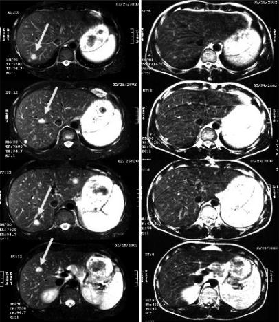
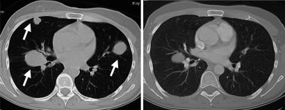
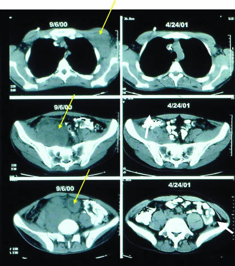
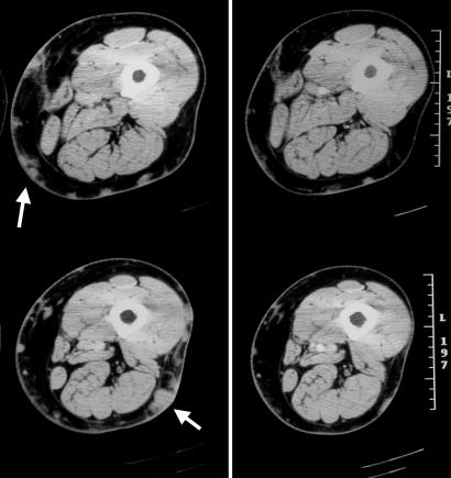
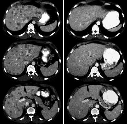
A clinical protocol was conducted in which we administered the nonmyeloablative chemotherapy at the highest doses achieved in the phase I study followed by the administration of heterogeneous TIL populations often containing both CD4+ and CD8+ cells (11). Because TIL populations were grown from tumors and not cloned it was often possible to administer the cells after just a single expansion with anti-CD3 antibody and IL-2. The characteristics of the first 13 patients treated in this trial are shown in Table 4, and the reactivity of cells administered is shown in Table 5 (11). All 13 patients had previously been refractory to treatment with high-dose IL-2, and eight were refractory to aggressive chemotherapy. Six patients showed an objective cancer response, and four additional patients showed mixed responses; three of the responding patients had previously received the same nonmyeloablative chemotherapy with cloned cells plus IL-2 and had not responded. It thus appeared that the combination of the lymphodepleting chemotherapy in conjunction with the more heterogeneous TILs cultured for shorter periods was responsible for the objective response rate in these patients with metastatic melanoma. Examples of responses of cancer at a variety of sites including skin, s.c. tissue, liver, and lung are shown in Figs. 1, 2, 3, 4, 5.
Table 4. Patient demographics, treatments received, and clinical outcomes.
| Treatment
|
||||||||
|---|---|---|---|---|---|---|---|---|
| Patient | Age/sex | Cells infused,* × 10-10 | CD8/CD4 phenotype,† % | Antigen specificity‡ | IL-2, doses | Sites of evaluable metastases | Response duration§ (months) | Autoimmunity |
| 1 | 18/male | 2.3 | 11/39 | Other | 9 | Lymph nodes (axillary, mesenteric, pelvic) | PR¶ (24+) | None |
| 2 | 30/female | 3.5 | 83/15 | MART-1, gp100 | 8 | Cutaneous, subcutaneous | PR (8) | Vitiligo |
| 3 | 43/female | 4.0 | 44/58 | gp100 | 5 | Brain, cutaneous, liver, lung | NR | None |
| 4 | 57/female | 3.4 | 56/52 | gp100 | 9 | Cutaneous, subcutaneous | PR (2) | None |
| 5 | 53/male | 3.0 | 16/85 | Other | 7 | Brain, lung, lymph nodes | NR-mixed | None |
| 6 | 37/female | 9.2 | 65/35 | Other | 6 | Lung, intraperitoneal, subcutaneous | PR (15+) | None |
| 7 | 44/male | 12.3 | 61/41 | MART-1 | 7 | Lymph nodes, subcutaneous | NR-mixed | Vitiligo |
| 8 | 48/male | 9.5 | 48/52 | gp100 | 12 | Subcutaneous | NR | None |
| 9 | 57/male | 9.6 | 84/13 | MART-1 | 10 | Cutaneous, subcutaneous | PR (10+) | Vitiligo |
| 10 | 55/male | 10.7 | 96/2 | MART-1 | 12 | Lymph nodes, cutaneous, subcutaneous | PR¶ (9+) | Uveitis |
| 11 | 29/male | 13.0 | 96/3 | MART-1 | 12 | Liver, pericardial subcutaneous | NR-mixed | Vitiligo |
| 12 | 37/male | 13.7 | 72/24 | MART-1 | 11 | Liver, lung, gallbladder, lymph nodes | NR-mixed | None |
| 13 | 41/female | 7.7 | 92/8 | MART-1 | 11 | Subcutaneous | NR | None |
Each patient was treated with chemotherapy starting 7 days before cell administration, consisting of 2 days of cyclophosphamide at 60 mg per kg of body weight, followed by 5 days of fludarabine at 25 mg/m2. On the day after the final dose of fludarabine, when circulating lymphocyte and neutrophil counts had dropped to <20/mm3, each patient received an i.v. infusion of autologous lymphocytes over ≈30–60 min. After cell infusion, patients received high-dose IL-2 therapy consisting of 720,000 units per kg bolus i.v. infusion every 8 hours to tolerance. Some patients with mixed or responding lesions received an additional course of cell transfer therapy.
T cell cultures for infusion were derived from TILs by minor modifications of established techniques. Multiple cultures were started from each resected melanoma specimen and were screened independently by cytokine secretion assay for recognition of autologous tumor cells (if available) and HLA-A2+ tumor cell lines. TIL cultures that exhibited specific tumor cell recognition were expanded for treatment by using one or two cycles of a rapid expansion protocol with irradiated allogeneic feeder cells, OKT3 (anti-CD3) antibody, and 6,000 units per ml of IL-2
Percent of lymphocyte-gated cells from the infusion sample that stained with each antibody. Values do not add up to 100% if a significant fraction of infused cells was negative or double positive for CD4 and CD8 antigen expression
Antigen specificity was determined by cytokine release assay. Other: recognition of autologous tumor cells but no HLA-A2-restricted epitope derived from MART-1, tyrosinase, tyrosinase-related protein 1(TRP1), TRP2, NY-ESO-1, gp100, MAGE1, or MAGE3
NR, no response; PR, partial response
Microscopic residual focus of disease resected; patient remains free of disease. Patient 1 had a 6-mm brain density that increased to 8 mm at 8 months. After localized stereotactic radiotherapy, the density disappeared
Table 5. Activity and specificity of infused lymphocyte cultures (IFN-γ pg/ml).
| Stimulation by tumor lines (HLA-A2)*
|
Stimulation by 293-A2 gene transfectants†
|
||||||||||||
|---|---|---|---|---|---|---|---|---|---|---|---|---|---|
| Patient | None | 938 (-) | 526 (+) | Autol. (+) | GFP | MART-1 | TRP1 | TRP2 | TYR | NY-ESO-1 | gp100 | MAGE1 | MAGE3 |
| 1 | 204 | 143 | 155 | 5,140 | 155 | 151 | 123 | 137 | 126 | 128 | 110 | 133 | 151 |
| 2 | 9 | 7 | 9,850 | 7,020 | 0 | 6,850 | 53 | 0 | 0 | 65 | 950 | 0 | 0 |
| 3 | 1 | 3 | 20,420 | 615 | 0 | 0 | 0 | 0 | 2 | 0 | 7,100 | 0 | 0 |
| 4 | 0 | 4 | 9,090 | nd | 0 | 0 | 75 | 0 | 0 | 0 | 4,390 | 0 | 0 |
| 5 | 0 | 2 | 19 | 3,535 | 0 | 0 | 0 | 0 | 2 | 0 | 0 | 0 | 0 |
| 6 | 0 | 9 | 798 | 2,620 | 68 | 121 | 94 | 87 | 121 | 104 | 118 | 96 | 104 |
| 7 | 0 | 279 | 5,170 | 5,230 | 0 | 3,790 | 0 | 0 | 0 | 0 | 28 | 23 | 83 |
| 8 | 0 | 849 | 4,990 | nd | 0 | 0 | 0 | 2 | 0 | 0 | 458 | 0 | 0 |
| 9 | 11 | 43 | 2,528 | nd | 12 | >1,361 | 17 | 13 | 17 | 30 | 17 | 14 | 19 |
| 10 | 0 | 0 | 15,150 | nd | 0 | 13,360 | 13 | 0 | 0 | 0 | 20 | 0 | 33 |
| 11 | 0 | 0 | 9,025 | nd | 0 | 12,110 | 10 | 0 | 7 | 65 | 287 | 0 | 70 |
| 12 | 11 | 72 | 1,200 | 1,006 | 126 | 1,970 | 120 | 80 | 96 | 60 | 145 | 88 | 209 |
| 13 | 0 | 234 | 3,545 | 703 | 75 | 1,730 | 0 | 0 | 0 | 0 | 19 | 0 | 0 |
All samples for infusion demonstrated significant cytokine release when stimulated with autologous or HLA-A2+ tumor cell lines. Six infusion samples recognized MART-1, five recognized gp100, and three recognized unidentified antigens expressed by the autologous tumor cell line. There was no correlation between the antigen specificity of the infused cells and objective response, the onset of autoimmunity, or treatment toxicity. The results are combined from three different experiments. Values indicated pg/ml IFN-γ. Significant T cell reactivity was defined by values that are at least two times all controls and >100 pg/ml (bold).
Melanoma cell lines were derived from tumor specimens obtained at the Surgery Branch, National Cancer Institute. If a patient had no autologous melanoma cell line available at the time of the assay, the value is listed as nd (not done)
The human embroyonic kidney cell line 293 was first stably transfected with an expression construct for HLA-A2, then transiently transfected with a panel of plasmids encoding shared melanoma antigens. Efficient transfection was confirmed in each experiment by visual assessment of GFP expression in GFP-transfected cells and in some assays by the stimulation of T cell clones by the appropriate transfectants including MART-1, tyrosinase-related protein (TRP)2, tyrosinase (TYR), NY-ESO-1, and gp100
Fig. 1.
MRI images before treatment (Left) and 2 months after cell transfer (Right) demonstrated the regression of multiple liver metastases (arrows) of patient 11 who had an overall mixed response.
Fig. 2.
Multiple lung metastases (arrows) showed dramatic shrinkage in computed tomographic scans of patient 6 who had an overall partial response to treatment. (Left) Before treatment. (Right) Seven months after cell transfer.
Fig. 3.
Regression of metastases in axillary (Top), pelvic (Middle), and mesenteric (Bottom) nodes was evident in computed tomographic scans taken before treatment (Left) or 7 months after cell transfer (Right) of patient 1, who had an overall partial response.
Fig. 4.
Patient 9 experienced an objective partial response with >95% reduction in cutaneous and s.c. disease (arrows) as seen in computed tomographic scans take before treatment (Left) or 3 months after cell transfer (Right).
Fig. 5.
Patient 31 exhibited an overall complete response with regression of multiple liver metastases as seen in computed tomographic images taken before treatment (Left) or 1 month after cell transfer (Right).
We observed a substantial ability of the transferred cells to survive and grow in the patients for many months after adoptive transfer. Examples of data from six patients are shown in Table 6. Two patients showed pronounced persistence of the administered antitumor T cells at levels of 70–80% of peripheral blood lymphocytes (PBLs) that persisted for >4 months. One of these patients has been followed for >2 years with persistence of >70% of all circulating CD8+ cells of a single Vβ7 clonotype with antitumor activity. We are now updating our experience with the treatment of 35 patients, 18 of whom (51%) have achieved an objective response, including four patients with a complete response. Analysis of T cell persistence in PBL of 25 of these patients showed a statistically significant correlation between the persistence if clonotypes from transferred TIL and objective tumor regression (P. Robbins, personal communication). In this treatment approach the antitumor T cells continue to numerically expand after i.v. injection and thus magnify their antitumor activity. In this way cell transfer therapies differ substantially from chemotherapeutic approaches whose antitumor effects are eliminated shortly after chemotherapy administration.
Table 6. TCR Vβ gene usage in CD8+ cells from TIL and posttreatment PBLs.
| Vβ family, percent of CD8+ cells
|
||||||||||||||||||||||
|---|---|---|---|---|---|---|---|---|---|---|---|---|---|---|---|---|---|---|---|---|---|---|
| Patient | Day* | CD82 | 1 | 2 | 3 | 5a | 5c | 6.7 | 7 | 8 | 9 | 11 | 12 | 13 | 14 | 16 | 17 | 18 | 20 | 21 | 22 | 23 |
| 6 | Pre | 1222 | 6 | 12 | 1 | 3 | 1 | 2 | 3 | 4 | 2 | 3 | 1 | 3 | 5 | 1 | 4 | 0 | 3 | 3 | 2 | 2 |
| TIL | — | 12 | 7 | 0 | 6 | 0 | 2 | 2 | 0 | 0 | 0 | 0 | 2 | 2 | 0 | 5 | 0 | 19 | 1 | 1 | 1 | |
| 8 | 115 | 4 | 11 | 1 | 4 | 0 | 2 | 1 | 4 | 0 | 0 | 1 | 3 | 2 | 1 | 7 | 0 | 10 | 2 | 1 | 0 | |
| 43 | 527 | 3 | 9 | 3 | 1 | 1 | 1 | 3 | 8 | 1 | 1 | 2 | 1 | 13 | 2 | 6 | 0 | 1 | 2 | 2 | 2 | |
| 9 | Pre | 390 | 2 | 4 | 2 | 3 | 1 | 1 | 11 | 12 | 1 | 0 | 1 | 2 | 4 | 0 | nd | 0 | 4 | 2 | 3 | 1 |
| TIL | — | 0 | 1 | 0 | 1 | 1 | 1 | 0 | 0 | 0 | 0 | 14 | 0 | 2 | 0 | nd | 0 | 0 | 0 | 0 | 1 | |
| 7 | 20,150 | 0 | 0 | 0 | 0 | 0 | 0 | 0 | 0 | 0 | 0 | 63 | 0 | 8 | 0 | nd | 0 | 0 | 0 | 0 | 0 | |
| 37 | nd | 0 | 0 | 0 | 1 | 0 | 0 | 0 | 0 | 0 | 0 | 69 | 0 | 6 | 0 | nd | 0 | 0 | 0 | 0 | 0 | |
| 97 | 1,090 | 1 | 0 | 1 | 0 | 1 | 1 | 1 | 0 | 0 | 0 | 82 | 0 | 6 | 0 | 0 | 0 | 0 | 0 | 4 | 0 | |
| 123 | 593 | 1 | 0 | 4 | 1 | 1 | 1 | 1 | 1 | 1 | 0 | 72 | 0 | 8 | 0 | 0 | 0 | 0 | 0 | 4 | 0 | |
| 10 | Pre | 1,092 | 2 | 8 | 3 | 2 | 4 | 3 | 2 | 2 | 2 | 1 | 1 | 3 | 3 | 2 | 3 | nd | 0 | 1 | 2 | 1 |
| TIL | — | 0 | 3 | 1 | 3 | 9 | 5 | 89 | 1 | 11 | 1 | 0 | 7 | 1 | 0 | 2 | nd | 1 | 0 | 0 | 1 | |
| 7 | 11,664 | 0 | 0 | 0 | 0 | 1 | 0 | 97 | 0 | 0 | 0 | 1 | 1 | 0 | 0 | 0 | nd | 0 | 0 | 0 | 0 | |
| 23 | 3,035 | 1 | 1 | 0 | 1 | 3 | 0 | 86 | 0 | 0 | 0 | 7 | 2 | 0 | 0 | 1 | nd | 2 | 0 | 1 | 0 | |
| 30 | 2,873 | 1 | 1 | 0 | 1 | 2 | 1 | 87 | 0 | 0 | 0 | 2 | 0 | 0 | 2 | 0 | 0 | 0 | 0 | 0 | 0 | |
| 159 | 697 | 1 | 1 | 0 | 1 | 3 | 1 | 75 | 1 | 1 | 0 | 2 | 1 | 2 | 3 | 1 | 0 | 1 | 1 | 1 | 1 | |
| 11 | Pre | 853 | 2 | 5 | 3 | 4 | 3 | 1 | 3 | 4 | 3 | 1 | 3 | 6 | 7 | 2 | 11 | 1 | 1 | 4 | 5 | 2 |
| TIL | — | 0 | 2 | 89 | 4 | 4 | 4 | 3 | 1 | 0 | 0 | 1 | 0 | 6 | 0 | 0 | 0 | 0 | 0 | 0 | 3 | |
| 6 | 298 | 0 | 4 | 57 | 1 | 1 | 0 | 0 | 0 | 2 | 2 | 3 | 2 | 14 | 1 | 1 | 2 | 3 | 3 | 3 | 3 | |
| 39 | 81 | 2 | 6 | 3 | 6 | 1 | 1 | 2 | 1 | 0 | 0 | 5 | 3 | 3 | 2 | 32 | 2 | 2 | 7 | 4 | 0 | |
| 12 | Pre | 787 | 5 | 6 | 2 | 3 | 2 | 1 | 5 | 3 | 2 | 2 | 2 | 3 | 3 | 2 | 3 | 0 | 1 | 2 | 3 | 2 |
| TIL | — | 6 | 4 | 4 | 6 | 3 | 5 | 6 | 1 | 4 | 2 | 2 | 6 | 0 | 1 | 5 | 2 | 2 | 4 | 5 | 4 | |
| 6 | 572 | 7 | 3 | 2 | 2 | 2 | 1 | 3 | 2 | 3 | 1 | 1 | 5 | 3 | 2 | 3 | 0 | 1 | 3 | 4 | 2 | |
| 30 | 327 | 9 | 3 | 6 | 1 | 4 | 4 | 2 | 6 | 1 | 1 | 16 | 7 | 4 | 3 | 4 | 1 | 1 | 3 | 3 | 1 | |
| 13 | Pre | 634 | 3 | 6 | 8 | 6 | 5 | 19 | 16 | 3 | 4 | 1 | 2 | 1 | 3 | 3 | 3 | 1 | 5 | 2 | 1 | 4 |
| TIL | — | 1 | 6 | 1 | 3 | 3 | 4 | 5 | 3 | 1 | 0 | 1 | 2 | 2 | 0 | 5 | 0 | 0 | 0 | 50 | 20 | |
| 8 | 400 | 1 | 2 | 1 | 1 | 1 | 1 | 1 | 7 | 0 | 0 | 1 | 0 | 2 | 0 | 3 | 0 | 0 | 1 | 15 | 43 | |
| 15 | nd | 8 | 7 | 5 | 10 | 12 | 11 | 13 | 2 | 2 | 0 | 3 | 2 | 4 | 2 | 3 | 0 | 1 | 1 | 4 | 5 | |
To investigate the function and fate of the transferred T cell populations, TCR expression was examined by using a panel of beta chain variable region (Vβ)-specific antibodies in all patients for whom peripheral blood samples were available at 1 week and ≈1 month postcell transfer. Vβ expression was highly skewed (bold) in five of the six administered TILs, and some of these same Vβ families were also overrepresented in the peripheral blood of the patients at 1 week after cell transfer, suggesting that those T cells in the PBLs were derived from the infused TIL (underlined). Patients 9 and 10 demonstrated a persistent high level engraftment of individual T cell clones. nd, not done.
†Absolute CD8 counts were calculated as the product of the absolute lymphocyte count on the indicated day and the percent of CD8+ cells determined by fluorescence-activated cell sorting analysis.
Pretreatment PBLs (Pre) were harvested before the start of the conditioning chemotherapy. TILs were sampled before infusion. Postinfusion PBL were sampled on the indicated days after cell transfer
Future Directions for the Development of Cell Transfer Immunotherapies
In 1990, we reported studies inserting the gene encoding neomycin phosphotransferase into TILs used for adoptive transfer to monitor the in vivo distribution and survival of the transferred cells (12). Shortly thereafter, pilot studies were performed inserting the gene for tumor necrosis factor into TILs (13). The poor levels of gene transduction available at that time led us to discontinue those studies. However, improvements in gene vector design and transduction techniques now enable the transduction of human T cells at efficiencies routinely in excess of 30%. We have thus begun efforts to genetically modify lymphocytes to increase their antitumor impact by introducing genes encoding cytokines, T cell receptors (TCRs), and antiapoptotic molecules.
The survival of lymphocytes with antitumor activity in vitro depends on the availability of sufficient local concentrations of the growth factor IL-2. In animal models the ability of adoptively transferred cells to mediate tumor destruction depends on the supply of an exogenous source of IL-2 (3, 14). The toxicity of the systemic administration of IL-2 in humans, however, limits the administration of IL-2 to only 2–3 days after cell transfer (15). We thus initiated studies to introduce the gene for IL-2 secretion into TILs, hypothesizing that the local production of IL-2 would obviate the need for its systemic administration. Retroviral vectors encoding human IL-2 were used to transduce peripheral blood mononuclear cells and cloned CD8+ T cells. Cells transduced with these retroviral vectors produced IL-2 and maintained viability after IL-2 withdrawal (16, 17). Upon antigen stimulation, IL-2-transduced lymphocytes proliferated and could be actively grown and maintained without the addition of exogenous IL-2 for >3 months (Fig. 6). Transduced cells maintained specific recognition against melanoma targets. GMP-quality retroviral vectors encoding IL-2 have now been produced, and pilot trials have recently been initiated in patients who are receiving autologous antigen-specific TILs transduced with the gene encoding IL-2.
Fig. 6.
IL-2 gene transduced TILs exhibited enhanced survival in vitro. (Left) TILs transduced with the IL-2 gene linked to yellow fluorescent protein (IL-2YFP, ♦) exhibited IL-2-independent growth and survival compared to control TILs transduced with YFP alone (▪). No difference in growth rate was seen when cells were rapidly expanded with OKT3 in the presence of exogenous IL-2 (Right). In the absence of exogenously added IL-2 (Center), the IL-2-transduced cells increased in number after OKT3 stimulation, whereas the YFP-transduced control cells did not. When cells were initially expanded in OKT3 and IL-2, followed by IL-2 withdrawal (Left), the IL-2-transduced cells increased to greater numbers and remained viable for longer then the control YFP-transduced cells.
It is not always possible to resect sufficient tumor samples from melanoma patients and even when tumor is available, it is not always possible to obtain melanoma-reactive TIL cultures. As a potential alternative to the requirement to establish TIL cultures from tumor we sought methods that could be used to obtain a polyclonal population of T cells with antitumor reactivity by transfer of antigen-specific TCR genes into PBLs. We recently reported a TCR vector encoding an anti-gp100 TCR obtained from the T cell clone R6C12, derived from a melanoma patient's PBLs that were obtained after multiple gp100 vaccinations in vivo (18). After retroviral transduction of PBL, expression of this TCR gene was confirmed by Western blot analysis, immunocytometric analysis, and HLA antigen tetramer staining. Gene transfer efficiencies of >50% into primary lymphocytes were obtained by using a method of prebinding retroviral vectors to cell culture vessels with Retronectin before the addition of lymphocytes. The biological activity of transduced cells was confirmed by cytokine production after coculture with stimulator cells pulsed with gp100 peptides but not with unrelated peptides and had activities similar to highly avid antimelanoma TILs, by recognition of low levels of peptide (<200 pM) and HLA class I-restricted recognition and lysis of melanoma tumor cell lines (Table 7). CD4+ T cells engineered with this anti-gp100 TCR gene were antigen reactive, suggesting CD8-independent activity of the expressed TCR. Nonmelanoma-reactive TIL cultures also developed antimelanoma activity after anti-gp100 TCR gene transfer. In addition, TIL with reactivity against non-gp100 melanoma antigens acquired gp100 reactivity and did not lose the recognition of autologous melanoma antigens after gp100 TCR gene transfer.
Table 7. Recognition of melanoma by PBLs transduced with gp100 TCR.
| Stimulators
|
|||||
|---|---|---|---|---|---|
| Responder cells | None | 526 Mel (A2+), pg/ml IFN-γ released | 624 Mel (A2+), pg/ml IFN-γ released | 888 Mel (A2-), pg/ml IFN-γ released | 938 Mel (A2-), pg/ml IFN-γ released |
| No vector | 9 (12) | 30 (1.4) | 101 (91) | 91 (22) | 28 (40) |
| YFP* | 8 (9.7) | 35 (12) | 56 (18) | 150 (75) | 80 (98) |
| TCR† | 80 (62) | 2,528 (305) | 1,614 (298) | 245 (100) | 63 (72) |
| TIL | 56 (20) | 2,713 (717) | 2,240 (235) | 10 (14) | 21 (30) |
Retroviral vector encoding YFP
Retroviral vector encoding TCR that recognizes gp100:209–217 epitope
This paper results from the Arthur M. Sackler Colloquium of the National Academy of Sciences, “Therapeutic Vaccines: Realities of Today and Hopes for Tomorrow,” held April 1–3, 2004, at the National Academy of Sciences in Washington, DC.
Abbreviations: TIL, tumor-infiltrating lymphocyte; PBL, peripheral blood lymphocyte; TCR, T cell receptor; YFP, yellow fluorescent protein.
References
- 1.Rosenberg, S. A. (2001) Nature 411, 380-384. [DOI] [PubMed] [Google Scholar]
- 2.Rosenberg, S. A. (1999) Immunity 10, 281-287. [DOI] [PubMed] [Google Scholar]
- 3.Rosenberg, S. A., Spiess, P. & Lafreniere, R. (1986) Science 233, 1318-1321. [DOI] [PubMed] [Google Scholar]
- 4.Kawakami, Y., Eliyahu, S., Delgado, C. H., Robbins, P. F., Sakaguchi, K., Appella, E., Yannelli, J. R., Adema, G. J., Miki, T. & Rosenberg, S. A. (1994) Proc. Natl. Acad. Sci. USA 91, 6458-6462. [DOI] [PMC free article] [PubMed] [Google Scholar]
- 5.Kawakami, Y., Eliyahu, S., Delgado, C. H., Robbins, P. F., Rivoltini, L., Topalian, S. L., Miki, T. & Rosenberg, S. A. (1994) Proc. Natl. Acad. Sci. USA 91, 3515-3519. [DOI] [PMC free article] [PubMed] [Google Scholar]
- 6.Rosenberg, S. A., Yang, J. C., Schwartzentruber, D. J., Hwu, P., Marincola, F. M., Topalian, S. L., Restifo, N. P., Dudley, M. E., Schwarz, S. L., Spiess, P. J., et al. (1998) Nat. Med. 4, 321-327. [DOI] [PMC free article] [PubMed] [Google Scholar]
- 7.Dudley, M. E. & Rosenberg, S. A. (2003) Nat. Rev. Cancer 3, 666-675. [DOI] [PMC free article] [PubMed] [Google Scholar]
- 8.Dudley, M. E., Wunderlich, J., Nishimura, M. I., Yu, D., Yang, J. C., Topalian, S. L., Schwarztentruber, D. J., Hwu, P., Marincola, F. M., Sherry, R., et al. (2001) J. Immunother. 24, 363-373. [DOI] [PubMed] [Google Scholar]
- 9.Dudley, M., Wunderlich, J., Yang, J. C., Hwu, P., Schwartzentruber, D. J., Topalian, S. L., Sherry, R., Marincola, F. M., Leitman, S. F., Seipp, C. A., et al. (2002) J. Immunother. 25, 243-251. [DOI] [PMC free article] [PubMed] [Google Scholar]
- 10.Dudley, M. E., Wunderlich, J. R., Shelton, T. E., Even, J. & Rosenberg, S. A. (2003) J. Immunother. 26, 332-342. [DOI] [PMC free article] [PubMed] [Google Scholar]
- 11.Dudley, M. E., Wunderlich, J. R., Robbins, P. F., Yang, J. C., Hwu, P., Schwartzentruber, D. J., Topalian, S. L., Sherry, R., Restifo, N. P., Hubicki, A. M., et al. (2002) Science 298, 850-854. [DOI] [PMC free article] [PubMed] [Google Scholar]
- 12.Rosenberg, S. A., Aebersold, P. M., Cornetta, K., Kasid, A., Morgan, R. A., Moen, R., Karson, E. M., Lotze, M. T., Yang, J. C., Topalian, S. L., et al. (1990) N. Engl. J. Med. 323, 570-578. [DOI] [PubMed] [Google Scholar]
- 13.Rosenberg, S. A. (1992) J. Am. Med. Assoc. 268, 2416-2419. [Google Scholar]
- 14.Overwijk, W. W., Theoret, M. R., Finkelstein, S. E., Surman, D. R., de Jong, L. A., Vyth-Dreese, F. A., Dellemijn, T. A., Antony, P. A., Spiess, P. J., Palmer, D. C., et al. (2003) J. Exp. Med. 198, 569-580. [DOI] [PMC free article] [PubMed] [Google Scholar]
- 15.Kammula, U. S., White, D. E. & Rosenberg, S. A. (1998) Cancer 83, 797-805. [PubMed] [Google Scholar]
- 16.Liu, K. & Rosenberg, S. A. (2001) J. Immunol. 167, 6356-6365. [DOI] [PMC free article] [PubMed] [Google Scholar]
- 17.Liu, K. & Rosenberg, S. A. (2003) J. Immunother. 26, 190-201. [DOI] [PMC free article] [PubMed] [Google Scholar]
- 18.Morgan, R. A., Dudley, M. E., Yu, Y. Y. L., Zheng, Z., Robbins, P. F., Theoret, M. R., Wunderlich, J. R., Hughes, M. S., Restifo, N. P. & Rosenberg, S. A. (2003) J. Immunol. 171, 3287-3295. [DOI] [PMC free article] [PubMed] [Google Scholar]


