Summary
Our understanding of human pre-implantation development is limited by the availability of human embryos and cannot completely rely on mouse studies. Petropoulos et al. (2016, in this issue) now provide an extensive transcriptome analysis of a large number of human pre-implantation embryos at single-cell resolution, revealing previously unrecognized features unique to early human development.
Pre-implantation development initiates from the zygote and, through a series of cell divisions, gives rise to the blastocyst with its three distinct cell lineages. So far, our knowledge about lineage segregation in the human pre-implantation embryo has been based on the analysis of only a handful of embryos or of a few marker genes. The paper by Lanner and colleagues in this issue of Cell (Petropoulos et al., 2016) fundamentally advances our understanding of human pre-implantation development, uncovering a unique transcriptional state and an unexpected dosage compensation process of the X chromosome in female blastocysts.
Lanner and colleagues report thus far the most extensive study of human pre-implantation embryo development at single-cell resolution by transcriptome analysis (Petropoulos et al., 2016). Their data are based on 88 embryos ranging from embryonic day 3 (E3), when the embryo is at the 8–16-cell stage, to E7, when the late blastocyst has formed. They reinforce previously reported developmental milestones of gene expression changes such as zygotic gene activation at around E3/E4 and the specific expression of key lineage markers, validating the reliability of their approach.
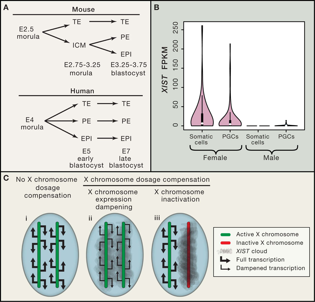
This study reveals that the lineage specification path in the developing human pre-implantation embryo is distinct from that in the mouse. In the mouse, lineage segregation occurs in two waves: first in the morula, where trophectoderm (TE) and the inner cell mass (ICM) segregate, then in the blastocyst, where epiblast cells (EPI) and primitive endoderm (PE) are specified from the ICM (Chazaud and Yamanaka, 2016) (Figure 1A). However, Lanner and colleagues find that lineage segregation into TE, EPI, and PE in human embryos occurs simultaneously at around E5 rather than stepwise (Figure 1A). Interestingly, prior to the definition of distinct cell identities, TE-, EPI-, and PE-specific genes are co-expressed, revealing a unique transcriptional plasticity of these cells before lineage commitment. These results provide a strong foundation for future studies of lineage segregations in early human development to elucidate how a cell co-expressing different lineage markers can subsequently commit to different identities as the blastocyst forms.
Figure 1. Lineage specification and X chromosome status in human pre-implantation development.
A. Mouse and human lineage specification starting from the morula. Mouse pre-implantation development is more rapid than that in human, resulting in different timing of events (E represents embryonic day post fertilization). In early mouse development, lineage segregation is stepwise. First the trophectoderm (TE) and the inner cell mass (ICM) segregate from the morula, later the emergence of the epiblast (EPI) and the primitive endoderm (PE) from the ICM follow. However, humans display a concurrent rather than stepwise lineage segregation where the TE, PE and EPI emerge simultaneously. The mouse time-course is adapted from Chazaud and Yamanaka (2016). B. Violin plots of transcript levels of the long non-coding RNA XIST in gonadal somatic cells or primordial germ cells (PGCs) from female and male embryos using data from Guo et al 2015. Female PGCs express XIST, while male PGCs have detectable but significantly lower XIST expression. C. Schematic depiction of different X chromosome dosage compensation states: (i) in female cells of E3 human pre-implantation embryos, where both X chromosomes are active (green), undergo full transcription (double-headed arrows), display no dosage compensation and no XIST expression; (ii) in E4–E7 cells of the human pre-implantation embryos, exhibiting the newly described dampened X chromosome expression state where both X chromosomes are active, express XIST (gray cloud), but instead of full transcription there is dampened, or lowered, transcription from both X chromosomes (single-headed arrows); (iii) in somatic cells with conventional X chromosome inactivation state, where XIST RNA is expressed from the inactive X chromosome (red), while the other X chromosome is active (green) with full transcription (double-headed arrows).
Although previous studies have employed single-cell RNA sequencing techniques to examine human pre-implantation embryos (Blakeley et al., 2015; Yan et al., 2013), this is the first study that could address sex-specific differences due to the large number of embryos analyzed (43 male, 45 female). Based on increased X-linked gene expression in female compared to male embryos and the detection of transcripts from both X chromosomes in individual cells, Lanner and colleagues show that all cells in female human embryos, i.e. across TE, PE and EPI, actively express both X chromosomes. These data, for the first time, demonstrate the chromosome-wide transcriptional activity of both X chromosomes in female human pre-implantation embryos, in agreement with previous reports based on select genes (Okamoto et al., 2011). Notably, this is different from the mouse, where the paternally-inherited X chromosome becomes silenced at the 4−8-cell stage due to imprinted X chromosome inactivation (XCI) and then, at the blastocyst state, specifically reactivates in the ICM (Brown and Chow, 2003). Therefore, lack of imprinted XCI is a distinguishing feature between mouse and human.
Another finding exposing critical differences between mouse and human is the expression of the long non-coding RNA (lncRNA) XIST. Xist is the master regulator of XCI, and, in the mouse, its expression is closely coupled to X chromosome silencing. However, in both female and male human embryos, XIST is expressed from the active X chromosomes. In 2011, Edith Heard’s group was the first to describe this unique XIST expression pattern from an active X chromosome in human pre-implantation blastocyst of both sexes based on RNA fluorescent in situ hybridization experiments (Okamoto et al., 2011). Notably, this study now uncovers that XIST expression levels are much lower in males compared to females, suggesting a critical difference in the regulation of XIST transcription or stability between the sexes. Interestingly, the lncRNA Tsix, which partially overlaps with and is antisense to Xist, is critical for the repression of Xist in early mouse development. There have been conflicting reports of whether humans also employ this lncRNA to regulate XIST (Brown and Chow, 2003). This study carefully addresses the presence of TSIX transcripts but does not detect them in any of the cells analyzed, agreeing with the conclusion that human cells do not express TSIX, which may be related to the fact that XIST is expressed from the active X chromosome in early human development.
Remarkably, the XIST-expressing active X chromosome is not restricted to cells of the pre-implantation embryo. XIST expression is also found in human primordial germ cells (PGCs), which give rise to gametes, as reported by Clark and colleagues (Gkountela et al., 2015) and also detected in single cell RNA sequencing data generated by Guo et al. (2015), where the female PGCs analyzed harbor two active X chromosomes (Figure 1B). Thus, in the only two known instances in human development where female cells have two active X chromosomes, there is also XIST expression. This underscores the importance of future studies on the role of XIST on an active X chromosome. Similar to the pre-implantation embryos, male PGCs have detectable, but significantly lower XIST expression compared to their female counterparts (Figure 1B).
A surprising finding from this study is the female-specific downregulation of X-linked gene expression during pre-implantation development. This occurs equally on both active X chromosomes and was coined “dampening” of X-linked gene expression. It appears to be mechanistically different from XCI, which leads to the transcriptional silencing of one of the two X chromosomes (Figure 1C). Interestingly, various forms of X chromosome dosage compensation between the sexes have evolved in different species. For example, in C. elegans, X chromosome dosage is compensated in XX hermaphrodites to their XO male counterparts by reducing gene expression levels of both X chromosomes to 50% (Crane et al., 2015). This is highly similar to what is now observed in female human embryos. However, two correlative observations suggest that the X-linked gene expression dampening in humans might be dependent on XIST, which is restricted to placental mammals: first, in females, the dampening of X-linked gene expression occurs gradually over time, which negatively correlates with XIST upregulation from E4 to E7; second, dampening is not observed on the male X chromosome, which expresses very low levels of XIST. Dissecting the molecular mechanism of this novel X chromosome dosage compensation in human development will be very interesting and perhaps will indicate some similarities to the mechanism employed by worms. The lack of X chromosome dosage compensation in naïve pluripotent cells of the mouse, that similarly carry two active X chromosomes, might be tolerated in vivo due to the more rapid pre-implantation development in mice.
Studies of human pre-implantation embryos are critical for our understanding of not only human developmental biology, but also for developing the correct mindset of what human embryonic stem cells should resemble at ground state (naïve) pluripotency. This is especially critical for our analysis of the recently developed naïve pluripotent culture conditions (Theunissen and Jaenisch, 2014). As elegantly demonstrated by the specific X chromosome state in human blastocysts, studies in mice, although very powerful, cannot be directly applied to human cells. Therefore, the extensive information provided by Lanner and colleagues in this issue of Cell will serve as a transcriptome encyclopedia of human pre-implantation blastocyst.
Footnotes
Publisher's Disclaimer: This is a PDF file of an unedited manuscript that has been accepted for publication. As a service to our customers we are providing this early version of the manuscript. The manuscript will undergo copyediting, typesetting, and review of the resulting proof before it is published in its final citable form. Please note that during the production process errors may be discovered which could affect the content, and all legal disclaimers that apply to the journal pertain.
References
- Blakeley P, Fogarty NME, del Valle I, Wamaitha SE, Hu TX, Elder K, Snell P, Christie L, Robson P, Niakan KK. Defining the three cell lineages of the human blastocyst by single-cell RNA-seq. Development. 2015;142:3613–3613. doi: 10.1242/dev.131235. [DOI] [PMC free article] [PubMed] [Google Scholar]
- Brown CJ, Chow JC. Beyond sense: the role of antisense RNA in controlling Xist expression. Semin. Cell Dev. Biol. 2003;14:341–347. doi: 10.1016/j.semcdb.2003.09.013. [DOI] [PubMed] [Google Scholar]
- Chazaud C, Yamanaka Y. Lineage specification in the mouse preimplantation embryo. Development. 2016;143:1063–1074. doi: 10.1242/dev.128314. [DOI] [PubMed] [Google Scholar]
- Crane E, Bian Q, McCord RP, Lajoie BR, Wheeler BS, Ralston EJ, Uzawa S, Dekker J, Meyer BJ. Condensin-driven remodelling of X chromosome topology during dosage compensation. Nature. 2015;523:240–244. doi: 10.1038/nature14450. [DOI] [PMC free article] [PubMed] [Google Scholar]
- Gkountela S, Zhang KX, Shafiq TA, Liao W-W, Hargan-Calvopiña J, Chen P-Y, Clark AT. DNA Demethylation Dynamics in the Human Prenatal Germline. Cell. 2015;161:1425–1436. doi: 10.1016/j.cell.2015.05.012. [DOI] [PMC free article] [PubMed] [Google Scholar]
- Guo F, Yan L, Guo H, Li L, Hu B, Zhao Y, Yong J, Hu Y, Wang X, Wei Y, et al. The Transcriptome and DNA Methylome Landscapes of Human Primordial Germ Cells. Cell. 2015;161:1437–1452. doi: 10.1016/j.cell.2015.05.015. [DOI] [PubMed] [Google Scholar]
- Okamoto I, Patrat C, Thépot D, Peynot N, Fauque P, Daniel N, Diabangouaya P, Wolf J-P, Renard J-P, Duranthon V, et al. Eutherian mammals use diverse strategies to initiate X-chromosome inactivation during development. Nature. 2011;472:370–374. doi: 10.1038/nature09872. [DOI] [PubMed] [Google Scholar]
- Petropoulos S, Edsgärd D, Reinius B, Deng Q, Panula SP, Codeluppi S, Plaza Reyes A, Linnarsson S, Sandberg R, Lanner F. Single-Cell RNA-Seq Reveals Lineage and X Chromosome Dynamics in Human Preimplantation Embryos. Cell. 2016 doi: 10.1016/j.cell.2016.03.023. This issue. [DOI] [PMC free article] [PubMed] [Google Scholar]
- Theunissen TW, Jaenisch R. Molecular Control of Induced Pluripotency. Cell Stem Cell. 2014;14:720–734. doi: 10.1016/j.stem.2014.05.002. [DOI] [PMC free article] [PubMed] [Google Scholar]
- Yan L, Yang M, Guo H, Yang L, Wu J, Li R, Liu P, Lian Y, Zheng X, Yan J, et al. Single-cell RNA-Seq profiling of human preimplantation embryos and embryonic stem cells. Nat. Struct. Mol. Biol. 2013;20:1131–1139. doi: 10.1038/nsmb.2660. [DOI] [PubMed] [Google Scholar]


