Abstract
One of the most significant paradigm shifts in membrane remodeling is the emerging view that membrane transformation is not exclusively controlled by cytoskeletal rearrangement, but also by biophysical constraints, adhesive forces, membrane curvature and compaction. One of the most exquisite examples of membrane remodeling is myelination. The advent of myelin was instrumental in advancing the nervous system during vertebrate evolution. With more rapid and efficient communication between neurons, faster and more complex computations could be performed in a given time and space. Our knowledge of how myelin-forming oligodendrocytes select and wrap axons has been limited by insufficient spatial and temporal resolution. By virtue of recent technological advances, progress has clarified longstanding controversies in the field. Here we review insights into myelination, from target selection to axon wrapping and membrane compaction, and discuss how understanding these processes has unexpectedly opened new avenues of insight into myelination-centered mechanisms of neural plasticity.
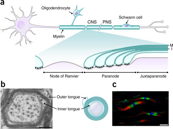
As the nervous system grew more computationally powerful and increasingly complex, the evolution of glial myelination allowed jawed vertebrates to overcome the pressure of increasing nervous system size for faster conduction speed and dramatically advanced the functional efficiency and complexity of the nervous system1,2. Myelin sheaths are made of glial plasma membranes that wrap around axons in a compact multilamellar spiral (Fig. 1a,b)3,4. These compact membrane layers serve as an insulator by increasing the resistance and decreasing the capacitance across the axonal membrane. Myelinating glia further potentiate rapid saltatory conduction by actively clustering voltage-gated sodium channels at the gaps between myelin sheaths1,5,6, called nodes of Ranvier (Fig. 1a,c). Myelin sheath thickness, length and axonal coverage patterns can affect the conduction velocity of action potentials7–9. Nodal length and channel density at the node may also influence the efficiency and velocity of the action potential. Perhaps unsurprisingly, then, much attention has been devoted to exploring the possibility that neuronal activity may influence myelination by oligodendroglia and regulate these parameters to modulate the conduction velocity in each underlying axon. It is an appealing concept that such dynamic myelination throughout the CNS might provide an additional mechanism for neural circuit plasticity by modulating timing and coordinating network synchrony and oscillations10,11. Without understanding myelination, we cannot fully appreciate how the nervous system develops and functions.
Figure 1.
Structure of myelin and molecular domains along myelinated axons. (a) A neuron and the myelin sheaths along its axon. Myelin sheaths are made by oligodendrocytes in the CNS and by Schwann cells in the PNS. A single oligodendrocyte can generate multiple myelin sheaths, whereas an individual Schwann cell only makes one. The magnified view (bottom) shows the ultrastructure around the node of Ranvier. Glial membranes at the ends of the sheaths are attached to the axonal membrane flanking the node, forming paranodes. Paranodal loops contain cytoplasm and are not compacted. Neuron-glia interactions at paranodes form paranodal axoglial junctions with the characteristic electron-dense transverse bands under EM. M indicates the major dense line, I the intraperiod line. (b) An electron micrograph from a cross section of an adult mouse optic nerve, and its illustration. The major dense lines are clearly visualized, but the intraperiod lines are not obvious under this magnification. The ends of the myelin spiral are the outer and inner tongues, which contain cytoplasm and are not compacted. (c) Immunostaining of a postnatal day 22 mouse optic nerve shows three molecular domains around nodes of Ranvier. Blue, nodes positive for βIV spectrin; green, paranodal junctions positive for Caspr; red, juxtaparanodes positive for potassium channel Kv1.2. Most of the myelinated region is between two juxtaparanodes and not visualized here. Scale bars: 200 nm (b); 3 μm (c). Panel a adapted from ref. 89, Elsevier; micrograph in b courtesy of K. Susuki. For clarity, the g-ratio in b is not drawn to scale.
Through recent advancement in technologies, our understanding of how myelin is formed and regulated has been greatly enhanced. In this Review, we focus on the most recent findings that together draw a mechanistic sketch of how oligodendrocytes select their targets, how they elaborate spiral layers of myelin membranes, and how these membrane layers compact to form mature sheaths. Finally, we take these mechanistic insights and consider how the formation and even remodeling of myelin may be harnessed as a new tool contributing to neural plasticity in the CNS.
Where to wrap? The biophysical and molecular settings
There is a close correlation between the myelination status of a CNS axon and whether or not it is above a threshold diameter (≥0.2–0.4 μm)12,13. What is the instructive signal that dictates this diameter requirement? Is it simply a matter of permissive geometry or is it transduced by dynamic molecular signaling? These questions have been addressed in the PNS, where Schwann cell ErbB receptors sense axonal levels of neuregulin 1 type III (Nrg1-III). Although it remains unclear how the level of Nrg1-III is normally regulated to precisely reflect an axon's diameter, suprathreshold Nrg1–ErbB signaling is the well-accepted determinant essential for myelination in the PNS, which can even override the biophysical parameter of axon diameter14,15. Surprisingly, Nrg1–ErbB signaling is largely dispensable for CNS myelination16, and several observations now collectively indicate that oligodendrocytes may not need an instructive signal to initiate myelination. Unlike Schwann cells, oligodendrocytes can differentiate and spread membrane sheets containing myelin proteins and lipids in the absence of neurons in vitro17,18. Oligodendrocytes myelinate paraformaldehyde-fixed axons, suggesting that dynamic neuron–glial interactions are not required for CNS myelination19. More recently, it was demonstrated that oligodendrocytes can even myelinate electrospun nanofibers above a threshold diameter between 0.3 and 0.4 μm (ref. 20), comparable to observed myelinated axon calibers in vivo, where the frequency of myelinated axons below 0.3 μm in diameter is extremely low12,13. These findings indicate that an overly small target diameter may represent a biophysical barrier to myelination of axons.
Taken together, oligodendrocytes appear to exhibit intrinsically broad target selection and wrapping activities, defined at least in part by unknown molecular sensors of fiber curvature. However, highly stereotyped spatiotemporal myelination patterns observed in the developing CNS21–23 leave plenty of room for molecular regulation and fine-tuning of these ‘default’ settings to better suit local needs in the oligodendrocyte microenvironment. Take, for instance, nonaxonal structures such as neuronal dendrites and somata, or existing myelinated axons that meet threshold diameter requirements yet are overwhelmingly avoided by differentiating oligodendrocytes. Inhibitory signaling to oligodendrocytes could curb myelination of these structures. While not absolutely required for selection and wrapping, the observation that oligodendrocytes prefer axons to nanofibers (S.A.R. and J.R.C., unpublished data) suggests the existence of attractive cues on axons. For instance, a cell adhesion molecule could be considered attractive if it preferentially stabilizes the initial contact between the oligodendrocyte and axon, resulting in an increased likelihood for subsequent myelination. Interestingly, both myelinated and unmyelinated axons of near-threshold diameters (0.2–0.8 μm) are observed in the CNS12,13. This suggests that the intrinsic property of oligodendrocytes can be adjusted by other, undefined fine-tuning cues and mechanisms that induce oligodendrocytes to myelinate subthreshold axons and prevent wrapping of otherwise biophysically permissive ones. Such cues include attractive and repulsive molecules, which could potentially be regulated in a neuronal activity–dependent manner. Identification of these molecules and how they function has been of substantial interest in the myelin field24,25. Recently, reconstruction of large electron microscopy (EM) data sets revealed scattered examples of long unmyelinated stretches of otherwise myelinated axons in the cortex26. This suggests that, even along the very same axon, myelination might be differentially regulated. It is tempting to speculate that graded expression and localization of repulsive and attractive cues along an axon could pattern myelin sheath length and spacing, which in turn can affect action potential conduction velocity and the timing of neurotransmitter release to postsynaptic targets. These parameters could vary within a given neural circuit to establish functionally desirable synchrony and oscillation patterns, and could be changed through myelin remodeling in the adult as an additional mechanism contributing to neural plasticity.
Transforming a cellular process into a spiraled sheet
Formation of myelin by wrapping glial plasma membranes around axons is one of the most remarkable biological events in membrane remodeling. To generate a 100-μm-long myelin sheath of ten spiral wraps around a 1-μm-diameter axon, myelinating glia need to generate over 6,400 μm2 of membrane! However, the delicate and elaborate nature of compact myelin and technical difficulties in resolving it at the nanometer level markedly hinder our detailed understanding of the wrapping process. Even Ramón y Cajal misinterpreted myelin as an axonal adjunct “entirely foreign to Schwann cells” in the PNS27.
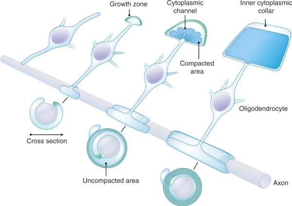
Our understanding of the active wrapping process during myelination began with studies on Schwann cells3,28. Continuous observation of the myelination process in culture by light microscopy and subsequent EM analysis excluded a cell-body-driven wrapping mechanism29. These observations resulted in the ‘carpet crawler’ model of myelination, whereby wrapping occurs by the inward crawling of the inner tongue around the axonal circumference, leaving a trail of the spirally extending myelin membrane sheet behind. This classic model would predict that the front of the inner tongue advances as a straight line along the axon during wrapping, resulting in a homogeneous number of wraps along the length of the forming sheath. Although the carpet crawler model may be still extrapolated to CNS myelination, it has turned out to be inconsistent with some early EM observations. For example, the number of wraps along the length was found to be greater in the middle and fewer toward the ends of a forming sheath30–32. This suggests the front of the inner tongue advances as a convex curve (Fig. 2), instead of a straight line. It also suggests that the radial growth (increase in wraps) and longitudinal growth (membrane sheet widening) take place simultaneously. To fill the gap between the carpet crawler model and observations such as variable wrap number, more recent work using cell culture systems, fluorescent proteins, and light and electron microscopy led to the ‘ofiomosaic’33 and ‘liquid croissant’34 models. Both models propose very different pictures of early wrapping events, and a fundamental difference among all three models lies in the way the membrane extends: from the innermost layer (carpet crawler), from the outermost layer (liquid croissant), or through lateral expansion of a glial process spiraling along an axon (ofiomosaic). Efforts to reconcile the different models and resolve the manner in which the membrane remodels during wrapping were hindered by limitations in spatial resolution of light microscopy, cell culture conditions, selective membrane labeling and EM artifacts produced during sample processing.
Figure 2.
The current model of myelination in the CNS. An illustration of a virtually unrolled myelin sheet at each stage is shown at the top process of the oligodendrocyte. The corresponding cross-sections are displayed below the axon. From left to right: a newly differentiated oligodendrocyte extends a process to an unmyelinated axon. After contacting the axon, it starts wrapping by spreading its membrane. The growth zone for active membrane incorporation is shown in green. The myelin membrane grows both radially (more wraps) and longitudinally (longer sheath). When there are about three layers of myelin membrane, intracellular compaction initiates from the outermost layer toward the inner layers, preserving uncompacted cytoplasmic channels for material transport between the oligodendrocyte process and the growth zone. If a mature myelin sheath were unrolled from the axon, it would consist of a trapezoid of compacted membrane lined with a continuous cytoplasmic collar. When coiled around the axon, the lateral cytoplasmic collars form loops located at the paranodes (see Fig. 1a). Myelin retraction may occur during active myelination (represented by one missing process in the right-most oligodendrocyte). Adapted from ref. 38, Elsevier.
Recent breakthrough studies have taken definitive and narrow approaches to closely describe both early myelination events and the elaboration of the growing myelin sheath. The advent of high-pressure freezing with subsequent freeze substitution preserves native tissue architecture, eliminating many of the EM artifacts associated with fixation and processing35. When combined with focused ion beam milling-coupled scanning EM for serial block-face imaging, the methods provide high-resolution three-dimensional images of nanoscale structures36. These techniques and high-resolution in vivo imaging of transgenic zebrafish larvae combined to generate the most current and complete model of the myelination process yet (Fig. 2)37,38. Exploratory oligodendrocyte processes were found to transform into short but elongating myelin sheaths37. The number of wraps is greatest at the site where the oligodendrocyte process is connected to the growing myelin sheath and gradually decreases toward the ends38, consistent with the early EM findings30–32. The longitudinal length of the membrane lamellae is shortest for the innermost layer and gradually longer toward the outermost one; the inner layers are covered completely by the respective outer ones. Thus, the inner tongue winds the axon spirally during active wrapping and may account for the helical coil patterns observed by light microscopy underlying the ofiomosaic and liquid croissant models. In addition, each layer has cytoplasm-containing edges, which are part of the continuous cytoplasmic collar of the membrane sheet and are always attached to the axon. As development progresses, these lateral cytoplasmic loops move toward the edges of the myelin sheath while each layer is extending longitudinally, and they finally meet and align as paranodal loops (Figs. 1a and 2).
Where and how new myelin membranes are incorporated into the growing myelin sheath had not been experimentally demonstrated until recently. Localization of newly synthesized vesicular stomatitis virus glycoprotein G (VSV-G) was used as a readout of new membrane deposition in infected oligodendroglia. As it turned out, new VSV-G accumulated at the inner tongue, reinforcing the idea that the inner tongue is the primary growth zone in growing myelin sheaths38. Interestingly, flanked by already compacted areas, transient cytoplasmic channels containing vesicular structures were preserved in CNS myelin with the high-pressure freezing EM techniques38. These findings suggest that, to fuel membrane growth, the cytoplasmic channels serve as the thruways embedded in compact areas to deliver new membranes in vesicles to the inner tongue (Fig. 2). It is noteworthy that these channels gradually close as compaction spreads during development, but can be re-formed in existing myelin sheaths that are stimulated to add more myelin wraps38.
What then is the driving force that powers membrane growth between the axon and the previous myelin layers? Intriguingly, two recent studies revealed that actin cytoskeleton dynamics is used by oligodendrocytes to push the inner tongues forward39,40. Actin filaments were found to be present in the growing myelin sheaths but depolymerized in the mature compact myelin38–40. Studies using cultured oligodendrocytes showed that actin filament depolymerization releases the membrane surface tension and promotes membrane spreading39,40. Pharmacological treatment inducing actin depolymerization facilitated myelin wrapping in the mouse spinal cords40. Furthermore, knockout mice lacking two of the actin filament disassembly factors exhibit fewer myelin wraps39. Taken together, these findings suggest that actin filament depolymerization may provide a driving force for myelin membrane wrapping around axons.
Presumably, oligodendrocytes need to coordinate the delivery of membrane vesicles and modulate actin cytoskeleton dynamics in a site-specific manner. According to the new model (Fig. 2), the growth rates of various sites among the advancing growth zone need to be differentially regulated as a myelin sheath is being formed. For the whole myelin membrane sheet to reach the final trapezoidal shape (when unrolled), radial growth must gradually attenuate when the final number of wraps is reached, while the longitudinal growth persists such that the newer and shorter inner layers catch up with the more mature, longer outer layers. However, how this differential regulation is achieved along a continuous inner tongue throughout the formation of a myelin sheath remains unknown.
Once the proper number of wraps is reached around an axon, the radial growth rate of a myelin sheath attenuates. How is this number determined and how does an oligodendrocyte sense that this number of wraps has been achieved? Most myelinated axons tend to have g-ratios (the diameter of the axon divided by the diameter of the axon and its myelin) close to the value optimal for conduction and nervous system efficiency41, and thicker axons have more wraps12. Intriguingly, the outer and inner tongues of mature CNS myelin are frequently located within the same quadrant in cross-sections42,43, indicating that oligodendrocytes often make nearly integral numbers of wraps. Remarkably, typical g-ratio values were not observed on myelinated nanofibers20, suggesting the existence of axonal molecular cues that instruct diameter-dependent, and even neuronal activity-dependent, radial growth requirements. Nrg1–ErbB signaling is a good candidate pathway because the manipulation of axonal Nrg1-III levels directly correlates with the total number of wraps produced by Schwann cells44. Although Nrg1–ErbB signaling may play a part in the development of CNS myelin, other surface cues should also exist, considering the normal CNS myelination observed in Nrg1 conditional knockout mice15,16. Signal transduction by these cues may converge on the regulation of membrane vesicle delivery and actin filament turnover to control wrapping. Interestingly, live imaging studies using zebrafish larvae observed retraction of some myelin sheaths by oligodendrocytes while other myelin sheaths were maintained37. This raises an important open question: what are the mechanisms underlying myelin retraction? Presumably, growth zones may also represent collapsing zones of endocytosis and membrane removal. Cytoplasmic channels may then serve as the routes for retrograde trafficking of vesicles out of the myelin sheath. If growth and retraction machinery are differentially adjustable, it is tempting to propose that bidirectional myelin sheath length and radial thickness modulation can be additional mechanisms contributing to circuit-level neural plasticity.
How do the myelin lamellae stack compactly?
Myelin achieves its insulating function by extruding most of the cytoplasm and extracellular space to obtain a low dielectric permittivity, which is vital for lowering axolemmal capacitance. In compact myelin, plasma membranes are brought into extremely close apposition (~1–3 nm) along a large surface area, representing an extraordinary example of membrane remodeling. There are two types of compaction in myelin: compaction between two cytoplasmic membrane leaflets, forming major dense lines under EM, and compaction between two extracellular leaflets, forming intraperiod lines (Figs. 1a,b and 3). It was traditionally thought that the compact myelin components PLP (proteolipid protein) and MBP (myelin basic protein) made up ~80% of CNS myelin proteins and might play vital roles in these compaction processes. The mechanisms underlying myelin compaction, however, turn out to be more complex than originally thought.
Figure 3.
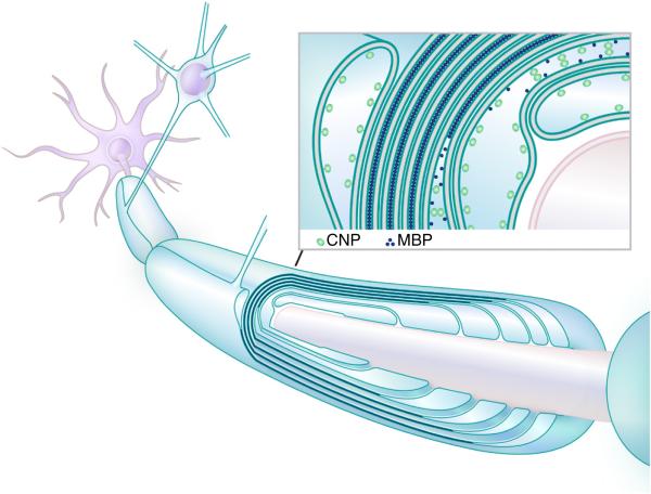
Intracellular compaction of myelin membranes. The illustration shows an actively forming myelin sheath with six lamellae. Compaction is completed for the two outermost layers and has not started for the two innermost layers. Compaction progresses both radially and longitudinally in parallel with membrane growth (see Fig. 2). In the longitudinal view, compaction of the two middle layers is still proceeding toward the paranodal loops. The inset shows a cross section of an area covering the outer tongue, compacted and uncompacted layers, and the inner tongue. In the region dominated by uncompacted myelin components, including CNP, the concentration of MBP is relatively low and the membranes are separated by the cytoplasm. After reaching a certain concentration, MBP closely binds two apposed membranes together and uncompacted myelin components and the cytoplasm are extruded; membranes are thereby compacted in a zipper-like fashion. Inset adapted from ref. 90, The Company of Biologists.
MBP is a small cytoplasmic protein that constitutes ~8% of proteins in myelin45. In shiverer mice, which lack MBP, the few myelin sheaths, if any, frequently lack major dense lines and fail to be compacted46–48. This suggests that MBP participates in compaction between two cytoplasmic membrane leaflets. MBP can bear up to 19 net positive charges at neutral pH and has been shown to bind negatively charged lipid head groups49–51. MBP also contains amphipathic α-helices that can be partly embedded in the membrane leaflet49,50. Therefore, MBP can associate with membranes as a peripheral membrane protein through both electrostatic and hydrophobic interactions. MBP neutralizes repulsive negative charges on the membranes and, given its small size, can potentially bring two cytoplasmic leaflets into close apposition (Fig. 3). Additionally, upon the interaction of MBP with membranes, it can self-associate into a high-order ensemble18,52,53. While MBP is an overall intrinsically disordered protein, its interaction with membranes induces formation of α-helices and β-sheets18,49. Through charge neutralization by elevating pH in vitro, purified MBP was shown to form micrometer-scale droplets in a reversible way, and this may involve a mechanism similar to amyloid-like β-aggregation53. Accordingly, the idea of phase transition of low-complexity disordered proteins into a hydrogel-like cohesive mesh-work has been introduced for MBP. Consistent with the requirement of a critical concentration for a phase transition, recent studies using surface forces apparatus described a cooperative membrane adsorption of MBP when its concentration exceeds a certain level54. When MBP is deposited between two membranes in heterologous cell lines, in the membrane sheets of cultured oligodendrocytes, or in cell-free membrane systems, proteins with 30 amino acid residues or more on the cytosolic side are excluded from MBP-occupied domains18,53. Assembly of a meshwork by phase transition may be the mechanistic basis of the observed diffusion barrier activity. Taking these results together, MBP promotes myelin compaction on the cytoplasmic side by interaction with membranes, self-assembly into a cohesive network and extrusion of proteins over a certain size (Fig. 3).
On the extracellular side of the membrane, transmembrane protein PLP exhibits membrane adhesion activities in vitro55–57. Because of its high abundance in CNS myelin (~17% of total protein45), it was assumed to be important to formation of compact myelin. Surprisingly, PLP-null mice produce nearly normal amounts of myelin58. Upon high-pressure freezing with freeze substitution, PLP-null myelin exhibits no conspicuous intraperiod line defects59, implying that PLP plays only a minor role in the maximal stability of compact myelin or that its role is functionally redundant with those of other molecules. Unfortunately, this leaves the detailed mechanisms crucial for the extracellular membrane compaction in the CNS mostly unclear. It has been proposed that intraperiod line formation may not require specific adhesive molecules but, alternatively, may be mediated by nonspecific van der Waals interactions and hydrogen bonding between membranes57,60. The main forces opposing extracellular compaction that need to be overcome include electrostatic repulsion and steric hindrance exerted by the glycocalyx on the membrane surface57. In this regard, extracellular compaction may be facilitated by the intracellular compaction as many transmembrane glycoproteins are excluded from the MBP-occupied domain53. However, intraperiod lines still form in shiverer mice47,61, indicating that the extracellular compaction can happen independently of intracellular compaction. Overexpression of a polysialyltransferase in myelinating oligodendrocytes induces increased surface levels of polysialic acids, which are highly anionic and have a large hydration volume57,62. Interestingly, in such cells, myelin lamellae are split when examined via conventional fixation and EM techniques57. Together with the observation of a decrease in the surface sugar residues during the course of oligodendrocyte differentiation, these findings suggest that removal of the glycocalyx via the normal oligodendrocyte differentiation program may be a prerequisite for extracellular compaction, serving to remove the force opposing it and unmask the weak interactions enabling it57.
One important aspect that remains unclear is how all the aforementioned findings on the intracellular and extracellular compaction can be fit into the spatiotemporal progression of myelin compaction in vivo. Early EM studies observed that compaction starts before there are more than three myelin layers12. A recent study found that the intraperiod lines can be observed between layers that still contain cytoplasm during early development38. Thus the differentiation program may downregulate the enzymes and membrane proteins contributing to the glycocalyx and upregulate PLP early, before the intracellular compaction, uncovering and enhancing the weak interactions for the extracellular compaction.
The same recent study38 further demonstrated that the intracellular compaction starts from the outermost layers and extends inward (Figs. 2 and 3). Intriguingly, the progression of the intracellular compaction is largely affected by the relative amounts of MBP and the most abundant component of uncompacted myelin, CNP (2′,3′-cyclic nucleotide 3′-phosphodiesterase; ~4% of total myelin proteins45). Intracellular compaction is more advanced in Cnp+/− mice38. Conversely, in heterozygous shiverer mice, there is a delay in intracellular compaction38. Moreover, overexpression of CNP inhibits intracellular compaction; in uncompacted regions, CNP far outnumbers MBP, and vice versa in compacted regions63,64. These observations imply that the dynamics of the intracellular compaction may be governed by the mutual exclusion between MBP and components of uncompacted myelin, including CNP (Fig. 3). This is reminiscent of the intra-axonal boundary resulting from the mutual exclusion of different cytoskeletal scaffold proteins, resulting in the assembly of a distinct molecular domain at the axon initial segment6,65. Remarkably, the length and location of the axon initial segment can be adjusted as a result of changes in neuronal activity66. A dynamic CNP–MBP competition could also be used to reopen the cytoplasmic channels in mature sheaths to change their thickness or even retract them. Therefore, further understanding of CNP–MBP dynamics will be required to elucidate not only the spatiotemporal onset and progression patterns of intracellular compaction during development, but also the potential mechanisms of activity-dependent myelin remodeling.
Our current understanding of the mechanisms regulating compaction is still not sufficient to explain how cytoplasmic channels are left open during active myelination and later buried when myelin matures. Along the same lines, another open question is how paranodal loops, inner cytoplasmic collars and outer tongues remain uncompacted (Fig. 2). It is tempting to propose that the cooperative regulation of dynamic molecular competition and membrane apposition may be important. Furthermore, the potential for the process of decompaction to participate in myelin retraction and thickness modulation is wholly unexplored. Future studies will be required to address all of these questions.
‘Plastic’ wrap: optimization and the call of activity
Myelin is not simply a static structure in place to ensure rapid conduction. In our nervous systems, functional optimization cannot be fulfilled merely by maximizing conduction velocity8,67. One prominent example is the avian auditory system68. In a specific group of neurons in the chicken auditory brainstem, each axon branches to propagate action potentials to both ipsilateral and contralateral targets in the brainstem. While the contralateral path is about twice as long as the ipsilateral one, the axon diameter and myelin sheath lengths of the ipsilateral axon are smaller and shorter, respectively, halving the conduction velocity. The overall effect is isochronicity of the conduction time between the two paths, down to sub-millisecond precision. Therefore, not all the myelin sheath parameters are optimized for the fastest action potential conduction. Oligodendrocytes appear to be responsive to individual axons and adjust myelination parameters according to their needs. Such plastic regulation of myeli-nation has been appreciated recently (reviewed in refs. 69–72), but the mechanisms underlying this active regulation of myelination remain poorly understood.
Myelination can modulate information flow in neural circuits by several potential mechanisms, including (i) de novo myelination of unmyelinated axons or unmyelinated segments of myelinated axons, (ii) myelin replacement, where an old sheath is replaced with one or more new sheaths, (iii) thickening or thinning of existing sheaths, (iv) lengthening or shortening of existing sheaths, and (v) myelin retraction, where sheaths are removed and are not replaced. Each step of myelination, from target selection and wrapping to myelin compaction during sheath maturation, could be coordinately regulated to mediate these processes and achieve a functional change in a neural circuit. A future challenge will be to determine whether these mechanisms of myelin-based neural plasticity are actually used in vivo, and if so, under what conditions.
There is emerging and strengthening evidence that myelin-based mechanisms of neural circuit regulation are active in the mouse brain. During development, many unmyelinated axons or unmyelinated segments of axons are present while normal developmental myeli-nation is proceeding. A recent mouse study indicates that juvenile social isolation (for 2 weeks after weaning) leads to thinner and fewer myelin sheaths made by individual oligodendrocytes specifically in the medial prefrontal cortex73. The affected mice exhibit defects in sociability and working memory. These results suggest that myelination by oligodendrocytes can be adjusted by their local environment, probably shaped by neuronal activity resulting from the external stimuli experienced by the organism. Complementing these findings, another study that deprived adult mice of social interaction for 8 weeks observed thinner myelin and decreased myelin gene expression specifically in the prefrontal cortex74. Remarkably, 4 weeks of social reintegration restored myelin gene expression and reversed behavioral deficits. The recovery after social reintegration might result from the thickening of the thinner myelin sheaths or resumed wrapping processes. The surprising effects of social isolation on myelination patterns and behavior in both adolescence and adulthood beg the question: what is the potential for myelin remodeling in the adult CNS?
In the adult CNS, oligodendrocyte precursor cells are broadly distributed and serve as a source of adult-born myelinating oligodendrocytes69–72. Early EM studies observed many unmyelinated axons 0.4–0.8 μm in diameter in the CNS12, and the recent study that reconstructed three-dimensional structure from large EM data sets even revealed long unmyelinated sections of otherwise myelinated axons in the adult cortex26. These findings suggest that unmyelinated and incompletely myelinated axons could be substrates for de novo myelination in the adult brain. Considering the findings that newly formed oligodendrocytes generate myelin sheaths only within a short time37,75, generation of new myelin sheaths in adulthood may require the generation of new oligodendrocytes. In accordance, by tracing the 14C concentration in purified oligodendrocyte DNA from postmortem human brains in combination with mathematical modeling, the population of oligodendrocytes in the cortex was estimated to expand until the end of the fourth decade of life and have an annual turnover rate of 2.5% afterwards76. Furthermore, another recent study showed that learning to run on a complex wheel with irregular rung spacing enhanced the proliferation of oligodendrocyte precursor cells and increased the number of newly formed oligodendrocytes in adult mice77. When the generation of new myelin-forming oligodendrocytes was genetically abolished in adult mice, they performed worse in mastering the complex wheel running than the control mice77. These results imply that adult myelination by newly formed oligodendrocytes is important to motor learning.
The findings in development and adulthood are consistent with the idea that environmental stimuli increase neuronal activity, which in turn stimulates oligodendrogenesis and myelination. The idea that activity modulates myelination has been proposed for decades10. Several studies have directly tested this in vivo. One study showed that unilateral stimulation of the rat corticospinal tract selectively induces proliferation and differentiation of oligodendrocyte precursor cells on the stimulated side78. Another recent study used channelrhodopsin to increase neuronal firing activity in the mouse M2 motor cortex. The authors observed increased oligodendrogenesis as well as thicker myelin sheaths in the area near the stimulated neurons and their axons79.
Taken together, the social isolation and directed neural stimulation studies suggest that myelin segment thickness can be modulated by neuronal activity. The particular mechanisms translating environmental experience into oligodendrocyte regulation of myelin thickness remains poorly understood. In these particular studies, whether the thinner or thicker myelin observed represents remodeled or de novo myelination remains unclear. However, increasing the thickness of existing myelin sheaths has been proven feasible. The inducible conditional knockout of Pten (phosphatase and tensin homolog) in mature oligodendrocytes of adult mice was reported to make the myelin much thicker by means of reopening cytoplasmic channels in the myelin sheaths, resulting in more wrapping38,80. By contrast, much less is known about thinning of existing myelin sheaths. However, myelin sheath retraction has been observed during live imaging of developing zebrafish larvae37. Myelin thinning may be achieved by modulating the same machinery used in myelin retraction.
Myelin retraction observed during zebrafish development unexpectedly reveals some uncertainty of target selection by oligodendrocytes in vivo. It suggests that dynamic regulation may edit the decision made during initial contact and target selection. It is unclear how an oligodendrocyte selects and stably myelinates only a subset of biophysically suitable substrates in the local environment. Two recent studies provide compelling evidence that this process may not require but can be affected by neuronal activity81,82. When neurons in zebrafish larvae express tetanus neurotoxin to inhibit synaptic vesicle release, individual oligodendrocytes form fewer myelin sheaths81,82. Though differences in study design seemed to lead to different mechanistic interpretations, this deficit is likely due to decreased nascent sheath stability. It appears that more of the ensheathments on silent axons may be ≤5 μm long and sustained for ≤10 min, and there may be more sheaths on silent axons that are stable for 6 h but retract afterwards. Therefore, neuronal activity may modulate myelination by influencing the stable growth and subsequent maintenance of nascent myelin sheaths. Notably, oligodendrocyte precursor cells form synapses with axons and express neurotransmitter receptors. These synapses directly connect neurons with oligodendrocyte precursor cells in all gray and white matter areas explored so far and allow oligodendrocyte precursor cells to sense most aspects of neuronal activity83–86. The function of these neuron-glial synapses and how oligodendroglia respond to neuronal activity by modulating myelination in vivo are under vigorous investigation.
In the emerging field of myelin plasticity, several key challenges await to be addressed to understand its role in modulating neural plasticity and circuit-level activity patterns. First, whether activity- or experience-dependent changes in myelination patterns are a specific or general phenomenon must be addressed. Directly linking changes in a neuron's activity with its own altered myelination profile, while technically challenging, would provide compelling support of specificity. Second, identifying neuronal and glial molecular regulators of myelination underlying how oligodendroglia respond to neuronal activity in vivo has been of long-standing interest in the field of myelination, and this interest only continues to grow as our appreciation of myelin's role in affecting neural function increases. Finally, demonstrating that changes in myelination patterns of specific circuits have functional behavioral consequences will go far in cementing myelin's role as a major, potentially bidirectional regulator of neural function and plasticity. While this experimental wish list may be addressable at present in part, full answers await the future development of tools and techniques able to overcome current limitations in spatiotemporal resolution.
Since its discovery over 160 years ago87, the study of myelin has endured despite myelin's simplistic reputation as a passive sidekick insulator of axons, with oligodendroglia once being overlooked among the non-neuronal, non-glial “third element” of the nervous system88. Now, oligodendrocyte myelination is starting to reveal itself as the dark horse in the race to understand the subtle and sophisticated inner workings of the nervous system. If restrictive preconceptions of myelin's potential can be released and this enigmatic structure given the respect it now so clearly deserves, only time will tell how fast and how far it will carry our understanding of the brain.
ACKNOWLEDGMENTS
The electron micrograph in Figure 1 is courtesy of K. Susuki at Wright State University with technical assistance from D. Townley and support from the Integrated Microscopy Core at Baylor College of Medicine, with funding from the US National Institutes of Health (NIH) (HD007495, DK56338 and CA125123), the Dan L. Duncan Cancer Center and the John S. Dunn Gulf Coast Consortium for Chemical Genomics. We thank P.-J. Lee for her insight, creativity and efforts in making the illustrations. We also thank A.J. Green and the members of the Chan laboratory for critical reading of the manuscript and insightful comments. This review was supported by National Multiple Sclerosis Society research grants (RG4541A3 and RG5203A4), NIH National Institute of Neurological Disorders and Stroke (NINDS) (R01NS062796) and the Rachleff Endowment to J.R.C. S.A.R. is supported by an NIH NINDS National Research Service Award (F31NS081905).
Footnotes
COMPETING FINANCIAL INTERESTS
The authors declare no competing financial interests.
References
- 1.Hartline DK, Colman DR. Rapid conduction and the evolution of giant axons and myelinated fibers. Curr. Biol. 2007;17:R29–R35. doi: 10.1016/j.cub.2006.11.042. [DOI] [PubMed] [Google Scholar]
- 2.Zalc B, Goujet D, Colman D. The origin of the myelination program in vertebrates. Curr. Biol. 2008;18:R511–R512. doi: 10.1016/j.cub.2008.04.010. [DOI] [PubMed] [Google Scholar]
- 3.Ben Geren B. The formation from the Schwann cell surface of myelin in the peripheral nerves of chick embryos. Exp. Cell Res. 1954;7:558–562. doi: 10.1016/s0014-4827(54)80098-x. [DOI] [PubMed] [Google Scholar]
- 4.Bunge MB, Bunge RP, Pappas GD. Electron microscopic demonstration of connections between glia and myelin sheaths in the developing mammalian central nervous system. J. Cell Biol. 1962;12:448–453. doi: 10.1083/jcb.12.2.448. [DOI] [PubMed] [Google Scholar]
- 5.Eshed-Eisenbach Y, Peles E. The making of a node: a co-production of neurons and glia. Curr. Opin. Neurobiol. 2013;23:1049–1056. doi: 10.1016/j.conb.2013.06.003. [DOI] [PMC free article] [PubMed] [Google Scholar]
- 6.Normand EA, Rasband MN. Subcellular patterning: axonal domains with specialized structure and function. Dev. Cell. 2015;32:459–468. doi: 10.1016/j.devcel.2015.01.017. [DOI] [PMC free article] [PubMed] [Google Scholar]
- 7.Waxman SG. Determinants of conduction velocity in myelinated nerve fibers. Muscle Nerve. 1980;3:141–150. doi: 10.1002/mus.880030207. [DOI] [PubMed] [Google Scholar]
- 8.Waxman SG. Axon-glia interactions: building a smart nerve fiber. Curr. Biol. 1997;7:R406–R410. doi: 10.1016/s0960-9822(06)00203-x. [DOI] [PubMed] [Google Scholar]
- 9.Babbs CF, Shi R. Subtle paranodal injury slows impulse conduction in a mathematical model of myelinated axons. PLoS One. 2013;8:e67767. doi: 10.1371/journal.pone.0067767. [DOI] [PMC free article] [PubMed] [Google Scholar]
- 10.Fields RD. White matter in learning, cognition and psychiatric disorders. Trends Neurosci. 2008;31:361–370. doi: 10.1016/j.tins.2008.04.001. [DOI] [PMC free article] [PubMed] [Google Scholar]
- 11.Pajevic S, Basser PJ, Fields RD. Role of myelin plasticity in oscillations and synchrony of neuronal activity. Neuroscience. 2014;276:135–147. doi: 10.1016/j.neuroscience.2013.11.007. [DOI] [PMC free article] [PubMed] [Google Scholar]
- 12.Hildebrand C, Remahl S, Persson H, Bjartmar C. Myelinated nerve fibres in the CNS. Prog. Neurobiol. 1993;40:319–384. doi: 10.1016/0301-0082(93)90015-k. [DOI] [PubMed] [Google Scholar]
- 13.Wang SS, et al. Functional trade-offs in white matter axonal scaling. J. Neurosci. 2008;28:4047–4056. doi: 10.1523/JNEUROSCI.5559-05.2008. [DOI] [PMC free article] [PubMed] [Google Scholar]
- 14.Salzer JL. Schwann cell myelination. Cold Spring Harb. Perspect. Biol. 2015;7:a020529. doi: 10.1101/cshperspect.a020529. [DOI] [PMC free article] [PubMed] [Google Scholar]
- 15.Mei L, Nave KA. Neuregulin-ERBB signaling in the nervous system and neuropsychiatric diseases. Neuron. 2014;83:27–49. doi: 10.1016/j.neuron.2014.06.007. [DOI] [PMC free article] [PubMed] [Google Scholar]
- 16.Brinkmann BG, et al. Neuregulin-1/ErbB signaling serves distinct functions in myelination of the peripheral and central nervous system. Neuron. 2008;59:581–595. doi: 10.1016/j.neuron.2008.06.028. [DOI] [PMC free article] [PubMed] [Google Scholar]
- 17.Mirsky R, et al. Myelin-specific proteins and glycolipids in rat Schwann cells and oligodendrocytes in culture. J. Cell Biol. 1980;84:483–494. doi: 10.1083/jcb.84.3.483. [DOI] [PMC free article] [PubMed] [Google Scholar]
- 18.Aggarwal S, et al. A size barrier limits protein diffusion at the cell surface to generate lipid-rich myelin-membrane sheets. Dev. Cell. 2011;21:445–456. doi: 10.1016/j.devcel.2011.08.001. [DOI] [PubMed] [Google Scholar]
- 19.Rosenberg SS, Kelland EE, Tokar E, De la Torre AR, Chan JR. The geometric and spatial constraints of the microenvironment induce oligodendrocyte differentiation. Proc. Natl. Acad. Sci. USA. 2008;105:14662–14667. doi: 10.1073/pnas.0805640105. [DOI] [PMC free article] [PubMed] [Google Scholar]
- 20.Lee S, et al. A culture system to study oligodendrocyte myelination processes using engineered nanofibers. Nat. Methods. 2012;9:917–922. doi: 10.1038/nmeth.2105. [DOI] [PMC free article] [PubMed] [Google Scholar]
- 21.Foran DR, Peterson AC. Myelin acquisition in the central nervous system of the mouse revealed by an MBP-Lac Z transgene. J. Neurosci. 1992;12:4890–4897. doi: 10.1523/JNEUROSCI.12-12-04890.1992. [DOI] [PMC free article] [PubMed] [Google Scholar]
- 22.Brody BA, Kinney HC, Kloman AS, Gilles FH. Sequence of central nervous system myelination in human infancy. I. An autopsy study of myelination. J. Neuropathol. Exp. Neurol. 1987;46:283–301. doi: 10.1097/00005072-198705000-00005. [DOI] [PubMed] [Google Scholar]
- 23.Kinney HC, Brody BA, Kloman AS, Gilles FH. Sequence of central nervous system myelination in human infancy. II. Patterns of myelination in autopsied infants. J. Neuropathol. Exp. Neurol. 1988;47:217–234. doi: 10.1097/00005072-198805000-00003. [DOI] [PubMed] [Google Scholar]
- 24.Piaton G, Gould RM, Lubetzki C. Axon-oligodendrocyte interactions during developmental myelination, demyelination and repair. J. Neurochem. 2010;114:1243–1260. doi: 10.1111/j.1471-4159.2010.06831.x. [DOI] [PubMed] [Google Scholar]
- 25.Taveggia C, Feltri ML, Wrabetz L. Signals to promote myelin formation and repair. Nat. Rev. Neurol. 2010;6:276–287. doi: 10.1038/nrneurol.2010.37. [DOI] [PMC free article] [PubMed] [Google Scholar]
- 26.Tomassy GS, et al. Distinct profiles of myelin distribution along single axons of pyramidal neurons in the neocortex. Science. 2014;344:319–324. doi: 10.1126/science.1249766. [DOI] [PMC free article] [PubMed] [Google Scholar]
- 27.Ramón y Cajal S. Degeneration & Regeneration of the Nervous System. Oxford Univ. Press; London: 1928. [Google Scholar]
- 28.Robertson JD. The ultrastructure of adult vertebrate peripheral myelinated nerve fibers in relation to myelinogenesis. J. Biophys. Biochem. Cytol. 1955;1:271–278. doi: 10.1083/jcb.1.4.271. [DOI] [PMC free article] [PubMed] [Google Scholar]
- 29.Bunge RP, Bunge MB, Bates M. Movements of the Schwann cell nucleus implicate progression of the inner (axon-related) Schwann cell process during myelination. J. Cell Biol. 1989;109:273–284. doi: 10.1083/jcb.109.1.273. [DOI] [PMC free article] [PubMed] [Google Scholar]
- 30.Webster HD. The geometry of peripheral myelin sheaths during their formation and growth in rat sciatic nerves. J. Cell Biol. 1971;48:348–367. doi: 10.1083/jcb.48.2.348. [DOI] [PMC free article] [PubMed] [Google Scholar]
- 31.Fraher JP. A quantitative study of anterior root fibres during early myelination. II. Longitudinal variation in sheath thickness and axon circumference. J. Anat. 1973;115:421–444. [PMC free article] [PubMed] [Google Scholar]
- 32.Fraher JP. Quantitative studies on the maturation of central and peripheral parts of individual ventral motoneuron axons. I. Myelin sheath and axon calibre. J. Anat. 1978;126:509–533. [PMC free article] [PubMed] [Google Scholar]
- 33.Ioannidou K, Anderson KI, Strachan D, Edgar JM, Barnett SC. Time-lapse imaging of the dynamics of CNS glial-axonal interactions in vitro and ex vivo. PLoS One. 2012;7:e30775. doi: 10.1371/journal.pone.0030775. [DOI] [PMC free article] [PubMed] [Google Scholar]
- 34.Sobottka B, Ziegler U, Kaech A, Becher B, Goebels N. CNS live imaging reveals a new mechanism of myelination: the liquid croissant model. Glia. 2011;59:1841–1849. doi: 10.1002/glia.21228. [DOI] [PubMed] [Google Scholar]
- 35.Möbius W, et al. Electron microscopy of the mouse central nervous system. Methods Cell Biol. 2010;96:475–512. doi: 10.1016/S0091-679X(10)96020-2. [DOI] [PubMed] [Google Scholar]
- 36.Peddie CJ, Collinson LM. Exploring the third dimension: volume electron microscopy comes of age. Micron. 2014;61:9–19. doi: 10.1016/j.micron.2014.01.009. [DOI] [PubMed] [Google Scholar]
- 37.Czopka T, Ffrench-Constant C, Lyons DA. Individual oligodendrocytes have only a few hours in which to generate new myelin sheaths in vivo. Dev. Cell. 2013;25:599–609. doi: 10.1016/j.devcel.2013.05.013. [DOI] [PMC free article] [PubMed] [Google Scholar]
- 38.Snaidero N, et al. Myelin membrane wrapping of CNS axons by PI(3,4,5)P3-dependent polarized growth at the inner tongue. Cell. 2014;156:277–290. doi: 10.1016/j.cell.2013.11.044. [DOI] [PMC free article] [PubMed] [Google Scholar]
- 39.Nawaz S, et al. Actin filament turnover drives leading edge growth during myelin sheath formation in the central nervous system. Dev. Cell. 2015;34:139–151. doi: 10.1016/j.devcel.2015.05.013. [DOI] [PMC free article] [PubMed] [Google Scholar]
- 40.Zuchero JB, et al. CNS myelin wrapping is driven by actin disassembly. Dev. Cell. 2015;34:152–167. doi: 10.1016/j.devcel.2015.06.011. [DOI] [PMC free article] [PubMed] [Google Scholar]
- 41.Chomiak T, Hu B. What is the optimal value of the g-ratio for myelinated fibers in the rat CNS? A theoretical approach. PLoS One. 2009;4:e7754. doi: 10.1371/journal.pone.0007754. [DOI] [PMC free article] [PubMed] [Google Scholar]
- 42.Peters A. Further observations on the structure of myelin sheaths in the central nervous system. J. Cell Biol. 1964;20:281–296. doi: 10.1083/jcb.20.2.281. [DOI] [PMC free article] [PubMed] [Google Scholar]
- 43.Waxman SG, Swadlow HA. Ultrastructure of visual callosal axons in the rabbit. Exp. Neurol. 1976;53:115–127. doi: 10.1016/0014-4886(76)90287-9. [DOI] [PubMed] [Google Scholar]
- 44.Nave KA, Salzer JL. Axonal regulation of myelination by neuregulin 1. Curr. Opin. Neurobiol. 2006;16:492–500. doi: 10.1016/j.conb.2006.08.008. [DOI] [PubMed] [Google Scholar]
- 45.Jahn O, Tenzer S, Werner HB. Myelin proteomics: molecular anatomy of an insulating sheath. Mol. Neurobiol. 2009;40:55–72. doi: 10.1007/s12035-009-8071-2. [DOI] [PMC free article] [PubMed] [Google Scholar]
- 46.Bird TD, Farrell DF, Sumi SM. Brain lipid composition of the shiverer mouse: (genetic defect in myelin development). J. Neurochem. 1978;31:387–391. doi: 10.1111/j.1471-4159.1978.tb12479.x. [DOI] [PubMed] [Google Scholar]
- 47.Privat A, Jacque C, Bourre JM, Dupouey P, Baumann N. Absence of the major dense line in myelin of the mutant mouse “shiverer”. Neurosci. Lett. 1979;12:107–112. doi: 10.1016/0304-3940(79)91489-7. [DOI] [PubMed] [Google Scholar]
- 48.Rosenbluth J. Central myelin in the mouse mutant shiverer. J. Comp. Neurol. 1980;194:639–648. doi: 10.1002/cne.901940310. [DOI] [PubMed] [Google Scholar]
- 49.Harauz G, Ladizhansky V, Boggs JM. Structural polymorphism and multifunctionality of myelin basic protein. Biochemistry. 2009;48:8094–8104. doi: 10.1021/bi901005f. [DOI] [PubMed] [Google Scholar]
- 50.Harauz G, Libich DS. The classic basic protein of myelin–conserved structural motifs and the dynamic molecular barcode involved in membrane adhesion and protein-protein interactions. Curr. Protein Pept. Sci. 2009;10:196–215. doi: 10.2174/138920309788452218. [DOI] [PubMed] [Google Scholar]
- 51.Nawaz S, et al. Phosphatidylinositol 4,5-bisphosphate-dependent interaction of myelin basic protein with the plasma membrane in oligodendroglial cells and its rapid perturbation by elevated calcium. J. Neurosci. 2009;29:4794–4807. doi: 10.1523/JNEUROSCI.3955-08.2009. [DOI] [PMC free article] [PubMed] [Google Scholar]
- 52.Kattnig DR, Bund T, Boggs JM, Harauz G, Hinderberger D. Lateral self-assembly of 18.5-kDa myelin basic protein (MBP) charge component-C1 on membranes. Biochim. Biophys. Acta. 2012;1818:2636–2647. doi: 10.1016/j.bbamem.2012.06.010. [DOI] [PubMed] [Google Scholar]
- 53.Aggarwal S, et al. Myelin membrane assembly is driven by a phase transition of myelin basic proteins into a cohesive protein meshwork. PLoS Biol. 2013;11:e1001577. doi: 10.1371/journal.pbio.1001577. [DOI] [PMC free article] [PubMed] [Google Scholar]
- 54.Lee DW, et al. Lipid domains control myelin basic protein adsorption and membrane interactions between model myelin lipid bilayers. Proc. Natl. Acad. Sci. USA. 2014;111:E768–E775. doi: 10.1073/pnas.1401165111. [DOI] [PMC free article] [PubMed] [Google Scholar]
- 55.Palaniyar N, Semotok JL, Wood DD, Moscarello MA, Harauz G. Human proteolipid protein (PLP) mediates winding and adhesion of phospholipid membranes but prevents their fusion. Biochim. Biophys. Acta. 1998;1415:85–100. doi: 10.1016/s0005-2736(98)00180-1. [DOI] [PubMed] [Google Scholar]
- 56.Bizzozero OA, Howard TA. Myelin proteolipid protein-induced aggregation of lipid vesicles: efficacy of the various molecular species. Neurochem. Res. 2002;27:1269–1277. doi: 10.1023/a:1021659313213. [DOI] [PubMed] [Google Scholar]
- 57.Bakhti M, et al. Loss of electrostatic cell-surface repulsion mediates myelin membrane adhesion and compaction in the central nervous system. Proc. Natl. Acad. Sci. USA. 2013;110:3143–3148. doi: 10.1073/pnas.1220104110. [DOI] [PMC free article] [PubMed] [Google Scholar]
- 58.Klugmann M, et al. Assembly of CNS myelin in the absence of proteolipid protein. Neuron. 1997;18:59–70. doi: 10.1016/s0896-6273(01)80046-5. [DOI] [PubMed] [Google Scholar]
- 59.Möbius W, Patzig J, Nave KA, Werner HB. Phylogeny of proteolipid proteins: divergence, constraints, and the evolution of novel functions in myelination and neuroprotection. Neuron Glia Biol. 2008;4:111–127. doi: 10.1017/S1740925X0900009X. [DOI] [PubMed] [Google Scholar]
- 60.Coetzee T, Suzuki K, Nave KA, Popko B. Myelination in the absence of galactolipids and proteolipid proteins. Mol. Cell. Neurosci. 1999;14:41–51. doi: 10.1006/mcne.1999.0768. [DOI] [PubMed] [Google Scholar]
- 61.Inoue Y, Nakamura R, Mikoshiba K, Tsukada Y. Fine structure of the central myelin sheath in the myelin deficient mutant Shiverer mouse, with special reference to the pattern of myelin formation by oligodendroglia. Brain Res. 1981;219:85–94. doi: 10.1016/0006-8993(81)90269-9. [DOI] [PubMed] [Google Scholar]
- 62.Schnaar RL, Gerardy-Schahn R, Hildebrandt H. Sialic acids in the brain: gangliosides and polysialic acid in nervous system development, stability, disease, and regeneration. Physiol. Rev. 2014;94:461–518. doi: 10.1152/physrev.00033.2013. [DOI] [PMC free article] [PubMed] [Google Scholar]
- 63.Gravel M, et al. Overexpression of 2′,3′-cyclic nucleotide 3′-phosphodiesterase in transgenic mice alters oligodendrocyte development and produces aberrant myelination. Mol. Cell. Neurosci. 1996;7:453–466. doi: 10.1006/mcne.1996.0033. [DOI] [PubMed] [Google Scholar]
- 64.Yin X, Peterson J, Gravel M, Braun PE, Trapp BD. CNP overexpression induces aberrant oligodendrocyte membranes and inhibits MBP accumulation and myelin compaction. J. Neurosci. Res. 1997;50:238–247. doi: 10.1002/(SICI)1097-4547(19971015)50:2<238::AID-JNR12>3.0.CO;2-4. [DOI] [PubMed] [Google Scholar]
- 65.Galiano MR, et al. A distal axonal cytoskeleton forms an intra-axonal boundary that controls axon initial segment assembly. Cell. 2012;149:1125–1139. doi: 10.1016/j.cell.2012.03.039. [DOI] [PMC free article] [PubMed] [Google Scholar]
- 66.Yoshimura T, Rasband MN. Axon initial segments: diverse and dynamic neuronal compartments. Curr. Opin. Neurobiol. 2014;27:96–102. doi: 10.1016/j.conb.2014.03.004. [DOI] [PMC free article] [PubMed] [Google Scholar]
- 67.Seidl AH. Regulation of conduction time along axons. Neuroscience. 2014;276:126–134. doi: 10.1016/j.neuroscience.2013.06.047. [DOI] [PMC free article] [PubMed] [Google Scholar]
- 68.Seidl AH, Rubel EW, Harris DM. Mechanisms for adjusting interaural time differences to achieve binaural coincidence detection. J. Neurosci. 2010;30:70–80. doi: 10.1523/JNEUROSCI.3464-09.2010. [DOI] [PMC free article] [PubMed] [Google Scholar]
- 69.Richardson WD, Young KM, Tripathi RB, McKenzie I. NG2-glia as multipotent neural stem cells: fact or fantasy? Neuron. 2011;70:661–673. doi: 10.1016/j.neuron.2011.05.013. [DOI] [PMC free article] [PubMed] [Google Scholar]
- 70.Zatorre RJ, Fields RD, Johansen-Berg H. Plasticity in gray and white: neuroimaging changes in brain structure during learning. Nat. Neurosci. 2012;15:528–536. doi: 10.1038/nn.3045. [DOI] [PMC free article] [PubMed] [Google Scholar]
- 71.Wang S, Young KM. White matter plasticity in adulthood. Neuroscience. 2014;276:148–160. doi: 10.1016/j.neuroscience.2013.10.018. [DOI] [PubMed] [Google Scholar]
- 72.de Hoz L, Simons M. The emerging functions of oligodendrocytes in regulating neuronal network behaviour. Bioessays. 2015;37:60–69. doi: 10.1002/bies.201400127. [DOI] [PubMed] [Google Scholar]
- 73.Makinodan M, Rosen KM, Ito S, Corfas G. A critical period for social experience-dependent oligodendrocyte maturation and myelination. Science. 2012;337:1357–1360. doi: 10.1126/science.1220845. [DOI] [PMC free article] [PubMed] [Google Scholar]
- 74.Liu J, et al. Impaired adult myelination in the prefrontal cortex of socially isolated mice. Nat. Neurosci. 2012;15:1621–1623. doi: 10.1038/nn.3263. [DOI] [PMC free article] [PubMed] [Google Scholar]
- 75.Watkins TA, Emery B, Mulinyawe S, Barres BA. Distinct stages of myelination regulated by γ-secretase and astrocytes in a rapidly myelinating CNS coculture system. Neuron. 2008;60:555–569. doi: 10.1016/j.neuron.2008.09.011. [DOI] [PMC free article] [PubMed] [Google Scholar]
- 76.Yeung MS, et al. Dynamics of oligodendrocyte generation and myelination in the human brain. Cell. 2014;159:766–774. doi: 10.1016/j.cell.2014.10.011. [DOI] [PubMed] [Google Scholar]
- 77.McKenzie IA, et al. Motor skill learning requires active central myelination. Science. 2014;346:318–322. doi: 10.1126/science.1254960. [DOI] [PMC free article] [PubMed] [Google Scholar]
- 78.Li Q, Brus-Ramer M, Martin JH, McDonald JW. Electrical stimulation of the medullary pyramid promotes proliferation and differentiation of oligodendrocyte progenitor cells in the corticospinal tract of the adult rat. Neurosci. Lett. 2010;479:128–133. doi: 10.1016/j.neulet.2010.05.043. [DOI] [PMC free article] [PubMed] [Google Scholar]
- 79.Gibson EM, et al. Neuronal activity promotes oligodendrogenesis and adaptive myelination in the mammalian brain. Science. 2014;344:1252304. doi: 10.1126/science.1252304. [DOI] [PMC free article] [PubMed] [Google Scholar]
- 80.Goebbels S, et al. Elevated phosphatidylinositol 3,4,5-trisphosphate in glia triggers cell-autonomous membrane wrapping and myelination. J. Neurosci. 2010;30:8953–8964. doi: 10.1523/JNEUROSCI.0219-10.2010. [DOI] [PMC free article] [PubMed] [Google Scholar]
- 81.Hines JH, Ravanelli AM, Schwindt R, Scott EK, Appel B. Neuronal activity biases axon selection for myelination in vivo. Nat. Neurosci. 2015;18:683–689. doi: 10.1038/nn.3992. [DOI] [PMC free article] [PubMed] [Google Scholar]
- 82.Mensch S, et al. Synaptic vesicle release regulates myelin sheath number of individual oligodendrocytes in vivo. Nat. Neurosci. 2015;18:628–630. doi: 10.1038/nn.3991. [DOI] [PMC free article] [PubMed] [Google Scholar]
- 83.Bergles DE, Roberts JD, Somogyi P, Jahr CE. Glutamatergic synapses on oligodendrocyte precursor cells in the hippocampus. Nature. 2000;405:187–191. doi: 10.1038/35012083. [DOI] [PubMed] [Google Scholar]
- 84.Lin SC, Bergles DE. Synaptic signaling between GABAergic interneurons and oligodendrocyte precursor cells in the hippocampus. Nat. Neurosci. 2004;7:24–32. doi: 10.1038/nn1162. [DOI] [PubMed] [Google Scholar]
- 85.Kukley M, Capetillo-Zarate E, Dietrich D. Vesicular glutamate release from axons in white matter. Nat. Neurosci. 2007;10:311–320. doi: 10.1038/nn1850. [DOI] [PubMed] [Google Scholar]
- 86.Ziskin JL, Nishiyama A, Rubio M, Fukaya M, Bergles DE. Vesicular release of glutamate from unmyelinated axons in white matter. Nat. Neurosci. 2007;10:321–330. doi: 10.1038/nn1854. [DOI] [PMC free article] [PubMed] [Google Scholar]
- 87.Virchow R. Über das ausgebreitete Vorkommen einer dem Nervenmark analogen Substanz in den tierischen Geweben. Virchows Arch. Pathol. Anat. 1854;6:562–572. [Google Scholar]
- 88.Pérez-Cerdá F, Sánchez-Gómez MV, Matute C. Pío del Río Hortega and the discovery of the oligodendrocytes. Front. Neuroanat. 2015;9:92. doi: 10.3389/fnana.2015.00092. [DOI] [PMC free article] [PubMed] [Google Scholar]
- 89.Chang KJ, Rasband MN. Excitable domains of myelinated nerves: axon initial segments and nodes of Ranvier. Curr. Top. Membr. 2013;72:159–192. doi: 10.1016/B978-0-12-417027-8.00005-2. [DOI] [PubMed] [Google Scholar]
- 90.Snaidero N, Simons M. Myelination at a glance. J. Cell Sci. 2014;127:2999–3004. doi: 10.1242/jcs.151043. [DOI] [PubMed] [Google Scholar]


