Abstract
Background:
SIRT4, which is localised in the mitochondria, is one of the least characterised members of the sirtuin family of nicotinamide adenine dinucleotide-dependent enzymes that play key roles in multiple cellular processes such as metabolism, stress response and longevity. There are only a few studies that have characterised its function and assessed its clinical significance in human cancers.
Methods:
We established colorectal cancer cell lines (SW480, HCT116, and HT29) overexpressing SIRT4 and investigated their effects on proliferation, migration and invasion, as well as E-cadherin expression, that negatively regulates tumour invasion and metastases. The associations between SIRT4 expression in colorectal cancer specimens and clinicopathological features including prognosis were assessed by immunohistochemistry.
Results:
SIRT4 upregulated E-cadherin expression and suppressed proliferation, migration and invasion through inhibition of glutamine metabolism in colorectal cancer cells. Moreover, SIRT4 expression in colorectal cancer decreased with the progression of invasion and metastasis, and a low expression level of SIRT4 was correlated with a worse prognosis.
Conclusions:
SIRT4 has a tumour-suppressive function and may serve as a novel therapeutic target in colorectal cancer.
Keywords: SIRT4, colorectal cancer, tumour suppressor, E-cadherin, invasion, epithelial–mesenchymal transition
Colorectal cancer is the second most frequently diagnosed cancer and the fourth leading cause of cancer death worldwide, accounting for 1.2 million new cases and 608 000 deaths annually (Jemal et al, 2011). Although early diagnosis and effective treatment, including surgery and chemotherapy, have led to improvements in survival, colorectal cancer remains a major global health problem. Thus, it is still essential to explore the molecular mechanisms underlying colorectal cancer in order to develop novel and effective treatments.
Sirtuins (SIRT1–7) are proteins with deacetylase and ADP-ribosylase activities and are involved in multiple cellular processes such as metabolism, stress response, and longevity (Haigis et al, 2006; Liu et al, 2013). SIRT1, SIRT6, and SIRT7 are located in the nucleus, SIRT2 in the cytoplasm, and SIRT3, SIRT4, and SIRT5 in the mitochondria (Laurent et al, 2013). SIRT4 is one of the least well-characterised members of the sirtuin family. Unlike other sirtuins, it lacks nicotinamide adenine dinucleotide-dependent deacetylase activity, but has ADP-ribosyltransferase activity on histones (Mahlknecht and Voelter-Mahlknecht, 2009). SIRT4 also represses mitochondrial glutamine metabolism by inhibiting glutamate dehydrogenase (GDH) activity (Haigis et al, 2006). It negatively regulates cell proliferation by inhibiting the uptake of glutamine, an essential metabolite for proliferating cells (DeBerardinis et al, 2008; Csibi et al, 2013). Furthermore, the inhibition of glutamine metabolism is reported to lead to the phosphorylation and activation of p53 that can prevent genomic instability (Reid et al, 2013). Jeong et al (2013) reported that SIRT4 itself was induced by DNA damage including chemotherapy and γ-irradiation and is capable of arresting the cell cycle by inhibiting mitochondrial glutamine metabolism, thus operating as a critical regulator of genome fidelity. In all, these findings point to a role for SIRT4 as a tumour suppressor. However, there are only a few studies that have analysed the tumour-suppressive function of SIRT4 in human cancers.
In the present study, we demonstrate that SIRT4 regulation of E-cadherin expression suppresses colorectal cancer malignancy via inhibition of glutamine metabolism. Moreover, the decrease in SIRT4 expression is closely associated with the progression and recurrence of colorectal cancer. Thus, maintaining SIRT4 function could be a novel therapeutic strategy against colorectal cancer.
Materials and Methods
Cell lines and culture
The human colorectal cancer cell lines SW480, HCT116, and HT29 were obtained from the ATCC (Manassas, VA, USA). Cells were cultured in Dulbecco's modified Eagle's medium (DMEM D6046; Sigma Aldrich, St Louis, MO, USA) containing 10% fetal bovine serum, 100 U ml−1 penicillin, and 100 μg ml−1 streptomycin (Life Technologies, Carlsbad, CA, USA) at 37 °C in a humidified incubator with 5% CO2.
Transfection of vector
Cells overexpressing SIRT4 were generated by lentiviral infection using the pLenti-C-Myc-DDK-IRES-Puro vector (OriGene, Rockville, MD, USA) with Lenti-vpak Packaging Kit (OriGene) following the manufacturer's instructions. Control cells were transfected using the same method but with an empty control vector. SW480, HCT116, and HT29 were selected with 2, 0.5, and 3 μg ml−1 puromycin to establish stable cell lines, respectively.
Quantitative real-time PCR analysis
Total cellular RNA was purified from cultured cells using the RNeasy mini kit (Qiagen, Valencia, CA, USA) following the manufacturer's protocol. For quantitative real-time PCR (qRT–PCR), RNA was reverse-transcribed using the ReverTra Ace qPCR RT kit (TOYOBO, Osaka, Japan). The resulting cDNA was analysed by qRT–PCR using the LightCycler FastStart DNA Master SYBR Green 1 (Roche Diagnostics, GmbH, Mannheim, Germany). The cDNA products were amplified using primers specific for SIRT4 (forward: 5′-CAGCAAGTCCTCCTCTGGAC-3′ and reverse: 5′-CCAGCCTACGAAGTTTCTCG-3′) and CDH1 (forward: 5′-ACACCATCCTCAGCCAAGA-3′ and reverse: 5′-CGTAGGGAAACTCTCTCGGT-3′). ACTB primers (forward: 5′-GATGAGATTGGCATGGCTTT-3′ and reverse: 5′-CACCTTCACCGTTCCAGTTT-3′) were used as a normalisation control. All reactions were run on a LightCycler 2.0 Instrument (Roche Diagnostics) with a 10-min hot start at 95 °C followed by 45 cycles of a 3-step thermocycling programme: denaturation, 10 s at 95 °C; annealing, 10 s at 60 °C; and extension, 10 s at 72 °C.
Western blot analysis
Total protein was extracted from the cell lines in radioimmunoprecipitation assay buffer (Thermo Fisher Scientific, Waltham, MA, USA). Aliquots of protein were electrophoresed on SDS–PAGE Tris-HCl gels (Bio-Rad, Hercules, CA, USA). The separated proteins were transferred to Immun-Blot PVDF membrane (Bio-Rad) using a wet transfer system (Bio-Rad) and incubated with SIRT4 antibody (ab105039; Abcam, Cambridge, MA, USA; 1 : 1000 dilution) or ACTB antibody (A2066; Sigma Aldrich; 1 : 2000 dilution) at 4 °C overnight, followed by incubation with HRP-linked anti-rabbit IgG (GE Healthcare Biosciences, Piscataway, NJ, USA) in a dilution of 1 : 100 000 for 1 h at room temperature. The antigen–antibody complex was detected with the ECL Prime Western Blotting Detection Kit (GE Healthcare Biosciences).
Biochemical assay
Biochemical activity of these cell lines was analysed using the GDH Activity Colorimetric Assay Kit (Biovision, Milpitas, CA, USA) for GDH activity according to the manufacturer's instructions.
Cell proliferation assay
Cells were plated in 24-well plates at 10 000 cells per well in 0.5 ml of media. At the indicated time points, cells were fixed in 80% methanol and stained with 0.2% crystal violet. The relative proliferation was determined by the absorbance at 595 nm.
Invasion assay
Invasion assays were performed as described previously (Hu et al, 2012). Briefly, this assay was carried out using the BioCoat Matrigel Invasion Chamber with 8 μm pores (Becton Dickinson, Bedford, MA, USA), according to the manufacturer's instructions. A total of 0.5 ml of the cell suspension (2 × 10⁵ cells per ml SW480 or 3 × 10⁵ cells per ml HCT116 in DMEM) was added to the upper 24-well chambers. In the lower chamber, DMEM containing 10% FBS was placed. The chambers were incubated for 48 h (37 °C, 5% CO2 atmosphere). After the noninvading cells were removed from the upper surface of the membrane, the invading cells on the lower surface were fixed and stained with Diff-Quik (Sysmex, Kobe, Japan) and counted.
Wound healing assay
Migratory ability was assessed by a wound healing assay as described previously (Zhao et al, 2012). Cells were seeded into a 6-well plate and allowed to grow to 90–100% confluence in DMEM, and the cell monolayers were then wounded by a 200 μl pipette tip. The wounded monolayers were washed four times with phosphate-buffered saline and incubated in serum-deprived DMEM with or without dimethyl-α-ketoglutarate (α-KG; Sigma Aldrich) for the indicated times (Csibi et al, 2013). Distances covered by migrated cells were quantified.
Cell immunofluorescence
Immunofluorescence studies were performed as described previously (Ma et al, 2010), with an anti-E-cadherin primary antibody (#3195, 1 : 200 dilution; Cell Signaling Technology, Danvers, MA, USA) and an Alexa Fluor-647-conjugated anti-rabbit secondary antibody (#4414, 1 : 1000 dilution; Cell Signaling Technology). For staining of nuclei, DAPI was used.
Tissue samples
Colorectal cancer tissue samples were obtained from 142 consecutive patients who underwent surgery at the Osaka University Hospital between October 2006 and August 2009. All patients provided their written informed consent regarding the use of the resected specimen in this study according to the guidelines approved by the Institutional Review Board for co-principal investigators, H Yamamoto, Y Doki, M Mori, and H Ishii (the approved protocol numbers were #08226 and #213). None of the patients had received chemotherapy or radiotherapy before surgery. A total of 134 patients underwent curative resection, and 8 patients underwent palliative surgery. Samples were fixed in buffered formalin at 4 °C overnight, processed through graded ethanol solutions, and embedded in paraffin. Parts of the adjacent normal colorectal tissues that were available from the resected specimens and 38 colorectal adenoma tissues were also included in this study. Various clinicopathological parameters such as age, sex, histology, depth of tumour invasion, lymph node metastasis, distant metastasis (pTNM stage according to TNM Classification System of Malignant Tumors, Seventh Edition) (Edge et al, 2010), venous invasion, and lymphatic invasion were evaluated by reviewing the medical and pathologic reports.
Immunohistochemistry
Immunohistochemical staining was performed with the VECTASTAIN Elite ABC kit (Vector Laboratories, Burlingame, CA, USA). Briefly, tissue sections (3.5 μm thick) were prepared from paraffin-embedded blocks. After deparaffinisation in xylene and dehydration in graded ethanol solutions, tissue sections were heated at 110 °C for 15 min in 10 mM citrate buffer (pH 6.0). The endogenous peroxidase activity was then blocked by incubation with 0.3% hydrogen peroxide for 20 min. The sections were incubated with SIRT4 antibody (ab105039; Abcam; 1 : 250 dilution) at 4 °C overnight. After incubation with a biotinylated secondary antibody solution for 30 min, the sections were incubated with ABC Reagent for 30 min. Immunostaining was developed with diaminobenzidine; haematoxylin was used for counterstaining. Phosphate-buffered saline instead of the antibodies was used as a negative control. Representative SIRT4-positive normal colorectal tissue was used as a positive control.
Evaluation of staining
The staining score was defined as the intensity of staining covering the widest region: (−) indicates negative staining; (+) weak; (++) moderate; and (+++) strong. To evaluate clinicopathological features and survival analysis, SIRT4 expression level was denoted as low expression (− and +) and high expression (++ and +++).
Statistical analysis
Data were expressed as mean±s.d. Statistical analysis was carried out using JMP Pro 10 software (SAS Institute, Cary, NC, USA). Statistically significant differences were determined by Student's t-test, the χ2 test, and Fisher's exact test as appropriate. The Cochran–Armitage test for trend was used to examine trends of SIRT4 expression in the depth of tumour invasion and stage. Recurrence-free survival and overall survival were analysed by the Kaplan–Meier method, and the statistical significance was evaluated by the log-rank test. P<0.05 was considered to indicate significant differences.
Results
Establishment of SIRT4 overexpression in colorectal cancer cell lines
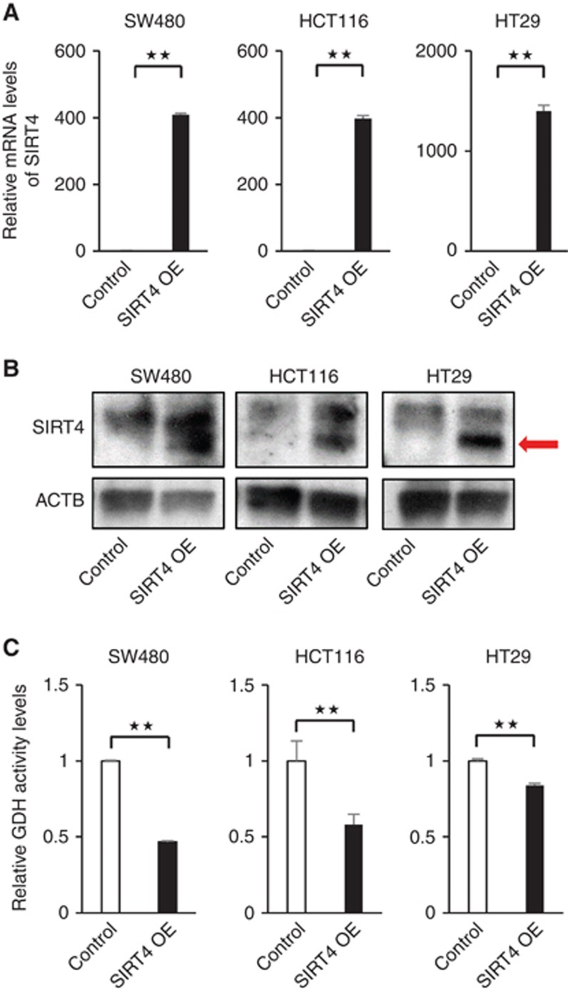
To assess the role of SIRT4 in colorectal cancer, SIRT4 was overexpressed in colorectal cancer cell lines with low mRNA expression levels (SW480, HCT116, and HT29; Supplementary Figure 1). The SIRT4 overexpression was performed using lentiviral infection and confirmed at both the transcript and protein levels (Figure 1A and B). Several studies have reported that SIRT4 represses the enzymatic activity of GDH, limiting the metabolism of glutamate and glutamine to generate ATP (Haigis et al, 2006). The GDH activity was also found to be significantly suppressed by SIRT4 overexpression in colorectal cancer cell lines (Figure 1C).
Figure 1.
Establishment of colorectal cancer cell lines overexpressing SIRT4. (A and B) Colorectal cancer cells (SW480, HCT116, and HT29) transfected with control or SIRT4 overexpression (OE) vector were analysed for SIRT4 expression by qRT–PCR and western blot analysis. (C) Glutamate dehydrogenase activity in control and cells overexpressing SIRT4. Data indicate mean±s.d. of at least three independent experiments (★★P<0.01).
Effect of SIRT4 on proliferation, invasion, and migration
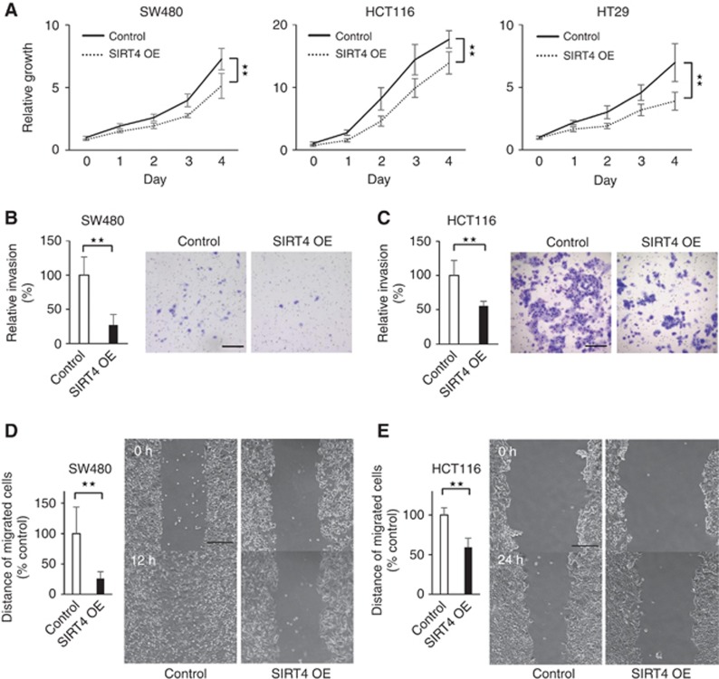
To shed light on the function of SIRT4 in colorectal cancer, proliferation assays were first performed in the cell lines mentioned above. Cell lines overexpressing SIRT4 grew significantly more slowly than control cell lines (Figure 2A). Metastatic disease remains a major problem in the management of colorectal cancer; the influence of SIRT4 on metastatic phenotypes in colorectal cancer cell lines was confirmed by significant inhibition of invasion (Figure 2B and C) and migration (Figure 2D and E) in SW480 and HCT116 cells. Consistent with the above, SIRT4 knockdown increased cell growth, cell invasion, and cell migration in colorectal cancer cells (Supplementary Figure 2A–D). These data demonstrate that SIRT4 suppresses the malignant potential of colorectal cancer cells.
Figure 2.
Inhibition of colorectal cancer cellular malignancy by SIRT4. (A) Overexpression of SIRT4 in SW480, HCT116, and HT29 significantly inhibited their growth. Cell numbers were determined from absorbance at 595 nm (OD 595). (B and C) Relative invasion ability of SIRT4-expressing cells decreased compared with control cells in SW480 (B) and HCT116 (C). The representative images of invasive cells are shown on the right (scale bar, 200 μm). (D and E) The wound healing assay showed that SIRT4 expression inhibited migration of SW480 (D) and HCT116 (E) after incubation for 12 and 24 h, respectively. Representative images at the indicated times are on the right (scale bar, 200 μm). Data are presented as mean±s.d. of at least three independent experiments (★★P<0.01).
Association between SIRT4 and E-cadherin
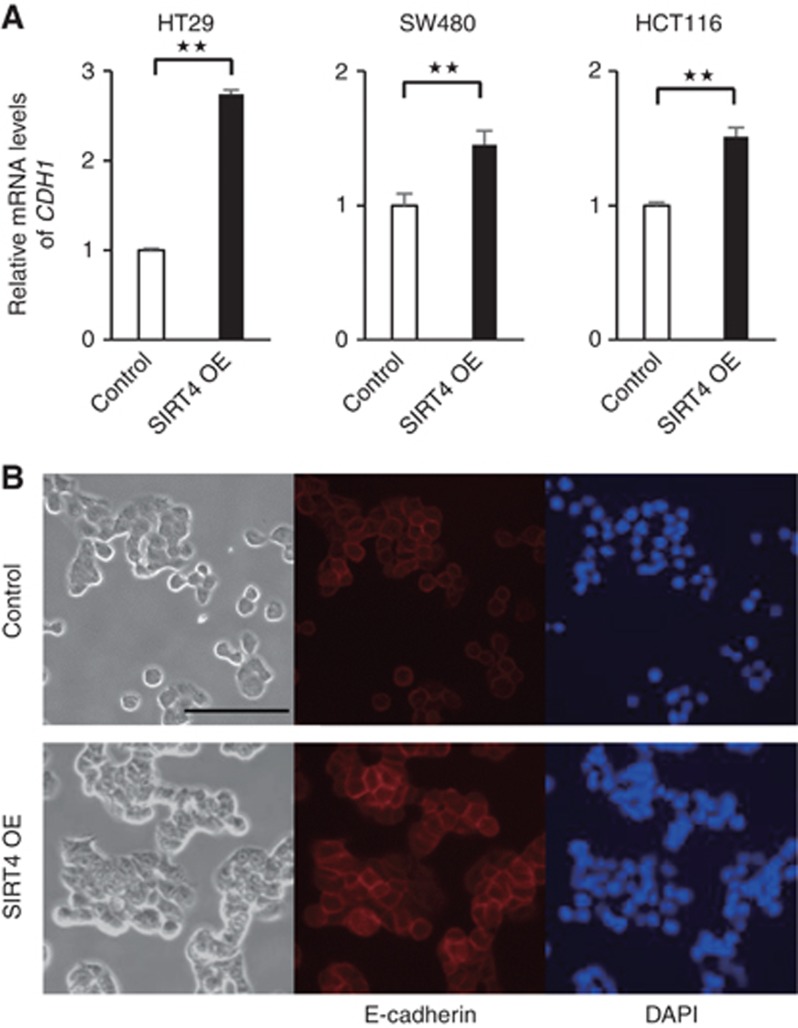
Loss of cell–cell adhesion is an important step associated with tumour invasion and metastases that is frequently accompanied by downregulation of the epithelial molecule E-cadherin (Rachagani et al, 2011). To determine the mechanism by which SIRT4 regulates cellular invasion and migration, we investigated the correlation between SIRT4 and E-cadherin. We found that the E-cadherin gene (CDH1) was significantly upregulated with SIRT4 overexpression in HT29, SW480, and HCT116 cells and downregulated with SIRT4 knockdown in the colorectal cancer cell line CaR1 (Figure 3A and Supplementary Figure 2E). Immunofluorescence staining also confirmed that E-cadherin expression was increased by SIRT4 overexpression (Figure 3B). Furthermore, we found that vimentin (VIM) was significantly downregulated with SIRT4 overexpression in HT29, SW480, and HCT116 cells (Supplementary Figure 3A). The expression of N-cadherin (CDH2) was not detected in these cell lines (Supplementary Figure 3B). Previous reports suggest that E-cadherin expression is suppressed by zinc-finger e-box binding homeobox (ZEB) family that itself is suppressed by miR-200c. However, miR-200c expression was suppressed by SIRT4, and SIRT4 had an inconsistent effect on ZEB1 expression (Supplementary Figure 3C and D), suggesting that different mechanisms might be responsible for the regulation of E-cadherin expression by SIRT4.
Figure 3.
SIRT4 regulates E-cadherin expression in colorectal cancer cell lines. (A) Quantitative RT–PCR showing the expression level of E-cadherin encoded by CDH1 in control and SIRT4 continuously expressing colorectal cancer cell lines (HT29, SW480, and HCT116). (B) Immunofluorescence analysis of E-cadherin in control and SIRT4-expressing HT29 cells (scale bar, 100 μm). Data are presented as mean±s.d. of at least three independent experiments (★★P<0.01).
SIRT4 regulates cellular mobility and E-cadherin expression via suppression of glutamine metabolism
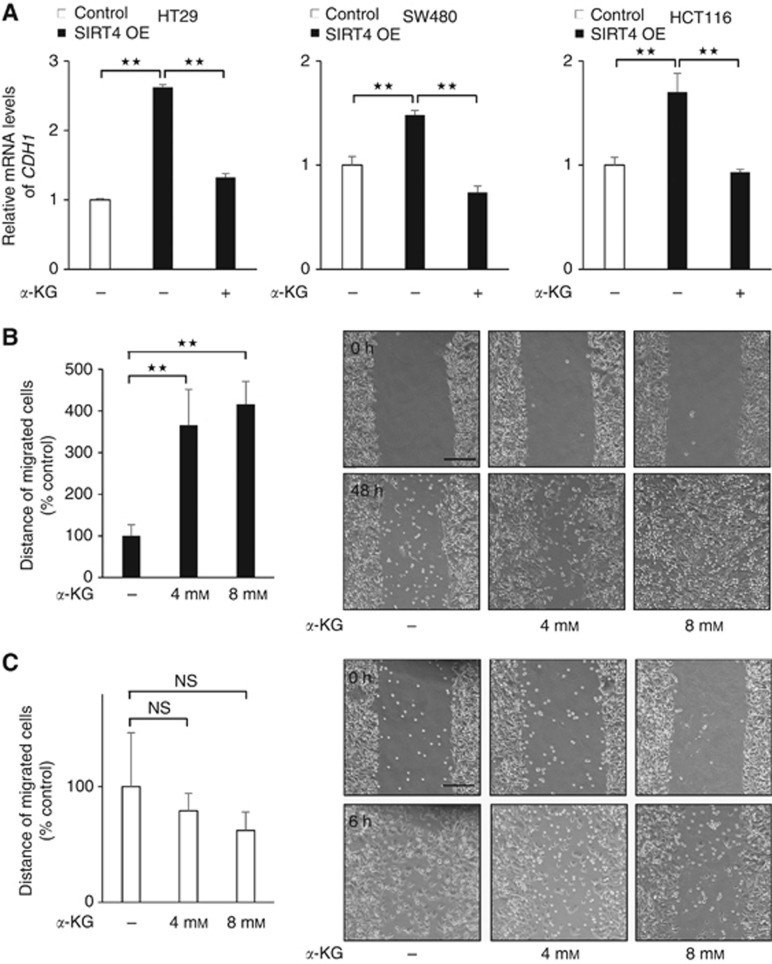
Glutamine is converted into glutamate by glutaminase and then into α-KG by either GDH or, less prominently, by transamination-coupled reactions (DeBerardinis et al, 2008). We observed that SIRT4 represses the enzymatic activity of GDH (Figure 1C). Thus, suppressed glutamine metabolism in colorectal cancer cell lines expressing SIRT4 might result in upregulated E-cadherin expression and decreased mobility. Indeed, α-KG, an important product of glutamine metabolism, abrogated the upregulation of E-cadherin expression by SIRT4 (Figure 4A). Migration was dramatically increased by the presence of α-KG in cells overexpressing SIRT4, but not in cells with basal expression of SIRT4 (Figure 4B and C). These results indicate that repressed glutamine metabolism drove the upregulation of E-cadherin expression and reduced the mobility of cell lines overexpressing SIRT4.
Figure 4.
Regulation of E-cadherin expression and cellular mobility by SIRT4 via suppression of glutamine metabolism. (A) The expression level of E-cadherin encoded by CDH1 in control and colorectal cancer cell lines continuously expressing SIRT4 with or without α-KG (4 mM). (B and C) The wound healing assay in SIRT4-overexpressing SW480 cells (B) and control cells (C) treated with α-KG after incubation for 48 and 6 h, respectively. Representative images at the indicated times are on the right (scale bar, 200 μm). Data are presented as mean±s.d. of at least three independent experiments (★★P<0.01; NS, not significant).
Immunohistochemistry for SIRT4 in colorectal cancer, adenoma, and normal tissue
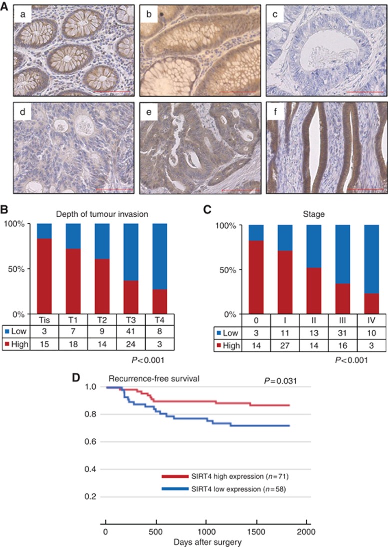
A recent meta-analysis revealed that the SIRT4 mRNA expression in human tumours was significantly lower than that of normal tissues in bladder, breast, colon, gastric, ovarian, and thyroid carcinomas (Csibi et al, 2013). In order to further clarify at which stage of carcinogenesis SIRT4 expression was lost at the protein level, we collected samples from colorectal cancer patients and analysed by immunohistochemistry the expression of SIRT4 in normal colorectal tissue, adenoma, and colorectal cancer. The SIRT4 expression was observed in the glandular cells of colorectal tissue (Figure 5A). The SIRT4 expression was positive in >90% of normal colorectal tissue, adenoma, and colorectal cancer (Supplementary Table 1). The proportions of those with high expression of SIRT4 were 61.1% (77 out of 126), 52.6% (20 out of 38), and 52.5% (75 out of 142) in normal colorectal tissue, adenoma, and colorectal cancer, respectively. The proportion of specimens with high expression of SIRT4 was greater in normal tissue when compared with adenoma and colorectal cancer (P=0.354 and P=0.177, respectively).
Figure 5.
SIRT4 expression by immunohistochemistry. (Aa) Representative SIRT4-positive normal colorectal tissue. (Ab) Representative SIRT4-positive colorectal adenoma. (Ac) Negative staining (−), (Ad) weak staining (+), (Ae) moderate staining (++), and (Af) strong staining (+++) for SIRT4 in colorectal cancer tissue. Scale bar indicates 100 μm. (B and C) The SIRT4 expression at each depth of tumour invasion (B) and each stage (C). The proportion of specimens with high expression of SIRT4 decreased with the depth of tumour invasion (P<0.001, the Cochran–Armitage test for trend) and advancing stage (P<0.001). (D) Kaplan–Meier curves for recurrence-free survival according to SIRT4 expression. Differences between the two groups were evaluated by the log-rank test. Ordinate: survival rate, abscissa: days after surgery.
Correlation between SIRT4 expression and clinicopathological features
Subsequently, we analysed the correlation between SIRT4 expression and clinicopathological features of 142 patients (Table 1). With respect to the depth of tumour invasion, Tis–T2 tumours were associated with a larger proportion of high expression of SIRT4 than T3–T4 tumours (P<0.001). Furthermore, SIRT4 expression decreased progressively from Tis to T4 (P<0.001) (Figure 5B). The SIRT4 expression was also negatively associated with lymph node metastasis, lymphatic invasion, and distant metastasis (P=0.001, P=0.013, and P=0.039, respectively). Consequently, SIRT4 expression was associated with the TNM stage (stages 0, I, and II vs III and IV; P<0.001), decreasing progressively from stage 0 to stage IV (P<0.001) (Figure 5C). The SIRT4 expression in colorectal cancer did not correlate with sex, age, venous invasion, or histological type.
Table 1. Statistical results of immunohistochemistry for SIRT4 in colorectal cancer.
| Clinicopathological factors | Classification | N | SIRT4 high expressiona | SIRT4 low expressionb | P-value |
|---|---|---|---|---|---|
|
Patient background | |||||
| Sex | Male | 89 | 47 | 42 | NS |
| Female | 53 | 27 | 26 | ||
| Age | <65 | 72 | 33 | 39 | NS |
| ⩾65 | 70 | 41 | 29 | ||
|
Tumour characteristics | |||||
| Histological type | tub1, tub2, pap | 128 | 67 | 61 | NS |
| por, muc | 14 | 7 | 7 | ||
| Depth of tumour invasion | Tis, T1, T2 | 66 | 47 | 19 | <0.001 |
| T3, T4 | 76 | 27 | 49 | ||
| Lymph node metastasis | Positive | 57 | 20 | 37 | 0.001 |
| Negative | 85 | 54 | 31 | ||
| Distant metastasis | Positive | 13 | 3 | 10 | 0.039 |
| Negative | 129 | 71 | 58 | ||
| Lymphatic invasion | Positive | 93 | 41 | 52 | 0.013 |
| Negative | 49 | 33 | 16 | ||
| Venous invasion | Positive | 28 | 12 | 16 | NS |
| Negative | 114 | 62 | 52 | ||
| Stage | 0, I, II | 82 | 55 | 27 | <0.001 |
| III, IV | 60 | 19 | 41 | ||
Abbreviations: muc=mucinous carcinoma; NS=not significant; pap=papillary adenocarcinoma; por=poorly differentiated adenocarcinoma; tub1=well-differentiated adenocarcinoma; tub2=moderately differentiated adenocarcinoma.
Percentage of positive cases with (++) and (+++) staining score.
Percentage of positive cases with (−) and (+) staining score.
Expression of SIRT4 is associated with recurrence-free survival of patients with colorectal cancer
The median follow-up duration of patients was 1755 days (335–2482 days). The median recurrence-free survival duration of all eligible patients was 1734 days (133–2482 days). Recurrence was diagnosed after surgery in 25 of 129 patients (19.3%), and the median length of time from surgery to recurrence was 436 days (133–1430 days). The recurrence rate in patients with low expression of SIRT4 was significantly higher than in patients with high expression of SIRT4 (Figures 5D, P=0.031). In addition, depth of tumour invasion (P<0.001), lymph node metastasis (P=0.008), venous invasion (P=0.002), and lymphatic invasion (P=0.001) were also significantly associated with recurrence-free survival, but sex, age, and histological type were not (data not shown). There was a trend for higher rate of 5-year overall survival in patients with high expression of SIRT4 than in patients with low expression of SIRT4 (Supplementary Figure 4, P=0.324).
Discussion
In tumour invasion, epithelial cells lose their epithelial characteristics and acquire mesenchymal properties in the process of epithelial–mesenchymal transition (EMT). The EMT, which allows cancer cells to dissociate from the bulk of the tumour and migrate to other tissues, is characterised by loss of the epithelial cell adhesion molecule E-cadherin, disrupting intercellular contacts and enabling cells to increase motility and invasiveness. The mechanism of induction and regulation of E-cadherin has been elucidated, and recently some reports have indicated the relevance of glycolysis in this process. High glucose suppresses the mRNA expression of E-cadherin compared with low glucose in pancreatic cancer (Han et al, 2013). Hamabe et al (2014) indicated that pyruvate kinase M2, an alternatively spliced variant of the pyruvate kinase gene, mediates E-cadherin in colorectal cancer. However, the relationship between E-cadherin and glutamine metabolism has not been reported to date in the literature. In this study, we first demonstrated that suppression of glutamine metabolism by SIRT4 resulted in positive regulation of E-cadherin expression. Furthermore, the fact that α-KG abrogated the induction of E-cadherin expression by SIRT4 suggested that SIRT4 inhibits EMT through reducing levels of intracellular a-KG, via inactivation of GDH. Additional studies are required to address the mechanism of regulation of E-cadherin by glutamine metabolism.
Colorectal cancer patients surviving for ⩾5 years exhibit significantly higher levels of E-cadherin mRNA than those surviving for <5 years (Dorudi et al, 1995). Jie et al (2013) revealed that E-cadherin was significantly downregulated in high TNM stage lesions and distant metastasis. Thus, loss of E-cadherin, which contributes to cancer development and progression, is associated with poor prognosis in human cancer (Al-Saad et al, 2008). In this study, we found that SIRT4 inhibited proliferation, invasion, and migration in colorectal cancer cells and that a low expression level of SIRT4 was correlated with a worse prognosis. Furthermore, SIRT4 expression decreased with the progression of invasion and metastasis in colorectal cancer. Similar to E-cadherin, SIRT4 plays a role in the suppression of the malignant phenotype in colorectal cancer, and the effect of SIRT4 may partially result from its regulation of E-cadherin. SIRT4 may be an important factor that determines the degree of malignancy of colorectal cancer.
Although understanding the mechanisms by which glutamine supports cancer metabolism is now an area of active investigation, the role of glutamine metabolism in colon cancer cell migration and invasion also deserves attention. Wang et al (2010) reported that in cell invasion assays, the migratory activity of the transformed fibroblasts and cancer cells is severely compromised by treatment with glutaminase inhibitor 968, suggesting a contribution of glutamine metabolism to cancer cell migration. Moreover, Fu et al (2004) indicated that glutamine restriction inhibited attachment, spreading, and migration of melanoma cell lines via inhibition of specific integrin expression and modulation of actin cytoskeleton remodelling. Our results shed light on other molecular mechanisms of glutamine metabolism modulation, in this case effects on E-cadherin expression through SIRT4 and consequently on cell mobility. Thus, glutamine metabolism may be important for cancer cell mobility.
In conclusion, we found that SIRT4 regulation of E-cadherin expression negatively modulated colorectal cancer cellular malignancy through suppression of glutamine metabolism and that SIRT4 expression decreased with the progression of invasion and metastasis in colorectal cancer. These findings suggest that SIRT4 has a tumour-suppressive activity and may serve as a novel therapeutic target in colorectal tumours.
Acknowledgments
We thank the members of our laboratories for their fruitful discussions. This work received the following financial support: a Grant-in-Aid for Scientific Research from the Ministry of Education, Culture, Sports, Science, and Technology (to MK, HI, and MM); a Grant-in-Aid from the Ministry of Health, Labor, and Welfare (to MK, HI, and MM); a grant from the Kobayashi Cancer Research Foundation (to HI); a grant from the Princess Takamatsu Cancer Research Fund, Japan (to HI); a grant from the National Institute of Biomedical Innovation (to MK, HI, and MM); and a grant from the Osaka University Drug Discovery Funds (to MK, HI, and MM). Partial support was received from Takeda Science and Medical Research Foundation through institutional endowments (to MM and HI), Princess Takamatsu Cancer Research Fund (to MM and HI), Kobayashi Cancer Research Foundation (to MM and HI), Suzuken Memorial Foundation (to MK), Yasuda Medical Foundation (to NN), Pancreas Research Foundation (to KK), Nakatani Foundation (to HI), and Nakatomi Foundation of Japan (to MK).
Institutional endowments were received partially from Taiho Pharmaceutical Co., Ltd, Evidence Based Medical (EBM) Research Center, Yakult Honsha Co, Ltd, Chugai Co., Ltd, and Merck Co., Ltd; these funders had no role in the main experimental equipment, supplies expenses, study design, data collection and analysis, decision to publish, or preparation of the manuscript for this work.
Footnotes
Supplementary Information accompanies this paper on British Journal of Cancer website (http://www.nature.com/bjc)
This work is published under the standard license to publish agreement. After 12 months the work will become freely available and the license terms will switch to a Creative Commons Attribution-NonCommercial-Share Alike 4.0 Unported License
Supplementary Material
References
- Al-Saad S, Al-Shibli K, Donnem T, Persson M, Bremnes RM, Busund LT. The prognostic impact of NF-kappaB p105, vimentin, E-cadherin and Par6 expression in epithelial and stromal compartment in non-small-cell lung cancer. Br J Cancer. 2008;99:1476–1483. doi: 10.1038/sj.bjc.6604713. [DOI] [PMC free article] [PubMed] [Google Scholar]
- Csibi A, Fendt SM, Li C, Poulogiannis G, Choo AY, Chapski DJ, Jeong SM, Dempsey JM, Parkhitko A, Morrison T, Henske EP, Haigis MC, Cantley LC, Stephanopoulos G, Yu J, Blenis J. The mTORC1 pathway stimulates glutamine metabolism and cell proliferation by repressing SIRT4. Cell. 2013;153:840–854. doi: 10.1016/j.cell.2013.04.023. [DOI] [PMC free article] [PubMed] [Google Scholar]
- DeBerardinis RJ, Lum JJ, Hatzivassiliou G, Thompson CB. The biology of cancer: metabolic reprogramming fuels cell growth and proliferation. Cell Metab. 2008;7:11–20. doi: 10.1016/j.cmet.2007.10.002. [DOI] [PubMed] [Google Scholar]
- Dorudi S, Hanby AM, Poulsom R, Northover J, Hart IR. Level of expression of E-cadherin mRNA in colorectal cancer correlates with clinical outcome. Br J Cancer. 1995;71:614–616. doi: 10.1038/bjc.1995.119. [DOI] [PMC free article] [PubMed] [Google Scholar]
- Edge SB, Byrd DR, Compton CC, Fritz AG, Greene FL, Trotti A.(eds) (2010AJCC Cancer Staging Manual7th edn.Springer: New York [Google Scholar]
- Fu YM, Zhang H, Ding M, Li YQ, Fu X, Yu ZX, Meadows GG. Specific amino acid restriction inhibits attachment and spreading of human melanoma via modulation of the integrin/focal adhesion kinase pathway and actin cytoskeleton remodeling. Clin Exp Metastasis. 2004;21:587–598. doi: 10.1007/s10585-004-5515-y. [DOI] [PubMed] [Google Scholar]
- Haigis MC, Mostoslavsky R, Haigis KM, Fahie K, Christodoulou DC, Murphy AJ, Valenzuela DM, Yancopoulos GD, Karow M, Blander G, Wolberger C, Prolla TA, Weindruch R, Alt FW, Guarente L. SIRT4 inhibits glutamate dehydrogenase and opposes the effects of calorie restriction in pancreatic beta cells. Cell. 2006;126:941–954. doi: 10.1016/j.cell.2006.06.057. [DOI] [PubMed] [Google Scholar]
- Hamabe A, Konno M, Tanuma N, Shima H, Tsunekuni K, Kawamoto K, Nishida N, Koseki J, Mimori K, Gotoh N, Yamamoto H, Doki Y, Mori M, Ishii H. Role of pyruvate kinase M2 in transcriptional regulation leading to epithelial-mesenchymal transition. Proc Natl Acad Sci USA. 2014;111:15526–15531. doi: 10.1073/pnas.1407717111. [DOI] [PMC free article] [PubMed] [Google Scholar]
- Han L, Peng B, Ma Q, Ma J, Li J, Li W, Duan W, Chen C, Liu J, Xu Q, Laporte K, Li Z, Wu E. Indometacin ameliorates high glucose-induced proliferation and invasion via modulation of e-cadherin in pancreatic cancer cells. Curr Med Chem. 2013;20:4142–4152. doi: 10.2174/09298673113209990249. [DOI] [PMC free article] [PubMed] [Google Scholar]
- Hu H, Zhang H, Ge W, Liu X, Loera S, Chu P, Chen H, Peng J, Zhou L, Yu S, Yuan Y, Zhang S, Lai L, Yen Y, Zheng S. Secreted protein acidic and rich in cysteines-like 1 suppresses aggressiveness and predicts better survival in colorectal cancers. Clin Cancer Res. 2012;18:5438–5448. doi: 10.1158/1078-0432.CCR-12-0124. [DOI] [PubMed] [Google Scholar]
- Jemal A, Bray F, Center MM, Ferlay J, Ward E, Forman D. Global cancer statistics. CA Cancer J Clin. 2011;61:69–90. doi: 10.3322/caac.20107. [DOI] [PubMed] [Google Scholar]
- Jeong SM, Xiao C, Finley LW, Lahusen T, Souza AL, Pierce K, Li YH, Wang X, Laurent G, German NJ, Xu X, Li C, Wang RH, Lee J, Csibi A, Cerione R, Blenis J, Clish CB, Kimmelman A, Deng CX, Haigis MC. SIRT4 has tumor-suppressive activity and regulates the cellular metabolic response to DNA damage by inhibiting mitochondrial glutamine metabolism. Cancer Cell. 2013;23:450–463. doi: 10.1016/j.ccr.2013.02.024. [DOI] [PMC free article] [PubMed] [Google Scholar]
- Jie D, Zhongmin Z, Guoqing L, Sheng L, Yi Z, Jing W, Liang Z. Positive expression of LSD1 and negative expression of E-cadherin correlate with metastasis and poor prognosis of colon cancer. Dig Dis Sci. 2013;58:1581–1589. doi: 10.1007/s10620-012-2552-2. [DOI] [PubMed] [Google Scholar]
- Laurent G, German NJ, Saha AK, de Boer VC, Davies M, Koves TR, Dephoure N, Fischer F, Boanca G, Vaitheesvaran B, Lovitch SB, Sharpe AH, Kurland IJ, Steegborn C, Gygi SP, Muoio DM, Ruderman NB, Haigis MC. SIRT4 coordinates the balance between lipid synthesis and catabolism by repressing malonyl CoA decarboxylase. Mol Cell. 2013;50:686–698. doi: 10.1016/j.molcel.2013.05.012. [DOI] [PMC free article] [PubMed] [Google Scholar]
- Liu B, Che W, Xue J, Zheng C, Tang K, Zhang J, Wen J, Xu Y. SIRT4 prevents hypoxia-induced apoptosis in H9c2 cardiomyoblast cells. Cell Physiol Biochem. 2013;32:655–662. doi: 10.1159/000354469. [DOI] [PubMed] [Google Scholar]
- Mahlknecht U, Voelter-Mahlknecht S. Fluorescence in situ hybridization and chromosomal organization of the sirtuin 4 gene (Sirt4) in the mouse. Biochem Biophys Res Commun. 2009;382:685–690. doi: 10.1016/j.bbrc.2009.03.092. [DOI] [PubMed] [Google Scholar]
- Ma L, Young J, Prabhala H, Pan E, Mestdagh P, Muth D, Teruya-Feldstein J, Reinhardt F, Onder TT, Valastyan S, Westermann F, Speleman F, Vandesompele J, Weinberg RA. miR-9, a MYC/MYCN-activated microRNA, regulates E-cadherin and cancer metastasis. Nat Cell Biol. 2010;12:247–256. doi: 10.1038/ncb2024. [DOI] [PMC free article] [PubMed] [Google Scholar]
- Rachagani S, Senapati S, Chakraborty S, Ponnusamy MP, Kumar S, Smith LM, Jain M, Batra SK. Activated KrasG12D is associated with invasion and metastasis of pancreatic cancer cells through inhibition of E-cadherin. Br J Cancer. 2011;104:1038–1048. doi: 10.1038/bjc.2011.31. [DOI] [PMC free article] [PubMed] [Google Scholar]
- Reid MA, Wang WI, Rosales KR, Welliver MX, Pan M, Kong M. The B55α subunit of PP2A drives a p53-dependent metabolic adaptation to glutamine deprivation. Mol Cell. 2013;50:200–211. doi: 10.1016/j.molcel.2013.02.008. [DOI] [PubMed] [Google Scholar]
- Wang JB, Erickson JW, Fuji R, Ramachandran S, Gao P, Dinavahi R, Wilson KF, Ambrosio AL, Dias SM, Dang CV, Cerione RA. Targeting mitochondrial glutaminase activity inhibits oncogenic transformation. Cancer Cell. 2010;18:207–219. doi: 10.1016/j.ccr.2010.08.009. [DOI] [PMC free article] [PubMed] [Google Scholar]
- Zhao QJ, Yu YB, Zuo XL, Dong YY, Li YQ. Milk fat globule-epidermal growth factor 8 is decreased in intestinal epithelium of ulcerative colitis patients and thereby causes increased apoptosis and impaired wound healing. Mol Med. 2012;18:497–506. doi: 10.2119/molmed.2011.00369. [DOI] [PMC free article] [PubMed] [Google Scholar]
Associated Data
This section collects any data citations, data availability statements, or supplementary materials included in this article.


