Abstract
When skeletal muscle fibers are injured, they regenerate and grow until their sizes are adjusted to surrounding muscle fibers and other relevant organs. In this study, we examined whether Mest, one of paternally expressed imprinted genes that regulates body size during development, and miR-335 located in the second intron of the Mest gene play roles in muscle regeneration. We generated miR-335-deficient mice, and found that miR-335 is a paternally expressed imprinted microRNA. Although both Mest and miR-335 are highly expressed during muscle development and regeneration, only Mest+/- (maternal/paternal) mice show retardation of body growth. In addition to reduced body weight in Mest+/-; DMD-null mice, decreased muscle growth was observed in Mest+/- mice during cardiotoxin-induced regeneration, suggesting roles of Mest in muscle regeneration. Moreover, expressions of H19 and Igf2r, maternally expressed imprinted genes were affected in tibialis anterior muscle of Mest+/-; DMD-null mice compared to DMD-null mice. Thus, Mest likely mediates muscle regeneration through regulation of imprinted gene networks in skeletal muscle.
Introduction
Skeletal muscle increases its mass during fetal, neonatal, and juvenile development. It has been known that IGF1-AKT-mTOR and Myostatin-Smad pathways are involved in positive and negative regulation of skeletal muscle mass, respectively [1]. These signaling pathways likely undertake muscle mass maintenance and plasticity in adult in addition to regulation of muscle growth during development. On the other hand, lengths of skeletal muscle fibers should be coordinately regulated with other relevant organs such as bones and tendons during development. Although underlying genetic and epigenetic mechanisms and gene networks for such a coordinated growth of skeletal muscle with body size still need to be elucidated, genetic evidence suggests that many genomic imprinted genes are involved in that process [2–8]. Under this scenario, some of these imprinted genes should also regulate muscle regeneration in which muscle stem cells expand and fuse with each other or with damaged myofibers to form adequate-sized regenerating muscle that fits surrounding tissues.
In this study, we examined whether the Mest (Mesoderm specific transcript) gene, a paternally expressed imprinted gene that regulates body size and miR-335, a microRNA (miRNA) located in intron 2 of Mest, are involved in muscle regeneration. miRNAs are a class of small noncoding RNAs of ~23 nucleotides that inhibit gene expression by binding 3’UTR of target mRNAs, and have emerged as a class of key post-transcriptional regulators of gene expression [9,10]. Recent studies have revealed the involvement of miRNAs in elaborate regulation of cell proliferation and differentiation [11]. We sought to determine whether miR-335 was co-regulated with Mest during postnatal development and skeletal muscle regeneration. When Mest-deficient mice were generated previously, miR-335 had not been identified in the Mest locus [2]. Although miR-335 genomic locus was left intact in the Mest +/- (maternal/paternal) mice, our finding that expression of miR-335 was significantly decreased in those mice lead us to examine which is responsible for the regulation of body size, Mest, miR-335, or both. miR-335 mutant mice revealed that Mest, but not miR-335, is responsible for the control of body and muscle sizes and for the regulation of muscle growth during regeneration. Although miR-335 was co-regulated with Mest as a paternally expressed imprinted miRNA, neither body growth nor muscle regeneration was affected by deleting miR-335.
Two types of experimental muscle regenerating models were used in this study. One is to analyze the acute phase of regeneration induced by cardiotoxin (CTX) administration into skeletal muscle, and the other is to investigate the chronic phase of regeneration in Dystrophin gene-deleted (DMD-null) mice [12], useful model for Duchenne Muscular Dystrophy (DMD). DMD is one of the most common and severe forms of muscular dystrophy [13–15]. DMD muscle fibers are highly susceptible to mechanical damage and therefore exhibit chronic or repetitive regeneration. Mest +/- ; DMD-null mice exhibited a significant reduction in body growth after 1 week compared to DMD-null mice, and showed the dysregulation of H19 and Igf2r genes, maternally expressed imprinted genes. Moreover Mest +/- mice resulted in decrease in muscle fiber thickness when skeletal muscle regeneration was induced with CTX. Roles of Mest in muscle regeneration are discussed based on these results.
Results
Mest and miR-335 are coordinately expressed during skeletal muscle development and regeneration
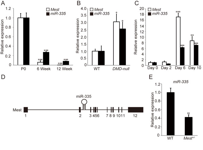
Both Mest and miR-335 were highly expressed in tibialis anterior (TA) muscles at postnatal day 0 (P0), and decreased gradually as mice grew up (Fig 1A). A previous report showed that miR-335 was up-regulated in adductor muscle of mdx mice and skeletal muscle biopsy of DMD patients in common [16]. We found the up-regulation of Mest as well as miR-335 in TA muscles of 3 months old DMD–null compared with that of wild type (WT) mice (Fig 1B). Since DMD-null mice undergo repetitive myofiber degeneration and regeneration, we next examined whether Mest and miR-335 were up-regulated either in degenerating or in regenerating phases upon skeletal muscle injury induced by the administration of CTX into TA muscles. Degenerating and regenerating muscles were isolated 2 days and 6–10 days after the CTX injection, respectively. Both Mest and miR-335 were remarkably activated in the regenerating phase but not in the degenerating phase (Fig 1C). Thus, Mest and miR-335 are expressed coordinately during skeletal muscle growth and regeneration, and in the pathological condition of DMD. The Mest mutant mice had been established by deleting exons 3–8 and part of exon 9 previously [2], which left the miR-335 genomic locus locating within the intron 2 of the Mest gene intact (Fig 1D). Interestingly, however, quantitative RT-PCR (qRT-PCR) analyses revealed that expression of miR-335 was significantly lower in Mest +/- than in WT mice (Fig 1E).
Fig 1. Mest and miR-335 are coordinately expressed in skeletal muscle during postnatal development and regeneration.
(A) qRT-PCRs for Mest mRNA and miR-335 were performed with TA muscles of P0, 6 weeks, and 12 weeks old WT mice (n = 3 per time point). (B) qRT-PCRs for Mest mRNA and miR-335 were performed with TA muscles of 3 months old WT (n = 4) and DMD–null mice (n = 7). (C) qRT-PCRs for Mest mRNA and miR-335 were performed with TA muscles from day 0 to day 10 after CTX injection (n = 3 per time point). (D) A schematic diagram of the Mest and miR-335 genomic region on chromosome 6 in mouse. (E) qRT-PCR for miR-335 was performed in TA muscles of WT and Mest +/- mice (n = 3 per genotype). Expression of Mest and that of miR-335 are normalized to Gapdh and snoRNA-202, respectively. Error bars indicate the s.e.m. *P < 0.05, **P < 0.01, ***P < 0.001 compared with P0 (A), WT (B and E), and day 0 (C).
miR-335 is a paternally expressed imprinted gene
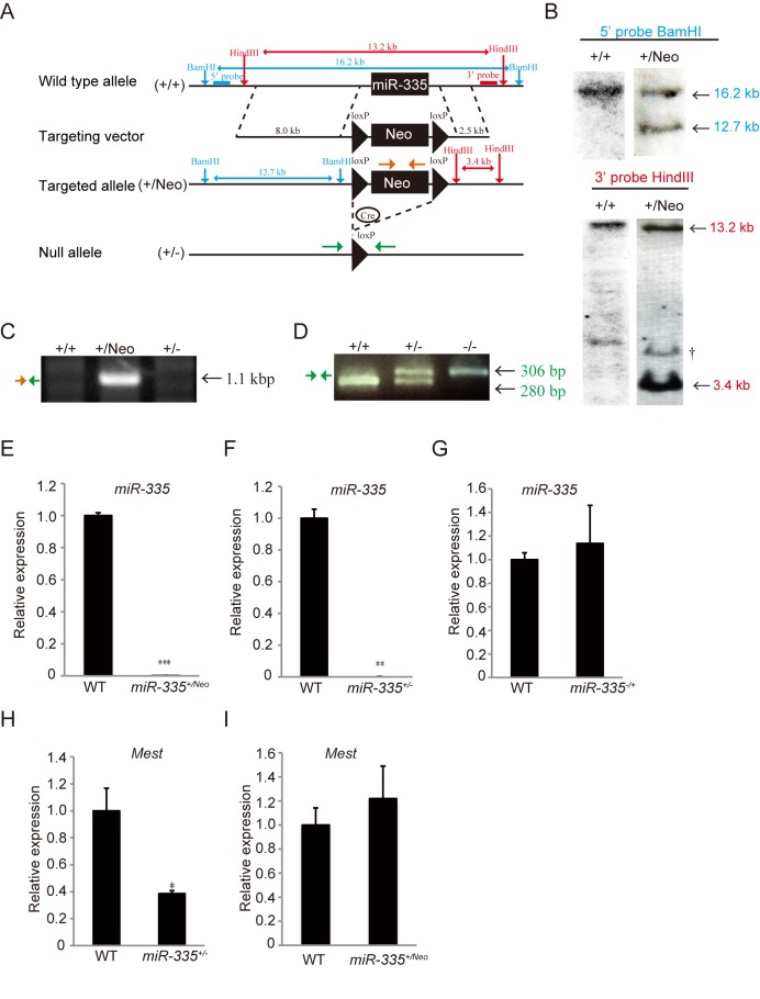
To distinguish roles of Mest from those of miR-335, we generated miR-335 deficient mice by homologous recombination in embryonic stem (ES) cells. The miR-335 sequence was replaced with a neomycin resistance cassette flanked by loxP sites (Fig 2A). Targeted ES clones were confirmed by Southern blotting using 5’ and 3’ probes against mouse genomic DNA digested with BamHI and HindIII, respectively (Fig 2B). The absence of random integration was verified with a neomycin probe (data not shown). Removal of the neomycin resistance cassette by mating male miR-335 +/Neo mice to female CAG-cre transgenic mice resulted in the deletion of short flanking sequences together with the neomycin cassette (miR-335 +/- mice) (Fig 2C and 2D). Mest is an imprinted gene that is expressed essentially from the paternal allele during development [2]. We investigated whether miR-335 is also paternally expressed. qRT-PCR analysis revealed that miR-335 +/Neo (maternal/paternal) mice obtained by crossing male miR-335 +/Neo mice with female WT mice scarcely expressed miR-335 in skeletal muscle (Fig 2E). Similarly, miR-335 +/- (maternal/paternal) mice obtained by crossing male miR-335 +/- mice with female WT mice scarcely expressed miR-335 in skeletal muscle (Fig 2F). On the contrary, miR-335 -/+ (maternal/paternal) mice obtained by crossing male WT mice with female miR-335 +/- mice expressed miR-335 in skeletal muscle comparable to WT mice (Fig 2G). Thus, these results indicate that miR-335 is a paternally expressed imprinted miRNA. In addition, while the expression of Mest was decreased in miR-335 +/- mice, it was not altered in miR-335 +/Neo mice (Fig 2H and 2I), suggesting that the region surrounding the neomycin resistance cassette flanked by loxP sites, but not the miR-335 itself, is involved in transcriptional regulation of Mest. To evaluate roles of miR-335, further analyses were mainly performed with miR-335 +/Neo mice.
Fig 2. Generation of miR-335 deficient mice.
(A) Design of constructs used for generation of miR-335 deficient mice. The miR-335 genomic locus was replaced by a floxed neomycin-resistance cassette (loxP-Neo-loxP) to obtain miR-335 +/Neo mice. miR-335 +/- mice (+/-) were generated by crossing male miR-335 +/Neo mice with female CAG-cre transgenic mice. (B) Southern blot analysis of WT and G418 resistant ES clones with 5’ and 3’ probes. †: Non-specific band. (C and D) PCR analysis for targeted allele with genomic DNA in tails of WT (+/+), miR-335 +/Neo (+/Neo), and miR-335 +/- mice (+/-). In (C), an insertion and a deletion of a floxed neomycin-resistance cassette in genomic DNA of miR-335 +/Neo (+/Neo) and miR-335 +/- mice (+/-), respectively, were detected with PCRs amplified with primers shown in Fig 2A (Orange and Green arrows). In (D), a PCR analysis to distinguish alleles for WT (+/+), miR-335 +/- (+/-), and miR-335 -/- mice (-/-) was shown. The WT allele-specific (280 bp) and the mutant allele-specific (306 bp) bands were amplified with the primers shown in Fig 2A (Green arrow). (E, F and G) qRT-PCR for miR-335 was performed in TA muscles isolated from WT, miR-335 +/Neo, and miR-335 +/- or miR-335 -/+ mice (n = 3 per genotype). (H and I) qRT-PCR for Mest mRNA was performed in TA muscles of WT, miR-335 +/-, and miR-335 +/Neo mice (n = 3 per genotype). Expression of Mest mRNA and that of miR-335 are normalized to Gapdh and snoRNA-202, respectively. Error bars indicate the s.e.m. *P < 0.05, **P < 0.01, ***P < 0.001.
Mest but not miR-335 affects body growth
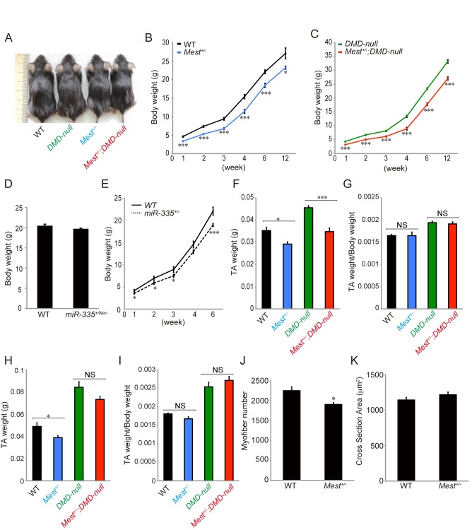
Mest +/- mice show growth retardation ([2], Fig 3A and 3B). miR-335 +/Neo mice, in contrast, did not show a reduction in body weight compared to WT mice significantly (Fig 3D). miR-335 +/- mice exhibited mild growth defects (Fig 3E), which is likely due to decreased expression of Mest in miR-335 +/- mice (Fig 2H). Based on these results, we concluded that Mest, but not miR-335, is involved in regulation of body size during development. When TA muscles of WT and Mest +/- mice were compared at 6 and 11–13 weeks of age, those of the latter were significantly smaller than those of the former (Fig 3F and 3H). Moreover, numbers of muscle fibers are significantly less in Mest +/- mice (Fig 3J). In contrast, the ratio of TA weight/Body weight and the average cross section areas of TA muscles of both WT and Mest +/- mice were similar to each other (Fig 3G, 3I and 3K).
Fig 3. Mest is required for body and skeletal muscle growth.
(A) Representative images of 4 weeks old mice in individual genotypes. (B and C) Body weights of male littermate WT (n = 3–22), Mest +/- (n = 6–15), DMD-null (n = 4–25), and Mest +/- ; DMD-null mice (n = 4–22) from 1 to 12 (11–13) weeks old. (D) Body weights of WT (n = 15) and miR-335 +/Neo (n = 12) mice at 6 weeks. (E) Body weights of WT (n = 8–15) and miR-335 +/- mice (n = 15–18) from 1 to 6 weeks old. (F) TA muscle weights of male littermate WT (n = 13), Mest +/- (n = 6), DMD-null (n = 18), and Mest +/- ; DMD-null mice (n = 11) at 6 weeks old. (G) TA/Body weights of male littermate WT, Mest +/-, DMD-null, and Mest +/- ; DMD-null mice at 6 weeks old. (H) TA muscle weights of male littermate WT (n = 3), Mest +/- (n = 6), DMD-null (n = 4), and Mest +/- ; DMD-null mice (n = 4) at 11–13 weeks old. (I) TA/Body weights of male littermate WT, Mest +/-, DMD-null, and Mest +/- ; DMD-null mice at 11–13 weeks old. (J and K) The numbers and average cross section areas of TA muscle fibers of male littermate WT (n = 7) and Mest +/- mice (n = 4) at 6 weeks. Error bars indicate the s.e.m. *P < 0.05, ***P < 0.001. NS = Not significant.
Mest affects skeletal muscle growth during regeneration
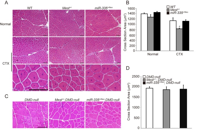
Although the expression of Mest is up-regulated in regenerating skeletal muscle induced by injection of CTX and in skeletal muscle of adult DMD–null mice (Fig 1B and 1C), roles of Mest in muscle regeneration and pathology of DMD remain unknown. In order to clarify roles of Mest in muscle regeneration, we examined whether muscle regeneration induced by CTX was affected in Mest +/- mice. Ratios of myofibers with central nuclei in Mest +/- mice were comparable to those in WT mice, indicating that muscle regeneration occurs in the absence of Mest (Fig 4A). However, regenerating muscle fibers were significantly thinner in Mest +/- than those in WT mice when analyzed 14 days after CTX-injection while average cross section areas of intact TA muscle fibers of both WT and Mest +/- mice were similar to each other (Fig 4A and 4B). These results indicate that Mest is required for efficient muscle growth during muscle regeneration. Next, we investigated whether Mest affects pathology of DMD-null mice. There is no significant difference in Cross Section Areas (CSA) between DMD-null and Mest +/- ; DMD-null mice (Fig 4C and 4D). We analyzed body weight of DMD-null and Mest +/- ; DMD-null mice. Fig 3C shows that body weights of Mest +/- ; DMD-null mice were consistently smaller than those of DMD-null mice from 1 week to 11–13 weeks. If Mest promotes muscle regeneration in addition to body growth during postnatal development, the difference in body weights between DMD-null and Mest +/- ; DMD-null mice should be accelerated after muscle degeneration and regeneration start in them. In contrast, such an accelerated difference in body weights would not be observed between WT and Mest +/- mice, which no regeneration occurs. As shown in S1 Fig, the ratio of Mest +/- ; DMD-null/ DMD-null indeed became slightly lower than that of Mest +/-/WT after 4 weeks of age when muscle degeneration and regeneration start in those mice. The result implies contribution of Mest in body growth during muscle regeneration as well as contribution in body growth during postnatal development. Furthermore, we analyzed the relation between body and TA weight in 6 and 11–13 weeks DMD-null and Mest +/- ; DMD-null mice. TA weights of Mest +/- ; DMD-null mice were significantly smaller at 6 weeks and slightly smaller at 11–13 weeks compared to those of DMD-null mice although statistical significance could not be obtained with the latter probably due to small numbers of mice we analyzed (Fig 3F and 3H). TA/body weight was more or less constant among these mice (Fig 3G and 3I). In contrast, there was no significant difference in muscle growth during regeneration induced by CTX between WT and miR-335 +/Neo mice and in growth of Dystrophin-deficient muscle between DMD-null and miR-335 +/Neo ; DMD-null mice (Fig 4A–4D). Therefore, these result suggest that Mest but not miR-335 is involved in skeletal muscle growth during regeneration.
Fig 4. Mest is required for skeletal muscle growth during regeneration.
(A) H&E staining of TA muscles under normal condition (top panel) and 14 days after CTX-induced injury (middle and bottom panels). Bottom panel shows the extended images of a part of middle panel. (B) Average cross section areas of TA muscles in WT (n = 3), Mest +/- (n = 4) and miR-335 +/Neo mice (n = 6) under normal condition and WT (n = 7), Mest +/- (n = 4) and miR-335 +/Neo mice (n = 6) 14 days after CTX injury. (C) H&E staining of TA muscles of DMD-null, Mest +/- ; DMD-null and miR-335 +/Neo ; DMD-null mice at 11–13 weeks old. (D) Average cross section areas of TA muscles in DMD-null (n = 8), Mest +/- ; DMD-null (n = 4) and miR-335 +/Neo ; DMD-null mice (n = 4) at 11–13 weeks old. Error bars indicate the s.e.m. # P = 0.0549 compared with WT mice. Scale bar: 100 μm.
H19 and Igf2r are affected in Mest +/- ; DMD-null mice
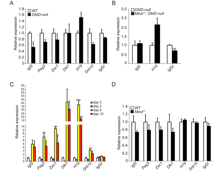
Imprinted gene networks have been reported to control somatic growth [5, 17, 18]. Such imprinted gene networks might also regulate growth of muscle fibers during skeletal muscle regeneration. Indeed Dlk1, a paternally expressed imprinted gene, is involved in skeletal muscle regeneration [19]. To investigate whether Mest has the relation with some paternally and maternally expressed imprinted genes in regenerating TA muscles, we focused on paternally (Igf2, Peg3, Zac1, and Dlk1) and maternally (H19, Grb10, and Igf2r) expressed imprinted genes. qRT-PCR revealed that the expression of Igf2 and H19 was altered in TA muscles at 6 weeks old DMD–null mice compared with WT mice (Fig 5A). Expression of H19 was further augmented while expression of Igf2 was not affected in Mest +/- ; DMD-null mice (Fig 5B). Instead, Igf2r expression was attenuated in Mest +/- ; DMD-null mice (Fig 5B). Thus, H19 and Igf2r, maternally expressed imprinted genes, were affected in pathologically regenerating muscle of Mest +/- ; DMD-null mice. Next, we examined whether some imprinted genes were up-regulated either in degenerating or regenerating phase upon skeletal muscle injury by CTX. These genes except for Igf2r were remarkably activated in the regenerating phase (Fig 5C). However, there was no significant difference based on the expression of these imprinted genes between WT and Mest +/- mice at 6 days after CTX (Fig 5D).
Fig 5. The expression level of imprinted genes is altered in DMD-null mice and CTX injury.
(A) qRT-PCRs for paternally expressed (Igf2, Peg3, Zac1, Dlk1) and maternally expressed (H19, Grb10, Igf2r) imprinted genes were performed in TA muscles of 6 weeks old WT and DMD-null mice (n = 3 per genotype). (B) qRT-PCRs for Igf2, H19, and Igf2r mRNA were performed in TA muscles of 6 weeks old DMD-null (n = 4) and Mest +/- ; DMD-null (n = 6). (C) qRT-PCRs for imprinted genes were performed with TA muscles obtained from WT mice from day 0 to day 10 after CTX injection (n = 3 per time point). (D) qRT-PCRs for imprinted genes were performed in TA muscles of 12–15 weeks old WT (n = 5) and Mest +/- mice (n = 3) 6 days after CTX. Error bars indicate the s.e.m. *P < 0.05, **P < 0.01, ***P < 0.001.
Muscle satellite cells are not affected in Mest +/- mice
The decrease of TA weight and the thin CSA in Mest +/- mice could be due to some defects in muscle satellite cells, skeletal muscle-specific stem cells. In order to investigate functions of Mest in muscle satellite cells, we compared numbers of muscle satellite cells in TA muscles of WT and Mest +/- mice in normal and CTX injured condition. As a result, there was no significant difference in numbers of Pax7-positive muscle satellite cells per myofiber between WT and Mest +/- mice (Fig 6A). Next, in order to evaluate maintenance and differentiation of muscle satellite cells, we examined whether expressions of Pax7 for maintenance and MyoD for differentiation are affected in Mest +/- mice. As a result, there was no significant difference for expression of Pax7 and MyoD both in normal (Fig 6B) and CTX injured condition (S2 Fig) in Mest +/- mice compared to WT mice. In addition, although miR-335 +/Neo mice show no significant difference in number of Pax7-positive muscle satellite cells per myofiber, the expression of Pax7 was decreased (Fig 6A and 6B).
Fig 6. Muscle satellite cells are not affected in Mest +/- mice.
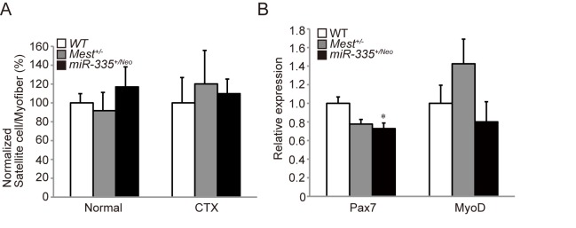
(A) Numbers of Pax7 positive-satellite cells per muscle fiber in sections of TA muscles of WT, Mest +/-, and miR-335 +/Neo mice (n = 3 per genotype) at 10 weeks old in normal condition and 14 day after CTX. (B) qRT-PCRs for Pax7 and MyoD mRNA were performed in normal TA muscles of 11–13 weeks old WT, Mest +/-, and miR-335 +/Neo mice (n = 3 per genotype). Error bars indicate the s.e.m. *P < 0.05 compared with WT mice.
Discussion
Mest is a positive regulator of muscle growth during development and regeneration
In this study, we showed roles of the Mest gene in control of body size and skeletal muscle growth during postnatal development and regeneration. Because the Mest includes miR-335 in its second intron, we examined a possibility whether miR-335 is also involved in body and skeletal muscle growth and in muscle regeneration. Although Mest +/- mice have miR-335 genomic locus, the expression of miR-335 was down-regulated in Mest +/- mice compared to WT mice (Fig 1E). Thus, it was necessary to analyze miR-335 deficient mice to conclude that the Mest gene, not miR-335, is responsible for control of body growth. miR-335 +/Neo mice showed normal body weight and expression of Mest while miR-335 +/- mice showed reduced body weight and expression of Mest (Figs 2H, 2I, 3D and 3E). These results suggest that decreased expression of Mest in miR-335 +/- mice, but not the deletion of miR-335 itself, causes reduced body weight.
Although previous studies pointed out high-level expression of Mest together with other imprinted genes in skeletal muscle [20], its roles in muscle growth and regeneration have been elusive. It is noteworthy that the average cross section areas in Mest +/- mice was significantly smaller in CTX-induced regeneration model, but not in Mest +/- ; DMD-null mice at 11–13 weeks. Mest was much more prominently enhanced in muscle after CTX injection than DMD muscle (Fig 1B and 1C). Therefore, different molecular mechanisms could regulate skeletal muscle regeneration induced by CTX and by Dystrophin-deficiency. Alternatively, synchronous progression of CTX-induced regeneration could be affected in Mest +/- mice more clearly than regeneration caused by Dystrophin-deficiency that occurs randomly within muscle tissues. Although the decrease of TA weight and the thin CSA in injured Mest +/- mice are observed (Figs 3F, 3H and 4B), there was no significant difference in numbers of Pax7-positive muscle satellite cells per myofiber between WT and Mest +/- mice (Fig 6A), suggesting that quiescent satellite cells are generated and maintained similarly in WT and Mest +/- mice. Moreover, Pax7 and MyoD genes were similarly expressed in normal (Fig 6B) and CTX-induced injured conditions (S2 Fig) in Mest +/- mice compared to those in WT mice, suggesting that expression of myogenic transcription factors in muscle satellite cells was not affected by the lack of Mest during CTX-induced myogenic activation. Thus, the reduction of muscle growth by the lack of Mest in CTX-induced regeneration model is likely due to decreased proliferation of muscle satellite cells after their activation and/or decreased thickening of muscle fibers after myotube formation. Taken similarity in CSAs of intact TA muscle fibers of both WT and Mest +/- mice into account, involvement of Mest in proliferation of muscle satellite cells rather than that in hypertrophic growth of muscle fibers is favorable for the interpretation of these data. In contrast, expression of Pax7 was affected in miR-335 +/Neo mice. However, more precise analyses need to be performed in order to know whether proliferation and differentiation of muscle satellite cells during muscle regeneration are affected in Mest +/- and miR-335 +/Neo mice.
Expressions of H19 and Igf2r, maternally expressed imprinted genes, are affected in Mest +/- ; DMD-null mice
Functions of Mest still remain unknown. Previous studies have demonstrated regulatory networks of imprinted genes [5]. We found that expressions of H19 and Igf2r genes, maternally expressed imprinted genes, were affected significantly in Mest +/- ; DMD-null mice, suggesting that Mest affects these genes during muscle regeneration in DMD-null mice. We especially note augmented expression of H19 in Mest +/- ; DMD-null mice. The H19 gene is located in a specialized domain in the genome governed by gene imprinting [21]. In this domain, the H19 gene is co-regulated negatively with the Igf2 gene by the imprinting center called differentially methylated region (DMR)/ICR localized between these two genes. Interestingly, expressions of both genes were affected just in DMD-null mice. The enhanced expression of H19, but not that of Igf2, in TA muscles of DMD-null mice was further augmented in Mest +/- ; DMD-null mice, implying that Mest suppresses the upregulation of H19 during muscle regeneration in DMD-null mice. Mice lac king H19 show overgrowth phenotype while overexpression of H19 results in postnatal growth retardation [6,18]. Taking these previous studies into account, enhanced expression of H19 in Mest +/- ; DMD-null mice might be associated with their reduced body growth after 4 weeks compared to DMD-null mice.
H19 is a long non-coding RNA that produces no known protein in mice. Venkatraman et al. showed that maternal deletion of the DMR upstream of the H19 gene results in depletion of H19, which reduces the quiescence of hematopoietic stem cells and activation of Igf2-Igf1r signaling [22]. On the other hand, Kallen et al. showed that H19 antagonizes let-7 miRNAs; either depletion of H19 or overexpression of let-7 causes precocious muscle differentiation in cultured C2C12 myoblasts [23]. Thus, imprinting gene network including H19 and Igf2 likely affects maintenance and differentiation of muscle stem cells.
In this study, we could not assign any roles to miR-335 in skeletal muscle development and regeneration in vivo. It has been reported that miR-335 suppresses breast cancer metastasis by targeting SOX4 and Tenascin C [24], small cell lung cancer metastasis by targeting IGF1R and RANKL [25], and induces apoptosis of breast cancer cells by regulating ERα, IGF1R, SP1, and ID4 [26]. miR-335 is also involved in the induction of p53 tumor suppressor pathway by targeting Rb-1 [27], and regulates cell proliferation and differentiation of mesenchymal stem cells by targeting RUNX2 [28]. Thus, roles and functions of miR-335 likely vary in different contexts. Analyses of miR-335-deficient mice we generated would elucidate physiological or pathological significance of miR-335 in vivo.
Materials and Methods
Ethical Statement
This study was carried out according to the Regulations of Animal Experimentation at Kyoto University. The protocol was approved by the Animal Research Committee of Kyoto University (Permit Number: J-6, J-7). CTX injections were performed under anesthesia using isoflurane inhalation. Mice were sacrificed by cervical dislocation prior to tissue collection, and all efforts were made to minimize suffering.
Mice
Generation of Mest +/- (maternal/paternal) mice was reported previously [2]. Mest +/- mice were backcrossed 4–7 times with C57BL/6 mice. CAG-cre transgenic [29] mice expressing Cre recombinase under the control of the CAG (CMV enhancer and chicken β-actin promoter) and DMD-null [12] mice were kindly provided by Dr. Jun-ichi Miyazaki and Dr. Kazunori Hanaoka, respectively. DMD-null mice were crossed with and maintained on a C57BL/6 background. miR-335 +/Neo mice (Accession No. CDB1107K: http://www.clst.riken.jp/arg/mutant%20mice%20list.html) were produced as follows and were backcrossed 1–4 times with C57BL/6 mice. To construct a miR-335 targeting vector, we inserted a floxed neomycin-resistance cassette (fNeo) into the miR-335 genomic locus using multisite Gateway system (Invitrogen). First, we generated three independent constructs of pENTR-fNeo, pENTR-5’, and pENTR-3’ with the latter two containing miR-335 genomic sequences. For the generation of pENTR-fNeo, fNeo fragment was amplified from pBS-fNeo plasmid by PCR and cloned into the pDONR221 using BP Clonase II enzyme (Invitrogen). For pENTR-5’ sequence of miR-335, the 8.0 kb of 5’ arm was retrieved from BAC DNA RP24-211G11 (CHORI) by using amplified DNA derived from pDONR P4-P1R. For ENTR-3’ sequence of miR-335, the 2.5 kb of 3’ arm was amplified from BAC DNA RP24-211G11 by PCR and cloned into the pDONR P2R-P3 using BP Clonase II enzyme. Second, the three plasmids were cloned into pDEST R4-R3 plasmid using LR Clonase II Plus enzyme (Invitrogen). The miR-335 targeting vector was linearized and introduced into TT2 ES cells by electroporation [30]. Briefly, ES cells were trypsinized and suspended at a concentration of 1x10⁷/ml in 0.4 ml HBS. Sixty nM of linearized DNA in 0.1 ml HBS was added to the ES cell suspension and mixed, followed by a single pulse of electroporation at room temperature using Bio-Rad Gene Pulser II (0.8 kV, 3.0 uF). Screening of ES cells with homologous recombination and production of chimera mice were performed as described (http://www.clst.riken.jp/arg/Methods.html). Briefly, ES cells with homologous recombination at the target site were screened by PCR. Homologous recombination was confirmed by Southern blotting with 5’, 3’, and neomycin probe, prepared using DIG DNA labeling mix (Roche) (Fig 2B). The primers used to generate the probes 5’, 3’, and neomycin are shown in S1 Table. The 5’ probe that was amplified from BAC DNA RP24-211G11 by PCR detected a 16.2 kb and a 12.7 kb band for WT and the targeted allele, respectively, when genomic DNA was digested with BamHI. The 3’ probe that was amplified from genomic DNA by PCR detected a 13.2 kb and a 3.4 kb band for WT and the targeted allele, respectively, when genomic DNA was digested with HindIII. The neomycin probe that was amplified from pBS-fNeo plasmid detected a 5.0 kb band for the targeted allele of genomic DNA digested with BamHI. The established mice, miR-335 +/Neo, were crossed with CAG-cre transgenic mice to delete neomycin resistance cassette (miR-335 +/-, Fig 2A). Sequences of the primers are listed in S1 Table. For P0 mice, gender was determined by the presence or absence of Sry gene, assessed by PCR of genomic DNA in tails (Forward: 5’ TTGTCTAGAGAGCATGGAGGGCCATGTCAA3’ Reverse: 5’CCACTCCTCTGTGACACT TTAGCCCTCCGA3’, http://mgc.wustl.edu/Protocols/PCRGenotypingPrimerPairs/tabid/154/Default.aspx).
Southern Blotting
Southern blotting was performed as follows. Briefly, genomic DNA that was isolated from ES cells and digested with BamHI or HindIII was resolved on 0.7% agarose gels, transferred to Hybond-N+ membrane (GE Healthcare Life Sciences), and was exposed under the UV light using a UV crosslinker (FUNA-UV-LINKER FS-800, Funakoshi). Next, the membrane was hybridized with 5’, 3’, and neomycin probes labeled with DIG-dUTP at 40°C overnight, incubated with stringency solution and hybridized with Anti-Digoxigenin-AP, Fab fragments (Roche). Signals were detected using CDP-Star (Roche) in automatic processor (Fujifilm).
PCR
Genomic DNA in tails was isolated using 100 μg/ml Proteinase K in lysis buffer (150 mM NaCl, 10 mM Tris-HCl pH 8.0, 10 mM EDTA, and 0.5% SDS) at 55°C overnight. PCR was performed using KOD FX (TOYOBO) according to the manufacture’s instructions. Briefly, genomic DNA including reaction mixture preheated to the denaturation temperature (94°C) for 2 minutes was amplified in denaturation (98°C) for 15 seconds, annealing (58–60°C) for 30 seconds, and extention (68°C) for 60 seconds/kb of expected product in thermocycler (Biometra). The number of denaturation-annealing-extention cycle was 35.
qRT-PCR analyses
Total RNA was isolated from skeletal muscles that were treated in 0.2% Collagenase Type 2 (Worthington) using miRNeasy Micro Kit (QIAGEN) according to the manufacture’s instructions. For analyses of mRNA expression, cDNA synthesis with oligo dT primers (Invitrogen) was performed using SuperScript III Reverse Transcriptase (Invitrogen). For analyses of miR-335 expression, reverse transcription was performed using TaqMan MicroRNA Reverse Transcription Kit (Applied Biosystems) according to the manufacture’s instructions. qRT-PCR was performed using Power SYBR Green PCR Master Mix for mRNAs or TaqMan Universal PCR Master Mix (Applied Biosystems) for miRNAs on StepOnePlus (Applied Biosystems). Sequences of the primers are listed in S2 Table [5,31–33], snoRNA-202 (Assay ID: 001232, Applied Biosystems) and miR-335 (Assay ID: 000546, Applied Biosystems).
Histological analysis
To cause the injury in TA muscles of 8–15 weeks old male mice (n = 36), 100 μl of 10 μM CTX (SIGMA) was injected into TA muscles under isoflurane anesthesia (Mylan). TA muscles were excised and frozen in liquid nitrogen-cooled isopentane (Nacalai Tesque). Transverse sections (10 μm) were cut using cryostats (CM3050S, Leica Microsystems) and collected onto MAS coated glass slides (Matsunami). H&E staining was carried out on these sections. Photomicrographs were obtained with a microscope (BX50), a digital camera (DP72) and DP2 BSW software (all from Olympus). Cross Section Areas (CSA) (Fig 4B) was evaluated on pictures showing H&E stained skeletal muscle using DP2 BSW software. CSA (Fig 4D) was evaluated on pictures showing Laminin-stained (not shown) skeletal muscle using ImageJ. The number of myofibers was counted on pictures showing Laminin-stained (not shown) skeletal muscles using Keyence analysis application.
Immunohistochemistry
Sections of TA muscles were fixed in 4% PFA, permeabilized with cold methanol, blocked with 50 mM NH4Cl, reacted with 0.01 M citric acid (pH 6.0) at 80°C for 10 minutes for antigen retrieval, and blocked in M.O.M. kit (Vectorlabs). Primary antibodies against Pax7 (Mouse, 1/200, Developmental Studies Hybridoma Bank) and Laminin α2 (Rat, 1/400, ALEXIS) were used. Secondary antibodies conjugated with Alexa Fluor 488 or 594 (1/500, Molecular Probe) were used. DAPI was used to stain nucleic acid. Fluorescence was obtained with fluorescence microscopes (AF6000: Leica Microsystems and BIOREVO: Keyence).
Statistical analysis
Each error bars indicate standard error of the mean (s.e.m.). Statistical analyses were carried out using Student’s t test between two groups and Dunnett’s test among more than two groups. A value of P < 0.05 was considered statistically significant unless otherwise specified.
Supporting Information
The ratio in body weights of male littermate mutant mice from 1 to 12 (11–13) weeks old.
(TIF)
qRT-PCRs for Pax7 and MyoD mRNA were performed in TA muscles of 12–15 weeks old WT (n = 5) and Mest +/- mice (n = 3) 6 days after CTX. Error bars indicate the s.e.m.
(TIF)
(DOCX)
(DOCX)
Acknowledgments
We thank Dr Kazunori Hanaoka for providing DMD-null mice and Dr Jun-ichi Miyazaki for providing CAG-cre transgenic mice.
Data Availability
Data of miR-335 knockout mice is available on the following site: (Accession No. CDB1107K: http://www.clst.riken.jp/arg/mutant%20mice%20list.html).
Funding Statement
This work was supported by grants-in-aid from the Ministry of Education, Culture, Sports, Science and Technology (ASF: grant numbers 22122007), a grant from Nervous and Mental Disorders from the Ministry of Health, Labour and Welfare (ASF), molecular mechanisms underlying reconstruction of 3D structures during regeneration (TS: grant number 23124504), a grant from Takeda Science Foundation (TS), a grant from Uehara Memorial Foundation (TS). A Grant from Platform for Dynamic Approaches to Living System from the Ministry of Education, Culture, Sports, Science and Technology, Japan (YH and ASF). YH and TS were supported by a research assistantship and postdoctoral fellowship from the Center for Frontier Medicine of the Kyoto University Global COE Program, respectively. The funders had no role in study design, data collection and analysis, decision to publish, or preparation of the manuscript.
References
- 1. Schiaffino S, Dyar KA, Ciciliot S, Blaauw B, Sandri M (2013) Mechanisms regulating skeletal muscle growth and atrophy. FEBS J 280: 4294–4314. 10.1111/febs.12253 [DOI] [PubMed] [Google Scholar]
- 2. Lefebvre L, Viville S, Barton SC, Ishino F, Keverne EB, Surani MA (1998) Abnormal maternal behaviour and growth retardation associated with loss of the imprinted gene Mest. Nat Genet 20: 163–169. [DOI] [PubMed] [Google Scholar]
- 3. DeChiara TM, Efstratiadis A, Robertson EJ (1990) A growth-deficiency phenotype in heterozygous mice carrying an insulin-like growth factor II gene disrupted by targeting. Nature 345: 78–80. [DOI] [PubMed] [Google Scholar]
- 4. Li L, Keverne EB, Aparicio SA, Ishino F, Barton SC, Surani MA (1999) Regulation of Maternal Behavior and Offspring Growth by Paternally Expressed Peg3. Science 284: 330–334. [DOI] [PubMed] [Google Scholar]
- 5. Varrault A, Gueydan C, Delalbre A, Bellmann A, Houssami S, Aknin C, et al. (2006) Zac1 regulates an imprinted gene network critically involved in the control of embryonic growth. Dev Cell 11: 711–722. [DOI] [PubMed] [Google Scholar]
- 6. Leighton PA, Ingram RS, Eggenschwiler J, Efstratiadis A, Tilghman SM (1995) Disruption of imprinting caused by deletion of the H19 gene region in mice. Nature 375: 34–39. [DOI] [PubMed] [Google Scholar]
- 7. Charalambous M, Smith FM, Bennett WR, Crew TE, Mackenzie F, Ward A. (2003) Disruption of the imprinted Grb10 gene leads to disproportionate overgrowth by an Igf2-independent mechanism. Proc Natl Acad Sci U S A 100: 8292–8297. [DOI] [PMC free article] [PubMed] [Google Scholar]
- 8. Wang ZQ, Fung MR, Barlow DP, Wagner EF (1994) Regulation of embryonic growth and lysosomal targeting by the imprinted Igf2/Mpr gene. Nature 372: 464–467. [DOI] [PubMed] [Google Scholar]
- 9. Girardot M, Cavaillé J, Feil R (2012) Small regulatory RNAs controlled by genomic imprinting and their contribution to human disease. Epigenetics 7: 1341–1348. 10.4161/epi.22884 [DOI] [PMC free article] [PubMed] [Google Scholar]
- 10. Bartel DP (2009) MicroRNAs: target recognition and regulatory functions. Cell 136: 215–233. 10.1016/j.cell.2009.01.002 [DOI] [PMC free article] [PubMed] [Google Scholar]
- 11. Shenoy A, Blelloch RH (2014) Regulation of microRNA function in somatic stem cell proliferation and differentiation. Nat Rev Mol Cell Biol 15: 565–576. 10.1038/nrm3854 [DOI] [PMC free article] [PubMed] [Google Scholar]
- 12. Kudoh H, Ikeda H, Kakitani M, Ueda A, Hayasaka M, Tomizuka K, et al. (2005) A new model mouse for Duchenne muscular dystrophy produced by 2.4 Mb deletion of dystrophin gene using Cre-loxP recombination system. Biochem Biophys Res Commun 328: 507–516. [DOI] [PubMed] [Google Scholar]
- 13. Hoffman EP, Brown RH, Kunkel LM (1987) Dystmphin: The Protein Product of the Duchenne Muscular Dystrophy Locus. Cell 51: 919–928. [DOI] [PubMed] [Google Scholar]
- 14. Emery AE (2002) The muscular dystrophies. Lancet 359: 687–695. [DOI] [PubMed] [Google Scholar]
- 15. Muntoni F, Torelli S, Ferlini A (2003) Dystrophin and mutations: one gene, several proteins, multiple phenotypes. Lancet Neurol 2: 731–740. [DOI] [PubMed] [Google Scholar]
- 16. Greco S, De Simone M, Colussi C, Zaccagnini G, Fasanaro P, Pescatori M, et al. (2009) Common micro-RNA signature in skeletal muscle damage and regeneration induced by Duchenne muscular dystrophy and acute ischemia. FASEB J 23: 3335–3346. 10.1096/fj.08-128579 [DOI] [PubMed] [Google Scholar]
- 17. Lui JC, Finkielstain GP, Barnes KM, Baron J (2008) An imprinted gene network that controls mammalian somatic growth is down-regulated during postnatal growth deceleration in multiple organs. Am J Physiol Regul Integr Comp Physiol 295: R189–96. 10.1152/ajpregu.00182.2008 [DOI] [PMC free article] [PubMed] [Google Scholar]
- 18. Gabory A, Ripoche M-A, Le Digarcher A, Watrin F, Ziyyat A, Forné T, et al. (2009) H19 acts as a trans regulator of the imprinted gene network controlling growth in mice. Development 136: 3413–3421. 10.1242/dev.036061 [DOI] [PubMed] [Google Scholar]
- 19. Andersen DC, Laborda J, Baladron V, Kassem M, Sheikh SP, Jensen CH (2013) Dual role of delta-like 1 homolog (DLK1) in skeletal muscle development and adult muscle regeneration. Development 140: 3743–3753. 10.1242/dev.095810 [DOI] [PubMed] [Google Scholar]
- 20. Yan Z, Choi S, Liu X, Zhang M, Schageman JJ, Lee SY, et al. (2003) Highly coordinated gene regulation in mouse skeletal muscle regeneration. J Biol Chem 278: 8826–8836. [DOI] [PubMed] [Google Scholar]
- 21. Gabory A, Jammes H, Dandolo L (2010) The H19 locus: role of an imprinted non-coding RNA in growth and development. Bioessays 32: 473–480. 10.1002/bies.200900170 [DOI] [PubMed] [Google Scholar]
- 22. Venkatraman A, He XC, Thorvaldsen JL, Sugimura R, Perry JM, Tao F, et al. (2013) Maternal imprinting at the H19-Igf2 locus maintains adult haematopoietic stem cell quiescence. Nature 500: 345–349. 10.1038/nature12303 [DOI] [PMC free article] [PubMed] [Google Scholar]
- 23. Kallen AN, Zhou XB, Xu J, Qiao C, Ma J, Yan L, et al. (2013) The imprinted H19 lncRNA antagonizes let-7 microRNAs. Mol Cell 52: 101–112. 10.1016/j.molcel.2013.08.027 [DOI] [PMC free article] [PubMed] [Google Scholar]
- 24. Tavazoie SF, Alarcón C, Oskarsson T, Padua D, Wang Q, Bos PD, et al. (2008) Endogenous human microRNAs that suppress breast cancer metastasis. Nature 451: 147–152. 10.1038/nature06487 [DOI] [PMC free article] [PubMed] [Google Scholar]
- 25. Gong M, Ma J, Guillemette R, Zhou M, Yang Y, Yang Y, et al. (2014) miR-335 inhibits small cell lung cancer bone metastases via IGF-IR and RANKL pathways. Mol Cancer Res 12: 101–110. 10.1158/1541-7786.MCR-13-0136 [DOI] [PubMed] [Google Scholar]
- 26. Heyn H, Engelmann M, Schreek S, Ahrens P, Lehmann U, Kreipe H, et al. (2011) MicroRNA miR-335 is crucial for the BRCA1 regulatory cascade in breast cancer development. Int J Cancer 129: 2797–2806. 10.1002/ijc.25962 [DOI] [PubMed] [Google Scholar]
- 27. Scarola M, Schoeftner S, Schneider C, Benetti R (2010) miR-335 directly targets Rb1 (pRb/p105) in a proximal connection to p53-dependent stress response. Cancer Res 70: 6925–6933. 10.1158/0008-5472.CAN-10-0141 [DOI] [PubMed] [Google Scholar]
- 28. Tomé M, López-Romero P, Albo C, Sepúlveda JC, Fernández-Gutiérrez B, Dopazo A, et al. (2011) miR-335 orchestrates cell proliferation, migration and differentiation in human mesenchymal stem cells. Cell Death Differ 18: 985–995. 10.1038/cdd.2010.167 [DOI] [PMC free article] [PubMed] [Google Scholar]
- 29. Sakai K, Miyazaki J (1997) A Transgenic Mouse Line That Retains Cre Recombinase Activity in Mature Oocytes Irrespective of the cre Transgene Transmission. Biochem Biophys Res Commun 237: 318–324. [DOI] [PubMed] [Google Scholar]
- 30. Yagi T, Tokunaga T, Furuta Y, Nada S, Yoshida M, Tsukada T, et al. (1993) A novel ES cell line, TT2, with high germline-differentiating potency. Anal Biochem 214:70–76. [DOI] [PubMed] [Google Scholar]
- 31. Sato T, Yamamoto T, Sehara-Fujisawa A (2014) miR-195/497 induce postnatal quiescence of skeletal muscle stem cells. Nat Commun 5: 4597 10.1038/ncomms5597 [DOI] [PubMed] [Google Scholar]
- 32. Lagha M, Brunelli S, Messina G, Cumano A, Kume T, Relaix F, et al. (2009) Pax3:Foxc2 reciprocal repression in the somite modulates muscular versus vascular cell fate choice in multipotent progenitors. Dev Cell 17: 892–899. 10.1016/j.devcel.2009.10.021 [DOI] [PubMed] [Google Scholar]
- 33. Riclet R, Chendeb M, Vonesch J-L, Koczan D, Thiesen H-J, Losson R, et al. (2009) Disruption of the interaction between transcriptional intermediary factor 1{beta} and heterochromatin protein 1 leads to a switch from DNA hyper- to hypomethylation and H3K9 to H3K27 trimethylation on the MEST promoter correlating with gene reactivation. Mol Biol Cell 20: 296–305. 10.1091/mbc.E08-05-0510 [DOI] [PMC free article] [PubMed] [Google Scholar]
Associated Data
This section collects any data citations, data availability statements, or supplementary materials included in this article.
Supplementary Materials
The ratio in body weights of male littermate mutant mice from 1 to 12 (11–13) weeks old.
(TIF)
qRT-PCRs for Pax7 and MyoD mRNA were performed in TA muscles of 12–15 weeks old WT (n = 5) and Mest +/- mice (n = 3) 6 days after CTX. Error bars indicate the s.e.m.
(TIF)
(DOCX)
(DOCX)
Data Availability Statement
Data of miR-335 knockout mice is available on the following site: (Accession No. CDB1107K: http://www.clst.riken.jp/arg/mutant%20mice%20list.html).


