Abstract
Chronic nonhealing wounds in the elderly population are associated with a prolonged and excessive inflammatory response, which is widely hypothesized to impede healing. Previous studies have linked alterations in local L-arginine metabolism, principally mediated by the enzymes arginase (Arg) and inducible nitric oxide synthase (iNOS), to pathological wound healing. Over subsequent years, interest in Arg/iNOS has focused on the classical versus alternatively activated (M1/M2) macrophage paradigm. Although the role of iNOS during healing has been studied, Arg contribution to healing remains unclear. Here, we report that Arg is dynamically regulated during acute wound healing. Pharmacological inhibition of local Arg activity directly perturbed healing, as did Tie2-cre-mediated deletion of Arg1, revealing the importance of Arg1 during healing. Inhibition or depletion of Arg did not alter alternatively activated macrophage numbers but instead was associated with increased inflammation, including increased influx of iNOS+ cells and defects in matrix deposition. Finally, we reveal that in preclinical murine models reduced Arg expression directly correlates with delayed healing, and as such may represent an important future therapeutic target.
Introduction
Expanding global elderly and diabetic populations combined with a continued lack of effective treatment modalities means the incidence of chronic wounds is increasing. Chronic wounds are associated with an excessive inflammatory response, which is widely accepted to be a major causative factor in the multifactorial healing pathology (Loots et al., 1998; Diegelmann, 2003). Macrophages, the key mediators of the inflammatory response to infection and repair, display clear plasticity that permits development into a spectrum of phenotypes depending on environmental and cytokine signals. Seminal studies have classified the major macrophage subtypes that lie at the polar ends of the spectrum, these include (a) Th1-induced classically activated macrophages (CAMs)—IFN-γ and tumor necrosis factor-α induced with enhanced antimicrobial capacity and proinflammatory cytokine production (Mosser and Zhang, 2008) and (b) Th2-induced alternatively activated macrophages (AAMs)—IL-4 and/or IL-13 induced with anti-inflammatory “tissue” repair functions (Gordon and Martinez, 2010). Although the disease relevance of macrophage polarization (or lack of) has been demonstrated in numerous tissue pathologies and clearly linked to disease progression (Hesse et al., 2001; Pesce et al., 2009; Sindrilaru et al., 2011), the contribution of these macrophage subtypes to chronic wound pathology remains unclear.
CAMs and AAMs are phenotypically different with AAMs identified through the expression of cell surface receptors IL4Rα chain and mannose receptor and intracellular enzymes Retnla (encoding Fizz1/RELMα), Chi3l3 (Ym1), and Arg1 (Gordon and Martinez, 2010). CAMs and AAMs are thought to have different functions during the host response, mediated partly by the upregulation of intracellular enzymes, inducible nitric oxide synthase (iNOS) in CAMs, and arginase (Arg) in AAMs. Although Arg1 is predominantly associated with AAMs, its expression has also been observed in CAMs in chronic parasitic and bacterial infection (El Kasmi et al., 2008; Gordon and Martinez, 2010). Interestingly, iNOS and Arg can compete for their common substrate, the amino acid L-arginine, which is a key component of the urea cycle. L-arginine metabolism by iNOS, through substrate competition with Arg, produces L-citrulline and nitric oxide, a critical mediator of immunological and physiological aspects of tissue repair. It is noteworthy that iNOS-deficient mice display altered epithelial and endothelial cell proliferation and migration (Ziche et al., 1994; Yamasaki et al., 1998). Arg exists as two isoforms with Arg1 previously linked to tissue regeneration (Peranzoni et al., 2007). Both Arg1 and Arg2 metabolize L-arginine into L-ornithine and urea. L-ornithine is a precursor of proline and polyamines, which promote collagen synthesis and cell proliferation, respectively, key aspects of tissue regeneration (Jenkinson et al., 1996; Witte et al., 2002). The expression and activity of Arg and iNOS must therefore be tightly regulated to provide tissues with the appropriate biological mediators. Indeed, a dysregulated balance between the local iNOS and Arg activity has been suggested to promote chronic disease (Unal et al., 2005; Maarsingh et al., 2006; Naura et al., 2010; Redente et al., 2010) and potentially impair wound healing in elderly subjects (Childress et al., 2008; Debats et al., 2009).
Recent studies have begun to focus on the role of macrophage activation/polarization during healing, with Miao et al., (2012), reporting altered macrophage activation in diabetic mouse wounds. Data in this area remain somewhat contentious with iNOS-deficient mice displaying delayed healing or no effect on healing depending on the wound model investigated (Yamasaki et al., 1998; Most et al., 2002). Surprisingly, although Arg has been found to be functionally important in multiple disease pathologies (Abeyakirthi et al., 2010; Maarsingh et al., 2006; Pesce et al., 2009), little is known about the role of Arg1 in normal skin repair. Here, we report the effects of both functional Arg inhibition (via local nor-NOHA treatment) and genetic ablation of Arg1 (cell-specific deletion T2C;Arg1fl/fl) during skin repair. In both models, Arg deficiency delays healing associated with an altered inflammatory response and abnormal matrix deposition.
Results
Arg1 is dynamically regulated during acute healing
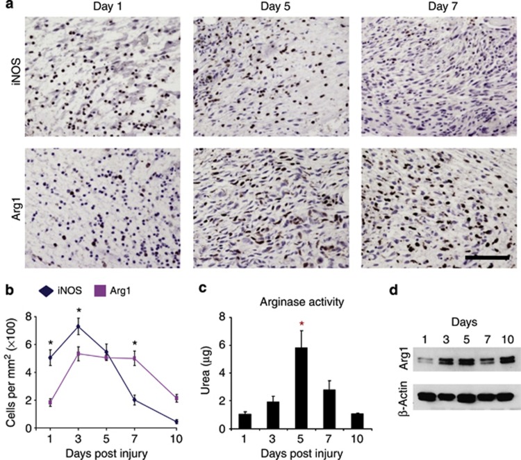
Previous studies have suggested that macrophage phenotype is temporally regulated during wound healing, with CAMs present at early stages and AAMs more dominant during later stages (Albina et al., 1990; Daley et al., 2010). We confirmed this temporal profile in our C57/Bl6 excisional wound model using immunohistochemistry for Arg1 and iNOS, widely accepted markers of CAM and AAM activation, respectively (Gordon and Martinez, 2010). In the acute healing model, iNOS levels peaked at 3 days post wounding, whereas Arg1 remained high until 7 days (Figure 1). These time points correlate with the transition from a proinflammatory extracellular milieu to a phase of matrix deposition (Shaw and Martin, 2009). To corroborate these findings further, we analyzed Arg enzymatic activity, which provides a functionally relevant measure (Witte et al., 2002). We report a strong peak in wound tissue Arg activity 5 days post wounding (Figure 1c), corresponding to the midpoint of the peak in wound granulation tissue Arg1+ macrophages (Figure 1b). Finally, we profiled global Arg1 protein levels in isolated wound tissue over a healing time course. Total protein also peaked at day 5 in line with total tissue Arg activity (Figure 1d). It is to be noted that the subsequent increase in Arg1 at 10 days post wounding likely reflects the previously observed latter-stage induction in wound fibroblasts (Witte et al., 2002).
Figure 1.
Arginase1 (Arg1) is dynamically regulated during healing. (a) Images representing the experimental group mean for inducible nitric oxide synthase-positive (iNOS+) and Arg1+ cells in 1-, 5-, and 7-day excisional wound granulation tissue. (b) Quantification of iNOS+ and Arg1+ dermal inflammatory cells reveals differing temporal profiles. Immunohistochemical quantification data are derived from the mean of five randomly selected high-powered fields per wound and two wounds per mouse. (c) Arginase activity from isolated excisional wound tissue (measured through urea production) peaks at 5 days post wounding. (d) Western blot analysis of total Arg1 protein in excisional wounds reveals increased expression at 3, 5, and 10 days post wounding. (b) Data presented indicate mean+SEM of n=5–6 mice per group or (c, d) three replicates per group across two individual experiments. Bar=100 μm. (b) *P<0.05 comparing iNOS with Arg1, (c) *P<0.05 compared with days 1, 3, and 10.
Local inhibition of Arg activity significantly delays cutaneous healing
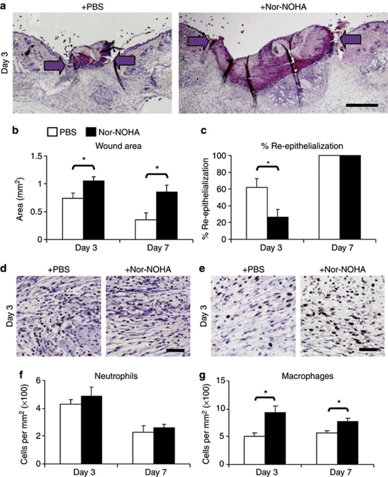
To explore the functional role of Arg during healing, a commonly used inhibitor of Arg activity, nor-NOHA (Tenu et al., 1999; Takahashi et al., 2010), was locally applied to incisional wounds. Nor-NOHA treatment significantly delayed healing, demonstrated by an increased histological wound area versus vehicle-treated wounds at both 3 and 7 days post wounding accompanied by reduced re-epithelialization (Figure 2a–c). Interestingly, delayed healing in nor-NOHA-treated wounds was accompanied by increased numbers of local macrophages (Figure 2e) maintained across days 3 and 7 post wounding. To determine the contribution of recruitment versus removal, we assessed local-wound chemokine levels and apoptosis. Delayed healing in nor-NOHA-treated wounds are associated with increased levels of inflammatory chemokines at 3 days post wounding with elevated apoptosis at 7 days post wounding (Supplementary Figure S1 online). Thus, pharmacological inhibition of Arg (Arg1 and Arg2) delays repair associated with altered local macrophage numbers. To confirm a specific role for Arg1, we next studied a cell-specific conditional Arg1 knockout model.
Figure 2.
Inhibition of arginase activity significantly delays cutaneous healing. (a) Representative hematoxylin and eosin–stained incisional wounds (day 3) from control and N(omega)-hydroxy-nor-l-arginine (nor-NOHA)-treated mice. Arrows indicate wound margins. Nor-NOHA treatment significantly delays wound closure quantified by (b) increased wound area at 3 and 7 days post wounding and (c) decreased re-epithelialization at 3 days post wounding. Images representing the experimental group mean for (d) wound neutrophils and (e) macrophages from control and nor-NOHA-treated mice. (f) Quantification reveals no effect of nor-NOHA on wound neutrophil numbers. (g) Quantification of wound macrophage numbers reveals a significant increase following nor-NOHA treatment at 3 and 7 days post wounding. Immunohistochemical quantification data are derived from the mean of five randomly selected high-powered fields per wound and two wounds per mouse. Data presented indicate mean+SEM, n=6 mice per group. Bar=400 μm (a), 50 μm (d, e). *P<0.05. PBS, phosphate-buffered saline.
Tie2-cre-mediated conditional ablation of Arg1 (T2C;Arg1 fl/fl ) reveals a cell-specific role for Arg during healing
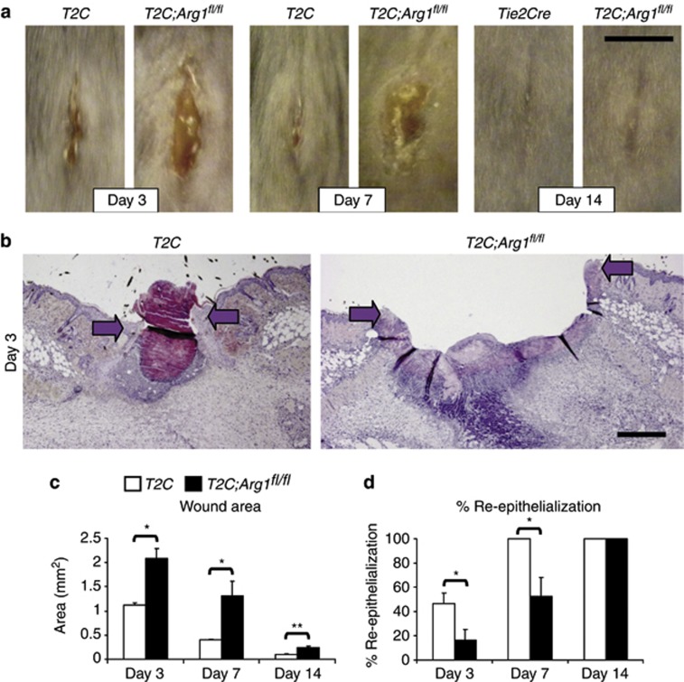
Arg1 is thought to be expressed in multiple cell types involved in the healing process, including keratinocytes, inflammatory cells, and fibroblasts (Albina et al., 1990; Witte et al., 2002; Kampfer et al., 2003). However, we hypothesized that macrophages would be the key Arg1-expressing cell type in the wound repair system. To test this idea, we used an Arg1 conditional allele crossed to Tie2-Cre, which is active in all hematopoietic and endothelial cells. As macrophages are the main Arg1-expressing cell type, Tie2-Cre deletion provides a convenient way to ablate macrophage Arg1, noting that some endothelial cells may also express the gene (El Kasmi et al., 2008; Pesce et al., 2009). Here, we report that Tie2-cre-mediated deletion of Arg1 (T2C;Arg1fl/fl) resulted in a pronounced healing delay compared with T2C;Arg1+/+ littermate controls, depicted macroscopically at 3, 7, and 14 days post wounding (Figure 3a). Subsequent histological analysis revealed a substantial increase in wound area and reduction in re-epithelialization in T2C;Arg1fl/fl wounds. This wound phenotype reveals an important role for macrophage/endothelial-derived Arg1 during skin repair (Figure 3b–d).
Figure 3.
Conditional ablation of Arg1 (T2C;Arg1fl/fl) significantly delays cutaneous repair. (a) Representative macroscopic images of control (T2C) and T2C;Arg1fl/fl wounds at 3, 7, and 14 days post wounding show delayed healing in T2C;Arg1fl/fl wounds. (b) Representative hematoxylin and eosin–stained day-3 histology from T2C and T2C;Arg1fl/fl wounds. Arrows indicate wound margins. Dermal cell–specific deletion of arginase 1 significantly delays wound closure quantified by (c) increased wound area and (d) delayed re-epithelialization over multiple time points. Bar=5 mm (a) and 400 μm (b). Mean+SEM, n=6–7 mice per group. **P<0.01, *P<0.05.
T2C;Arg1 fl/fl mice exhibit alterations in wound inflammatory cell recruitment
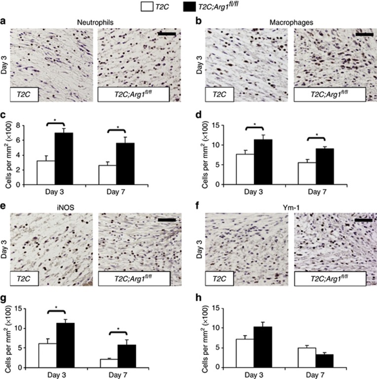
An excessive inflammatory response is a common theme in pathological healing (Martin and Leibovich, 2005; Emmerson et al., 2010). In light of the link of macrophage Arg1 to inhibition of inflammation, we assessed the inflammatory cell profile in delayed healing T2C;Arg1fl/fl wounds. Tie2-cre-mediated Arg1 ablation led to an increased and extended influx of not only macrophages (Figure 4b and d) but also neutrophils (Figure 4a and c). We next assessed markers of macrophage polarization (Daley et al., 2010). Interestingly, wound granulation tissue iNOS+ cells (CAM marker) were increased in T2C;Arg1fl/fl at both 3 and 7 days post wounding, suggesting a maintained proinflammatory environment (Figure 4e and g). These data were confirmed through increased nitrotyrosine staining (used as a marker of cell damage and inflammation through NO) in T2C;Arg1fl/fl at both 3 and 7 days post wounding (data not shown). In contrast, no difference was observed in the number of wound cells expressing the AAM marker, Ym1, at either 3 or 7 days post wounding (Figure 4f and h). Thus, the macrophage component of the wound-healing phenotype in T2C;Arg1fl/fl mice involves excessive numbers of iNOS+ cells rather than a defect in the ability of macrophages to become polarized in the skin microenvironment. Intriguingly, previous studies have shown that isolated Arg-deficient macrophages have increased NO production in response to lipopolysaccharide stimulation (El Kasmi et al., 2008; Pesce et al., 2009). Crucially, our data suggest that despite Arg1 being associated with AAMs, Arg1-deficient macrophages remain able to adopt an AAM phenotype.
Figure 4.
Conditional deletion of arginase 1 (Arg1) results in excessively prolonged and local inflammation. Images representing the experimental group mean for (a) neutrophil and (b) macrophage immunohistochemical analysis from day-3 wound granulation tissue. T2C;Arg1fl/fl wounds display significantly increased numbers of (c) neutrophils and (d) macrophages at 3 and 7 days post wounding. Images representing the experimental group mean for (e) inducible nitric oxide synthase-positive (iNOS+) and (f) Ym1+ cells. T2C;Arg1fl/fl wounds have increased numbers of (g) iNOS+ cells, with no difference in (h) Ym1+ cells. Immunohistochemical quantification data are derived from the mean of five randomly selected high-powered fields per wound and two wounds per mouse. Data presented indicate the mean+SEM of n=6–7 mice per group. Bar=50 μm. *P<0.05.
Excessive protease activity and reduced matrix deposition contribute to delayed healing in T2C;Arg1 fl/fl wounds
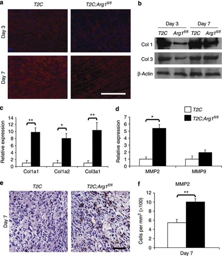
Arg-mediated L-arginine metabolism produces L-ornithine, which is an important component of collagen synthesis (Morris, 2009). We thus hypothesized that T2C;Arg1fl/fl mice would display alterations in wound matrix deposition, synthesis, and/or remodeling. Indeed, delayed healing in T2C;Arg1fl/fl mice was associated with reduced wound granulation tissue collagen deposition, assessed by collagen 1 immunofluorescence (Figure 5a) and substantially reduced total wound collagen 1 and 3 levels measured by western blot (Figure 5b). Conversely, at the level of gene expression, the major skin collagen species are increased in T2C;Arg1fl/fl wounds. This fits with our previous studies, where delayed healing is phenotypically linked to increased collagen gene expression, presumably as a compensatory mechanism (Hardman et al., 2005; Hardman and Ashcroft, 2008). Gelatinases (MMP2 and MMP9) have an important role in wound granulation tissue remodeling. However, excessive gelatinase activity has been linked to the reduced matrix deposition observed in chronic wounds (Lobmann et al., 2002). Analysis of T2C;Arg1fl/fl wounds at 7 days post wounding reveals a significant and selective increase in MMP2 at both the gene expression (Figure 5d) and protein (Figure 5e and f) levels.
Figure 5.
T2C;Arg1fl/fl wounds display altered matrix deposition and protease activity. Wound protein collagen 1 content determined by (a) immunofluorescence with corresponding western blot analysis for (b) collagen (Col) 1 and 3 revealed a reduction in T2C;Arg1fl/fl wounds. (c, d) T2C;Arg1fl/fl wounds display increased expression of collagen species and MMP2 (quantitative PCR) at 7 days post wounding. (e) Images representing the experimental group mean for MMP2+ dermal cells at 7 days post wounding. (f) Quantification of MMP2+ dermal cells reveals significantly increased numbers in T2C;Arg1fl/fl day 7 wounds. Immunohistochemical quantification data are derived from the mean of five randomly selected high-powered fields per wound and two wounds per mouse. Data presented indicate (f) mean+SEM of n=6–7 mice per group, (a) n=4 mice per group, or (c, d) three replicates per group and two individual experiments. Bar=200 μm (a) and 50 μm (e). **P<0.01, *P<0.05.
Reduced Arg1 is a conserved feature of delayed-healing mouse wounds
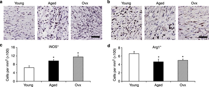
Data presented thus far reveal that either pharmacological inhibition or genetic ablation of Arg in vivo leads to a significant delay in skin healing. To confirm the functional relevance to pathological healing, we turned to preclinical delayed-healing mouse models that have been extensively validated by our group and others (Hardman et al., 2008; Holcomb et al., 2009). We report significantly altered granulation tissue levels of iNOS+ and Arg1+ (Figure 6) cells in both aged and ovariectomized delayed-healing models. In both models, the levels of Arg1 was reduced, whereas that of iNOS was increased, presumably reflecting a delayed switch in macrophage polarization.
Figure 6.
Reduced Arginase1 (Arg1) is a conserved feature of delayed healing in mouse and human wounds. Images representing the experimental group mean for (a) inducible nitric oxide synthase-positive (iNOS+) and (b) Arg1-positive (Arg1+) immunohistochemical analysis from control (young) and delayed healing aged and ovariectomized (Ovx) day-3 wounds. Aged and Ovx mice wounds are associated with increased numbers of (c) iNOS+ dermal cells and (d) reduced Arg1+ cells compared with control mice. Immunohistochemical quantification data are derived from the mean of five randomly selected high-powered fields per wound and two wounds per mouse. Data presented indicate the mean+SEM, n=6 mice per group. Bar=50 μm. *P<0.05.
Discussion
The contribution of macrophage polarization to cutaneous wound healing remains unclear. The situation is further complicated by the fact that markers of each macrophage phenotype, iNOS and Arg, compete for a common substrate, L-arginine, with products from each being important for healing (Childress et al., 2008). Previous studies have shown that L-arginine administration is able to promote acute healing in both rodents and humans (Seifter et al., 1978; Barbul et al., 1990; Williams et al., 2002), an effect attributed to increased nitric oxide production through iNOS metabolism of L-arginine (Shi et al., 2000). Subsequent studies on the importance of iNOS are conflicting, depending largely on the wound model used (Yamasaki et al., 1998; Most et al., 2002). Data presented in this study reveal an important role of the Arg arm in the L-arginine metabolism pathway during cutaneous healing. We report similar findings across pharmacological inhibition and cell-specific genetic ablation studies, confirming relevance in preclinical delayed-healing mouse models.
A previous study reported accelerated acute wound healing following Arg inhibition with the compound (2)-(S)-amino-6-boronohexanoic acid (ABH) (Kavalukas et al., 2011). The differing effects of Arg inhibition seen by Kavalukas et al., and in this current study, are most likely due to the use of different inhibitors. This study used the compound nor-NOHA, which has been shown to be a potent inhibitor of Arg1 and Arg2 in a number of in vitro/in vivo models (Tenu et al., 1999; Takahashi et al., 2010). Kavalukas et al., used the compound ABH, which is a more selective inhibitor of Arg2 compared with Arg1 in vivo (Baggio et al., 1999). A previous study has shown that both Arg1 and Arg2 are expressed in multiple cutaneous cell types (e.g., keratinocytes and inflammatory cells) (Kampfer et al., 2003). We confirmed the importance of Arg1 using T2C;Arg1fl/fl mice, which displayed an even more pronounced delayed-healing phenotype compared with that observed through nor-NOHA treatment. The clear correlation between these two independent models reinforces the importance of Arg1 during healing.
Arg1 is expressed across a range of cell types involved in wound healing including keratinocytes (Kampfer et al., 2003), fibroblasts (Witte et al., 2002), endothelial cells (Abd-El-Aleem et al., 2000), and inflammatory cells (Miao et al., 2012). Here, we have used the Tie2-cre mouse to selectively ablate Arg1 in all hematopoietic and endothelial cell lineages (El Kasmi et al., 2008). The fact that delayed healing in Tie2cre;Argfl/fl mice is associated with alterations to a range of cell functions primarily attributed to additional cell types, e.g., re-epithelialization and matrix deposition, implies an important role for paracrine signaling. It is noteworthy that iNOS+ cells are increased in Tie2cre;Argfl/fl wounds in line with the proposed role of Arg1-expressing Th2-activated macrophages as suppressor cells that help to dampen Th1-driven inflammation (Pesce et al., 2009). Indeed, this mechanism is most likely important in chronic wounds, which are widely accepted to be in a Th1 proinflammatory state (Sindrilaru et al., 2011).
Arg1-mediated metabolism of L-arginine is an important source of local ornithine, a proline precursor important for collagen synthesis. However, the main cellular source of wound collagen is fibroblasts (Singer and Clark, 1999). Thus, the observed defect in collagen deposition following Tie2-cre-mediated deletion of Arg1 is presumably a secondary effect of the delayed-healing environment. Indeed, the reduced collagen content of Tie2cre;Argfl/fl wounds is most likely a result of elevated wound gelatinase expression. Moreover, the increased local collagen gene expression may reflect a compensatory mechanism. It is noteworthy that we have previously reported (Hardman et al., 2005; Hardman and Ashcroft, 2008) a global compensatory upregulation of collagen gene expression in tandem with increased local gelatinase activity conserved across delayed-healing wounds in both humans and mice. Intriguingly, Arg1 is reportedly localized to gelatinase granules in human neutrophils (Jacobsen et al., 2007). The provisional matrix laid down following healing is essential to provide a scaffold for the migration of key cell types and for wound-vessel neogenesis (Shaw and Martin, 2009), and matrix changes may thus be an important causative factor to the observed healing delay.
Altered macrophage polarization is emerging as a common theme across murine delayed-healing acute wound models: aged and ovariectomized mice (this study) and diabetic mice (Miao et al., 2012). This fits with the hypothesis that delayed-healing wounds are unable to switch to a reparatory AAM environment required for normal healing. The finding that normal acute healing requires a temporal shift in macrophage polarization is supported by previous studies (Deonarine et al., 2007) and our own data (Figure 1). A failure to switch from a Th1 to a Th2 environment would have severe consequences for the healing of chronic wounds. Here, we note confusion in the literature as to whether chronic wounds per se have altered Arg expression; an initial study showed increased Arg expression in diabetic ulcers versus normal skin (Jude et al., 1999; Abd-El-Aleem et al., 2000) supported by two subsequent reports of increased Arg expression in diabetic murine models (Kampfer et al., 2003; Miao et al., 2012). Importantly, these studies measured global wound Arg expression failing to account for potential variation in cellular source. Our data currently reveal that age-associated delay in acute healing is accompanied by a local reduction in wound granulation tissue Arg1+ cells.
Data presented in this study clearly demonstrate an important and previously unappreciated role of Arg1 during cutaneous healing. These findings are particularly interesting in the context of previous studies demonstrating the beneficial effects of L-arginine supplementation on acute wound healing. An important next step will be to confirm potential beneficial effects of Arg1 and/or L-arginine in human chronic wounds. Indeed, we suggest that a combined L-arginine supplementation/local Arg induction therapeutic approach may have considerable clinical benefit.
Materials and methods
Animals and wounding
All animal studies were performed in accordance with Home Office regulations. Ten-week-old female C57BL/6, transgenic Tie2cre;Argfl/fl (El Kasmi et al., 2008), Tie2cre;Arg+/+ (wild-type controls), ovariectomized (bilateral ovariectomy performed 3 weeks before wounding), and aged mice (18-month old) were anesthetized and wounded following our established protocol (Ashcroft et al., 2003). N(omega)-hydroxy-nor-l-arginine (nor-NOHA—20 μg per 50 μl) (VWR International, Lutterworth, UK) or vehicle (phosphate-buffered saline) were locally injected in C57BL/6 mice at days 1 and 0 with respect to wounding and every subsequent day until collection. Two equidistant 1-cm full-thickness incisional or 6-mm excisional wounds were made and left to heal by secondary intention. Wounds were excised at either 1, 3, 5, 7, or 10 days post wounding and bisected, with half processed for histological analysis (wound midpoint). The remaining half of each wound was flash frozen and stored at −80 °C for biochemical analysis.
Histology and immunohistochemistry
Histological sections were prepared from wound tissue fixed in 10% buffered formalin saline and embedded in paraffin. Six-micrometer-thick sections were stained with hematoxylin and eosin or subjected to immunohistochemical analysis with the following antibodies: Arg-I goat polyclonal, NOS2 rabbit polyclonal and MMP2 goat polyclonal (Santa Cruz, Heidelberg, Germany), anti-neutrophil rat polyclonal (Fisher Scientific, Loughborough, UK), anti-Mac-3 rat polyclonal (BD Biosciences, Oxford, UK), Ym1 goat polyclonal (R&D Systems, Minneapolis, MN), and Collagen 1 rabbit polyclonal (Millipore, Billerica, MA). Bound primary antibody was detected using the VECTASTAIN ABC kit (Vector Laboratories, Peterborough, UK) combined with NovaRed substrate. Images were captured (Nikon eclipse E600/SPOT camera (Image solutions, Preston, UK)) and granulation tissue wound area and re-epithelialization quantified using Image Pro Plus software (MediaCybernetics, Rockville, MD) as previously described (Ashcroft and Mills, 2002). Total cell numbers (expressed as number of cells per mm2) were determined using five randomly assigned granulation tissue images per wound with Image Pro Plus software.
Arg activity assay
Arg activity was assessed by measuring the amount of urea production via the metabolism of L-arginine by Arg as previously described (Corraliza et al., 1994). In brief, wounded tissue was homogenized in 0.5 ml 0.1% Triton X-100 (Sigma-Aldrich, Cambridge, UK). After 30 minutes, 0.5 ml of assay buffer (10 mmol l−1 MnCl in 50 mmol l−1 Tris, pH 7.5) was added and the enzyme was activated by heating for 10 minutes at 55 °C. For the metabolism of L-arginine by Arg, triplicate cultures of 25 μl cell lysate in buffer were incubated with 25 μl of 0.5 m L-arginine (Sigma-Aldrich) for 60 minutes at 37 °C and the reaction was stopped by adding 400 μl of acid mixture. Twenty-five microliters of 9% α-isonitrosopropiophenone (Sigma-Aldrich) was added and incubated for 45 minutes at 100 °C in the dark. Absorbance was measured at 570 nm using a MRXII (Dynex Technologies, West Sussex, UK). To normalize the samples, the protein concentration in cell lysates was measured using a BCA Protein Assay Kit (Thermo Fisher Scientific, Runcorn, UK).
Protein extraction and immunoblotting
Total protein was extracted from unwounded and wounded tissue by boiling and homogenizing in SDS sample buffer, containing 5% β-mercaptoethanol, for 5 minutes. Protein samples (1 mg) were separated by SDS–PAGE and blotted onto a nitrocellulose membrane, which was blocked in 5% nonfat milk and 0.1% Tween for 16 hours at 4 °C, before incubation with primary antibody for 1 hours at room temperature. The bound primary antibodies were detected with peroxidase-labeled secondary antibodies (GE Healthcare, Hatfield, UK), followed by the ECL Plus detection system (GE Healthcare). Primary antibodies against Arg I and Collagen 3A1 (Santa Cruz), Collagen I (Millipore), and β-actin (Sigma-Aldrich) were used in conjunction with anti-goat, anti-rabbit, or anti-mouse secondary antibodies (GE Healthcare).
Quantitative real-time PCR
Total RNA was isolated from frozen tissue by homogenizing in Trizol reagent (Invitrogen, Paisley, UK). cDNA was transcribed from 1 μg of RNA (Promega RT kit, Madison, WI and AMV-reverse transcriptase; Roche, Welwyn Garden City, UK) and quantitative PCR performed using the MESA-green kit (Eurogentec, Southampton, UK) and an Opticon quantitative PCR thermal cycler (Bio-Rad, Hemel Hempstead, UK). For each primer set, an optimal dilution was determined and melting curves were used to determine amplification specificity. Each sample was serially diluted over three orders of magnitude, and expression ratios were normalized to the mean of two separate reference primers (Gapdh and Ywahz) with all samples analyzed concurrently. Full primer sequences are listed in Supplementary Table S1 online.
Statistical analysis
Statistical differences were determined using either Student's t-tests (Figure 5) (Mann–Whitney U tests for nonparametric data), one-way analysis of variance (Figures 1c and 6) or two-way (Figures 1b, 2, 3, and 4) analysis of variance (with appropriate post-hoc testing) (SimFit, William Bardsley, University of Manchester, Manchester, UK). A P-value of <0.05 was considered significant.
Acknowledgments
We thank Larisa Logunova and Yohko Kitagawa for valuable technical assistance. This research was supported by an AgeUK Senior Fellowship (M.J.H.) and an MRC–funded studentship (L.C.). P.J.M. is supported by the Hartwell Foundation and the American Lebanese Associated Charities.
Glossary
- AAM
alternatively activated macrophage
- Arg
arginase
- CAM
classically activated macrophage
- iNOS
inducible nitric oxide synthase
- nor-NOHA
N(omega)-hydroxy-nor-l-arginine
The authors state no conflict of interest.
Footnotes
SUPPLEMENTARY MATERIAL
Supplementary material is linked to the online version of the paper at http://www.nature.com/jid
Supplementary Material
References
- Abd-El-Aleem SA, Ferguson MW, Appleton I, et al. Expression of nitric oxide synthase isoforms and arginase in normal human skin and chronic venous leg ulcers. J Pathol. 2000;191:434–442. doi: 10.1002/1096-9896(2000)9999:9999<::AID-PATH654>3.0.CO;2-S. [DOI] [PubMed] [Google Scholar]
- Abeyakirthi S, Mowbray M, Bredenkamp N, et al. Arginase is overactive in psoriatic skin. Br J Dermatol. 2010;163:193–196. doi: 10.1111/j.1365-2133.2010.09766.x. [DOI] [PubMed] [Google Scholar]
- Albina JE, Mills CD, Henry WL, Jr., et al. Temporal expression of different pathways of 1-arginine metabolism in healing wounds. J Immunol. 1990;144:3877–3880. [PubMed] [Google Scholar]
- Ashcroft GS, Mills SJ. Androgen receptor-mediated inhibition of cutaneous wound healing. J Clin Invest. 2002;110:615–624. doi: 10.1172/JCI15704. [DOI] [PMC free article] [PubMed] [Google Scholar]
- Ashcroft GS, Mills SJ, Lei K, et al. Estrogen modulates cutaneous wound healing by downregulating macrophage migration inhibitory factor. J Clin Invest. 2003;111:1309–1318. doi: 10.1172/JCI16288. [DOI] [PMC free article] [PubMed] [Google Scholar]
- Baggio R, Emig FA, Christianson DW, et al. Biochemical and functional profile of a newly developed potent and isozyme-selective arginase inhibitor. J Pharmacol Exp Ther. 1999;290:1409–1416. [PubMed] [Google Scholar]
- Barbul A, Lazarou SA, Efron DT, et al. Arginine enhances wound healing and lymphocyte immune responses in humans. Surgery. 1990;108:331–336. [PubMed] [Google Scholar]
- Childress B, Stechmiller JK, Schultz GS. Arginine metabolites in wound fluids from pressure ulcers: a pilot study. Biol Res Nurs. 2008;10:87–92. doi: 10.1177/1099800408322215. [DOI] [PubMed] [Google Scholar]
- Corraliza IM, Campo ML, Soler G, et al. Determination of arginase activity in macrophages: a micromethod. J Immunol Methods. 1994;174:231–235. doi: 10.1016/0022-1759(94)90027-2. [DOI] [PubMed] [Google Scholar]
- Daley JM, Brancato SK, Thomay AA, et al. The phenotype of murine wound macrophages. J Leukoc Biol. 2010;87:59–67. doi: 10.1189/jlb.0409236. [DOI] [PMC free article] [PubMed] [Google Scholar]
- Debats IB, Wolfs TG, Gotoh T, et al. Role of arginine in superficial wound healing in man. Nitric Oxide. 2009;21:175–183. doi: 10.1016/j.niox.2009.07.006. [DOI] [PubMed] [Google Scholar]
- Deonarine K, Panelli MC, Stashower ME, et al. Gene expression profiling of cutaneous wound healing. J Transl Med. 2007;5:11. doi: 10.1186/1479-5876-5-11. [DOI] [PMC free article] [PubMed] [Google Scholar]
- Diegelmann RF. Excessive neutrophils characterize chronic pressure ulcers. Wound Repair Regen. 2003;11:490–495. doi: 10.1046/j.1524-475x.2003.11617.x. [DOI] [PubMed] [Google Scholar]
- El Kasmi KC, Qualls JE, Pesce JT, et al. Toll-like receptor-induced arginase 1 in macrophages thwarts effective immunity against intracellular pathogens. Nat Immunol. 2008;9:1399–1406. doi: 10.1038/ni.1671. [DOI] [PMC free article] [PubMed] [Google Scholar]
- Emmerson E, Campbell L, Ashcroft GS, et al. The phytoestrogen genistein promotes wound healing by multiple independent mechanisms. Mol Cell Endocrinol. 2010;321:184–193. doi: 10.1016/j.mce.2010.02.026. [DOI] [PubMed] [Google Scholar]
- Gordon S, Martinez FO. Alternative activation of macrophages: mechanism and functions. Immunity. 2010;32:593–604. doi: 10.1016/j.immuni.2010.05.007. [DOI] [PubMed] [Google Scholar]
- Hardman MJ, Ashcroft GS. Estrogen, not intrinsic aging, is the major regulator of delayed human wound healing in the elderly. Genome Biol. 2008;9:R80. doi: 10.1186/gb-2008-9-5-r80. [DOI] [PMC free article] [PubMed] [Google Scholar]
- Hardman MJ, Emmerson E, Campbell L, et al. Selective estrogen receptor modulators accelerate cutaneous wound healing in ovariectomized female mice. Endocrinology. 2008;149:551–557. doi: 10.1210/en.2007-1042. [DOI] [PubMed] [Google Scholar]
- Hardman MJ, Waite A, Zeef L, et al. Macrophage migration inhibitory factor: a central regulator of wound healing. Am J Pathol. 2005;167:1561–1574. doi: 10.1016/S0002-9440(10)61241-2. [DOI] [PMC free article] [PubMed] [Google Scholar]
- Hesse M, Modolell M, La Flamme AC, et al. Differential regulation of nitric oxide synthase-2 and arginase-1 by type 1/type 2 cytokines in vivo: granulomatous pathology is shaped by the pattern of L-arginine metabolism. J Immunol. 2001;167:6533–6544. doi: 10.4049/jimmunol.167.11.6533. [DOI] [PubMed] [Google Scholar]
- Holcomb VB, Keck VA, Barrett JC, et al. Obesity impairs wound healing in ovariectomized female mice. In Vivo. 2009;23:515–518. [PubMed] [Google Scholar]
- Jacobsen LC, Theilgaard-Monch K, Christensen EI, et al. Arginase 1 is expressed in myelocytes/metamyelocytes and localized in gelatinase granules of human neutrophils. Blood. 2007;109:3084–3087. doi: 10.1182/blood-2006-06-032599. [DOI] [PubMed] [Google Scholar]
- Jenkinson CP, Grody WW, Cederbaum SD. Comparative properties of arginases. Comp Biochem Physiol B Biochem Mol Biol. 1996;114:107–132. doi: 10.1016/0305-0491(95)02138-8. [DOI] [PubMed] [Google Scholar]
- Jude EB, Boulton AJ, Ferguson MW, et al. The role of nitric oxide synthase isoforms and arginase in the pathogenesis of diabetic foot ulcers: possible modulatory effects by transforming growth factor beta 1. Diabetologia. 1999;42:748–757. doi: 10.1007/s001250051224. [DOI] [PubMed] [Google Scholar]
- Kampfer H, Pfeilschifter J, Frank S. Expression and activity of arginase isoenzymes during normal and diabetes-impaired skin repair. J Invest Dermatol. 2003;121:1544–1551. doi: 10.1046/j.1523-1747.2003.12610.x. [DOI] [PubMed] [Google Scholar]
- Kavalukas SL, Uzgare AR, Bivalacqua TJ, et al. Arginase inhibition promotes wound healing in mice. Surgery. 2011;151:287–295. doi: 10.1016/j.surg.2011.07.012. [DOI] [PubMed] [Google Scholar]
- Lobmann R, Ambrosch A, Schultz G, et al. Expression of matrix-metalloproteinases and their inhibitors in the wounds of diabetic and non-diabetic patients. Diabetologia. 2002;45:1011–1016. doi: 10.1007/s00125-002-0868-8. [DOI] [PubMed] [Google Scholar]
- Loots MA, Lamme EN, Zeegelaar J, et al. Differences in cellular infiltrate and extracellular matrix of chronic diabetic and venous ulcers versus acute wounds. J Invest Dermatol. 1998;111:850–857. doi: 10.1046/j.1523-1747.1998.00381.x. [DOI] [PubMed] [Google Scholar]
- Maarsingh H, Leusink J, Bos IS, et al. Arginase strongly impairs neuronal nitric oxide-mediated airway smooth muscle relaxation in allergic asthma. Respir Res. 2006;7:6. doi: 10.1186/1465-9921-7-6. [DOI] [PMC free article] [PubMed] [Google Scholar]
- Martin P, Leibovich SJ. Inflammatory cells during wound repair: the good, the bad and the ugly. Trends Cell Biol. 2005;15:599–607. doi: 10.1016/j.tcb.2005.09.002. [DOI] [PubMed] [Google Scholar]
- Miao M, Niu Y, Xie T, et al. Diabetes-impaired wound healing and altered macrophage activation: a possible pathophysiologic correlation. Wound Repair Regen. 2012;20:203–213. doi: 10.1111/j.1524-475X.2012.00772.x. [DOI] [PubMed] [Google Scholar]
- Morris SM., Jr. Recent advances in arginine metabolism: roles and regulation of the arginases. Br J Pharmacol. 2009;157:922–930. doi: 10.1111/j.1476-5381.2009.00278.x. [DOI] [PMC free article] [PubMed] [Google Scholar]
- Mosser DM, Zhang X. Activation of murine macrophages. Curr Protoc Immunol. 2008;Chapter 14:Unit 14 2. doi: 10.1002/0471142735.im1402s83. [DOI] [PMC free article] [PubMed] [Google Scholar]
- Most D, Efron DT, Shi HP, et al. Characterization of incisional wound healing in inducible nitric oxide synthase knockout mice. Surgery. 2002;132:866–876. doi: 10.1067/msy.2002.127422. [DOI] [PubMed] [Google Scholar]
- Naura AS, Zerfaoui M, Kim H, et al. Requirement for inducible nitric oxide synthase in chronic allergen exposure-induced pulmonary fibrosis but not inflammation. J Immunol. 2010;185:3076–3085. doi: 10.4049/jimmunol.0904214. [DOI] [PMC free article] [PubMed] [Google Scholar]
- Peranzoni E, Marigo I, Dolcetti L, et al. Role of arginine metabolism in immunity and immunopathology. Immunobiology. 2007;212:795–812. doi: 10.1016/j.imbio.2007.09.008. [DOI] [PubMed] [Google Scholar]
- Pesce JT, Ramalingam TR, Mentink-Kane MM, et al. Arginase-1-expressing macrophages suppress Th2 cytokine-driven inflammation and fibrosis. PLoS Pathog. 2009;5:e1000371. doi: 10.1371/journal.ppat.1000371. [DOI] [PMC free article] [PubMed] [Google Scholar]
- Redente EF, Higgins DM, Dwyer-Nield LD, et al. Differential polarization of alveolar macrophages and bone marrow-derived monocytes following chemically and pathogen-induced chronic lung inflammation. J Leukoc Biol. 2010;88:159–168. doi: 10.1189/jlb.0609378. [DOI] [PMC free article] [PubMed] [Google Scholar]
- Seifter E, Rettura G, Barbul A, et al. Arginine: an essential amino acid for injured rats. Surgery. 1978;84:224–230. [PubMed] [Google Scholar]
- Shaw TJ, Martin P. Wound repair at a glance. J Cell Sci. 2009;122:3209–3213. doi: 10.1242/jcs.031187. [DOI] [PMC free article] [PubMed] [Google Scholar]
- Shi HP, Efron DT, Most D, et al. Supplemental dietary arginine enhances wound healing in normal but not inducible nitric oxide synthase knockout mice. Surgery. 2000;128:374–378. doi: 10.1067/msy.2000.107372. [DOI] [PubMed] [Google Scholar]
- Sindrilaru A, Peters T, Wieschalka S, et al. An unrestrained proinflammatory M1 macrophage population induced by iron impairs wound healing in humans and mice. J Clin Invest. 2011;121:985–997. doi: 10.1172/JCI44490. [DOI] [PMC free article] [PubMed] [Google Scholar]
- Singer AJ, Clark RA. Cutaneous wound healing. N Engl J Med. 1999;341:738–746. doi: 10.1056/NEJM199909023411006. [DOI] [PubMed] [Google Scholar]
- Takahashi N, Ogino K, Takemoto K, et al. Direct inhibition of arginase attenuated airway allergic reactions and inflammation in a Dermatophagoides farinae-induced NC/Nga mouse model. Am J Physiol Lung Cell Mol Physiol. 2010;299:L17–L24. doi: 10.1152/ajplung.00216.2009. [DOI] [PubMed] [Google Scholar]
- Tenu JP, Lepoivre M, Moali C, et al. Effects of the new arginase inhibitor N(omega)-hydroxy-nor-L-arginine on NO synthase activity in murine macrophages. Nitric Oxide. 1999;3:427–438. doi: 10.1006/niox.1999.0255. [DOI] [PubMed] [Google Scholar]
- Unal M, Cimen MY, Dogruer ZN, et al. The potential inflammatory role of arginase and iNOS in children with chronic adenotonsillar hypertrophy. Int J Pediatr Otorhinolaryngol. 2005;69:381–385. doi: 10.1016/j.ijporl.2004.11.003. [DOI] [PubMed] [Google Scholar]
- Williams JZ, Abumrad N, Barbul A. Effect of a specialized amino acid mixture on human collagen deposition. Ann Surg. 2002;236:369–374. doi: 10.1097/00000658-200209000-00013. [DOI] [PMC free article] [PubMed] [Google Scholar]
- Witte MB, Barbul A, Schick MA, et al. Upregulation of arginase expression in wound-derived fibroblasts. J Surg Res. 2002;105:35–42. doi: 10.1006/jsre.2002.6443. [DOI] [PubMed] [Google Scholar]
- Yamasaki K, Edington HD, McClosky C, et al. Reversal of impaired wound repair in iNOS-deficient mice by topical adenoviral-mediated iNOS gene transfer. J Clin Invest. 1998;101:967–971. doi: 10.1172/JCI2067. [DOI] [PMC free article] [PubMed] [Google Scholar]
- Ziche M, Morbidelli L, Masini E, et al. Nitric oxide mediates angiogenesis in vivo and endothelial cell growth and migration in vitro promoted by substance P. J Clin Invest. 1994;94:2036–2044. doi: 10.1172/JCI117557. [DOI] [PMC free article] [PubMed] [Google Scholar]
Associated Data
This section collects any data citations, data availability statements, or supplementary materials included in this article.


