Abstract
Direct reprogramming involves the enforced re-expression of key transcription factors to redefine a cellular state. The nephron progenitor population of the embryonic kidney gives rise to all cells within the nephron other than the collecting duct through a mesenchyme-to-epithelial transition, but this population is exhausted around the time of birth. Here, we sought to identify the conditions under which adult proximal tubule cells could be directly transcriptionally reprogrammed to nephron progenitors. Using a combinatorial screen for lineage-instructive transcription factors, we identified a pool of six genes (SIX1, SIX2, OSR1, EYA1, HOXA11, and SNAI2) that activated a network of genes consistent with a cap mesenchyme/nephron progenitor phenotype in the adult proximal tubule (HK2) cell line. Consistent with these reprogrammed cells being nephron progenitors, we observed differential contribution of the reprogrammed population into the Six2+ nephron progenitor fields of an embryonic kidney explant. Dereplication of the pool suggested that SNAI2 can suppress E-CADHERIN, presumably assisting in the epithelial-to-mesenchymal transition (EMT) required to form nephron progenitors. However, neither TGFβ-induced EMT nor SNAI2 overexpression alone was sufficient to create this phenotype, suggesting that additional factors are required. In conclusion, these results suggest that reinitiation of kidney development from a population of adult cells by generating embryonic progenitors may be feasible, opening the way for additional cellular and bioengineering approaches to renal repair and regeneration.
The direct conversion of one differentiated cell type to another through the forced expression of key transcription factors has been shown to be feasible for a number of distinct cell types, including the conversion of fibroblasts to neurons1 and cardiomyocytes.2 These advances and other similar advances provide the potential for cellular therapies in tissues, including heart, liver, pancreas, and the nervous system.1–7 Such lineage conversion is thought to require the reactivation of a critical endogenous gene regulatory network, with the introduced key genes removing epigenetic barriers to re-establish the attractor state of the cell type required.8,9 Although there have been impressive examples of direct reprogramming to well characterized and phenotypically identifiable mature cell types, for some tissues, it is the generation of stem/progenitor cells that may be required for the regeneration of complex structures.
Within the field of nephrology, there is considerable interest in regenerative medicine for the treatment of ESRD. However, the well characterized stem cell population responsible for giving rise to the functional units of the adult kidney, the nephrons, exists only during the embryonic state.10,11 These nephron progenitor (NP) cells are a mesenchymal population residing within the periphery of the developing kidney in a region termed the cap mesenchyme. These cells, in turn, are derivatives of the Odd-skipped-related 1 (Osr1)+ / Wilms tumor 1 (WT1)+ metanephric mesenchyme, which gives rise to both the Sine oculis homeobox homolog 2 (Six2)+ nephron progenitors and the Foxd1+ stromal progenitors of the kidney.12 Specification of these separate lineages seems to occur from Osr1+ intermediate mesoderm before the onset of kidney development, although Osr1 activity is only required for the formation of nephron progenitors and not the stromal progenitors.12 Throughout kidney development, WT1 continues to be expressed in the NPs as well as the developing nephrons.13 However, Six2 exclusively marks the NP compartment.14 Lineage tracing has shown that these Six2+ NP cells self-renew throughout development to give rise to all of the epithelial cells of the nephron other than the collecting ducts.15,16
In this study, we have used a combinatorial screening approach to identify the genes required to reprogram human adult proximal tubule cells to a kidney nephron progenitor phenotype. A major challenge to such a project is the successful identification of the nephron progenitor end point. Unlike a mature well characterized target cell type, the nephron progenitor has only been identified and characterized in vivo during development. Hence, a robust and stringent assay of nephron progenitor potential was required. We also show that our previously described ex vivo organoid recombination assay can be used to selectively identify the nephron progenitor capacity of introduced test cell populations. Using this recombination assay together with a multistage screen, including changes in cellular morphology and the reinduction of endogenous nephron progenitor gene and protein expression, we describe the identification of a pool of six genes, SIX1, SIX2, OSR1, Eyes absent homolog 1 (EYA1), Homeobox A11 (HOXA11) and Snail homolog 2 (SNAI2), able to reprogram a proportion of adult kidney epithelial cells to adopt a nephron progenitor-like phenotype. These results show the feasibility of the potential to reinitiate kidney development from an adult cell population.
Results
A Combinatorial Screening Approach for NP Reprogramming Factors
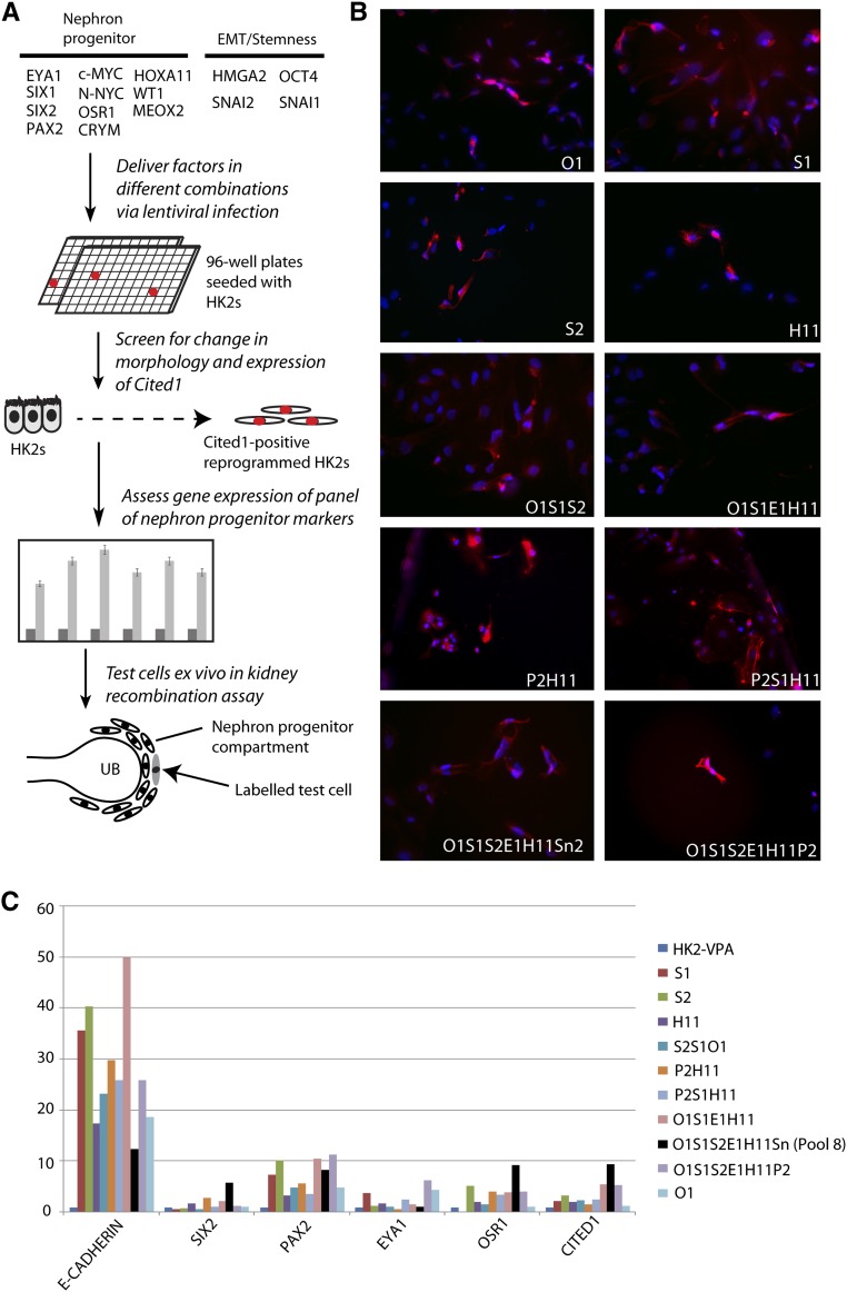
The efficiency of direct reprogramming correlates with the lineage relationship between the start and end cell type.17 Hence, adult progenitor cells, such as myeloid progenitor cells or pro-B cells, can be reprogrammed to induced pluripotent stem cells with 300 times greater efficiency than their more differentiated progeny under identical conditions.18 For this reason, we used the adult proximal tubule cell line human kidney 2 (HK2),19 which will have initially been derived from an embryonic nephron progenitor followed by subsequent patterning, segmentation, and differentiation (reviewed in ref. 20). HK2 cells are epithelial in morphology and show clear expression of the tight junction protein, zona occludens 1 (ZO-1), as well as cortical F-actin fibers typical of an epithelial morphology (Supplemental Figure 1, A–E). Based on the known transcription factor expression profile of the nephron progenitor/cap mesenchyme population defined in the mouse kidney,20,21 11 transcriptional regulators potentially playing a role in specification of the nephron progenitor phenotype were selected. Other genes included have previously been identified to either specify epithelial-to-mesenchymal transition (EMT; SNAI1 and SNAI2) and/or confer stemness (Octamer-binding transcription factor 4 [OCT4]/High mobility group AT-hook 2 [HMGA2]) (Figure 1A and Supplemental Table 1). Each gene was cloned separately into a lentiviral construct accompanied with green fluorescent protein (GFP) to identify successfully infected cells. To reprogram to a nephron progenitor phenotype, we undertook combinatorial screening of these 15 different transcriptional regulators through lentiviral transduction of HK2 cells with each transcription factor either alone or combined with other factors. Infection was performed in a 96-well plate format. Reprogramming can be facilitated through the active relaxation of the chromatin marks specifying lineage.22,23 Hence, epigenetic modifying agents, such as the histone deacetylase inhibitor valproic acid (VPA) or 5-azacytadine, can partially remove chromatin marks in fully differentiated cells, increasing the efficiency of reprogramming.24–26 For this reason, VPA was included during reprogramming to assist in chromatin relaxation.25 In the screen, results were compared back with HK2 cells infected with lentiviral cassette encoding GFP alone in the absence of VPA (HK2-VPA).
Figure 1.
Combinatorial screening for transcription factors is able to reprogram to a nephron progenitor. (A) Combinatorial screening with 15 different factors was performed in triplicate in 96-well plates. Criteria of morphologic EMT and detection of Cited1+ cells in two of three wells allowed progression to the qRT-PCR phase, which assessed a broader panel of NP markers. qRT-PCR results provided the basis for input populations for the recombination assay, which assessed the capacity of reprogrammed cell populations to behave as bona fide kidney progenitors. UB; ureteric bud. (B) Ten pools were identified based on induction of CITED1 protein as assessed by immunofluorescence. E1, EYA1; H11, HOXA11; O1, OSR1; P2, PAX2; S1, SIX1; S2, SIX2; Sn2, SNAI2. (C) qRT-PCR to determine the degree of reprogramming in HK2 cells infected with viral pools. qRT-PCR screening identified only one factor combination (pool 8; black) that showed consistent activation of a variety of NP markers coupled with downregulation of E-CADHERIN. Gene expression is normalized to glyceraldehyde-3-phosphate dehydrogenase, and it is expressed relative to HK2 cells infected with GFP-containing lentivirus and cultured without VPA.
Figure 1A outlines the process of selection for successful reprogramming events. This process first involved the assessment of a significant morphologic change consistent with EMT and the induction of Cbp/P300-interacting transactivator 1 (CITED1) protein, a specific marker of the NP phenotype that is not required for NP specification or survival.27 Only 10 different reprogramming pools induced consistent CITED1 protein, indicating putative induced NPs (Figure 1B). These 10 pools were then screened for the relative gene expression of key components of the NP gene regulatory network. In doing this screening, we focused on a coordinated upregulation of NP markers accompanied by low or decreased epithelial cadherin (E-CADHERIN) expression. To assess induction of endogenous gene expression rather than expression of the introduced reprogramming transcripts, we used primers located in the untranslated region of these genes (Supplemental Table 2), because only the coding sequence was delivered by lentivirus. All 10 pools displayed some level of endogenous NP gene expression activation. However, only one pool (pool 8), containing SIX1, SIX2, OSR1, HOXA11, EYA1, and SNAI2, showed lower E-CADHERIN coupled with coordinated activation of the NP gene regulatory network (Figure 1C). Pool 8 also showed the highest level of SIX2 and CITED1 expression, genes that exclusively mark the NP population in the developing kidney.15,16 Thus, we focused our additional analyses on pool 8 reprogramming.
Pool 8 Induced EMT and Specifically Activated the NP Gene Regulatory Network
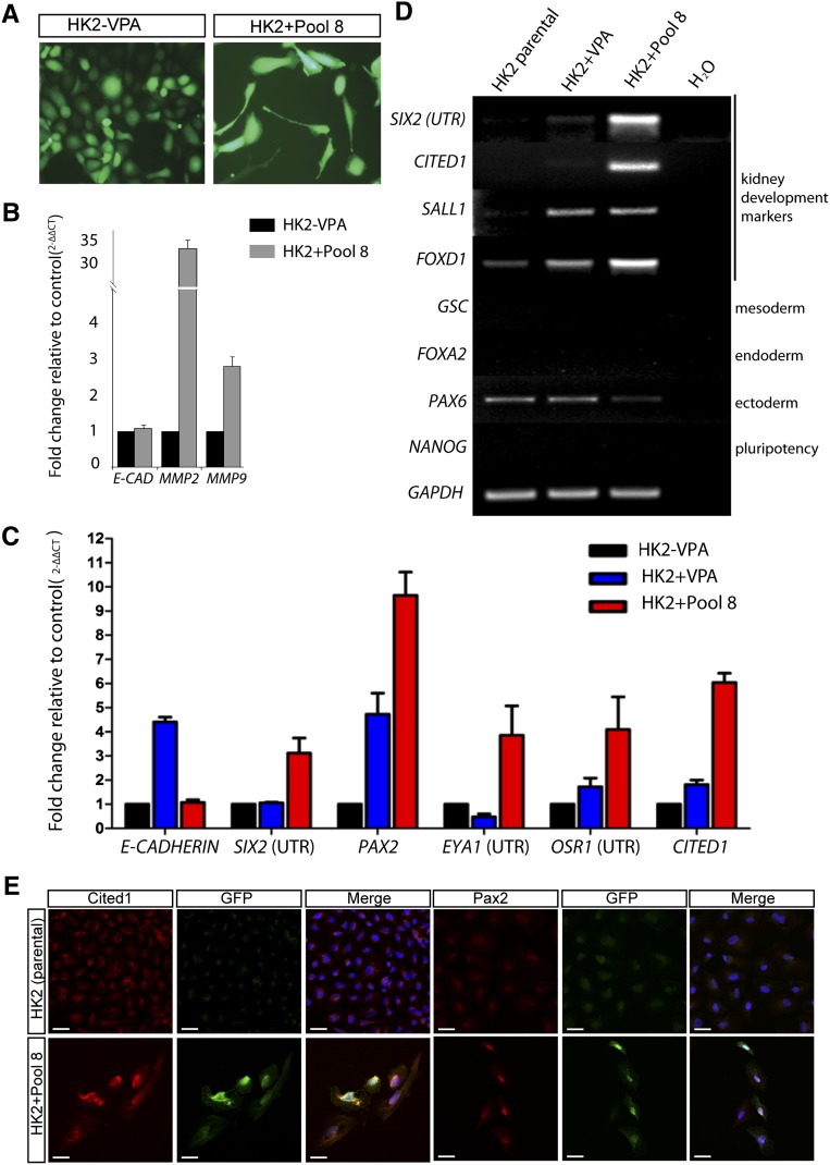
Reprogramming of HK2 cells using pool 8 was reanalyzed using morphology, RT-PCR/quantitative real-time PCR (qRT-PCR), and immunofluorescence. Results were compared back with either parental HK2 cells (HK2 parental) or HK2 cells infected with lentiviral cassette encoding GFP alone in the presence (HK2+VPA) or absence (HK2−VPA) of VPA. Ten days after HK2 transduction with pool 8 reprogramming factors, the cells lost their cobblestone morphology, and they became elongated and spindle-shaped (Figure 2A). EMT markers Matrix metalloproteinase 9 (MMP9) and MMP2 were upregulated approximately 3- and 30-fold, respectively (Figure 2B). The apparent reinduction of NP gene expression was further analyzed to examine the effect of VPA. The addition of VPA alone seemed to result in an upregulation of Paired box gene 2 (PAX2). However, the coordinate upregulation of other NP genes (SIX2, EYA1, OSR1, and CITED1) was restricted to HK2+Pool 8 (Figure 2C). It has been previously reported that VPA also derepresses histone deacetylase-mediated silencing of E-CADHERIN, leading to sustained E-CADHERIN expression.28,29 This result was seen as an upregulation of E-CADHERIN in HK2+VPA cells, with this expression repressed to the level of HK2-VPA in pool 8-treated cells (Figure 2C). This replicated set of pool 8 reprogrammed cells showed evidence of a more convincing EMT event than was seen in the initial screen. E-CADHERIN levels were on par with control cells as opposed to the 15-fold upregulation in the initial pass. This technical variability may be caused by the variation in response of the cultures to VPA.
Figure 2.
Identification of a pool of genes able to induce EMT and endogenous expression of NP markers. (A) Compared with HK2 cells infected with an empty lentiviral vector without the addition of VPA (HK2-VPA), pool 8-reprogrammed HK2 cells (HK2+Pool 8) became elongated and spindle-shaped by day 7 of reprogramming, consistent with an EMT. (B) qRT-PCR revealed that EMT markers MMP2 and MMP9 were upregulated ∼30- and 3-fold, respectively, in HK2+Pool 8 cells relative to HK2-VPA control cells, whereas E-CADHERIN levels were unchanged. (C) qRT-PCR showing coordinated upregulation of all NP markers in HK2+Pool 8 compared with HK2-VPA control cells, with simultaneous downregulation of E-CADHERIN expression. qRT-PCR results are expressed as the average relative fold change compared with control from three biologic replicates ± SEM. (D) RT-PCR analysis of the unmanipulated HK2 cell line (HK2 parental), HK2+VPA, and HK2+Pool 8 to examine (1) the endogenous expression level of markers of kidney development (SIX2, CITED1, SALL1, and FOXD1), (2) the marker of three germ layers (mesoderm [goosecoid; GSC], endoderm [FOXA1], and ectoderm [PAX6]), and (3) a pluripotency marker (NANOG). (E) Immunofluorescence analysis of the induction of PAX2 and CITED1 protein in the GFP+ lentivirally transfected cells within cultures of HK2+Pool 8 cells versus HK2 parental cells. Merge includes 4',6-diamidino-2-phenylindole to visualize nuclei. Scale bars, 30 µm.
Because HK2 cells are an immortalized cell line, we also investigated whether a similar gene expression profile could be induced in primary human renal proximal tubule cells (RPTECs). Despite exhibiting some variation in gene upregulation compared with HK2+Pool 8 cells, RPTEC+Pool 8 showed highly increased expression of the NP-specific genes SIX2 and CITED1 (approximately 200- and 20-fold, respectively) as well as SIX2 protein expression (Supplemental Figure 2, A and B). Furthermore, although PAX2 expression did not seem increased in RPTECs+Pool 8 when expressed relative to RPTECs-VPA, the level of PAX2 expression in unmanipulated RPTECs was found to be 410-fold higher than the expression in HK2 cells (Supplemental Figure 2C), suggesting that these cells already possess a high endogenous level of PAX2.
To validate the qRT-PCR and further investigate the specificity of this reprogramming event, we used RT-PCR (Figure 2D). Activation of NP markers SIX2 and CITED1 was differentially higher in HK2+Pool 8 cells, whereas sal-like 1 (SALL1) expression was detected at a similar level in the presence of VPA alone (HK2+VPA). HK2+Pool 8 cells also showed upregulation of forkhead box D1 (FOXD1), a marker of the renal stroma.30,31 However, this result was detected in both parental HK2 and HK2+VPA (Figure 2D). Because the VPA may randomly relax chromatin marks, we also investigated the expression of markers of the three germ layers, early mesoderm (Goosecoid, GSC), endoderm (FOXA2), and ectoderm (PAX6) as well as evidence for re-expression of a pluripotency marker (NANOG). Although we did detect weak expression of the ectodermal/neural marker, PAX6, this result was also detected in control conditions.
Finally, we examined whether the detectable levels of NP gene expression were accompanied by the endogenous production of proteins characteristic of the NP state but not in the lentiviral pool. Immunofluorescence for PAX2 and CITED1 proteins in parental HK2, HK2+VPA, and HK2+Pool 8 cells showed the specific induction of these proteins in GFP+ (lentivirally infected) HK2+Pool 8 cells (Figure 2E and Supplemental Figures 3 and 4).
The Recombination Assay Is a Stringent Assay for NP Potential
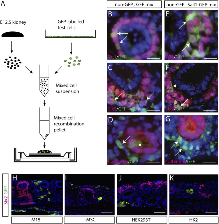
Because there are no NP cells within the postnatal kidney, there is no standard in vivo assay to test NP potential in an adult mouse model. In addition, because the expression constructs delivered continue to be expressed, successfully reprogrammed cells will face constant pressure to remain in an NP state and hence, must be assessed for this phenotype. We have previously developed an assay in which to test NP potential using ex vivo organoid cultures (Figure 3A).32,33 In this assay, embryonic day 12.5 (E12.5) kidneys were dissociated to single-cell suspensions, mixed with test cells to form the recombination pellet, and cultured above a feeder layer of wingless-type MMTV integration site family member 4 (Wnt4)-expressing National Institute of Health 3-day transfer, inoculum 3×10⁵ cells (NIH3T3).34 The recombined organoid culture recapitulates critical aspects of normal kidney development, such as retention of specific subcompartments and their spatial arrangement (Supplemental Figure 5). Specifically, the Six2+ NP compartment is present in these cultures (Figure 3, C and F and Supplemental Figure 5). For this approach to represent a stringent assay for NP potential, it was necessary to show (1) the presence of NP cells in such cultures, (2) the capacity for such endogenous NP cells to participate in nephrogenesis, and (3) the evidence that only populations with an NP phenotype are able to integrate into the endogenous NP field.
Figure 3.
Development of a stringent assay for NP potential. (A) E12.5 mouse kidneys were dissociated to single cells, mixed with GFP-labeled test cells, and processed to form a recombined metanephric kidney ex vivo. (B–D) Single cells from a GFP-labeled E12.5 kidney can be detected in the NP compartment marked by Wt1 and Six2 and also in the calbindin+ ureteric bud (UB) compartment. (E–G) Sall1-GFP NPs contribute to developing nephron compartments but not to the UB-derived calbindin+ compartment. Arrows highlight integration sites of the test cell population. (H–K) Non-NP cell lines M15, MSCs, HEK293Ts, and HK2s cannot contribute to the Six2+ NP compartment. Cell lines were identified by anti-GFP antibody. Immunofluorescence for Six2 was used to assess incorporation into NP compartments. Scale bars, 120 µm in B–G; 360 µm in H–K.
The ability to detect the origin of individual cells within a recombination was determined by mixing dissociated wild-type (unlabeled) and GFP-labeled E12.5 kidneys. GFP+ cells were able to contribute to all endogenous populations, including Wt1+ (metanephric mesenchyme, NP, and nephron), Six2+ (NP only), and Calbindin+ (ureteric epithelium only) populations (Figure 3, B–D). To address the stringency of this assay in determining the NP potential of test populations, GFP+ fractions were FACS-sorted from the dissociated E13 kidneys of Sall1 GFP mice35 and recombined with unlabeled wild-type embryonic kidney. GFP+ cells were readily detectable within the Wt1+ and Six2+ NP fields (Figure 3, E and F) but not in the Calbindin+ ureteric epithelial compartment (Figure 3G). The capacity of non-NP cells to integrate into the endogenous NP field was also examined to determine the stringency of the assay. Non-NP cell populations, including the mesonephric cell line M15,36 HEK293T, an immortalized cell line derived from human embryonic kidney,37 primary isolates of murine bone marrow-derived mesenchymal stem cells (MSCs), and the human proximal tubule cells used in this study (HK2),19 were assessed for their integration capacity in this assay. Each cell line was first infected with a GFP-containing lentivirus and FACS-sorted to isolate a pure population of GFP-labeled cells. All cell types were detected in the recombined organoids but showed negligible integration into the Six2+, Wt1+, or Calbindin+ compartments (Figure 3, H–K and Supplemental Figure 6A). Hence, the only population of cells able to robustly contribute to the NP field was bona fide NPs.
Pool 8 Reprogrammed HK2 Cells Differentially Integrate into the NP Population Ex Vivo
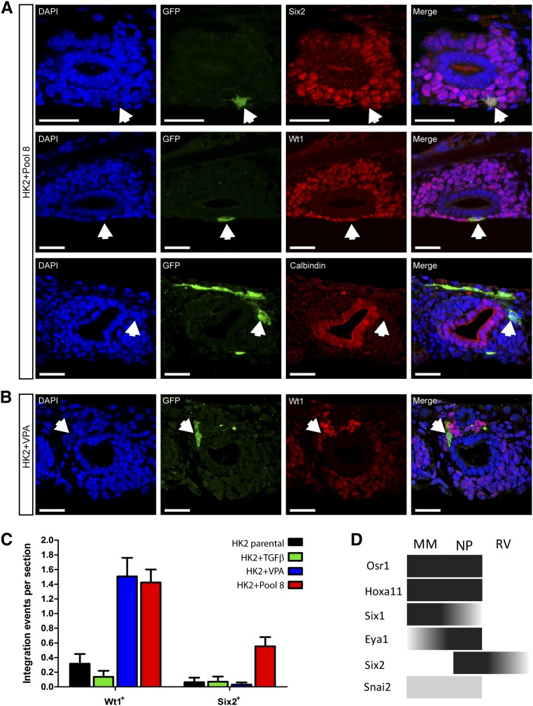
The recombination assay was subsequently used to assess the relative capacity of HK2+Pool 8 cells to integrate into the NP compartment comparison with parental HK2, HK2+VPA, and HK2 cells treated with recombinant TGFβ1 for 3 days to induce a fibrogenic transformation. HK2+Pool 8 cells integrated into both Wt1+ and Six2+ compartments in recombination assays, which was evidenced by the surrounding Six2+/GFP− cells (Figure 4A, arrows), and showed nuclear expression of both WT1 and SIX2 proteins. However, the latter can be partially attributed to the inclusion of SIX2 in the reprogramming pool. Note that both antibodies recognize murine and human forms of the protein. In contrast, neither HK2+VPA nor HK2+TGFβ1 cells showed robust integration into the Six2+ compartment, although HK2+VPA cells did integrate into Wt1hi structures likely to represent non-NP fields (Figure 4B). Relative quantitation of integration events revealed that HK2+Pool 8 cells were approximately fourfold more likely to integrate into a Six2+ NP compartment compared with all other populations tested (P<0.05) (Figure 4C). As anticipated, although able to integrate into these regions, pool 8-reprogrammed HK2 cells did not integrate into the ureteric bud-derived Calbindin+ compartment (Figure 4A). A fraction of these GFP+ cells were also positive for phosphohistone H3 (Supplemental Figure 7), indicating that pool 8-transduced cells not only integrate into endogenous renal structures but can also proliferate within the recombination assay.
Figure 4.
Contribution of reprogrammed cells to the NP compartment in recombination assays. (A) GFP+ HK2+Pool 8 cells (block arrows) contributed to both the Wt1+ and Six2+ NP compartments but not the Calbindin+ compartment ex vivo. Additional Six2+/GFP− cells surround the reprogrammed cells, indicating true integration into the NP compartment. Scale bars, 30 µm. (B) Although unable to efficiently integrate into the Six2+ compartment, HK2+VPA cells did integrate into Wt1+ structures likely to represent early nephrons. Scale bars, 30 µm. (C) Quantitation of integration events revealed that, although HK2 cells treated with VPA alone or together with pool 8 integrated into Wt1+ structures, HK2+Pool 8 cells preferentially integrated into the endogenous ex vivo Six2+ compartment. Both HK2 parental and HK2+TGFβ1-treated cells showed poor integration into either compartment. (n=3±SEM). (D) Schematic representation of the expression domain in the murine developing kidney of the six reprogramming factors in pool 8. MM, metanephric mesenchyme; RV, renal vesicle.
EMT Is Not Sufficient for Reprogramming to an NP Phenotype
EMT describes a phenotypic change that occurs both during normal development (type 1 EMT) and also in association with postnatal fibrosis and the initiation of metastasis in cancer (reviewed in ref. 38). This change uniformly involves a change in cellular morphology and the loss of expression of cadherins and cell–cell junction proteins accompanied by the re-expression of fibroblastic proteins, such as fibroblast-specific protein (FSP-1), vimentin, and MMPs.39,40 In culture, renal epithelial cells readily undergo EMT in response to various treatments, including TGFβ1 or cyclosporine A.41–43 Whereas in the kidney, this EMT has been associated with the pathologic progression to interstitial fibrosis,44 it has more recently been challenged.31 Certainly, reprogramming from renal epithelial cells to cap mesenchyme will require an EMT.
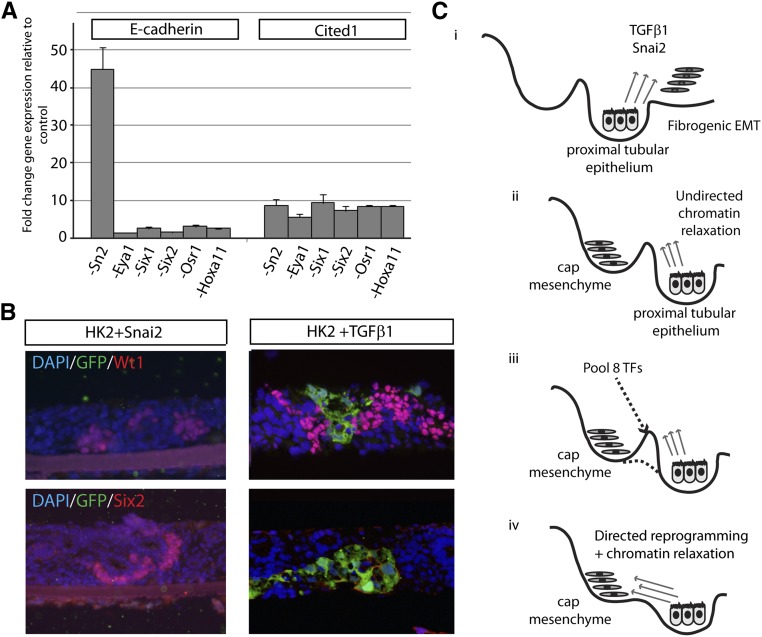
Although the murine orthologs of all six of the genes within pool 8 are expressed in the murine nephron progenitor population, many also show expression in preceding (MM) or subsequent (RV) structures (Figure 4D). In the mouse, Snai2 is expressed only weakly in the endogenous NP population, with expression more tightly associated with the cortical stroma (www.gudmap.org).45 SNAI2 is a potent inducer of EMT; hence, it was possible that transduction with SNAI2 alone in conjunction with VPA treatment may be sufficient for reprogramming to an NP phenotype. Sequential removal of one factor at a time from the pool revealed that SNAI2 was able to repress E-CADHERIN, because removal of SNAI2 from the pool led to a 40-fold upregulation of E-CADHERIN. In contrast, removal of any other reprogramming factor did not affect E-CADHERIN levels significantly (P<0.001) (Figure 5A). However, the expression of the NP marker CITED1 was unaffected by individual removal of any of the six factors, including SNAI2 (Figure 5A).
Figure 5.
The role of EMT in reprogramming to a NP phenotype. (A) Removal of SNAI2 from the reprogramming pool leads to dramatic upregulation of E-CADHERIN, with no effect on CITED1 levels. (B) Reprogramming using SNAI2 alone or HK2 cells induced to undergo EMT through treatment with TGFβ1 failed to generate cell populations able to integrate into embryonic kidney recombinations. (C) Schematic diagram illustrating the observed outcomes of the reprogramming screen. (C, i) Addition of either recombinant TGFβ1 or SNAI2 transduction alone resulted in a fibrogenic EMT, with the resulting cells unable to contribute to the nephron progenitor compartment of the developing explants. (C, ii) Undirected chromatin relaxation mediated by VPA treatment biases the HK2s to the NP/cap mesenchyme fate, but the cells cannot overcome existing epigenetic barriers; therefore, they are only partially reprogrammed. (C, iii) Lineage-instructive transcriptions factors together with SNAI2 reduce the existing epigenetic barriers, which then allows (C, iv) transition from the HK2 proximal tubular phenotype to the NP/cap mesenchyme phenotype.
Recently, the process of EMT has been linked to the acquisition of a stem cell-like phenotype,46,47 possibly reflecting the reversal of processes involved in the initial differentiation of such cells. We, therefore, investigated the possibility that the simple induction of EMT in HK2 cells would generate a nephron progenitor phenotype. HK2 cells infected with lentivirus containing SNAI2 alone underwent marked EMT, with a distinct transition to more elongated and spindle-shaped FSP-1+ cells (Supplemental Figure 8). However, when SNAI2-overexpressing cells were tested in the recombination assay, only very rare cells were detected (less than five cells per entire recombined kidney), none of which integrated into either the Wt1+ or Six2+ compartments (Figure 5B). Similarly, the treatment of parental HK2 cells with recombinant TGFβ1 for 3 days resulted in a fibroblastic transformation with the induction of FSP-1 protein and the loss of cortical F-actin (Supplemental Figure 1, F–J). Of note, no upregulation of PAX2 protein was observed. In this instance, HK2+TGFβ1 cells were seen within recombinations but did not contribute to the Wt1+, Six2+, or Calbindin+ compartments (Figures 4C and 5B and Supplemental Figure 6B). This result argues that EMT alone is not sufficient to endow a NP phenotype.
Discussion
The reprogramming of adult epithelial kidney cells to a NP phenotype represents a major advance for both kidney developmental biology and the reprogramming field. There are no previous reports of kidney cells reprogrammed to a progenitor cell type, whether by forced expression of lineage-instructive genes or manipulation of the local environment with growth factors, epigenetic modifying agents, or small molecules. We have shown that overexpression of SIX1, SIX2, OSR1, EYA1, HOXA11, and SNAI2 in HK2 cells can reprogram them to undergo an EMT and activate a gene regulatory network consistent with a cap mesenchyme/NP phenotype. Furthermore, a portion of HK2 cells transduced with this pool of genes showed a differential capacity to contribute to the NP compartment of a developing embryonic kidney ex vivo, showing the feasibility of direct reprogramming to generate nephron progenitors.
The specific combination of reprogramming factors identified in this study supports much of what is known in kidney developmental biology. All five kidney developmental genes in the pool (OSR1, SIX2, SIX1, HOXA11, and EYA1) are expressed in the cap mesenchyme and essential for formation of this population in vivo (Figure 4D).48–51 PAX2 is also expressed in the cap mesenchyme,52 and accordingly, PAX2 expression in HK2s was upregulated in response to pool 8. PAX2 may not have been required in the pool, because its expression may have been activated by EYA1, HOXA11, and/or SIX1, all of which complex with PAX2 to regulate kidney development.51,53,54 However, it may be able to substitute for one or more of these factors. Six2 is known to upregulate its own expression, and recent studies investigating Six2 target genes during murine kidney development suggest direct regulation of Osr1 and Hox11 paralog expression by Six2 in self-renewing undifferentiated cap mesenchyme,55 further reinforcing the interactions between this particular set of transcription factors in defining the nephron progenitor attractor state. This finding suggests the possibility for remaining redundancy in this pool. As such, additional dereplication may enable the substitution of genes to reduce the total number of transcription factors required.
SNAI2 was the only reprogramming factor without a previously shown role in kidney development. Removal of SNAI2 from the reprogramming pool suggested a role in the suppression of E-CADHERIN and subsequent destabilization of the epithelial phenotype. Consistent with this finding, SNAI2 has been reported to repress E-CADHERIN and is involved in EMT.56–59 In the context of Waddington’s epigenetic landscape,8 which depicts the dynamics of cellular differentiation during development, SNAI2 may serve to overcome epigenetic barriers and make a smoother and more efficient transition to a NP phenotypic state through this induction of EMT (Figure 5C). However, it is possible that SNAI2 has more specific roles to play in the NP state. Presumably, the remaining five genes in pool 8 are critical for ensuring survival and integration in the correct developmental context (Figure 5C). If the starting cell type was not epithelial, such as a skin fibroblast, SNAI2 may not have been required. Importantly, however, proximal tubule cells treated with either recombinant TGFβ1 or SNAI2 alone were not able to contribute to the NP lineage, suggesting that EMT alone is not sufficient to recapitulate the specific NP phenotype.
Although we assume that the chromatin relaxation provided by the addition of VPA assisted the initial reprogramming, one obstacle faced with its usage is the potential for nonspecific gene activation, which may result in an undesirable phenotypic outcome. Indeed, treatment with VPA alone induced the upregulation of FOXD1 and SALL1. The further upregulation of FOXD1 in the presence of pool 8 may even suggest that at least a portion of the population has adopted an even more primitive metanephric mesenchymal state or that both a nephron progenitor and stromal progenitor population are present. Better defining the hetereogeneity of the response will be important if this approach is to be harnessed for cellular therapy. Indeed, the induction of a heterogeneous, confused, or intermediate phenotype in a portion of cells undergoing reprogramming continues to represent a serious challenge in the reprogramming field as a whole and cannot be avoided in the absence of an appropriate selection marker. Thus, the development of a stringent functional assay was critical in this instance to enable detection of successful reprogramming to specific intermediate progenitor phenotypes. The recombination assay not only opened the way for a systematic quantitative analysis of putative kidney stem cell populations but also provided the specific niche requirements to support the NP population generated.
Although this assay allowed us to provide evidence that reprogramming to a nephron progenitor state is feasible, the approach used in this study has several limitations to overcome. Successful reprogramming is known to be an inefficient process, with only a portion of the treated cells adopting the desired end point without active selection.60 Based on an average of 0.5 integration events per section and approximately 140 slides per recombined kidney, we conservatively estimate a reprogramming efficiency of ∼0.875% in response to pool 8 reprogramming. This result represents a 43× higher rate of reprogramming than in the initial study showing the generation of induced pluripotent stem cells (1 in 5×10⁴ starting cells).60 In that instance, the identification of a successful outcome relied on a combination of genetic selection, predefined optimal culture conditions, and a characteristic colony phenotype for pluripotent cells in culture. None of these features was available within the screen described here. Having identified a key gene regulatory network capable of NP reprogramming, it will now be feasible to recreate this approach using inducible, polycistronic constructs. This result will ensure an increased efficiency of integration of all genes and a capacity to shut this program down in vitro or in vivo to show a subsequent capacity to move from nephron progenitor to nephron. Furthermore, reducing toxicity by removing the need for multiple lentiviral constructs may also improve reprogramming of primary human cells, for which our preliminary results suggest are amenable to reprogramming with pool 8 factors. However, regardless of the parental cell type, it will be necessary to optimize the cell culture conditions for the prolonged maintenance of a nephron progenitor population in culture.
Major challenges to the reprogramming of adult cells to nephron progenitors are the transient nature of this attractor state and the intricate reliance of this population on niche instructive cues for its identity. The NP population is a very transient embryonic population, existing in vivo for only 8–10 days of development in the mouse and unable to survive in the absence of an appropriate niche.61 It is also in a highly permissive state, ready to differentiate into nephron epithelia in response to Wnt9b-mediated canonical Wnt signaling,62 Wnt4-mediated noncanonical signaling,63,64 or even ectopic notch signaling.65 Hence, the challenge during reprogramming, like during kidney development, is maintaining the self-renewal of this population. The growth conditions optimal for maintenance of nephron progenitors/cap mesenchyme in vitro have not been completely defined, although there is evidence that it is mediated by fibroblast growth factor and bone morphogenetic protein signaling.66 Optimization of these conditions will be critical for increasing the efficiency of this reprogramming process.
Despite these remaining barriers, this study represents a major step forward to reprogramming in the renal field. Ultimately, the generation of such cells may lead to cellular therapies for adult renal disease. The observation in zebrafish that a NP population can be transplanted from one animal to another and generate neonephrons in an adult fish67 raises the possibility that a selected population of NPs may behave similarly in an adult mammalian kidney. Indeed, it has recently been shown that embryonic kidney organoids placed into adult rat kidney can undergo onward development and vascular development.68 The optimization of methods for the expansion of NP in culture may also allow the renewal generation of renal cells for nephrotoxicity screening. Finally, the generation of nephron progenitors from human adult cells may also assist us in developing approaches to prolong or reinitiate nephrogenesis based on our understanding of the genetic regulation of this population.
Concise Methods
Cell Culture and Reprogramming
HEK293T (CRL-11268; ATCC), HK2 (CRL-2190; ATCC), and primary human RPTE (CC-2553; Lonza Australia) cells were maintained as per the manufacturer’s instructions (HEK293Ts and RPTECs) or as previously described (HK2s) plus 100 U/ml penicillin, 100 g/ml streptomycin, 2 nmol/L l-glutamine, and 0.1 µM hydrocortisone.69 M15 cells were maintained as previously described with the addition of l-glutamine.36 Primary bone marrow-derived murine MSCs were isolated and maintained as previously described.70 For lentiviral reprogramming, the coding sequence of candidate genes (Supplemental Table 1) was cloned into the pLV101G vector upstream of a hrGFP reporter through the Gateway Cloning System (Invitrogen). Third generation lentivirus was packaged as described elsewhere using 5 µl/ml Lipofectamine 2000 (Invitrogen) instead of CaCl2.71 For reprogramming studies, HK2s were serum-deprived for 24 hours and then infected with lentivirus overnight before treatment with VPA (2 mM for 7 days; P4543; Sigma Aldrich). These cells were split into one-third pieces 48 and 96 hours after the initial treatment exposure. RPTECs were treated similarly to HK2 cells above but were not serum-deprived. Control HK2s and RPTECs underwent identical treatment, and they were infected with a GFP-only lentivirus and cultured minus VPA. For EMT studies, HK2s (4×10³ cells/cm) were serum-deprived for 24 hours and then treated with TGFβ1 (20 ng/ml for 3 days; 240-B; R&D).
qRT-PCR and RT-PCR
RNA was extracted from cell cultures using the RNeasy Mini Kit (QIAGEN) as per the manufacturer’s instructions. Equal amounts of RNA were used to make cDNA using the SuperScript III First Strand Synthesis System (Invitrogen). qRT-PCR reactions were performed using Platinum SYBR Green Qpcr Supermix UDG (Invitrogen) as per the manufacturer’s instruction. All qRT-PCR reactions were performed on the ABI Prism 7000 real-time machine with the manufacturer’s software. cDNA levels of target genes were analyzed using comparative Ct levels, normalized to glyceraldehyde-3-phosphate dehydrogenase, and expressed as fold change in treated cells relative to control as previously described.72 Means were calculated based on at least three biologic replicates per treatment and expressed ± SEM. P values were calculated using a two-tailed homoscedastic t test. Conventional RT-PCR analysis was performed using One Taq DNA polymerase (NEB) as per the manufacturer’s instruction. Primer sequences for qRT-PCR are supplied in Supplemental Table 2. Forward and reverse primer pairs for RT-PCR are supplied in Supplemental Table 3.
Immunofluorescence
Immunofluorescence on cells was performed as previously described.73 For the reprogramming screen, immunofluorescence was performed in 96-well black-walled clear bottom plates (Corning). For recombinations, slides were prepared for antibody staining as previously described.32,33 Antibody staining was performed overnight at 4°C for primary antibodies and for 1 hour for secondary antibodies. A list of antibodies and stains used and the manufacturers’ details are supplied in Supplemental Table 4; 4',6-diamidino-2-phenylindole was diluted to 1 in 1000 and incubated for 5 minutes before slides were mounted in Vectashield or Prolong Gold (Invitrogen) and photographed using an Olympus BX-51 upright or Zeiss Meta 510 confocal microscope.
Recombination Assay
Assays were performed as previously described.32,33 A total of 4×10⁵ single cells (5% exogenous cells for M15, HEK293T, and MSC cells and 2% exogenous cells for HK2s) was centrifuged to form a single recombination pellet. The chosen ratio of test cells to embryonic kidney cells was based on both our own (unpublished) findings and the findings of others74 that the addition of higher numbers of test cells can have a disruptive effect on the self-organization of disaggregated kidney cells. Quantification of integration events was calculated as the number of GFP+ cells associated with either a Wt1+ or Six2+ cluster (a cluster is defined as five or more positive cells) in a given section. Association was regarded as being in contact with more than one cell within a given cluster. Either 8 (Wt1) or 16 (Six2) sections were analyzed per biologic replicate, where the sections were spaced equally to cover the entire recombination pellet. Means were calculated based on three biologic replicates per cell line and expressed ± SEM. P values were calculated using a two-tailed homoscedastic t test.
Disclosures
None.
Acknowledgments
We thank Dr. Ryuichi Nishinakamura for the provision of the Sall1-eGFP mice, Prof. Kerry Atkinson for the provision of bone marrow-derived MSCs, Prof. Peter Koopman for access to constitutional GFP+ mice, Prof. Tom Gonda for lentiviral vectors, and Prof. Andy McMahon for provision of the Wnt4-expressing NIH3T3 cells.
This work was supported by Australian Stem Cell Centre Grant P067 and National Health and Medical Research Council, Australia Grant ID631362. Microscopy was performed at the Institute for Molecular Bioscience/Australian Cancer Research Foundation Cancer Biology Imaging Facility, which was established with the generous support of the Australian Cancer Research Foundation. C.E.H. is a Rosamond Siemon postgraduate scholar. M.H.L. is an National Health and Medical Research Council Senior Principal Research Fellow.
Footnotes
Published online ahead of print. Publication date available at www.jasn.org.
This article contains supplemental material online at http://jasn.asnjournals.org/lookup/suppl/doi:10.1681/ASN.2012121143/-/DCSupplemental.
References
- 1.Pang ZP, Yang N, Vierbuchen T, Ostermeier A, Fuentes DR, Yang TQ, Citri A, Sebastiano V, Marro S, Südhof TC, Wernig M: Induction of human neuronal cells by defined transcription factors. Nature 476: 220–223, 2011 [DOI] [PMC free article] [PubMed] [Google Scholar]
- 2.Qian L, Huang Y, Spencer CI, Foley A, Vedantham V, Liu L, Conway SJ, Fu JD, Srivastava D: In vivo reprogramming of murine cardiac fibroblasts into induced cardiomyocytes. Nature 485: 593–598, 2012 [DOI] [PMC free article] [PubMed] [Google Scholar]
- 3.Feng R, Desbordes SC, Xie H, Tillo ES, Pixley F, Stanley ER, Graf T: PU.1 and C/EBPalpha/beta convert fibroblasts into macrophage-like cells. Proc Natl Acad Sci U S A 105: 6057–6062, 2008 [DOI] [PMC free article] [PubMed] [Google Scholar]
- 4.Kajimura S, Seale P, Kubota K, Lunsford E, Frangioni JV, Gygi SP, Spiegelman BM: Initiation of myoblast to brown fat switch by a PRDM16-C/EBP-beta transcriptional complex. Nature 460: 1154–1158, 2009 [DOI] [PMC free article] [PubMed] [Google Scholar]
- 5.Huang P, He Z, Ji S, Sun H, Xiang D, Liu C, Hu Y, Wang X, Hui L: Induction of functional hepatocyte-like cells from mouse fibroblasts by defined factors. Nature 475: 386–389, 2011 [DOI] [PubMed] [Google Scholar]
- 6.Zhou Q, Brown J, Kanarek A, Rajagopal J, Melton DA: In vivo reprogramming of adult pancreatic exocrine cells to beta-cells. Nature 455: 627–632, 2008 [DOI] [PMC free article] [PubMed] [Google Scholar]
- 7.Jayawardena TM, Egemnazarov B, Finch EA, Zhang L, Payne JA, Pandya K, Zhang Z, Rosenberg P, Mirotsou M, Dzau VJ: MicroRNA-mediated in vitro and in vivo direct reprogramming of cardiac fibroblasts to cardiomyocytes. Circ Res 110: 1465–1473, 2012 [DOI] [PMC free article] [PubMed] [Google Scholar]
- 8.Waddington C: The Strategy of the Genes, London, Geo, Allen & Unwin, 1957 [Google Scholar]
- 9.Hochedlinger K, Plath K: Epigenetic reprogramming and induced pluripotency. Development 136: 509–523, 2009 [DOI] [PMC free article] [PubMed] [Google Scholar]
- 10.Rumballe BA, Georgas KM, Combes AN, Ju AL, Gilbert T, Little MH: Nephron formation adopts a novel spatial topology at cessation of nephrogenesis. Dev Biol 360: 110–122, 2011 [DOI] [PMC free article] [PubMed] [Google Scholar]
- 11.Hendry C, Rumballe B, Moritz K, Little MH: Defining and redefining the nephron progenitor population. Pediatr Nephrol 26: 1395–1406, 2011 [DOI] [PMC free article] [PubMed] [Google Scholar]
- 12.Mugford JW, Sipilä P, McMahon JA, McMahon AP: Osr1 expression demarcates a multi-potent population of intermediate mesoderm that undergoes progressive restriction to an Osr1-dependent nephron progenitor compartment within the mammalian kidney. Dev Biol 324: 88–98, 2008 [DOI] [PMC free article] [PubMed] [Google Scholar]
- 13.Pelletier J, Schalling M, Buckler AJ, Rogers A, Haber DA, Housman D: Expression of the Wilms’ tumor gene WT1 in the murine urogenital system. Genes Dev 5: 1345–1356, 1991 [DOI] [PubMed] [Google Scholar]
- 14.Self M, Lagutin OV, Bowling B, Hendrix J, Cai Y, Dressler GR, Oliver G: Six2 is required for suppression of nephrogenesis and progenitor renewal in the developing kidney. EMBO J 25: 5214–5228, 2006 [DOI] [PMC free article] [PubMed] [Google Scholar]
- 15.Boyle S, Misfeldt A, Chandler KJ, Deal KK, Southard-Smith EM, Mortlock DP, Baldwin HS, de Caestecker M: Fate mapping using Cited1-CreERT2 mice demonstrates that the cap mesenchyme contains self-renewing progenitor cells and gives rise exclusively to nephronic epithelia. Dev Biol 313: 234–245, 2008 [DOI] [PMC free article] [PubMed] [Google Scholar]
- 16.Kobayashi A, Valerius MT, Mugford JW, Carroll TJ, Self M, Oliver G, McMahon AP: Six2 defines and regulates a multipotent self-renewing nephron progenitor population throughout mammalian kidney development. Cell Stem Cell 3: 169–181, 2008 [DOI] [PMC free article] [PubMed] [Google Scholar]
- 17.Hendry CE, Little MH: Reprogramming the kidney: A novel approach for regeneration. Kidney Int 82: 138–146, 2012 [DOI] [PubMed] [Google Scholar]
- 18.Eminli S, Foudi A, Stadtfeld M, Maherali N, Ahfeldt T, Mostoslavsky G, Hock H, Hochedlinger K: Differentiation stage determines potential of hematopoietic cells for reprogramming into induced pluripotent stem cells. Nat Genet 41: 968–976, 2009 [DOI] [PMC free article] [PubMed] [Google Scholar]
- 19.Ryan MJ, Johnson G, Kirk J, Fuerstenberg SM, Zager RA, Torok-Storb B: HK-2: An immortalized proximal tubule epithelial cell line from normal adult human kidney. Kidney Int 45: 48–57, 1994 [DOI] [PubMed] [Google Scholar]
- 20.Little MH, McMahon AP: Mammalian kidney development: Principles, progress, and projections. Cold Spring Harb Perspect Biol 4: pii: a008300, 2012 [DOI] [PMC free article] [PubMed] [Google Scholar]
- 21.Brunskill EW, Aronow BJ, Georgas K, Rumballe B, Valerius MT, Aronow J, Kaimal V, Jegga AG, Yu J, Grimmond S, McMahon AP, Patterson LT, Little MH, Potter SS: Atlas of gene expression in the developing kidney at microanatomic resolution. Dev Cell 15: 781–791, 2008 [DOI] [PMC free article] [PubMed] [Google Scholar]
- 22.Lu R, Markowetz F, Unwin RD, Leek JT, Airoldi EM, MacArthur BD, Lachmann A, Rozov R, Ma’ayan A, Boyer LA, Troyanskaya OG, Whetton AD, Lemischka IR: Systems-level dynamic analyses of fate change in murine embryonic stem cells. Nature 462: 358–362, 2009 [DOI] [PMC free article] [PubMed] [Google Scholar]
- 23.Mikkelsen TS, Ku M, Jaffe DB, Issac B, Lieberman E, Giannoukos G, Alvarez P, Brockman W, Kim TK, Koche RP, Lee W, Mendenhall E, O’Donovan A, Presser A, Russ C, Xie X, Meissner A, Wernig M, Jaenisch R, Nusbaum C, Lander ES, Bernstein BE: Genome-wide maps of chromatin state in pluripotent and lineage-committed cells. Nature 448: 553–560, 2007 [DOI] [PMC free article] [PubMed] [Google Scholar]
- 24.Chiu CP, Blau HM: 5-Azacytidine permits gene activation in a previously noninducible cell type. Cell 40: 417–424, 1985 [DOI] [PubMed] [Google Scholar]
- 25.Huangfu D, Maehr R, Guo W, Eijkelenboom A, Snitow M, Chen AE, Melton DA: Induction of pluripotent stem cells by defined factors is greatly improved by small-molecule compounds. Nat Biotechnol 26: 795–797, 2008 [DOI] [PMC free article] [PubMed] [Google Scholar]
- 26.Mikkelsen TS, Hanna J, Zhang X, Ku M, Wernig M, Schorderet P, Bernstein BE, Jaenisch R, Lander ES, Meissner A: Dissecting direct reprogramming through integrative genomic analysis. Nature 454: 49–55, 2008 [DOI] [PMC free article] [PubMed] [Google Scholar]
- 27.Boyle S, Shioda T, Perantoni AO, de Caestecker M: Cited1 and Cited2 are differentially expressed in the developing kidney but are not required for nephrogenesis. Dev Dyn 236: 2321–2330, 2007 [DOI] [PubMed] [Google Scholar]
- 28.von Burstin J, Eser S, Paul MC, Seidler B, Brandl M, Messer M, von Werder A, Schmidt A, Mages J, Pagel P, Schnieke A, Schmid RM, Schneider G, Saur D: E-cadherin regulates metastasis of pancreatic cancer in vivo and is suppressed by a SNAIL/HDAC1/HDAC2 repressor complex. Gastroenterology 137: 361–371, 2009 [DOI] [PubMed] [Google Scholar]
- 29.Göttlicher M, Minucci S, Zhu P, Krämer OH, Schimpf A, Giavara S, Sleeman JP, Lo Coco F, Nervi C, Pelicci PG, Heinzel T: Valproic acid defines a novel class of HDAC inhibitors inducing differentiation of transformed cells. EMBO J 20: 6969–6978, 2001 [DOI] [PMC free article] [PubMed] [Google Scholar]
- 30.Hatini V, Huh SO, Herzlinger D, Soares VC, Lai E: Essential role of stromal mesenchyme in kidney morphogenesis revealed by targeted disruption of Winged Helix transcription factor BF-2. Genes Dev 10: 1467–1478, 1996 [DOI] [PubMed] [Google Scholar]
- 31.Humphreys BD, Lin SL, Kobayashi A, Hudson TE, Nowlin BT, Bonventre JV, Valerius MT, McMahon AP, Duffield JS: Fate tracing reveals the pericyte and not epithelial origin of myofibroblasts in kidney fibrosis. Am J Pathol 176: 85–97, 2010 [DOI] [PMC free article] [PubMed] [Google Scholar]
- 32.Lusis M, Li J, Ineson J, Christensen ME, Rice A, Little MH: Isolation of clonogenic, long-term self renewing embryonic renal stem cells. Stem Cell Res (Amst) 5: 23–39, 2010 [DOI] [PubMed] [Google Scholar]
- 33.Davies JA, Unbekandt M, Ineson J, Lusis M, Little MH: Dissociation of embryonic kidney followed by re-aggregation as a method for chimeric analysis. Methods Mol Biol 886: 135–146, 2012 [DOI] [PubMed] [Google Scholar]
- 34.Kispert A, Vainio S, McMahon AP: Wnt-4 is a mesenchymal signal for epithelial transformation of metanephric mesenchyme in the developing kidney. Development 125: 4225–4234, 1998 [DOI] [PubMed] [Google Scholar]
- 35.Takasato M, Osafune K, Matsumoto Y, Kataoka Y, Yoshida N, Meguro H, Aburatani H, Asashima M, Nishinakamura R: Identification of kidney mesenchymal genes by a combination of microarray analysis and Sall1-GFP knockin mice. Mech Dev 121: 547–557, 2004 [DOI] [PubMed] [Google Scholar]
- 36.Larsson SH, Charlieu JP, Miyagawa K, Engelkamp D, Rassoulzadegan M, Ross A, Cuzin F, van Heyningen V, Hastie ND: Subnuclear localization of WT1 in splicing or transcription factor domains is regulated by alternative splicing. Cell 81: 391–401, 1995 [DOI] [PubMed] [Google Scholar]
- 37.Graham FL, Smiley J, Russell WC, Nairn R: Characteristics of a human cell line transformed by DNA from human adenovirus type 5. J Gen Virol 36: 59–74, 1977 [DOI] [PubMed] [Google Scholar]
- 38.Thiery JP, Acloque H, Huang RY, Nieto MA: Epithelial-mesenchymal transitions in development and disease. Cell 139: 871–890, 2009 [DOI] [PubMed] [Google Scholar]
- 39.Zeisberg M, Neilson EG: Biomarkers for epithelial-mesenchymal transitions. J Clin Invest 119: 1429–1437, 2009 [DOI] [PMC free article] [PubMed] [Google Scholar]
- 40.Tian YC, Chen YC, Chang CT, Hung CC, Wu MS, Phillips A, Yang CW: Epidermal growth factor and transforming growth factor-beta1 enhance HK-2 cell migration through a synergistic increase of matrix metalloproteinase and sustained activation of ERK signaling pathway. Exp Cell Res 313: 2367–2377, 2007 [DOI] [PubMed] [Google Scholar]
- 41.Slattery C, Campbell E, McMorrow T, Ryan MP: Cyclosporine A-induced renal fibrosis: A role for epithelial-mesenchymal transition. Am J Pathol 167: 395–407, 2005 [DOI] [PMC free article] [PubMed] [Google Scholar]
- 42.Fan JM, Ng YY, Hill PA, Nikolic-Paterson DJ, Mu W, Atkins RC, Lan HY: Transforming growth factor-beta regulates tubular epithelial-myofibroblast transdifferentiation in vitro. Kidney Int 56: 1455–1467, 1999 [DOI] [PubMed] [Google Scholar]
- 43.Forino M, Torregrossa R, Ceol M, Murer L, Della Vella M, Del Prete D, D’Angelo A, Anglani F: TGFbeta1 induces epithelial-mesenchymal transition, but not myofibroblast transdifferentiation of human kidney tubular epithelial cells in primary culture. Int J Exp Pathol 87: 197–208, 2006 [DOI] [PMC free article] [PubMed] [Google Scholar]
- 44.Liu Y: New insights into epithelial-mesenchymal transition in kidney fibrosis. J Am Soc Nephrol 21: 212–222, 2010 [DOI] [PMC free article] [PubMed] [Google Scholar]
- 45.Harding SD, Armit C, Armstrong J, Brennan J, Cheng Y, Haggarty B, Houghton D, Lloyd-MacGilp S, Pi X, Roochun Y, Sharghi M, Tindal C, McMahon AP, Gottesman B, Little MH, Georgas K, Aronow BJ, Potter SS, Brunskill EW, Southard-Smith EM, Mendelsohn C, Baldock RA, Davies JA, Davidson D: The GUDMAP database—an online resource for genitourinary research. Development 138: 2845–2853, 2011 [DOI] [PMC free article] [PubMed] [Google Scholar]
- 46.Mani SA, Guo W, Liao MJ, Eaton EN, Ayyanan A, Zhou AY, Brooks M, Reinhard F, Zhang CC, Shipitsin M, Campbell LL, Polyak K, Brisken C, Yang J, Weinberg RA: The epithelial-mesenchymal transition generates cells with properties of stem cells. Cell 133: 704–715, 2008 [DOI] [PMC free article] [PubMed] [Google Scholar]
- 47.Battula VL, Evans KW, Hollier BG, Shi Y, Marini FC, Ayyanan A, Wang RY, Brisken C, Guerra R, Andreeff M, Mani SA: Epithelial-mesenchymal transition-derived cells exhibit multilineage differentiation potential similar to mesenchymal stem cells. Stem Cells 28: 1435–1445, 2010 [DOI] [PMC free article] [PubMed] [Google Scholar]
- 48.James RG, Kamei CN, Wang Q, Jiang R, Schultheiss TM: Odd-skipped related 1 is required for development of the metanephric kidney and regulates formation and differentiation of kidney precursor cells. Development 133: 2995–3004, 2006 [DOI] [PubMed] [Google Scholar]
- 49.Wellik DM, Hawkes PJ, Capecchi MR: Hox11 paralogous genes are essential for metanephric kidney induction. Genes Dev 16: 1423–1432, 2002 [DOI] [PMC free article] [PubMed] [Google Scholar]
- 50.Xu PX, Adams J, Peters H, Brown MC, Heaney S, Maas R: Eya1-deficient mice lack ears and kidneys and show abnormal apoptosis of organ primordia. Nat Genet 23: 113–117, 1999 [DOI] [PubMed] [Google Scholar]
- 51.Xu PX, Zheng W, Huang L, Maire P, Laclef C, Silvius D: Six1 is required for the early organogenesis of mammalian kidney. Development 130: 3085–3094, 2003 [DOI] [PMC free article] [PubMed] [Google Scholar]
- 52.Torres M, Gómez-Pardo E, Dressler GR, Gruss P: Pax-2 controls multiple steps of urogenital development. Development 121: 4057–4065, 1995 [DOI] [PubMed] [Google Scholar]
- 53.Sajithlal G, Zou D, Silvius D, Xu PX: Eya 1 acts as a critical regulator for specifying the metanephric mesenchyme. Dev Biol 284: 323–336, 2005 [DOI] [PMC free article] [PubMed] [Google Scholar]
- 54.Gong KQ, Yallowitz AR, Sun H, Dressler GR, Wellik DM: A Hox-Eya-Pax complex regulates early kidney developmental gene expression. Mol Cell Biol 27: 7661–7668, 2007 [DOI] [PMC free article] [PubMed] [Google Scholar]
- 55.Park JS, Ma W, O’Brien LL, Chung E, Guo JJ, Cheng JG, Valerius MT, McMahon JA, Wong WH, McMahon AP: Six2 and Wnt regulate self-renewal and commitment of nephron progenitors through shared gene regulatory networks. Dev Cell 23: 637–651, 2012 [DOI] [PMC free article] [PubMed] [Google Scholar]
- 56.Jayachandran A, Königshoff M, Yu H, Rupniewska E, Hecker M, Klepetko W, Seeger W, Eickelberg O: SNAI transcription factors mediate epithelial-mesenchymal transition in lung fibrosis. Thorax 64: 1053–1061, 2009 [DOI] [PubMed] [Google Scholar]
- 57.Alves CC, Carneiro F, Hoefler H, Becker KF: Role of the epithelial-mesenchymal transition regulator Slug in primary human cancers. Front Biosci 14: 3035–3050, 2009 [DOI] [PubMed] [Google Scholar]
- 58.Medici D, Hay ED, Olsen BR: Snail and Slug promote epithelial-mesenchymal transition through beta-catenin-T-cell factor-4-dependent expression of transforming growth factor-beta3. Mol Biol Cell 19: 4875–4887, 2008 [DOI] [PMC free article] [PubMed] [Google Scholar]
- 59.Leroy P, Mostov KE: Slug is required for cell survival during partial epithelial-mesenchymal transition of HGF-induced tubulogenesis. Mol Biol Cell 18: 1943–1952, 2007 [DOI] [PMC free article] [PubMed] [Google Scholar]
- 60.Takahashi K, Tanabe K, Ohnuki M, Narita M, Ichisaka T, Tomoda K, Yamanaka S: Induction of pluripotent stem cells from adult human fibroblasts by defined factors. Cell 131: 861–872, 2007 [DOI] [PubMed] [Google Scholar]
- 61.Kreidberg JA, Sariola H, Loring JM, Maeda M, Pelletier J, Housman D, Jaenisch R: WT-1 is required for early kidney development. Cell 74: 679–691, 1993 [DOI] [PubMed] [Google Scholar]
- 62.Carroll TJ, Park JS, Hayashi S, Majumdar A, McMahon AP: Wnt9b plays a central role in the regulation of mesenchymal to epithelial transitions underlying organogenesis of the mammalian urogenital system. Dev Cell 9: 283–292, 2005 [DOI] [PubMed] [Google Scholar]
- 63.Tanigawa S, Wang H, Yang Y, Sharma N, Tarasova N, Ajima R, Yamaguchi TP, Rodriguez LG, Perantoni AO: Wnt4 induces nephronic tubules in metanephric mesenchyme by a non-canonical mechanism. Dev Biol 352: 58–69, 2011 [DOI] [PMC free article] [PubMed] [Google Scholar]
- 64.Burn SF, Webb A, Berry RL, Davies JA, Ferrer-Vaquer A, Hadjantonakis AK, Hastie ND, Hohenstein P: Calcium/NFAT signalling promotes early nephrogenesis. Dev Biol 352: 288–298, 2011 [DOI] [PMC free article] [PubMed] [Google Scholar]
- 65.Boyle SC, Kim M, Valerius MT, McMahon AP, Kopan R: Notch pathway activation can replace the requirement for Wnt4 and Wnt9b in mesenchymal-to-epithelial transition of nephron stem cells. Development 138: 4245–4254, 2011 [DOI] [PMC free article] [PubMed] [Google Scholar]
- 66.Barak H, Huh SH, Chen S, Jeanpierre C, Martinovic J, Parisot M, Bole-Feysot C, Nitschké P, Salomon R, Antignac C, Ornitz DM, Kopan R: FGF9 and FGF20 maintain the stemness of nephron progenitors in mice and man. Dev Cell 22: 1191–1207, 2012 [DOI] [PMC free article] [PubMed] [Google Scholar]
- 67.Diep CQ, Ma D, Deo RC, Holm TM, Naylor RW, Arora N, Wingert RA, Bollig F, Djordjevic G, Lichman B, Zhu H, Ikenaga T, Ono F, Englert C, Cowan CA, Hukriede NA, Handin RI, Davidson AJ: Identification of adult nephron progenitors capable of kidney regeneration in zebrafish. Nature 470: 95–100, 2011 [DOI] [PMC free article] [PubMed] [Google Scholar]
- 68.Xinaris C, Benedetti V, Rizzo P, Abbate M, Corna D, Azzollini N, Conti S, Unbekandt M, Davies JA, Morigi M, Benigni A, Remuzzi G: In vivo maturation of functional renal organoids formed from embryonic cell suspensions. J Am Soc Nephrol 23: 1857–1868, 2012 [DOI] [PMC free article] [PubMed] [Google Scholar]
- 69.Jones SG, Ito T, Phillips AO: Regulation of proximal tubular epithelial cell CD44-mediated binding and internalisation of hyaluronan. Int J Biochem Cell Biol 35: 1361–1377, 2003 [DOI] [PubMed] [Google Scholar]
- 70.Pelekanos RA, Li J, Gongora M, Chandrakanthan V, Scown J, Suhaimi N, Brooke G, Christensen ME, Doan T, Rice AM, Osborne GW, Grimmond SM, Harvey RP, Atkinson K, Little MH: Comprehensive transcriptome and immunophenotype analysis of renal and cardiac MSC-like populations supports strong congruence with bone marrow MSC despite maintenance of distinct identities. Stem Cell Res (Amst) 8: 58–73, 2012 [DOI] [PubMed] [Google Scholar]
- 71.Dull T, Zufferey R, Kelly M, Mandel RJ, Nguyen M, Trono D, Naldini L: A third-generation lentivirus vector with a conditional packaging system. J Virol 72: 8463–8471, 1998 [DOI] [PMC free article] [PubMed] [Google Scholar]
- 72.Schmittgen TD, Livak KJ: Analyzing real-time PCR data by the comparative C(T) method. Nat Protoc 3: 1101–1108, 2008 [DOI] [PubMed] [Google Scholar]
- 73.Little MH, Wilkinson L, Brown DL, Piper M, Yamada T, Stow JL: Dual trafficking of Slit3 to mitochondria and cell surface demonstrates novel localization for Slit protein. Am J Physiol Cell Physiol 281: C486–C495, 2001 [DOI] [PubMed] [Google Scholar]
- 74.Rak-Raszewska A, Wilm B, Edgar D, Kenny S, Woolf AS, Murray P: Development of embryonic stem cells in recombinant kidneys. Organogenesis 8: 125–136, 2012 [DOI] [PMC free article] [PubMed] [Google Scholar]


