Abstract
Background
Anti-cytotoxic T-lymphocyte antigen-4 (CTLA-4) antibodies, such as ipilimumab, have generated measurable immune responses to Melan-A, NY-ESO-1, and gp100 antigens in metastatic melanoma. Vaccination against such targets has potential for immunogenicity and may produce an effector-memory T-cell response.
Methods
To determine the effect of CTLA-4 blockade on antigen-specific responses following vaccination, in-depth immune monitoring was performed on three ipilimumab-treated patients prevaccinated with gp100 DNA (IMF-24), gp100209–217 and tyrosinase peptides plus GM-CSF DNA (IMF-32), or NY-ESO-1 protein plus imiquimod (IMF-11); peripheral blood mononuclear cells were analyzed by tetramer and/or intracellular cytokine staining following 10-day culture with HLA-A*0201-restricted gp100209–217 (ITDQVPFSV), tyrosinase369–377 (YMDGTMSQV), or 20-mer NY-ESO-1 overlapping peptides, respectively. Tumors from IMF-32 were analyzed by immunohistochemistry to help elucidate mechanism(s) underlying tumor escape.
Results
Following vaccination, patients generated weak to no CD4+ or CD8+ T-cell response specific to the vaccine antigen but demonstrated increases in effector-memory (CCR7loCD45RAlo) tetramer+CD8+ T cells. After ipilimumab induction, patients experienced a robust, although sometimes transient, antigen-specific response for gp100 (IMF-32 and IMF-24) or NY-ESO-1 (IMF-11) and produced polyfunctional intracellular cytokines. Primary and metastatic tumors expressed tyrosinase but not gp100 or class I/II MHC molecules.
Conclusion
Vaccination induced a measurable antigen-specific T-cell response that increased following CTLA-4 blockade, potentially “boosting” the vaccine-primed response. Tumor escape may be related to antigen loss or lack of MHC expression necessary for immune activity. These results in a limited number of patients support the need for further research into combining vaccination with ipilimumab and provide insight into mechanisms underlying tumor escape.
Electronic supplementary material
The online version of this article (doi:10.1007/s00262-011-1011-9) contains supplementary material, which is available to authorized users.
Keywords: Melanoma, Cytotoxic T-lymphocyte antigen-4, Ipilimumab, T-cell response, Vaccines, PIVAC 10
Introduction
The incidence of melanoma has risen in the past several decades [1]. Unfortunately, this increase has not been accompanied by improved treatments for advanced disease; rather, conventional treatments remain toxic and relatively ineffective [2–4]. Most trials of melanoma therapies have reported objective responses in <15% of patients and these tend to be of short duration. In fact, a clear survival benefit for chemotherapy has yet to be demonstrated. The notable exception is high-dose interleukin-2 (IL-2), which has a low response rate and serious toxicity, but does lead to durable complete responses (CRs) in a small subset of patients [5]. Because of the marginal benefits of conventional therapy, there has been considerable interest in the use of vaccination strategies to stimulate the immune system. Such immune approaches are attractive because of the rare but durable responses noted with IL-2 therapy and anecdotal reports of complete tumor regression, both of which are thought to have an immune-based mechanism.
Vaccine strategies evaluated in pre-clinical and small clinical trials have been very heterogeneous with results demonstrating immunogenic variability and clinical uncertainty. Targets for vaccination include differentiation antigens expressed selectively on melanoma cells (e.g., gp100 and Melan-A/MART-1) and cancer-testis antigens expressed only on normal testes, placenta, and certain tumor tissues (e.g., NY-ESO-1). The vaccine constructs have variably consisted of peptide fragments, whole proteins, or DNA plasmids administered alone or with immune adjuvants (e.g., granulocyte macrophage colony-stimulating factor [GM-CSF]) [6–8] or viral vectors [9, 10]. Clinical benefits noted in these trials have been generally modest, even though many of these constructs have been shown to elicit specific immune responses against the antigen of interest. One possible explanation for the failure of vaccine strategies to date is the co-induction of suppressive regulatory T cells (Tregs), a hypothesis supported by pre-clinical models [11] and the observation that advanced melanoma itself induces an increase in peripheral blood Tregs [12].
Another target of interest is cytotoxic T-lymphocyte antigen-4 (CTLA-4), a co-inhibitory molecule expressed on activated T cells and Tregs. CTLA-4 is essential to maintenance of immune homeostasis [13–16] and contributes to tolerance to self-antigens by down-regulating T-cell response and proliferation [17–20]. Ipilimumab, a fully human IgG1 anti-CTLA-4 monoclonal antibody, has been found to induce objective tumor responses in phase I and II trials of patients with advanced melanoma [21, 22] and other tumor types [23, 24]. Additionally, ipilimumab monotherapy has been shown to induce polyfunctional NY-ESO-1 antigen-specific T cells and Melan-A antigen-specific CD8+ T cells in melanoma patients [25, 26]. Significant improvement in overall survival was demonstrated in MDX010-20, a phase III trial of ipilimumab as second-line therapy for advanced melanoma. In this trial, patients received one of three therapies: ipilimumab alone, a gp100 vaccine alone, or a combination of both agents. Clinical response was demonstrated in both ipilimumab-containing arms, but not with gp100 vaccine monotherapy; in fact, gp100 appeared to attenuate ipilimumab activity somewhat in the combination arm [27].
Given the positive results seen with anti-CTLA-4 monotherapy, investigators have sought to combine various vaccine strategies with CTLA-4 blockade and results have shown promise. Also, combination treatment with GM-CSF and ipilimumab has been shown to induce activation of T cells, including NY-ESO-1 specific CD8+ T-cell responses [28]. Furthermore, autologous dendritic cells pulsed with MART-1 peptides together with the anti-CTLA-4 antibody tremelimumab have shown a measurable tumor response, although there was no difference in MART-1 antigen specific response between patients with an objective tumor response and those without a response [29]. Based on these observations, patients treated with ipilimumab in clinical trials at Memorial Sloan-Kettering Cancer Center (MSKCC) who received prior cancer vaccinations were examined to determine whether a measurable antigen-specific immune response was generated, and if so, the response was characterized. If anti-CTLA-4 immunotherapy does in fact enhance antigen-specific responses after prior vaccination, this may help define why some melanoma patients experience a clinical response to CTLA-4 blockade whereas others do not. In turn, measurably enhanced immunogenicity would promote further research into the use of vaccines combined with other immunomodulatory therapies.
Methodology
Patients
The patients described in this report were treated with ipilimumab at MSKCC in a previously described clinical trial [25, 30]. Pathology was confirmed at MSKCC and all patients provided informed consent. Seventy patients with advanced melanoma received ipilimumab at either 0.3 mg/kg (n = 1), 3 mg/kg (n = 4), or 10 mg/kg (n = 65) every 3 weeks for four treatments. Those without dose-limiting toxicity and with evidence of clinical benefit (defined as an objective response or stable disease at week 24) could continue to receive ipilimumab at the original dose or 10 mg/kg every 12 weeks until disease progression, toxicity, withdrawal of consent, or death occurred. Responses were adjudicated by recently proposed immune-related response criteria [31]. Of the 70 ipilimumab-treated patients, 4 received prior cancer vaccines, of which 3 (IMF-11, -24, and -32) had specimens available for in-depth immune monitoring.
T-cell stimulation in vitro
Thawed peripheral blood mononuclear cells (PBMCs) were incubated at a 1:1 ratio with irradiated autologous PBMCs pulsed with one of the following HLA-A*0201-restricted peptides at 10 μg/ml: gp100209–217 (ITDQVPFSV), tyrosinase369–377 (YMDGTMSQV), or 20-mer NY-ESO-1 overlapping peptides (JPT Peptide Technologies, Berlin, Germany). T cells were cultured in vitro for 10 days with the cytokines IL-2 (10 IU/ml) and IL-15 (10 ng/ml). The culture medium was changed every 2–3 days during in vitro stimulation. The cells were harvested at day 10 and analyzed immediately for polyfunctionality by tetramer staining and intracellular cytokine staining (ICS). Details of this analysis were previously described [32].
Tetramer staining and ICS
HLA-A*0201-PE-labeled tetramers loaded with gp100209–217 (ITDQVPFSV) and tyrosinase369–377 (YMDGTMSQV) were provided by the Tetramer Core, Ludwig Institute for Cancer Research, Lausanne, Switzerland. Tetramer staining and ICS were performed on cells obtained after 10-day T-cell stimulation as previously described [33]. T-cell responses at post-vaccination or post-ipilimumab time points were considered positive if these were ≥3 standard deviations greater than the mean value at baseline and had an absolute value >0.1%.
Tumor sample processing
Tissue sections (5 μm) were prepared from formalin-fixed, paraffin-embedded material and collected on Superfrost®/plus microscope slides (Fisher Scientific, Fair Lawn, NJ). After deparaffinization and rehydration, the slides were boiled in 50 mM citrate buffer (pH 6) for 30 min to retrieve the antigens. After the slides were allowed to cool to room temperature, immunohistochemistry (IHC) was performed using the avidin-biotinylated enzyme complex (ABC) method. Before applying primary antibodies, the sections were blocked with 10% rabbit and horse normal serum (Santa Cruz Biotechnology, Santa Cruz, CA) for HLA DR and HLA class I, respectively. Rat anti-human monoclonal antibody against HLA DR (1:200 dilution, clone YE2/36HLK, Abcam Biotechnology, Cambridge, MA) and mouse anti-human monoclonal antibody against HLA class I (1:200 dilution, clone A4, eBioscience, San Diego, CA) were then applied at 4°C overnight. Biotinylated rabbit anti-rat and horse anti-mouse secondary antibodies (Vectastain® Elite ABC kit, Vector Labs, Burlingame, CA) were added on the second day and incubated for 30 min at room temperature. A tertiary reagent was applied according to the manufacturer’s instructions. Antigen detection was performed by a color reaction with 3,3-diaminobenzidine (DAB + chromogen, DakoCytomation, Hamburg, Germany). The sections were counterstained with hematoxylin and mounted with PermountTM media (Fisher Scientific, Fair Lawn, NJ). The slides were scanned with Mirax® scanner (Carl Zeiss, Chester, VA), and images were acquired with Mirax® viewer 1.11 software. IHC detection of gp100 and tyrosinase was performed using monoclonal antibodies HMB45 and T311, respectively, as previously described [33].
Case studies
Characteristics, treatment history, and immune responses for the three patients are summarized in Table 1.
Table 1.
Patient characteristics, treatment history, and immune response
| Patient identifier | IMF-32 | IMF-24 | IMF-11 |
|---|---|---|---|
| Age (years) | 74 | 43 | 62 |
| Gender | Male | Male | Female |
| Vaccine setting | Adjuvant | Adjuvant | Adjuvant |
| Vaccine antigen | gp100 + tyrosinase peptides | gp100 DNA | NY-ESO-1 protein |
| Vaccine-induced immune response | CD8 (gp100 only) | CD8 (gp100 tetramer) | CD4 |
| Prior therapy | Temozolomide, CVT | Temozolomide, RT, IL-2 | TKI, sorafenib, temozolomide, anti-integrin |
| Time from vaccination to ipilimumab treatment | 2.5 years | 1 year | 10 months |
| Ipilimumab-induced immune response | CD8 (gp100 only), polyfunctional T cells | CD8 gp100, polyfunctional T cells | CD4 and CD8, polyfunctional T cells |
| Tumor/metastasis antigen expression | Tyrosinase positive, gp100 negative | NA | NA |
CVT cisplatin + vinblastine + temozolomide, IL-2 interleukin-2, NA not available, RT radiation therapy, TKI tyrosine kinase inhibitor
Case 1—Patient IMF-32
In June 2005, this 74-year-old man underwent resection of a left thigh melanoma at MSKCC. He received post-operative experimental therapy in a clinical trial with an HLA-A*0201-specific gp100209–217 and tyrosinase369–377 peptide vaccine administered together with GM-CSF DNA. Since tumors can be heterogeneous within a patient, this trial did not require included patients to have a tumor biopsy demonstrating gp100 expression; it was learned later that his tumor was gp100 negative. In February 2006, 8 months after his initial surgery, he developed recurrent disease in the left groin that was surgically resected, followed by treatment with temozolomide. In April 2007, 6 months after completing chemotherapy, he developed further recurrence in the bilateral lungs with pathology confirmed by biopsy. He was treated with the CVT (cisplatin/vinblastine/temozolomide) regimen. In November 2007, following eight cycles of therapy, the patient underwent resection of left lung nodules that revealed viable tumor. Two months later, he developed further recurrence/enlargement of right lung nodules. In January 2008, he began therapy with ipilimumab (Supplemental Fig. 1), receiving induction doses of 10 mg/kg IV every 3 weeks for four doses. A computed tomography (CT) scan at week 12 demonstrated stability of the lung lesions, but recurrence was detected in the left groin. In April 2008, surgical resection of a left inguinal mass confirmed metastatic melanoma. Changes in these lesions are shown in Supplemental Fig. 1A. Ongoing clinical benefit at week 24 supported the decision to initiate maintenance therapy with ipilimumab administered every 12 weeks. Since that time, he has continued to receive maintenance ipilimumab. CT scans revealed a CR in the lung lesions without further recurrence. He remains without evidence of disease more than 3 years after initiation of ipilimumab.
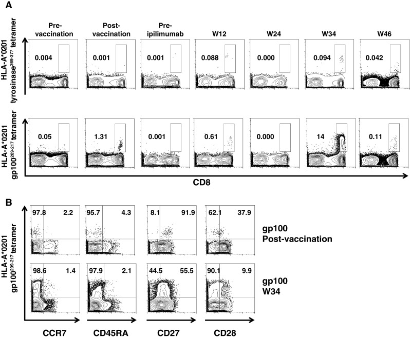
Although Patient IMF-32 received both gp100 and tyrosinase peptides (along with GM-CSF DNA), he developed gp100-specific, but not tyrosinase-specific, tetramer-reactive CD8+ cells in the peripheral blood immediately following vaccination [8]. As such, we were interested in quantifying gp100- and tyrosinase-specific T-cell responses following ipilimumab therapy (Supplemental Fig. 1E). Tetramer staining did not reveal a significant change in low levels of tyrosinase-specific tetramer-reactive CD8+ cells (below the defined threshold of ≥0.1% for tetramer positivity) with ipilimumab therapy (Fig. 1a). A significant increase in gp100-specific tetramer-reactive CD8+ cells was detected after ipilimumab administration (week 12) and peaked after the week 34 dose of ipilimumab therapy (the first maintenance treatment) and declined by week 48 (after the second maintenance treatment). He experienced rapid increases in ALC above 1,000/mm3 and CD4+, CD8+, and CD4+ICOShi T cells (Supplemental Fig. 1B,C,D), which confirmed previous observations [30, 34].
Fig. 1.

Changes in gp100- and tyrosinase-specific tetramer-reactive CD8+ T cells following ipilimumab therapy in Patient IMF-32. Thawed PBMCs from various time points were cultured for 10 days with either HLA-A*0201-restricted peptides gp100209–217 (ITDQVPFSV) or HLA-A*0201 tyrosinase369–377 (YMDGTMSQV) before undergoing tetramer staining. Panel A representative dot plots reveal a significant increase in gp100-specific but not tyrosinase-specific tetramer-reactive CD8+ T cells with ipilimumab therapy. Panel B further characterization of the week 34 gp100-specific tetramer-reactive CD8+ T cells reveals that most of these are CCR7−CD45RA−, consistent with an effector phenotype
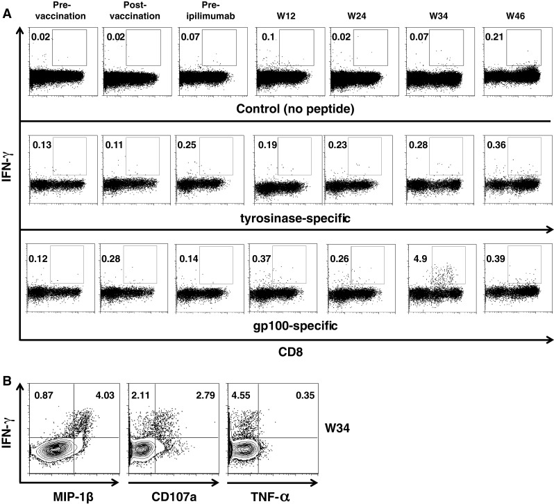
The gp100-specific tetramer-reactive CD8+ cells were analyzed at week 34 by characterizing CCR7 and CD45RA expression in these cells (Fig. 1b). The tetramer-reactive CD8+ cells were CCR7loCD45RAlo, consistent with an effector-memory phenotype. The CCR7−CD45RA− cells from this patient were also characterized as EM2 (CD27+CD28−) effector cells [35]. In addition to tetramer reactivity, the cultured cells were also analyzed for the expression of interferon gamma (IFN-γ). As with the previous tetramer response, Patient IMF-32 did not exhibit any increase in tyrosinase-specific CD8+IFN-γ+ cells following vaccination and subsequent ipilimumab therapy (Fig. 2a); however, there was a slight increase in gp100-specific CD8+IFN-γ+ cells after vaccination and a robust response following ipilimumab therapy, again peaking at week 34.
Fig. 2.

Changes in gp100- and tyrosinase-specific CD8+IFN-γ+ T cells and evidence of polyfunctional responses in Patient IMF-32. Thawed PBMCs from various time points were cultured for 10 days with either HLA-A*0201-restricted gp100209–217 (ITDQVPFSV) or HLA-A*0201 tyrosinase369–377 (YMDGTMSQV) peptides and were then restimulated with the corresponding peptides for 6 h prior to ICS. Panel A representative dot plots are provided for control [no peptide added] (top row) and show an increase in gp100-specific T cells (bottom row) but not tyrosinase-specific CD8+IFN-γ+ T cells (middle row) following ipilimumab therapy. Panel B representative dot plots show evidence of polyfunctionality wherein the CD8+ T cells express MIP-1β and CD107a
Although a single parameter, most commonly IFN-γ, has previously been used to assess the effector function of T cells, increasing evidence suggests that polyfunctional responses are associated with improved control of viral infections in preclinical models [36, 37], in patients infected with human immunodeficiency virus [38], and those immunized with vaccinia constructs [39]. As such, the week 34 gp100-specific CD8+ IFN-γ+ cells were also analyzed for, and found to express, a combination of tumor necrosis factor (TNF)-α, macrophage inflammatory protein (MIP)-1β, and the degranulation surface marker CD107a (Fig. 2b).
Tissue from the primary tumor resected in June 2005 was previously analyzed by IHC for expression of the melanoma differentiation antigens gp100 and tyrosinase. In addition, metastatic tumor tissue resected from left groin metastases in February 2006 and April 2008 and lung metastases in April and November 2007 were analyzed by IHC for gp100 and tyrosinase expression, as well as expression of class I and II major histocompatibility complex (MHC) molecules. The primary and metastatic tumors were found to express tyrosinase but not gp100. All of the metastatic tumors were also found to have low to no expression of class I and II MHC molecules. Immunohistochemical analyses are summarized in Table 2, and images are provided in Supplemental Figs. 2 and 3.
Table 2.
Summary of immunohistochemistry data—Patient IMF-32
| Tumor | gp100 | Tyrosinase | MHC class I | MHC class II |
|---|---|---|---|---|
| Primary tumor | – | + | N/A | N/A |
| Inguinal lymph node | – | + | Low | Low |
| Lung | – | + | Low | Low |
Case 2—Patient IMF-24
In June 2003, this 43-year-old man presented with melanoma of the left temporal region with satellite sites and a positive sentinel lymph node biopsy. Following excision of a left cheek cutaneous metastasis, he was treated with 6 months of temozolomide (75 mg/m2 daily) beginning in December 2003. In December 2004, he had a left neck lymph node dissection revealing 5/69 positive nodes and was given radiation therapy to his left temporal region, face, and neck. In July 2005, he was enrolled in a phase I clinical trial of a gp100 DNA vaccine at MSKCC. He received 3 doses of 500 μg human gp100 DNA followed by 3 doses of mouse gp100 DNA via intramuscular injection, with each dose separated by 3 weeks. He developed HLA-A*0201 gp100280–288 tetramer-reactive, but not HLA-A*0201gp100209–217 tetramer-reactive, CD8+ T cells in the peripheral blood immediately following vaccination [32 (Patient 8)].
In November 2005, a PET/CT scan revealed new mesenteric lymphadenopathy. He was treated with 6 weeks of chronic low-dose temozolomide but continued to demonstrate disease progression with the appearance of new retroperitoneal and hepatic metastases. In February 2006, the patient was treated with 5 weeks of high-dose IL-2 therapy at the National Cancer Institute. In August 2006, showing signs of stable disease, the patient initiated ipilimumab therapy at MSKCC in an effort to produce an objective response. He received the same ipilimumab induction regimen as Patient IMF-32. At week 24, ipilimumab maintenance therapy was initiated due to clinical benefit. In June 2007, the patient developed multiple small visceral metastases and a right occipital brain metastasis, the latter of which was resected in July 2007 followed by boost radiotherapy in October 2007. His disease remained stable until January 2009 when he was hospitalized for profound leukocytosis secondary to rapid disease progression and consequently died in March 2009.
Contrary to Patient IMF-32, Patient IMF-24 received only gp100 DNA prior to receiving immunotherapy and had no measurable immune response to HLA-A*0201 gp100209–217 following vaccination; however, on week 56 following ipilimumab initiation, the patient had a robust and durable gp100 antigen-specific tetramer response (Supplemental Fig. 4A). As with Patient IMF-32, these tetramer-positive cells were of the CCR7loCD45RAlo effector-memory phenotype (Supplemental Fig. 4B). Samples from time points immediately prior to and following ipilimumab treatment were not available for analysis.
Patterns of gp100-specific CD8+ tetramer+ and gp100-specific CD8+ IFN-γ+ T-cell production were similar in this patient, with both showing no significant increase following vaccination, but a strong and durable response after ipilimumab therapy (Supplemental Figs. 4A and 5, middle row). As with Patient IMF-32, the T cells cultured from Patient IMF-24 were also shown to express MIP-1β and CD107a, respectively (Supplemental Fig. 5, middle and bottom row). Together with the phenotypic characterization of the gp100-specific tetramer-reactive cells, the polyfunctionality of these cells suggests that they possess significant effector function.
Case 3—Patient IMF-11
In April 2005, this 62-year-old woman presented with a 0.49 mm nodular malignant melanoma of the left calf with a positive sentinel lymph node biopsy. She entered a vaccine trial at New York University Langone Medical Center of recombinant human NY-ESO-1 protein (100 μg; intradermal injection into 5% imiquimod cream pretreated skin) every 3 weeks for a total of four injections. In January 2006, the patient developed in-transit metastases on her leg. Despite treatment with sorafenib followed by two cycles of anti-integrin antibody therapy and then temozolomide chemotherapy, she continued to progress. In October 2006, a PET/CT scan demonstrated increasing size of her leg tumors along with an enlarged left external iliac lymph node.
In November 2006, with unresectable stage III cutaneous melanoma, she was initiated on ipilimumab therapy with the same dose regimen as the previous two patients. After only one treatment, she began to show a profound response evidenced by shrinking tumors, loss of associated pain, and improvement in overall quality of life. By February 2007, all tumors on both her leg and lymph node completely disappeared and her disease remains in remission for more than 3.5 years.
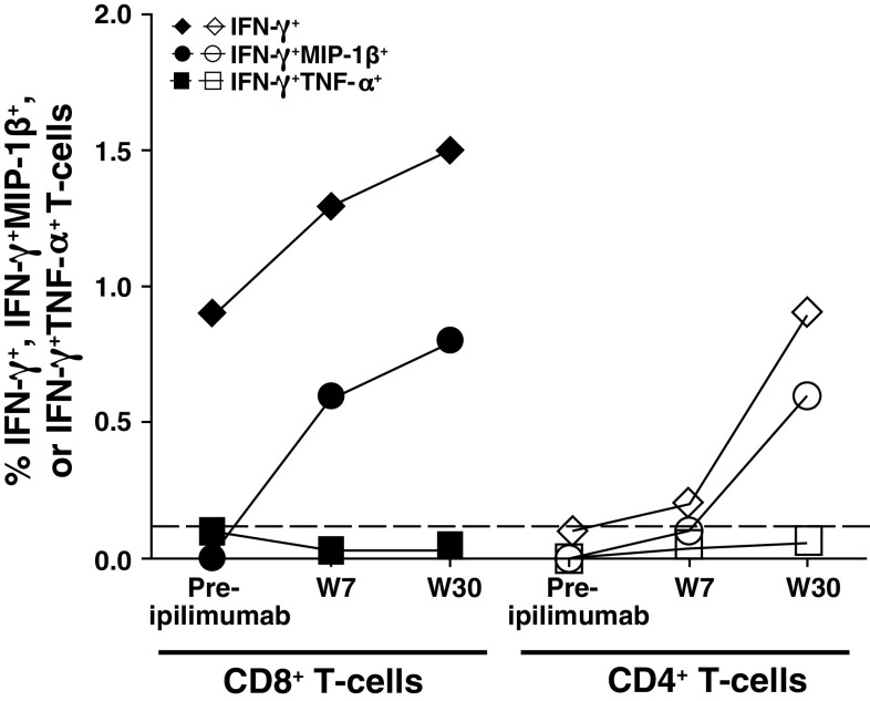
Tissue was biopsied from one of the cutaneous lesions that responded to ipilimumab therapy. Hematoxylin and eosin (H&E) staining indicated a tumorous melanin-pigmented nodule infiltrated by macrophages and lymphocytes, but with no evidence of tumor cells or melanocytes. Immunostaining demonstrated that the lymphocytes were predominantly of the CD8+ subtype, with few cells staining positive for CD4; the macrophages were also weakly CD4 positive (Supplemental Fig. 6). The patient developed NY-ESO-1 antigen-specific antibodies and a CD4+ T-cell response after vaccination with NY-ESO-1 protein. An NY-ESO-1-specific CD8+ T-cell response was not detectable utilizing different in vitro cell culture methodology [40 (Patient 6)] [40]. During the course of anti-CTLA-4 antibody treatment, Patient IMF-11 did not have an NY-ESO-1 antibody response to recombinant NY-ESO-1 protein; however, a low-titer antibody response to NY-ESO-1 peptides was detected (data not shown). Intracellular cytokine staining of PBMCs revealed an increase in NY-ESO-1-specific CD4+IFN-γ+ and CD8+IFN-γ+ T cells at week 30 following ipilimumab therapy (Fig. 3). Within these populations, there was also an increase in IFN-γ+MIP-1β+, but not IFN-γ+TNF-α+, T cells.
Fig. 3.
Ipilimumab induced NY-ESO-1 antigen-specific CD4+ and CD8+ T-cell response in Patient IMF-11. NY-ESO-1-specific CD8+ and CD4+ T cells secrete IFN-γ and MIP-1β, or IFN-γ and TNF-α, after 10-day culture and restimulation with 20-mer NY-ESO-1 overlapping peptides; CD8+IFN- γ+, CD8+IFN-γ+MIP-1β+, CD4+IFN- γ+, and CD4+IFN-γ+MIP-1β+ T cells increased after CTLA-4 blockade in Patient IMF-11
Discussion
Unique insights into the antigen-specific immune effects of ipilimumab were provided by intensive monitoring of patients with metastatic melanoma who received prior vaccination against melanoma-specific antigens and subsequently received ipilimumab therapy. This comprehensive analysis highlights several novel observations and raises interesting questions. Since the patients received prior vaccination with either gp100 peptide or DNA or NY-ESO-1 protein with or without tyrosinase peptide, we were also able to determine the antigen-specific immunogenicity of these vaccinations and whether ipilimumab therapy affected immune responses to these tumor antigens. Both Patients IMF-32 and IMF-24 did not develop any tyrosinase-specific tetramer-reactive or IFN-γ+ CD8+ responses following vaccination or at any time during ipilimumab therapy. In contrast, gp100-specific tetramer-reactive CD8+ cells, which were mildly detectable (Patient IMF-32; Patient IMF-24 [gp100280–288]) or absent (Patient IMF-24 [gp100209–217]) after vaccination, increased robustly during ipilimumab therapy, peaking at week 34 in Patient IMF-32 and remaining durable in Patient IMF-24 from week 56 to the most recent time point at week 123. This pattern was mirrored by a similar increase in CD4+IFN-γ+ and CD8+IFN-γ+ responses, which was also true for the NY-ESO-1 antigen-specific T-cell response of IMF-11. The CD8+tetramer+ populations for the phenotypic markers CCR7, CD45RA, CD27, and CD28 were analyzed after a short-term (10-day) in vitro culture stimulation to avoid the expansion of CD8+CCR7–CD45RA– effector T cells that would result from use of a longer culture period [41]. Results suggest that these cells represent an effector-memory population, an observation further substantiated by the polyfunctional nature of these cells as evidenced by the expression of CD107a, TNF-α, and/or the chemokine MIP-1β.
Patient IMF-32’s primary tumor expressed tyrosinase, but not gp100, by IHC staining. When he was treated in the adjuvant setting with a gp100 and tyrosinase peptide vaccine (administered with GM-CSF DNA), he developed gp100-reactive, but not tyrosinase tetramer-reactive, CD8+ T cells. The absence of a tyrosinase-specific immune response to vaccination may be related directly to the moderate immunogenicity of the tyrosinase antigen. For example, in our tyrosinase trial, 7/18 patients (39%) produced an antigen-specific T-cell response [7]. An intriguing but speculative possibility is that tyrosinase-specific Tregs induced by tyrosinase expression on the primary tumor may exist that dampened any immune response to the tyrosinase peptide vaccine. In contrast, since gp100 was not expressed on the primary melanoma tissue, gp100-specific Tregs may not have been present, thereby enabling an immune response to the gp100 peptide vaccination. In support of this hypothesis, recent evidence suggests that antigen-specific Tregs occur in patients with melanoma [42]. Regardless of the cause, the absence of an immune response against tyrosinase or a moderate response against gp100 is consistent with the recurrence of disease in Patient IMF-32 8 months after vaccination. IHC of tumor tissue from this and three subsequent recurrences confirmed persistent expression of tyrosinase, but not gp100. Another possible explanation for tumor escape from immune surveillance is the lack of expression of class I and II MHC molecules on the tumor tissue that would prevent effective antigen presentation to T cells.
Since none of the metastatic tumors analyzed expressed gp100, it is unclear why gp100-specific immune responses were only transiently enhanced by ipilimumab therapy. One possibility is that IHC was not sufficiently sensitive to detect the presence of low-level gp100 expression; real-time polymerase chain reaction, a more sensitive methodology, may have detected evidence of gp100 expression at the mRNA level. Another possibility is that regressing lesions, such as those seen in the lung, expressed gp100 which sustained the immune response whereas progressing lesions lost gp100, resulting in a dampened response when antigen was no longer being presented. A third possibility is that responses to gp100, normally expressed on melanocytes [43], may have been “primed” by the initial peptide vaccination and subsequently “boosted” by ipilimumab therapy. In this case, the induction of gp100-specific immune responses does not represent an anti-tumor mechanism per se, but merely reflects a non-specific activation of the immune system when CTLA-4 is inhibited. Whereas tumors from Patients IMF-24 and IMF-11 were not analyzed by IHC, the increase in T-cell response specific for the vaccine-targeted antigens seen following ipilimumab therapy may also lend support to the “prime-boost” hypothesis.
Currently, overall survival is the only validated endpoint for ipilimumab therapy. The clinical significance of the antigen-specific responses seen in this study is uncertain given the variability of outcomes in these three patients. Furthermore, although not tested, it is possible that the increase in cellular immune response was due to tumor shrinkage and loss of tumor-induced regulatory T cells rather than being a direct result of ipilimumab. Results of this study should also be interpreted with caution given the limited number of patients evaluated; however, the findings support the need for additional research to determine potential clinical implications of inducing immune responses by vaccination prior to administration of ipilimumab for treatment of advanced melanoma.
Although a gp100 vaccine did not demonstrate activity as monotherapy and did not enhance clinical response to ipilimumab in MDX010-20 [27], clinical trials evaluating different temporal combinations of ipilimumab with various vaccines are planned or ongoing in the adjuvant and metastatic setting in melanoma and prostate and pancreatic cancers [44]. Hopefully, these trials will shed light on if, how, and when ipilimumab should be combined with vaccine therapy.
Electronic supplementary material
Below is the link to the electronic supplementary material.
Acknowledgments
We thank Dr. Immanuel Luescher from Tetramer Core, Lausanne Branch, Ludwig Institute of Cancer Research, for providing the tetramers; Cora Mariano from the MSKCC Departments of Pathology for the collection of tumor tissues; and Hematology for the lymphocyte counts. This work was supported by Swim Across America, the Experimental Therapeutics Center of MSKCC and Ludwig Foundation. JDW was supported by a Damon Runyon-Lilly Clinical Investigator Award. JY was supported by MSKCC Pilot Grant of Grant number P50AT002779 from the National Center for complementary and Alternative Medicine (NCCAM) and the Office of Dietary Supplements (ODS). Editorial and writing assistance was provided by StemScientific with funding from Bristol-Myers Squibb Company.
Abbreviations
- ALC
Absolute lymphocyte count
- CTLA-4
Cytotoxic T-lymphocyte antigen-4
- GM-CSF
Granulocyte macrophage colony-stimulating factor
- H&E
Hematoxylin and eosin
- ICOS
Inducible co-stimulator
- ICS
Intracellular cytokine staining
- IHC
Immunohistochemistry
- PBMC
Peripheral blood mononuclear cell
- WBC
White blood cell
Footnotes
This paper is a Focussed Research Review based on a presentation given at the Tenth International Conference on Progress in Vaccination against Cancer (PIVAC 10), held in St. Catharine’s College, Cambridge, UK, September 27th–30th, 2010. It is part of a CII series of Focussed Research Reviews and meeting report.
References
- 1.Jemal A, Siegel R, Ward E, et al. Cancer statistics, 2008. CA Cancer J Clin. 2008;58:71–96. doi: 10.3322/CA.2007.0010. [DOI] [PubMed] [Google Scholar]
- 2.Atkins MB, Lotze MT, Dutcher JP, et al. High-dose recombinant interleukin 2 therapy for patients with metastatic melanoma: analysis of 270 patients treated between 1985 and 1993. J Clin Oncol. 1999;17:2105–2116. doi: 10.1200/JCO.1999.17.7.2105. [DOI] [PubMed] [Google Scholar]
- 3.Eigentler TK, Caroli UM, Radny P, Garbe C. Palliative therapy of disseminated malignant melanoma: a systematic review of 41 randomised clinical trials. Lancet Oncol. 2003;4:748–759. doi: 10.1016/S1470-2045(03)01280-4. [DOI] [PubMed] [Google Scholar]
- 4.Chapman PB, Einhorn LH, Meyers ML, et al. Phase III multicenter randomized trial of the Dartmouth regimen versus dacarbazine in patients with metastatic melanoma. J Clin Oncol. 1999;17:2745–2751. doi: 10.1200/JCO.1999.17.9.2745. [DOI] [PubMed] [Google Scholar]
- 5.Petrella T, Quirt I, Verma S, et al. Single-agent interleukin-2 in the treatment of metastatic melanoma: a systematic review. Cancer Treat Rev. 2007;33:484–496. doi: 10.1016/j.ctrv.2007.04.003. [DOI] [PubMed] [Google Scholar]
- 6.Karbach J, Gnjatic S, Bender A, et al. Tumor-reactive CD8+ T-cell responses after vaccination with NY-ESO-1 peptide, CpG 7909 and Montanide ISA-51: association with survival. Int J Cancer. 2010;126:909–918. doi: 10.1002/ijc.24850. [DOI] [PubMed] [Google Scholar]
- 7.Wolchok JD, Yuan J, Houghton AN, et al. Safety and immunogenicity of tyrosinase DNA vaccines in patients with melanoma. Mol Ther. 2007;15:2044–2050. doi: 10.1038/sj.mt.6300290. [DOI] [PubMed] [Google Scholar]
- 8.Perales MA, Yuan J, Powel S, et al. Phase I/II study of GM-CSF DNA as an adjuvant for a multipeptide cancer vaccine in patients with advanced melanoma. Mol Ther. 2008;16:2022–2029. doi: 10.1038/mt.2008.196. [DOI] [PMC free article] [PubMed] [Google Scholar]
- 9.Spaner DE, Astsaturov I, Vogel T, et al. Enhanced viral and tumor immunity with intranodal injection of canary pox viruses expressing the melanoma antigen, gp100. Cancer. 2006;106:890–899. doi: 10.1002/cncr.21669. [DOI] [PubMed] [Google Scholar]
- 10.Smith CL, Dunbar PR, Mirza F, et al. Recombinant modified vaccinia Ankara primes functionally activated CTL specific for a melanoma tumor antigen epitope in melanoma patients with a high risk of disease recurrence. Int J Cancer. 2005;113:259–266. doi: 10.1002/ijc.20569. [DOI] [PubMed] [Google Scholar]
- 11.Lacelle MG, Jensen SM, Fox BA. Partial CD4 depletion reduces regulatory T-cells induced by multiple vaccinations and restores therapeutic efficacy. Clin Cancer Res. 2009;15:6881–6890. doi: 10.1158/1078-0432.CCR-09-1113. [DOI] [PMC free article] [PubMed] [Google Scholar]
- 12.Nicholaou T, Ebert LM, Davis ID, et al. Regulatory T-cell-mediated attenuation of T-cell responses to the NY-ESO-1 ISCOMATRIX vaccine in patients with advanced malignant melanoma. Clin Cancer Res. 2009;15:2166–2173. doi: 10.1158/1078-0432.CCR-08-2484. [DOI] [PubMed] [Google Scholar]
- 13.Krummel MF, Allison JP. CD28 and CTLA-4 have opposing effects on the response of T-cells to stimulation. J Exp Med. 1995;182:459–465. doi: 10.1084/jem.182.2.459. [DOI] [PMC free article] [PubMed] [Google Scholar]
- 14.Salomon B, Lenschow DJ, Rhee L, et al. B7/CD28 costimulation is essential for the homeostasis of the CD4+CD25+ immunoregulatory T-cells that control autoimmune diabetes. Immunity. 2000;12:431–440. doi: 10.1016/S1074-7613(00)80195-8. [DOI] [PubMed] [Google Scholar]
- 15.Karandikar NJ, Vanderlugt CL, Walunas TL, et al. CTLA-4: a negative regulator of autoimmune disease. J Exp Med. 1996;184:783–788. doi: 10.1084/jem.184.2.783. [DOI] [PMC free article] [PubMed] [Google Scholar]
- 16.Brunner MC, Chambers CA, Chan FK, et al. CTLA-4-mediated inhibition of early events of T-cell proliferation. J Immunol. 1999;162:5813–5820. [PubMed] [Google Scholar]
- 17.van Elsas A, Hurwitz AA, Allison JP. Combination immunotherapy of B16 melanoma using anti-cytotoxic T lymphocyte-associated antigen 4 (CTLA-4) and granulocyte/macrophage colony-stimulating factor (GM-CSF)-producing vaccines induces rejection of subcutaneous and metastatic tumors accompanied by autoimmune depigmentation. J Exp Med. 1999;190:355–366. doi: 10.1084/jem.190.3.355. [DOI] [PMC free article] [PubMed] [Google Scholar]
- 18.Maker AV, Attia P, Rosenberg SA. Analysis of the cellular mechanism of antitumor responses and autoimmunity in patients treated with CTLA-4 blockade. J Immunol. 2005;175:7746–7754. doi: 10.4049/jimmunol.175.11.7746. [DOI] [PMC free article] [PubMed] [Google Scholar]
- 19.Quezada SA, Peggs KS, Curran MA, Allison JP. CTLA4 blockade and GM-CSF combination immunotherapy alters the intratumor balance of effector and regulatory T-cells. J Clin Invest. 2006;116:1935–1945. doi: 10.1172/JCI27745. [DOI] [PMC free article] [PubMed] [Google Scholar]
- 20.Quezada SA, Peggs KS, Simpson TR, et al. Limited tumor infiltration by activated T effector cells restricts the therapeutic activity of regulatory T-cell depletion against established melanoma. J Exp Med. 2008;205:2125–2138. doi: 10.1084/jem.20080099. [DOI] [PMC free article] [PubMed] [Google Scholar]
- 21.Hodi FS, Mihm MC, Soiffer RJ, et al. Biologic activity of cytotoxic T lymphocyte-associated antigen 4 antibody blockade in previously vaccinated metastatic melanoma and ovarian carcinoma patients. Proc Natl Acad Sci USA. 2003;100:4712–4717. doi: 10.1073/pnas.0830997100. [DOI] [PMC free article] [PubMed] [Google Scholar]
- 22.Phan GQ, Yang JC, Sherry RM, et al. Cancer regression and autoimmunity induced by cytotoxic T lymphocyte-associated antigen 4 blockade in patients with metastatic melanoma. Proc Natl Acad Sci USA. 2003;100:8372–8377. doi: 10.1073/pnas.1533209100. [DOI] [PMC free article] [PubMed] [Google Scholar]
- 23.Lynch T, Neal J, Bondarenko I et al (2010) Phase 2 trial of ipilimumab (IPI) and paclitaxel/carboplatin (P/C) in first-line stage IIIb/IV non-small cell lung cancer (NSCLC). Presented at the 2010 European Society for Clinical Oncology (ESMO) Annual Meeting; October 8–12, 2010, Milan, Italy. Abstract 375PD
- 24.Slovin SF, Beer TM, Higano CS et al (2009) Initial phase II experience of ipilimumab (IPI) alone and in combination with radiotherapy (XRT) in patients with metastatic castration resistant prostate cancer (mCRPC). Presented at the 2009 American Society for Clinical Oncology (ASCO) Meeting; May 29–June 2, 2009, Orlando, FL. Abstract 5138
- 25.Yuan J, Gnjatic S, Li H, et al. CTLA-4 blockade enhances polyfunctional NY-ESO-1 specific T-cell responses in metastatic melanoma patients with clinical benefit. Proc Natl Acad Sci USA. 2008;105:20410–20415. doi: 10.1073/pnas.0810114105. [DOI] [PMC free article] [PubMed] [Google Scholar]
- 26.Klein O, Ebert LM, Nicholaou T, et al. Melan-A-specific cytotoxic T-cells are associated with tumor regression and autoimmunity following treatment with anti-CTLA-4. Clin Cancer Res. 2009;15:2507–2513. doi: 10.1158/1078-0432.CCR-08-2424. [DOI] [PubMed] [Google Scholar]
- 27.Hodi FS, O’Day SJ, McDermott DF, et al. Improved survival with ipilimumab in patients with metastatic melanoma. N Engl J Med. 2010;363:711–723. doi: 10.1056/NEJMoa1003466. [DOI] [PMC free article] [PubMed] [Google Scholar]
- 28.Fong L, Kwek SS, O’Brien S, et al. Potentiating endogenous antitumor immunity to prostate cancer through combination immunotherapy with CTLA4 blockade and GM-CSF. Cancer Res. 2009;69:609–615. doi: 10.1158/0008-5472.CAN-08-3529. [DOI] [PubMed] [Google Scholar]
- 29.Ribas A, Comin-Anduix B, Chmielowski B, et al. Dendritic cell vaccination combined with CTLA4 blockade in patients with metastatic melanoma. Clin Cancer Res. 2009;15:6267–6276. doi: 10.1158/1078-0432.CCR-09-1254. [DOI] [PMC free article] [PubMed] [Google Scholar]
- 30.Ku GY, Yuan J, Page DB, et al. Single-institution experience with ipilimumab in advanced melanoma patients in the compassionate use setting: lymphocyte count after 2 doses correlates with survival. Cancer. 2010;116:1767–1775. doi: 10.1002/cncr.24951. [DOI] [PMC free article] [PubMed] [Google Scholar]
- 31.Wolchok JD, Hoos A, O’Day S, et al. Guidelines for the evaluation of immune therapy activity in solid tumors: immune-related response criteria. Clin Cancer Res. 2009;15:7412–7420. doi: 10.1158/1078-0432.CCR-09-1624. [DOI] [PubMed] [Google Scholar]
- 32.Yuan J, Ku GY, Gallardo HF, et al. Safety and immunogenicity of a human and mouse gp100 DNA vaccine in a phase I trial of patients with melanoma. Cancer Immun. 2009;9:5. [PMC free article] [PubMed] [Google Scholar]
- 33.Barrow C, Browning J, MacGregor D, et al. Tumor antigen expression in melanoma varies according to antigen and stage. Clin Cancer Res. 2006;12:764–771. doi: 10.1158/1078-0432.CCR-05-1544. [DOI] [PubMed] [Google Scholar]
- 34.Carthon BC, Wolchok JD, Yuan J, et al. Preoperative CTLA-4 blockade: tolerability and immune monitoring in the setting of a presurgical clinical trial. Clin Cancer Res. 2010;16:2861–2871. doi: 10.1158/1078-0432.CCR-10-0569. [DOI] [PMC free article] [PubMed] [Google Scholar]
- 35.Romero P, Zippelius A, Kurth I, et al. Four functionally distinct populations of human effector-memory CD8+ T lymphocytes. J Immunol. 2007;178:4112–4119. doi: 10.4049/jimmunol.178.7.4112. [DOI] [PubMed] [Google Scholar]
- 36.Casazza JP, Betts MR, Price DA, et al. Acquisition of direct antiviral effector functions by CMV-specific CD4+ T lymphocytes with cellular maturation. J Exp Med. 2006;203:2865–2877. doi: 10.1084/jem.20052246. [DOI] [PMC free article] [PubMed] [Google Scholar]
- 37.Genescà M, Rourke T, Li J, et al. Live attenuated lentivirus infection elicits polyfunctional simian immunodeficiency virus Gag-specific CD8+ T-cells with reduced apoptotic susceptibility in rhesus macaques that control virus replication after challenge with pathogenic SIVmac239. J Immunol. 2007;179:4732–4740. doi: 10.4049/jimmunol.179.7.4732. [DOI] [PMC free article] [PubMed] [Google Scholar]
- 38.Duvall MG, Precopio ML, Ambrozak DA, et al. Polyfunctional T-cell responses are a hallmark of HIV-2 infection. Eur J Immunol. 2008;38:350–363. doi: 10.1002/eji.200737768. [DOI] [PMC free article] [PubMed] [Google Scholar]
- 39.Precopio ML, Betts MR, Parrino J, et al. Immunization with vaccinia virus induces polyfunctional and phenotypically distinctive CD8+ T-cell responses. J Exp Med. 2007;204:1405–1416. doi: 10.1084/jem.20062363. [DOI] [PMC free article] [PubMed] [Google Scholar]
- 40.Adams S, O’Neill DW, Nonaka D, et al. Immunization of malignant melanoma patients with full-length NY-ESO-1 protein using TLR7 agonist imiquimod as vaccine adjuvant. J Immunol. 2008;181:776–784. doi: 10.4049/jimmunol.181.1.776. [DOI] [PMC free article] [PubMed] [Google Scholar]
- 41.Lin Y, Gallardo HF, Ku GY, et al. Optimization and validation of a robust human T-cell culture method for monitoring phenotypic and polyfunctional antigen-specific CD4 and CD8 T-cell responses. Cytotherapy. 2009;11:1–11. doi: 10.1080/14653240903136987. [DOI] [PMC free article] [PubMed] [Google Scholar]
- 42.Vence L, Palucka AK, Fay JW, et al. Circulating tumor antigen-specific regulatory T-cells in patients with metastatic melanoma. Proc Natl Acad Sci USA. 2007;104:20884–20889. doi: 10.1073/pnas.0710557105. [DOI] [PMC free article] [PubMed] [Google Scholar]
- 43.Thomson TM, Real FX, Murakami S, et al. Differentiation antigens of melanocytes and melanoma: analysis of melanosome and cell surface markers of human pigmented cells with monoclonal antibodies. J Invest Dermatol. 1988;90:459–466. doi: 10.1111/1523-1747.ep12460906. [DOI] [PubMed] [Google Scholar]
- 44.US National Institutes of Health. www.ClinicalTrials.gov. http://www.clinicaltrials.gov/ct2/show/NCT00836407?term=ipilimumab&rank=6; http://www.clinicaltrials.gov/ct2/show/NCT00357461?term=ipilimumab&rank=26; http://www.clinicaltrials.gov/ct2/show/NCT00124670?term=ipilimumab&rank=42
Associated Data
This section collects any data citations, data availability statements, or supplementary materials included in this article.


