Abstract
Psoriasis is a chronic inflammatory immune-mediated disorder affecting the skin and other organs including joints. Over 1,300 transcripts are altered in psoriatic involved skin compared to normal skin. However to our knowledge global epigenetic profiling of psoriatic skin is previously unreported. Here we describe a genome-wide study of altered CpG methylation in psoriatic skin. We determined the methylation levels at 27,578 CpG sites in skin samples from individuals with psoriasis (12 involved, 8 uninvolved) and 10 unaffected individuals. CpG methylation of involved skin differed from normal skin at 1,108 sites. Twelve mapped to the epidermal differentiation complex, upstream or within genes that are highly up-regulated in psoriasis. Hierarchical clustering of 50 of the top differentially methylated (DM) sites separated psoriatic from normal skin samples. CpG sites where methylation was correlated with gene expression are reported. Sites with inverse correlations between methylation and nearby gene expression include those of KYNU, OAS2, S100A12, and SERPINB3, whose strong transcriptional up-regulation are important discriminators of psoriasis. We observed intrinsic epigenetic differences in uninvolved skin. Pyrosequencing of bisulfite-treated DNA from skin biopsies at three DM loci confirmed earlier findings and revealed reversion of methylation levels towards the non-psoriatic state after one month of anti-TNF-α therapy.
Introduction
Psoriasis is a chronic, relapsing inflammatory skin disease affecting approximately 2% of the U.S. population and 125 million people worldwide (Bowcock and Krueger, 2005; Gudjonsson et al., 2010; Suarez-Farinas et al., 2010). It is a lifelong disease presenting predominantly before the age of 40 with spontaneous remissions infrequent. Flares can be exacerbated by stress, infection, medications, or other environmental triggers (Langley et al., 2005). In psoriasis, immune cell activation and altered epidermal differentiation are key pathogenic events (Lew et al., 2004; Zaba et al., 2009) and these are correlated with major changes in the transcriptome (Bowcock et al., 2001; Gudjonsson et al., 2010; Mee et al., 2007; Nomura et al., 2003; Quekenborn-Trinquet et al., 2005; Suarez-Farinas et al., 2010; Zhou et al., 2003).
Epigenetic alterations, such as DNA methylation and histone modification are correlated with gene expression changes (Champagne and Curley, 2009; Reik, 2007; Shi and Wu, 2009; Wilson et al., 2009). Such alterations may be part of normal developmental or differentiation processes but can also be triggered by environmental factors (Eckhardt et al., 2006; Morgan et al., 2005; Santos and Dean, 2004; Suter and Aagaard-Tillery, 2009; Weber et al., 2005). In mammals, DNA methylation commonly occurs at CpG dinucleotides (Bestor and Coxon, 1993). Approximately 70-80% of the CpG dinucleotides in the human genome are methylated, predominately in areas harboring repetitive sequences (Bird, 2002). However, regions rich in CpGs, termed CpG islands (CGIs), are also found in promoters of more than 70% of annotated genes (Bird et al., 1985; Saxonov et al., 2006). Approximately half of CGIs are associated with annotated gene transcription start sites (Bird, 2002), while others can have discrete sets of CpG sites within their promoters. The methylation of these sites has direct effects on transcriptional levels, where methylation levels typically demonstrate an inverse correlation with expression level (Bell et al., 2011).
There have been only a few studies of epigenetic alterations in diseased tissue. Many of these have involved cancerous tissue where the methylation status of tumor genomes are compared to matched normal tissue (Hu et al., 2005; Irizarry et al., 2009; Koga et al., 2009; Ordway et al., 2006). Studies of methylation changes in the diseased tissues of patients with complex diseases, including those leading to autoimmunity, are limited since diseased tissue is often difficult to access. A study on epigenetic changes in the blood of systemic lupus erythematosus patients revealed altered methylation of several genes contributing to T-cell autoreactivity, B-cell overstimulation and macrophage killing (Strickland and Richardson, 2008). Psoriasis has an advantage over many autoimmune diseases due to the accessibility of its main target organ: the skin. There have been a few reports of altered methylation within promoters of single genes in diseased skin. One example is the SHP-1 (PTPN6) promoter which is reported to be demethylated in psoriatic skin but not in skin from atopic dermatitis (AD) patients or healthy controls (Ruchusatsawat et al., 2006). However, genome-wide studies of methylation changes in psoriasis to our knowledge have not been previously described.
Here we describe global changes of methylation in involved psoriatic skin (PP) versus uninvolved psoriatic (PN) and normal (NN) skin. This was performed by querying 27,578 CpG sites with Illumina bead arrays with DNA derived from samples of each skin type. Many differences between PP versus NN skin were seen. Hierarchical clustering of 50 of the top differentially methylated sites demonstrated excellent power for differentiating PP versus NN skin. We also identified a subset of CpG sites where methylation was correlated with gene expression. Intermediate methylation at differentially methylated CpG sites was seen in PN skin, suggesting inherent epigenetic differences. Querying a subset of differentially methylated (DM) sites with an independent approach (pyrosequencing of bisulfite-treated DNA) confirmed the DM detected with the Illumina bead arrays, and also demonstrated that anti-TNF-α treatment in responders partially restores normal CpG methylation status at these loci.
Results
Differential CpG site methylation in psoriatic skin
We used the high throughput genome-wide bead-array (Infinium HumanMethylation27 Beadchip, Illumina, Inc., USA) to obtain a global, quantitative measure of the methylation status of CpG sites in PP, PN and NN skin (GSE31835). The array spanned 27,578 CpG loci selected from more than 14,000 genes, including more than 1,000 cancer-related genes and the promoter regions of 110 miRNAs. The vast majority of assayed CpG sites were located in the promoter regions of their cognate genes with an average distance of 365 bp (maximum ~1.5kb) from their transcription start sites.
PP skin samples were defined as skin biopsies collected from the site of an active psoriatic lesion. Conversely, PN skin samples were biopsies collected from skin that showed no evidence of macroscopic change. All psoriasis patient samples were collected at least 4 weeks after discontinuation of all systemic or topical therapy. Psoriasis Area and Severity Index (PASI) scores for psoriasis patients generally ranged from >10% to 30%. NN skin biopsies were defined as those biopsies collected from healthy volunteers with no clinically evident skin lesions and no self reported history of psoriatic outbreaks. Our study included 12 PP, 8 PN and 10 NN skin samples. The PN samples were derived from donors who also contributed a PP sample; hence there were 8 “paired” PP/PN samples and 4 additional PP samples without a matched PN sample.
The workflow used for analysis of the methylation data is presented in Supplementary Figure 1. For each CpG target on each array we calculated both percent methylation (β-value) and a methylation log-ratio (M-value; Methods; Figs S1 and S2). The M-values were used for tests of differential methylation since their standard deviations are more stable across a range of mean intensities than those of β –values (Supplementary Figure S3) (Du et al., 2010).
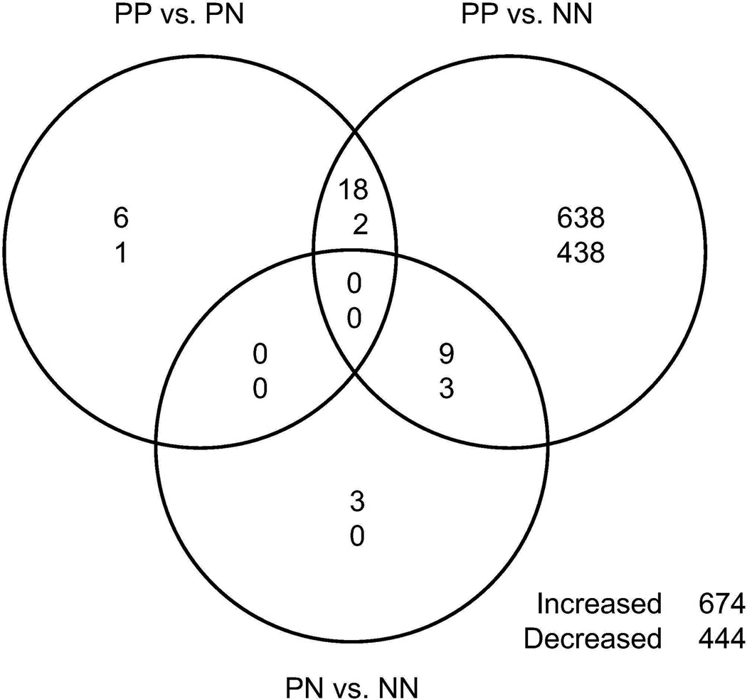
We defined a CpG as differentially methylated if it had a false-discovery rate (FDR) corrected p-value less than our significance threshold of 0.05 (Figure S1). CpG methylation in PP versus NN skin differed at 1,108 CpG sites, 88 of which demonstrated a greater than 2-fold change in M-value (Fig 1; Supplementary Table ST1). The top differentially methylated sites for this comparison are shown in Table 1. A total of 27 CpG sites demonstrated differential methylation in PP skin compared to PN skin from the same individual and 2 of those sites had a greater than 2-fold change in M-value (Fig 1; Supplementary Table ST2). Interestingly, PN skin compared to NN skin was differentially methylated at 15 CpGs, 8 of which had a greater than 2-fold change in M-value (Fig 1; Supplementary Table ST3). Additional loci may be discovered in follow-up studies with more samples in each group (Figure S4).
Figure 1.
Venn diagram of the CpG sites exhibiting differential methylation for each of three contrasts with a significance cutoff of 0.05 for the adjusted p-value. The contrasts are PP compared to PN (paired t-test), PP compared to NN and PN compared to NN. For each set the upper number is a count of the number of CpG sites with increased methylation, and the lower number is the count of CpG sites with decreased methylation. The total count of unique sites showing increased or decreased methylation in at least one comparison is shown at the bottom right.
Table 1.
The top 10 most significant CpG sites with differential methylation in PP versus NN skin. ID, Illumina CpG ID; BP GO Term, Gene Ontology Biological process; FDR p-value, false-discovery rate corrected p-value. β-values approximate percent DNA CpG methylation.
| Relevant GO | Raw β-values | ||||||
|---|---|---|---|---|---|---|---|
| ID | Symbol | BP Term Summary | Chrom | Position_hg18 | FDR p-value | PP | NN |
| cg03699566 | FOLR1 | Cell death | 11 | 71,578,300 | 5.89E-06 | 0.66 ± 0.04 | 0.43 ± 0.03 |
| cg11668844 | MCF2L | Apoptosis | 13 | 112,703,623 | 5.89E-06 | 0.45 ± 0.04 | 0.26 ± 0.02 |
| cg16139316 | S100A9 | Chronic inflammation | 1 | 151,597,382 | 5.89E-06 | 0.46 ± 0.08 | 0.84 ± 0.01 |
| cg04126866 | C10orf99 | - | 10 | 85,922,743 | 8.04E-06 | 0.65 ± 0.04 | 0.85 ± 0.01 |
| cg02813121 | S100A12 | Immune response; Positive regulation of NFKB cascade | 1 | 151,615,535 | 1.17E-05 | 0.72 ± 0.02 | 0.82 ± 0.01 |
| cg06131859 | KYNU | Response to INFgamma | 2 | 143,351,601 | 1.17E-05 | 0.32 ± 0.04 | 0.58 ± 0.03 |
| cg17582777 | EFNA3 | Cell-cell signaling | 1 | 153,316,724 | 1.58E-05 | 0.79 ± 0.02 | 0.68 ± 0.02 |
| cg13210534 | HSPB2 | Response to unfolded protein | 11 | 111,289,538 | 2.22E-05 | 0.41 ± 0.02 | 0.33 ± 0.02 |
| cg20161089 | IFI27 | Apoptosis; Type 1 interferon signaling | 14 | 93,647,267 | 2.49E-05 | 0.4 ± 0.04 | 0.63 ± 0.02 |
| cg16142218 | CHMP7 | Cell membrane organization; Endosome transport | 8 | 23,157,166 | 4.55E-05 | 0.29 ± 0.04 | 0.45 ± 0.03 |
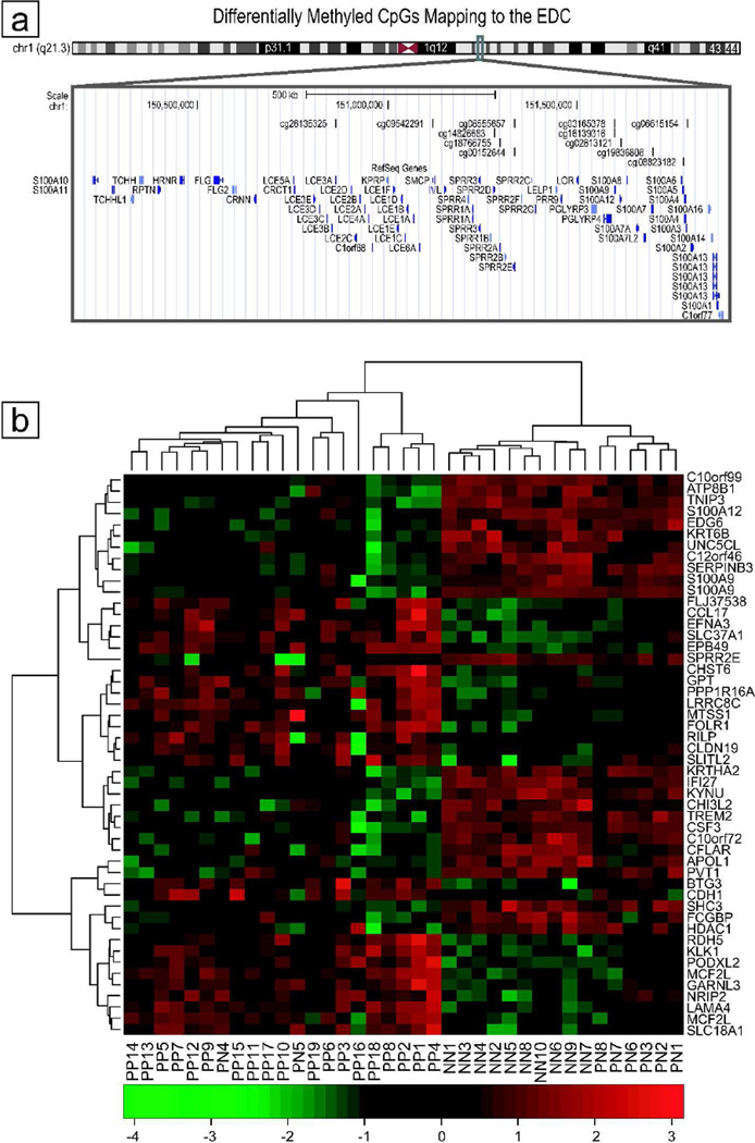
A total of 96 genes had at least two CpG sites in their vicinity where methylation levels were significant in the PP versus NN comparisons. CCND1 and GATA4 had 4 significant sites each, while GPX3 and SFRP4 had 3 significant sites each. The most extreme change was found in cg16139316, which lies upstream from S100A9 (p-value < 0.00001) within the epidermal differentiation complex (EDC), a region key to epidermal development (Volz et al., 1993). For this CpG site, methylation levels were 6.97 fold decreased in PP versus NN skin. S100A9 is strongly up-regulated in psoriatic skin (Benoit et al., 2006; Broome et al., 2003; Semprini et al., 2002; Suarez-Farinas et al., 2010; Zhou et al., 2003) and the decreased methylation in psoriatic skin is consistent with its enhanced expression. In total, there were twelve CpG sites from the EDC whose methylation levels was decreased in PP versus NN and which mapped close to genes upregulated in psoriasis (S100A3, S100A5, S100A7, S100A12, SMCP, SPRR2A, SPRR2D, SPRR2E, LCE3A; Figure 2a).
Figure 2.
A. Differentially methylated CpGs that map to the epidermal differentiation complex (EDC). Genes of the EDC are critical to epidermal development. Twelve differentially methylated CpG sites in PP compared to NN map to this region of chromosome 1. Shown are these sites and the nearby genes with chromosome ideogram. The image was adapted from a postscript generated with the UCSC genome browser (Fujita et al., 2010).
B. Heatmap showing PP, PN, and NN samples clustered with the top 50 CpG sites that differentiate PP from NN skin. Image was generated with normalized M-values. Red values indicate relatively increased methylation while green indicates relatively decreased expression.
The largest number of methylation differences and the differences of the largest magnitude were seen in the PP versus NN comparison (Supplementary Table ST1). There were comparatively few methylation changes in PP versus PN. These data contrast with expression analyses, where the PP versus PN skin comparisons are similar to the PP versus NN comparisons, though this may be an effect of small sample size (Gudjonsson et al., 2010; Zhou et al., 2003). The largest fold methylation increase in PP vs. PN skin was in sites upstream from MCF.2 cell line derived transforming sequence-like (MCF2L; FC = 2.40) and laminin alpha 4 (LAMA4; FC = 2.58). The largest fold decreases were in sites upstream from synaptopodin (SYNPO; FC = -1.91) and bone marrow stromal cell antigen 2 precursor (BST2; FC = -1.76). While the changes in methylation were significant, none of these genes have demonstrated differential expression in psoriasis.
Methylation differences in PN compared to NN skin were similarly few in number. The greatest fold changes (≥ 2) were all increases in methylation in PN versus NN skin. These included sites near GALR1, ZNF454, ZNF540, NEF3, RGS7, MLF1, FLJ42486 and NRIP2 (Supplementary Table ST3). MLF1 transcripts are down-regulated in psoriasis, consistent with the increase in methylation (Suarez-Farinas et al., 2010), but none of the other genes have been described as differentially expressed in psoriatic skin to our knowledge. The greatest decrease in methylation (−1.81 fold) was in a CpG site approximately 500bp upstream of the ZDHHC12 promoter.
Methylation levels correctly classify involved, uninvolved, and normal skin samples
We hypothesized that methylation levels of differentially methylated CpG sites could be used to classify the different skin groups. We performed between group analyses with principal component metrics and identified a subset of 50 sites (25 with increased methylation, 25 with decreased methylation) that differentiated PP from NN skin (Supplementary Table ST4). Data on an additional seven PP samples was obtained for cross-validation of clustering validity.
A heat-map of normalized M-values at the top 50 differentiating sites was generated with all PP, PN and NN samples (Figure 2b). The hierarchical clustering of these sites demonstrated excellent classifying power (Supplementary Table ST5). Classifications of psoriatic (PP or PN) versus NN were 100% accurate and 100% specific. PP clustered separately from both PN and NN skin, and performed well, with 100% sensitivity and 90% specificity. PN was classified with 75% sensitivity and 100% specificity. The lower sensitivity for PN samples was due to two PN samples (PN4, PN5) being classified as PP. Based on this dataset the classifying power of the global methylation data performed very well, especially at the classification of psoriatic versus normal, and may be as good a predictor of psoriasis as gene expression values.
Uninvolved skin exhibits intermediate levels of differential CpG methylation
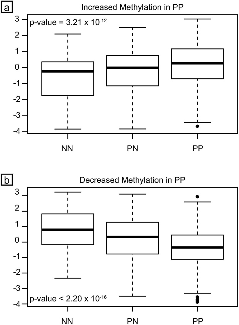
We prepared box-plots of the top 50 sites, separated by the direction of the methylation change observed in PP versus NN skin and by sample group. The medians of the three groups for sites with increased and decreased methylation were significantly different by the Kruskal-Wallis rank sum test. The trend is apparent for both the raw β-values and the normalized M-values (Supplementary Table ST4). We also observed that PN skin had a methylation level intermediate to that of the NN and PP skin for these top 50 sites (Figure 3). These intermediate methylation levels contrast with the expression levels of mRNA transcripts in PN skin which for many transcripts are usually very similar to that of normal skin (Bowcock et al., 2001). These differences may indicate intrinsic epigenetic differences in PN versus NN skin that may be reflective of a predisposition to psoriasis. However, the smaller differences in CpG methylation of PP vs. PN skin suggest that the number of samples available might have been too low (under-powered) to detect some of these alterations.
Figure 3.
Boxplots of methylation levels in three sample groups Shown are two boxplots of normalized M-value versus sample group (NN, PN, PP). The upper panel shows the methylation levels for the top 25 CpG sites that show increased methylation, and the lower panel shows the top 25 CpG sites with decreased methylation. Displayed p-values were derived from the nonparametric Kruskal-Wallis test for equality of medians among groups. Dark lines represent the median of each group. The bottom and top borders of each box are defined by the first and third quartiles. Whiskers reach out to data points up to 1.5 times the interquartile range above or below the appropriate quartile. Data points outside of that range are considered outliers and are represented by circles.
Correlation of methylation with gene expression
Nine PP, five PN, and six NN samples used for methylation analysis had also been used for global transcriptome analysis with the Affymetrix U95 arrays (Zhou et al., 2003). We were therefore able to perform a direct correlation between methylation at specific CpG loci and the level of expression of a downstream gene for these samples.
Correlations between methylation score values and gene expression levels were performed with R, and p-values were reported based on an FDR corrected p-value cutoff of 0.05. There were 12 CpG sites where methylation levels correlated significantly with gene expression levels at a nearby locus (adj. p-value ≤ 0.05; Supplementary Table ST6). Among these sites, 9 demonstrated negative correlations with nearby genes: C10orf99, OAS2 (3 sites), LGALS3BP, KYNU, IL1B, TRIM22, and PHYHIP. Three demonstrated positive correlations with nearby genes: GDPD3, TRIM14, and CCND1. Many of the genes that demonstrated a negative correlation between expression and methylation are highly up-regulated in psoriasis (C10orf99, OAS2, LGALS3BP, KYNU, IL1B, TRIM22; (Zhou et al., 2003), providing evidence of underlying methylation changes in the highly up-regulated genes in PP skin.
Overall, relatively few genes showed correlation between methylation status and gene expression. There are two possibilities for this. Firstly, the expression data used from previous generation expression arrays had fewer elements, covered fewer genes, and had less dynamic range than most modern arrays. A second reason may be low sample sizes (PP, n=9; PN, n=5; NN, n=6) which might have contributed to a lack of power to detect expression/methylation correlations. Therefore rather than directly correlating expression and methylation for the same samples we pursued a separate approach: A consensus list of 890 down-regulated and 732 up-regulated genes in psoriatic skin determined across expression studies was recently described (Suarez-Farinas et al., 2010). When this list was intersected with our methylation data, 128 differentially methylated CpG sites in PP compared to NN were less than 1.5 kilobases from the transcription start site of 113 genes in that consensus list (Supplementary Tables 1–3). For example, the genes CCL27, DDAH2, TNS1 and TRIM2 all showed consistent down-regulation in psoriatic skin and we found consistently increased methylation in and near these genes. By contrast, IFI27, KYNU, OAS2, S100A9, SERPINB3 and TNIP3 all showed significantly increased expression in psoriasis, and we found significantly decreased methylation for sites near them. There was only one gene in the consensus set where decreased expression correlated with decreased methylation: FCGBP is significantly down-regulated in psoriatic lesions, but we found significantly decreased CpG methylation approximately 430bp upstream of this gene at cg19103704.
Fine mapping of differential methylation by pyrosequencing and response to treatment with a TNFα inhibitor
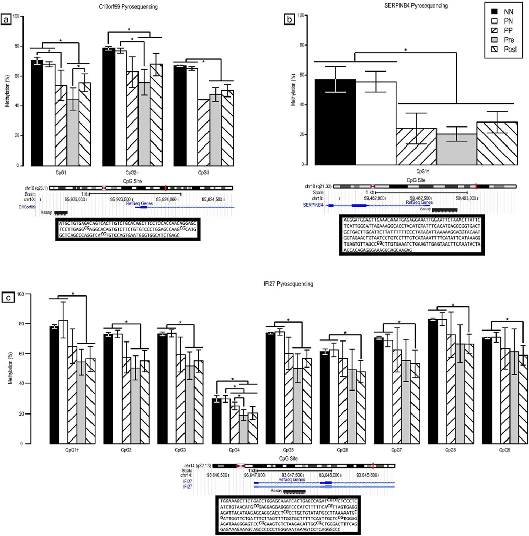
We targeted three regions for further methylation analyses. Each of these had exhibited a difference in CpG methylation in PP skin compared to NN skin (C10orf99 = −1.35; IFI27 = −2.74; SERPINB4 = −1.44). We used pyrosequencing as a separate approach to confirm these methylation differences and to investigate additional CpG sites within the c10orf99 and IFI27 intervals. In all cases, the original CpG site determined to be differentially methylated with the Illumina bead array was included in the pyrosequencing assay, along with nearby CpG sites. For all of these loci, the NN and PN samples demonstrated greater methylation than was seen in the PP samples (Figure 4). Many of these differences were statistically significant. Hence, we confirmed the differential methylation between PP and NN and/or PN skin detected by methylation bead arrays, and also showed that additional CpG sites in the differentially methylated regions exhibited similar methylation trends.
Figure 4.
Pyrosequencing data in PP, PN and NN skin biopsies at CpG sites near C10orf99 (a), SERPINB4 (b), and IFI27 (c). Methylation levels (%) with 95% confidence intervals are plotted for each CpG site by group. P-values were calculated with a two-sample t-test (unequal variance) or paired t-test as appropriate. Symbols: †, Infinium site; *, p-value < 0.05.
We also had access to PP skin biopsies from five psoriasis patients who were being treated with the anti-TNF-alpha monoclonal antibody adalimumab (Humira®). The standard dosing of 80 mg was applied by subcutaneous (SC) injection at week 0, 40 mg SC at week 1, and thereafter 40 mg SC every other week (Menter et al., 2008). Characteristics of the patients, including age, sex, and Psoriasis Area and Severity Index (PASI) score over time were ascertained for these patients (Supplemental Table ST7).
We obtained skin biopsies from these patients before treatment and after one month of adalimumab (post-treatment). The pre-treatment biopsies were taken from within a psoriatic plaque, and the post-treatment biopsies were taken either adjacent to the original biopsy site, or from a resolving psoriatic plaque contra-lateral to the original biopsy site. Four out of five patients responded well to adalimumab treatment and achieved a greater than 75% improvement in PASI score (PASI-75) at 6 months (ST7). Pyrosequencing at the same loci described above was also performed on the pre- and post-treatment samples. At one month plaques had not completely resolved. However, at each locus we observed that the mean methylation levels of treated samples increased, becoming more similar to that of uninvolved skin, though the difference was only statistically significant at CpG1 of C10orf99 (Figure 4). This suggests that methylation assays at a discrete set of loci might be a useful way to predict treatment response early in treatment.
Discussion
To our knowledge global CpG methylation changes in psoriatic versus normal skin have not previously been reported. We observed extensive differences in global methylation in PP skin compared to NN. These observations are similar to those we and others have made following expression comparisons of the same skin types (Bowcock et al., 2001; Gudjonsson et al., 2010; Oestreicher et al., 2001; Suarez-Farinas et al., 2010; Suomela et al., 2004). We identified a subset of differentially methylated CpG sites that correlated significantly with the differential expression of nearby genes. Many of these genes are highly up-regulated in psoriasis and a number map to the EDC. Some of the highly up-regulated genes, such as KYNU, OAS2, S100A12, and SERPINB3 are members of a set of genes whose high expression level differentiates psoriasis from other inflammatory skin diseases such as atopic dermatitis (Guttman-Yassky et al., 2009). Hence, altered CpG methylation near genes such as these is expected to be a good predictor of the psoriatic state. Many of the genes with the greatest methylation differences are expressed by keratinocytes. This is similar to the major alterations in mRNA levels from psoriatic versus normal skin (Bowcock et al., 2001; Gudjonsson et al., 2010; Mee et al., 2007; Nomura et al., 2003; Quekenborn-Trinquet et al., 2005; Suarez-Farinas et al., 2010; Zhou et al., 2003). Psoriatic blood has limited expression changes compared to blood from healthy controls (Lee et al., 2009) and we would expect similar findings from an investigation of methylation alterations in this tissue
We also identified methylation differences between PP compared to PN skin as well as between PN compared to NN skin. However, the number of differentially methylated sites in the PP versus PN comparisons was not nearly as great as those identified in the PP versus NN comparisons. This contrasts with expression studies where PP vs. NN and PP vs. PN comparisons yield some of the greatest alterations in transcript levels. In fact, PN skin frequently exhibited methylation levels that were intermediate with respect to PP and NN skin. This might be due to tissue heterogeneity in PN skin, but this difference has not been seen with expression studies to our knowledge. This observation needs to be explored further.
Although we observed correlations (primarily inverse relationships) between CpG methylation and expression of nearby genes, a significant number of differentially methylated CpG sites did not exhibit correlation with expression. This might be due to limited power based on the number of samples studied. Moreover, some differentially methylated genes might be expressed at low levels and have been missed by hybridization based microarray analysis. In these instances, non-hybridization strategies, such as RNA sequencing (RNA-Seq) may provide insight into less abundant transcripts in psoriasis. In other instances, these methylation differences might reflect altered methylation of noncoding RNAs, long range regulatory elements such as enhancers (Brideau et al., 2010; Hoivik et al., 2011; Lujambio et al., 2010; Shore et al., 2010; Yoon et al., 2005), or even elements mediating intra-chromosomal effects (Sharp et al., 2010).
It is unclear at this stage if the epigenetic differences described here are secondary to the altered signaling pathways of psoriasis, or are a stable predisposing event within psoriatic skin. A precedence for altered methylation predisposing to activation of the immune system is reported for interleukin-2 where demethylation at a specific CpG site in its promoter is associated with its transcriptional upregulation in mouse and humans (Bird, 2003; Bruniquel and Schwartz, 2003). This demethylation induces recruitment of Oct-1, and changes in histone modifications. Oct-1 remains on the enhancer region in a stable manner and leads to a faster and stronger induction upon subsequent stimulation. Hence, altered DNA methylation acts as a memory of the regulatory event (Murayama et al., 2006) and it is possible that similar types of epigenetic memory exist in psoriatic skin.
Multiple clinical trials have demonstrated the efficacy of TNF blockade for the treatment of psoriasis (Menter et al., 2007; Menter et al., 2008). When we examined the effect of adalimumab (Sladden et al., 2005) on global CpG methylation we observed that after a month of treatment, methylation levels had changed in the direction seen uninvolved skin. Hence, although altered methylation in psoriatic versus normal skin is not unexpected, the fact that it can be a surrogate for gene expression together with the relative ease with which it can be assayed makes it attractive as a possible predictor for diagnosing the status of activity in psoriatic skin, particularly when RNA from samples is inaccessible. Likewise, treatment response and remissions may be predicted, offering the opportunity to discontinue therapy for periods of time with significant cost saving to the patient.
Materials and Methods
Skin biopsy samples
The study was conducted according to Declaration of Helsinki Principles. Three to six millimeter punch biopsies were obtained from the PP and PN skin of psoriasis patients and NN skin from healthy controls (Supplementary Table ST8). The transcriptomes of some of these samples were previously analyzed and are described elsewhere (Bowcock et al., 2001; Zhou et al., 2003). Skin biopsies were obtained from collaborating dermatologists at Washington University School of Medicine (Saint Louis, MO), Psoriasis clinic, Baylor Hospital (Dallas, TX) or from the University of California in San Francisco (CA). Informed written consent was obtained from all individuals who donated skin biopsies. Protocols for obtaining patient biopsies were approved by Institutional Review Boards for the protection of human subjects.
DNA methylation profiling with Illumina bead arrays
Qiagen DNeasy Kits were used to isolate genomic DNA from skin biopsy samples according to the manufacturer's instructions. All samples were analyzed for DNA integrity, purity and concentration on a Nanodrop Spectrophotometer DN-100 (Nanodrop Techonologies). Bisulfite DNA conversion was by the EZ DNA methylation kit (Zymo Research) according to the manufacturer's recommendations (Bibikova et al., 2006). Bisulfite-converted genomic DNA was then interrogated with the Illumina Infinium HumanMethylation27 Beadchip, with the recommended protocols provided by the manufacturer. After hybridization, the arrays were imaged with a BeadArray Reader scanner. Image processing, intensity data extraction and analyses were conducted with the BeadArray Reader.
Differential Methylation Analysis
Non-normalized methylation data were analyzed with the R (v2.12.0) Bioconductor (Biobase v2.10.0) methylumi (v1.4.0) and lumi (v2.2.0) packages (Davis et al., 2010; Du et al., 2008; Gentleman et al., 2004; R Development Core Team, 2010). Supplementary Figure 1 provides a description of the workflow used for statistical analyses. Color channel intensities within each array were quantile normalized with the ‘lumiMethyC’ function, and data were globally normalized between arrays with simple scaling normalization via the ‘lumiMethyN’ function. In some tables we report β-values and M-values. β-values are intuitive, and M-values were used for statistical tests. Let the intensity of the methylated and unmethylated alleles be Imeth and Iunmeth, respectively.
Limma (v3.6.6) was used to fit linear models to each CpG (detection p-value ≤ 0.01 in at least one sample) (Smyth, 2005). Contrasts were defined for PP versus NN and PN versus NN. The log-odds of differential methylation were calculated for each CpG in each contrast with the ‘eBayes’ function.
Paired PP/PN samples were treated as paired samples. Linear models were fitted to only these samples, and the ‘eBayes’ function was used to calculate the moderated paired t-test. All p-values were adjusted for multiple tests with the false discovery rate method (Benjamini and Hochberg, 1995). Supplemental power calculations were performed in R 2.12.0 (Champely, 2009).
Selection of the top 50 group discriminating differentially methylated CpG sites
Between-group analysis was used to determine CpG sites that most differentiate PP from NN skin. Between-group analysis of PP vs. NN was performed using M-values for differentially methylated CpG sites with the ‘bga’ function of the MADE4 R package (Culhane et al., 2002; Culhane et al., 2005). The discriminating method used was principal components analysis. The sites were selected as the top 25 increased and top 25 decreased methylation sites on the first principal component axis. The top 50 sites were subsequently used to generate heatmaps showing the discriminatory power of these sites with Euclidean distance measures and complete hierarchical clustering. Heatmaps were generated with the Heatplus R package (v1.20.0) with a 50 color palette from the marray package maPalette function (v1.28.0).
Correlation with gene expression
Pearson correlations coefficients and their 95% confidence intervals were calculated to evaluate the strength of linear dependence between methylation at specific CpG loci and the level of expression of a downstream target. The FDR adjusted p-values were calculated to test the null hypothesis of zero correlation. All analyses were performed with R statistical programming language (v2.10.1).
Pyrosequencing
CpG methylation at and around sites flanking the statistically significant Illumina CpG loci were further validated by with pyrosequencing of bisulfite-treated DNA. This allowed us to quantify methylation at multiple CpG sites individually (Colella et al., 2003). Sample bisulfite treatment, PCR amplification, pyrosequencing, and extraction of percent methylation were performed at EpigenDx (Worcester, MA). Loci analyzed were promoter regions of IFI27, SERPINB4 and C10orf99 genes.
Supplementary Material
Acknowledgements
We thank Gabe Haller for correlations with methylation and expression at the start of this study. This work was supported in part by NIH grants AR050266 and 5RC1AR058681 to A.M.B. E.D.O.R was supported by NIH training grant T32AR007279.
Footnotes
Conflict of Interest
The authors declare no financial conflict of interest.
References
- Bell J, Pai A, Pickrell J, Gaffney D, Pique-Regi R, Degner J, et al. DNA methylation patterns associate with genetic and gene expression variation in HapMap cell lines. Genome Biology. 2011;12:R10. doi: 10.1186/gb-2011-12-1-r10. [DOI] [PMC free article] [PubMed] [Google Scholar]
- Benjamini Y, Hochberg Y. Controlling the False Discovery Rate: A Practical and Powerful Approach to Multiple Testing. Journal of the Royal Statistical Society Series B (Methodological) 1995;57:289–300. [Google Scholar]
- Benoit S, Toksoy A, Ahlmann M, Schmidt M, Sunderkotter C, Foell D, et al. Elevated serum levels of calcium-binding S100 proteins A8 and A9 reflect disease activity and abnormal differentiation of keratinocytes in psoriasis. Br J Dermatol. 2006;155:62–66. doi: 10.1111/j.1365-2133.2006.07198.x. [DOI] [PubMed] [Google Scholar]
- Bestor TH, Coxon A. Cytosine methylation: the pros and cons of DNA methylation. Curr Biol. 1993;3:384–386. doi: 10.1016/0960-9822(93)90209-7. [DOI] [PubMed] [Google Scholar]
- Bibikova M, Lin Z, Zhou L, Chudin E, Garcia EW, Wu B, et al. High-throughput DNA methylation profiling using universal bead arrays. Genome Res. 2006;16:383–393. doi: 10.1101/gr.4410706. [DOI] [PMC free article] [PubMed] [Google Scholar]
- Bird A. DNA methylation patterns and epigenetic memory. Genes Dev. 2002;16:6–21. doi: 10.1101/gad.947102. [DOI] [PubMed] [Google Scholar]
- Bird A. Il2 transcription unleashed by active DNA demethylation. Nat Immunol. 2003;4:208–209. doi: 10.1038/ni0303-208. [DOI] [PubMed] [Google Scholar]
- Bird A, Taggart M, Frommer M, Miller OJ, Macleod D. A fraction of the mouse genome that is derived from islands of nonmethylated, CpG-rich DNA. Cell. 1985;40:91–99. doi: 10.1016/0092-8674(85)90312-5. [DOI] [PubMed] [Google Scholar]
- Bowcock AM, Krueger JG. Getting under the skin: the immunogenetics of psoriasis. Nat Rev Immunol. 2005;5:699–711. doi: 10.1038/nri1689. [DOI] [PubMed] [Google Scholar]
- Bowcock AM, Shannon W, Du F, Duncan J, Cao K, Aftergut K, et al. Insights into psoriasis and other inflammatory diseases from large-scale gene expression studies. Hum Mol Genet. 2001;10:1793–1805. doi: 10.1093/hmg/10.17.1793. [DOI] [PubMed] [Google Scholar]
- Brideau CM, Kauppinen KP, Holmes R, Soloway PD. A Non-Coding RNA Within theRasgrf1Locus in Mouse Is Imprinted and Regulated by Its Homologous Chromosome inTrans . PLoS ONE. 2010;5:e13784. doi: 10.1371/journal.pone.0013784. [DOI] [PMC free article] [PubMed] [Google Scholar]
- Broome AM, Ryan D, Eckert RL. S100 protein subcellular localization during epidermal differentiation and psoriasis. J Histochem Cytochem. 2003;51:675–685. doi: 10.1177/002215540305100513. [DOI] [PMC free article] [PubMed] [Google Scholar]
- Bruniquel D, Schwartz RH. Selective, stable demethylation of the interleukin-2 gene enhances transcription by an active process. Nat Immunol. 2003;4:235–240. doi: 10.1038/ni887. [DOI] [PubMed] [Google Scholar]
- Champagne FA, Curley JP. Epigenetic mechanisms mediating the long-term effects of maternal care on development. Neurosci Biobehav Rev. 2009;33:593–600. doi: 10.1016/j.neubiorev.2007.10.009. [DOI] [PubMed] [Google Scholar]
- Champely S. pwr: Basic functions for power analysis (Version 1.1.1) [Software] 2009. [Google Scholar]
- Colella S, Shen L, Baggerly KA, Issa JP, Krahe R. Sensitive and quantitative universal Pyrosequencing methylation analysis of CpG sites. Biotechniques. 2003;35:146–150. doi: 10.2144/03351md01. [DOI] [PubMed] [Google Scholar]
- Culhane AC, Perriere G, Considine EC, Cotter TG, Higgins DG. Between-group analysis of microarray data. Bioinformatics. 2002;18:1600–1608. doi: 10.1093/bioinformatics/18.12.1600. [DOI] [PubMed] [Google Scholar]
- Culhane AC, Thioulouse J, Perriere G, Higgins DG. MADE4: an R package for multivariate analysis of gene expression data. Bioinformatics. 2005;21:2789–2790. doi: 10.1093/bioinformatics/bti394. [DOI] [PubMed] [Google Scholar]
- Davis S, Du P, Bilke S. methylumi: Handle Illumina methylation data (Version 1.4.0) [Software] 2010. [Google Scholar]
- Du P, Kibbe WA, Lin SM. lumi: a pipeline for processing Illumina microarray. Bioinformatics. 2008;24:1547–1548. doi: 10.1093/bioinformatics/btn224. [DOI] [PubMed] [Google Scholar]
- Du P, Zhang X, Huang CC, Jafari N, Kibbe WA, Hou L, et al. Comparison of Beta-value and M-value methods for quantifying methylation levels by microarray analysis. BMC Bioinformatics. 2010;11:587. doi: 10.1186/1471-2105-11-587. [DOI] [PMC free article] [PubMed] [Google Scholar]
- Eckhardt F, Lewin J, Cortese R, Rakyan VK, Attwood J, Burger M, et al. DNA methylation profiling of human chromosomes 6, 20 and 22. Nat Genet. 2006;38:1378–1385. doi: 10.1038/ng1909. [DOI] [PMC free article] [PubMed] [Google Scholar]
- Fujita PA, Rhead B, Zweig AS, Hinrichs AS, Karolchik D, Cline MS, et al. The UCSC Genome Browser database: update 2011. Nucleic Acids Research. 2010;39:D876–D882. doi: 10.1093/nar/gkq963. [DOI] [PMC free article] [PubMed] [Google Scholar]
- Gentleman R, Carey V, Bates D, Bolstad B, Dettling M, Dudoit S, et al. Bioconductor: open software development for computational biology and bioinformatics. Genome Biology. 2004;5:R80. doi: 10.1186/gb-2004-5-10-r80. [DOI] [PMC free article] [PubMed] [Google Scholar]
- Gudjonsson JE, Ding J, Johnston A, Tejasvi T, Guzman AM, Nair RP, et al. Assessment of the Psoriatic Transcriptome in a Large Sample: Additional Regulated Genes and Comparisons with In Vitro Models. J Invest Dermatol. 2010 doi: 10.1038/jid.2010.36. [DOI] [PMC free article] [PubMed] [Google Scholar]
- Guttman-Yassky E, Suarez-Farinas M, Chiricozzi A, Nograles KE, Shemer A, Fuentes-Duculan J, et al. Broad defects in epidermal cornification in atopic dermatitis identified through genomic analysis. J Allergy Clin Immunol. 2009;124:1235–1244 e1258. doi: 10.1016/j.jaci.2009.09.031. [DOI] [PubMed] [Google Scholar]
- Hoivik EA, Bjanesoy TE, Mai O, Okamoto S, Minokoshi Y, Shima Y, et al. DNA Methylation of Intronic Enhancers Directs Tissue-Specific Expression of Steroidogenic Factor 1/Adrenal 4 Binding Protein (SF-1/Ad4BP) Endocrinology. 2011;152:2100–2112. doi: 10.1210/en.2010-1305. [DOI] [PubMed] [Google Scholar]
- Hu M, Yao J, Cai L, Bachman KE, van den Brule F, Velculescu V, et al. Distinct epigenetic changes in the stromal cells of breast cancers. Nat Genet. 2005;37:899–905. doi: 10.1038/ng1596. [DOI] [PubMed] [Google Scholar]
- Irizarry RA, Ladd-Acosta C, Wen B, Wu Z, Montano C, Onyango P, et al. The human colon cancer methylome shows similar hypo- and hypermethylation at conserved tissue-specific CpG island shores. Nat Genet. 2009;41:178–186. doi: 10.1038/ng.298. [DOI] [PMC free article] [PubMed] [Google Scholar]
- Koga Y, Pelizzola M, Cheng E, Krauthammer M, Sznol M, Ariyan S, et al. Genome-wide screen of promoter methylation identifies novel markers in melanoma. Genome Res. 2009;19:1462–1470. doi: 10.1101/gr.091447.109. [DOI] [PMC free article] [PubMed] [Google Scholar]
- Langley RG, Krueger GG, Griffiths CE. Psoriasis: epidemiology, clinical features, and quality of life. Ann Rheum Dis. 2005;64(Suppl 2):ii18–ii23. doi: 10.1136/ard.2004.033217. discussion ii24-15. [DOI] [PMC free article] [PubMed] [Google Scholar]
- Lee S-K, Jeon E-K, Kim Y-J, Seo S-H, Kim C-D, Lim J-S, et al. A Global Gene Expression Analysis of the Peripheral Blood Mononuclear Cells Reveals the Gene Expression Signature in Psoriasis. Ann Dermatol. 2009;21:237–242. doi: 10.5021/ad.2009.21.3.237. [DOI] [PMC free article] [PubMed] [Google Scholar]
- Lew W, Bowcock AM, Krueger JG. Psoriasis vulgaris: cutaneous lymphoid tissue supports Tcell activation and"Type 1" inflammatory gene expression. Trends Immunol. 2004;25:295–305. doi: 10.1016/j.it.2004.03.006. [DOI] [PubMed] [Google Scholar]
- Lujambio A, Portela A, Liz J, Melo SA, Rossi S, Spizzo R, et al. CpG island hypermethylationassociated silencing of non-coding RNAs transcribed from ultraconserved regions in human cancer. Oncogene. 2010;29:6390–6401. doi: 10.1038/onc.2010.361. [DOI] [PMC free article] [PubMed] [Google Scholar]
- Mee JB, Johnson CM, Morar N, Burslem F, Groves RW. The psoriatic transcriptome closely resembles that induced by interleukin-1 in cultured keratinocytes: dominance of innate immune responses in psoriasis. Am J Pathol. 2007;171:32–42. doi: 10.2353/ajpath.2007.061067. [DOI] [PMC free article] [PubMed] [Google Scholar]
- Menter A, Feldman SR, Weinstein GD, Papp K, Evans R, Guzzo C, et al. A randomized comparison of continuous vs. intermittent infliximab maintenance regimens over 1 year in the treatment of moderate-to-severe plaque psoriasis. Journal of the American Academy of Dermatology. 2007;56:31.e31–31.e15. doi: 10.1016/j.jaad.2006.07.017. [DOI] [PubMed] [Google Scholar]
- Menter A, Tyring SK, Gordon K, Kimball AB, Leonardi CL, Langley RG, et al. Adalimumab therapy for moderate to severe psoriasis: A randomized, controlled phase III trial. Journal of the American Academy of Dermatology. 2008;58:106–115. doi: 10.1016/j.jaad.2007.09.010. [DOI] [PubMed] [Google Scholar]
- Morgan HD, Santos F, Green K, Dean W, Reik W. Epigenetic reprogramming in mammals. Hum Mol Genet. 2005;14(Spec No 1):R47–R58. doi: 10.1093/hmg/ddi114. [DOI] [PubMed] [Google Scholar]
- Murayama A, Sakura K, Nakama M, Yasuzawa-Tanaka K, Fujita E, Tateishi Y, et al. A specific CpG site demethylation in the human interleukin 2 gene promoter is an epigenetic memory. EMBO J. 2006;25:1081–1092. doi: 10.1038/sj.emboj.7601012. [DOI] [PMC free article] [PubMed] [Google Scholar]
- Nomura I, Gao B, Boguniewicz M, Darst MA, Travers JB, Leung DY. Distinct patterns of gene expression in the skin lesions of atopic dermatitis and psoriasis: a gene microarray analysis. J Allergy Clin Immunol. 2003;112:1195–1202. doi: 10.1016/j.jaci.2003.08.049. [DOI] [PubMed] [Google Scholar]
- Oestreicher JL, Walters IB, Kikuchi T, Gilleaudeau P, Surette J, Schwertschlag U, et al. Molecular classification of psoriasis disease-associated genes through pharmacogenomic expression profiling. Pharmacogenomics J. 2001;1:272–287. doi: 10.1038/sj.tpj.6500067. [DOI] [PubMed] [Google Scholar]
- Ordway JM, Bedell JA, Citek RW, Nunberg A, Garrido A, Kendall R, et al. Comprehensive DNA methylation profiling in a human cancer genome identifies novel epigenetic targets. Carcinogenesis. 2006;27:2409–2423. doi: 10.1093/carcin/bgl161. [DOI] [PubMed] [Google Scholar]
- Quekenborn-Trinquet V, Fogel P, Aldana-Jammayrac O, Ancian P, Demarchez M, Rossio P, et al. Gene expression profiles in psoriasis: analysis of impact of body site location and clinical severity. Br J Dermatol. 2005;152:489–504. doi: 10.1111/j.1365-2133.2005.06384.x. [DOI] [PubMed] [Google Scholar]
- R Development Core Team. R: A Language and Environment for Statistical Computing. Vienna, Austria: R Foundation For Statistical Computing; 2010. [Google Scholar]
- Reik W. Stability and flexibility of epigenetic gene regulation in mammalian development. Nature. 2007;447:425–432. doi: 10.1038/nature05918. [DOI] [PubMed] [Google Scholar]
- Ruchusatsawat K, Wongpiyabovorn J, Shuangshoti S, Hirankarn N, Mutirangura A. SHP-1 promoter 2 methylation in normal epithelial tissues and demethylation in psoriasis. J Mol Med. 2006;84:175–182. doi: 10.1007/s00109-005-0020-6. [DOI] [PubMed] [Google Scholar]
- Santos F, Dean W. Epigenetic reprogramming during early development in mammals. Reproduction. 2004;127:643–651. doi: 10.1530/rep.1.00221. [DOI] [PubMed] [Google Scholar]
- Saxonov S, Berg P, Brutlag DL. A genome-wide analysis of CpG dinucleotides in the human genome distinguishes two distinct classes of promoters. Proc Natl Acad Sci U S A. 2006;103:1412–1417. doi: 10.1073/pnas.0510310103. [DOI] [PMC free article] [PubMed] [Google Scholar]
- Semprini S, Capon F, Tacconelli A, Giardina E, Orecchia A, Mingarelli R, et al. Evidence for differential S100 gene over-expression in psoriatic patients from genetically heterogeneous pedigrees. Human Genetics. 2002;111:310–313. doi: 10.1007/s00439-002-0812-5. [DOI] [PubMed] [Google Scholar]
- Sharp AJ, Migliavacca E, Dupre Y, Stathaki E, Sailani MR, Baumer A, et al. Methylation profiling in individuals with uniparental disomy identifies novel differentially methylated regions on chromosome 15. Genome Research. 2010;20:1271–1278. doi: 10.1101/gr.108597.110. [DOI] [PMC free article] [PubMed] [Google Scholar]
- Shi L, Wu J. Epigenetic regulation in mammalian preimplantation embryo development. Reprod Biol Endocrinol. 2009;7:59. doi: 10.1186/1477-7827-7-59. [DOI] [PMC free article] [PubMed] [Google Scholar]
- Shore A, Karamitri A, Kemp P, Speakman J, Lomax M. Role ofUcp1enhancer methylation and chromatin remodelling in the control ofUcp1expression in murine adipose tissue. Diabetologia. 2010;53:1164–1173. doi: 10.1007/s00125-010-1701-4. [DOI] [PMC free article] [PubMed] [Google Scholar]
- Sladden MJ, Mortimer NJ, Hutchinson PE. Extensive plaque psoriasis successfully treated with adalimumab (Humira) Br J Dermatol. 2005;152:1091–1092. doi: 10.1111/j.1365-2133.2005.06582.x. [DOI] [PubMed] [Google Scholar]
- Smyth G. Limma: linear models for microarray data. In: Gentleman R, Carey V, Dudoit S, Irizarry R, Huber W, editors. Bioinformatics and Computational Biology Solutions using R and Bioconductor. New York: Springer; 2005. pp. 397–420. [Google Scholar]
- Strickland FM, Richardson BC. Epigenetics in human autoimmunity. Epigenetics in autoimmunity - DNA methylation in systemic lupus erythematosus and beyond. Autoimmunity. 2008;41:278–286. doi: 10.1080/08916930802024616. [DOI] [PMC free article] [PubMed] [Google Scholar]
- Suarez-Farinas M, Lowes MA, Zaba LC, Krueger JG. Evaluation of the Psoriasis Transcriptome across Different Studies by Gene Set Enrichment Analysis (GSEA) PLoS ONE. 2010;5:e10247. doi: 10.1371/journal.pone.0010247. [DOI] [PMC free article] [PubMed] [Google Scholar]
- Suomela S, Cao L, Bowcock A, Saarialho-Kere U. Interferon alpha-inducible protein 27 (IFI27) is upregulated in psoriatic skin and certain epithelial cancers. J Invest Dermatol. 2004;122:717–721. doi: 10.1111/j.0022-202X.2004.22322.x. [DOI] [PubMed] [Google Scholar]
- Suter MA, Aagaard-Tillery KM. Environmental influences on epigenetic profiles. Semin Reprod Med. 2009;27:380–390. doi: 10.1055/s-0029-1237426. [DOI] [PMC free article] [PubMed] [Google Scholar]
- Volz A, Korge BP, Compton JG, Ziegler A, Steinert PM, Mischke D. Physical Mapping of a Functional Cluster of Epidermal Differentiation Genes on Chromosome 1q21. Genomics. 1993;18:92–99. doi: 10.1006/geno.1993.1430. [DOI] [PubMed] [Google Scholar]
- Weber M, Davies JJ, Wittig D, Oakeley EJ, Haase M, Lam WL, et al. Chromosome-wide and promoter-specific analyses identify sites of differential DNA methylation in normal and transformed human cells. Nat Genet. 2005;37:853–862. doi: 10.1038/ng1598. [DOI] [PubMed] [Google Scholar]
- Wilson CB, Rowell E, Sekimata M. Epigenetic control of T-helper-cell differentiation. Nat Rev Immunol. 2009;9:91–105. doi: 10.1038/nri2487. [DOI] [PubMed] [Google Scholar]
- Yoon B, Herman H, Hu B, Park YJ, Lindroth A, Bell A, et al. Rasgrf1 Imprinting Is Regulated by a CTCF-Dependent Methylation-Sensitive Enhancer Blocker. Mol Cell Biol. 2005;25:11184–11190. doi: 10.1128/MCB.25.24.11184-11190.2005. [DOI] [PMC free article] [PubMed] [Google Scholar]
- Zaba LC, Fuentes-Duculan J, Eungdamrong NJ, Abello MV, Novitskaya I, Pierson KC, et al. Psoriasis is characterized by accumulation of immunostimulatory and Th1/Th17 cell-polarizing myeloid dendritic cells. J Invest Dermatol. 2009;129:79–88. doi: 10.1038/jid.2008.194. [DOI] [PMC free article] [PubMed] [Google Scholar]
- Zhou X, Krueger JG, Kao MC, Lee E, Du F, Menter A, et al. Novel mechanisms of T-cell and dendritic cell activation revealed by profiling of psoriasis on the 63,100-element oligonucleotide array. Physiol Genomics. 2003;13:69–78. doi: 10.1152/physiolgenomics.00157.2002. [DOI] [PubMed] [Google Scholar]
Associated Data
This section collects any data citations, data availability statements, or supplementary materials included in this article.


