Abstract
MicroRNAs (miRNAs) are small regulatory RNAs that have important regulatory roles in numerous developmental and metabolic processes in most eukaryotes. In Arabidopsis, DICER-LIKE1 (DCL1), HYPONASTIC LEAVES 1, SERRATE, HUA ENHANCER1 and HASTY are involved in processing of primary miRNAs (pri-miRNAs) to yield precursor miRNAs (pre-miRNAs) and eventually miRNAs. In addition to these components, mRNA cap-binding proteins, CBP80/ABA HYPERSENSITIVE1 and CBP20, also participate in miRNA biogenesis. Here, we show that STABILIZED1 (STA1), an Arabidopsis pre-mRNA processing factor 6 homolog, is also involved in the biogenesis of miRNAs. Similar to other miRNA biogenesis-defective mutants, sta1-1 accumulated significantly lower levels of mature miRNAs and concurrently higher levels of pri-miRNAs than wild type. The dramatic reductions of mature miRNAs were associated with the accumulation of their target gene transcripts and developmental defects. Furthermore, sta1-1 impaired splicing of intron containing pri-miRNAs and decreased transcript levels of DCL1. These results suggest that STA1 is involved in miRNA biogenesis directly by functioning in pri-miRNA splicing and indirectly by modulating the DCL1 transcript level.
INTRODUCTION
In plants, many growth and developmental processes are regulated by microRNA (miRNAs). These include development of rosette leaves, root development, apical dominancy of stem, auxin signaling, abscisic acid (ABA) responses, morphogenesis of flowers and arrangement of siliques (1–4). The responses of plants to abiotic stresses such as drought, high salinity, phosphate starvation and UV-B are also mediated by some miRNAs (5–10).
MiRNAs are generated from long primary transcripts [primary miRNAs (pri-miRNAs)] via precursor miRNAs (pre-miRNAs) through two-step processes involving at least five proteins [DICER-LIKE1 (DCL1), HYPONASTIC LEAVES 1 (HYL1), SERRATE (SE), HUA ENHANCER (HEN1) and HASTY (HST)] (11–16). DCL1 is the RNAse type III slicer that cleaves pri-miRNAs to produce pre-miRNAs and eventually miRNAs (17,18). HYL1 is a double-stranded RNA-binding protein with two RNA-binding domains for cooperative binding most likely to the miRNA/miRNA* duplex region. HYL1 interacts with SE and DCL1 through RNA-binding domain 2 (RBD2) for precise pri-miRNA processing (18–20). SE is a zinc-finger protein that enhances the accuracy of DCL1-dependent pri-miRNA processing together with HYL1 (18,21,22). HEN1 is a specific methyltransferase that adds a methyl group to the 2′-OH position of miRNA/miRNA* and siRNA/siRNA* duplexes. The HEN1-dependent methylation protects small RNA duplexes from unspecific addition of polyuridines and subsequent degradation (23). Finally, HST, the Arabidopsis homolog of the human nucleocytoplasmic transport factor Exportin-5 exports the miRNA/miRNA* duplex to cytoplasm (14).
Recently, two RNA-binding proteins were defined as components in miRNA biogenesis (24,25). One is DAWDLE (DDL), a type of fork-head associate domain (FHA) protein that interacts with DCL1 and functions in miRNA biogenesis, probably through stabilizing pri-miRNA transcript (24). Another is TOUGH (TGH) with G-patch and SWAP domains that promotes pri-miRNA recruitment to HYL1 and enhances the pri-miRNA processing efficiency as a component of the DCL1–HYL1–SE microprocessor complex (25).
The levels of pre-messenger RNAs (pre-mRNAs) in eukaryotes are tightly controlled by maturation (modification of nascent transcript with 5′-capping, splicing and 3′-polyadenylation) and stability. Likewise, pri-miRNAs are transcribed by RNA polymerase II and further modified by the addition of a 5′-seven methyl guanine cap and a polyadenosine tail at the 3′-end, and then processed by splicing in case of intron-containing nascent transcripts (26,27). Despite similarities in the maturation processes of pre-mRNAs and pri-miRNAs, their final destinations and functions are quite distinct. Mature mRNAs are exported to the cytoplasm and undergo a pioneer round of translation, and defective or improperly processed mRNAs are removed by the nonsense-mediated mRNA decaying pathway (28). In contrast, mature pri-miRNAs in plants are mostly retained in the nucleus for further processing into mature miRNAs and then exported to the cytoplasm (29,30). However, in plants very little is known about the maturation events from nascent transcripts to mature pri-miRNAs. Recently, a link between pre-mRNA processing and pri-miRNA processing emerged with the discovery of involvement of ABA HYPERSENSITIVE1 (ABH1)/CBP80 and CBP20 in pri-miRNA biogenesis (31). In addition to reduced pre-mRNA splicing efficiency, levels of pri-miRNAs are increased and mature miRNAs are reduced in abh1/cbp80 and cbp20 mutants as compared with wild-type (WT) plants (31). ABH1/CBP80 and CBP20 constitute a large complex, the nuclear cap-binding complex (CBC) that binds to the 5′-cap of pre-mRNAs (32). These results suggested that ABH1/CBP80 and CBP20 cap-binding proteins are involved in both pre-mRNA and pri-miRNA processing. Laubinger et al. (31) also suggested that SE has dual roles in pre-mRNA splicing and pri-miRNA processing and that SE might share a common mechanism with CBC or be a part of a larger cap-binding protein complex. Furthermore, inactivation of ABH1/CBP80 and CBP20 displayed significant reductions of ta-siRNAs along with their initiators, miR173 and miR390 (33). Despite the prominent reduction of ta-siRNAs that are important to sense post-transcriptional gene silencing, ABH1/CBP80 is not involved in the sense-TGS pathway, suggesting that their roles may be limited to miRNA biogenesis (33).
The involvement of ABH1/CBP80 and CBP20 in pri-miRNA processing raises interesting questions as to whether other pre-mRNA processing proteins are also involved in pri-miRNA processing. To address these questions, we focused on STABILIZED1 (STA1), a gene for pre-mRNA processing in Arabidopsis. STA1 encodes a protein that is homologous to human U5 snRNP-associated 102-kDa protein (PRPF6), and the yeast pre-mRNA splicing factors, PRP1p (fission yeast) and Prp6p (budding yeast), and was shown to be important in pre-mRNA splicing and mRNA stability (34). In addition, the pleiotropic defects of development, chilling sensitivity and hypersensitivity to ABA were observed in sta1-1, a weak allele of sta1 (34). Based on these initial clues, we investigated the possible role of STA1 in splicing of pri-miRNAs and the biogenesis of miRNAs through a series of molecular and bioinformatic analyses in this study.
MATERIALS AND METHODS
Plant materials and growth conditions
se-1 (35), hyl1-2 (SALK_ 064863) and abh1-285 (SALK_024285) mutants of the Colombial-0 (Col-0) background and sta1-1 with a single mutation in the Columbia-gl1 background harboring the stress-responsive RD29A promoter-driven luciferase (Col-RD29A-LUC) were used in this study (34). Col-0 and Col-RD29A-LUC were considered the WT. Seeds were grown on Murashige and Skoog medium (1% sucrose and 0.8% agarose) after surface sterilization with sodium hypochlorite (7%). The seeds were stratified at 4°C for 3 days in dark and transferred to a growth chamber (16-h light and 8-h dark at 22°C). For cold treatment, plants were placed in an ice-filled insulated box for 24 h in a 4°C cold room.
RNA extraction and cDNA synthesis from total RNAs
Total RNA was extracted from the seedlings using RNeasy Plant Mini Kits (QIAGEN) or IQeasy Plant RNA extraction Mini Kit (iNtRON). Total RNAs from Col-0, se-1, hyl1-2 and sta1-1 were used as templates for cDNA synthesis. PrimeScript Reverse Transcriptase (TaKaRa Bio) originating from Moloney Murine Leukemia Virus was used to synthesize the first-strand cDNA from denatured RNA using the manufacturer’s protocol. RNA for small RNA blot analysis was extracted using TRI Reagent Solution (Ambion). RNA concentrations were measured using a NanoDrop ND 1000 spectrophotometer.
Small RNA blot hybridization
The small RNA samples (15 μg) from Col-0, se-1, hyl1-2, abh1-285 and sta1-1 were mixed with 5 μl of gel loading buffer (Ambion) and resolved on denaturing 15% polyacrylamide gels containing 7.5 M urea. The separated RNA samples were transferred onto a positively charged Amersham Hybond-N+ nylon membrane (GE Healthcare) using a Trans-Blot SD Semi-Dry Electrophoretic Transfer Cell (Bio-Rad). γ-32ATP-radiolabeled single-stranded DNA oligonucleotide probes were used for detection of specific miRNAs. Sequences for probes are listed in Supplementary Table S4. Hybridized membranes were exposed to a storage phosphor screen (Amersham Biosciences) for 1–4 days and the screens were scanned using a Storm 860 phosphoimager (Molecular Dynamics).
Expression analysis of miRNA biogenesis genes
Total RNA from 12-day-old seedlings of WT or sta1-1 was isolated using Qiagen RNeasy Plant mini kit. The reverse transcription PCR (RT-PCR) was carried out with 50 ng of total RNA using HiPureoneStep RT-PCR kit (GENEPOLE). The RT-PCR programs used were as follows: reverse transcription at 45°C for 10 min, activation of DNA polymerase at 95°C for 2 min, followed by 29 cycles of denaturation at 95°C for 10 s, annealing at 55°C for 10 s and extension at 72°C for 30 s. The gene-specific primers are listed in Supplementary Table S5. All experiments were performed at least three times using seedlings grown independently. The expression level of UBQ10 was used for loading control.
Quantitative real-time PCR
Quantitative real-time PCR (qRT-PCR) was carried out using DyNAmo Flash SYBR Green qRT-PCR Kit (Finnzymes) in a RotorGene Q RT-PCR cycler (Qiagen). In each 0.1 ml qRT-PCR strip tube (Qiagen), 3 µl cDNA were mixed with 10 µl DyNAmo Flash SYBR Green master mix, 5 µl of Milli-Q H2O and 1 µl of gene-specific forward and reverse primer for a total volume of 20 µl. Gene-specific primers for miRNA biogenesis genes, miRNA target genes and pri-miRNAs are listed in Supplementary Tables S5, S6 and S7, respectively. Actin2 (At3g18780) or UBQ10 (At4g05320) was used as a reference gene using the primers 5′-GCACCCTGTTCTTCTTACCG-3′/5′-AACCCTCGTAGATTGGCACA-3′ (Actin2) and 5′-GATCTTTGCCGGAAAACAATTGGAGGATGG-3′/5′-CGACTTGTCATTAGAAAGAAAGAGATAACAGG-3′ (UBQ10). The following thermal cycle program was used for all amplifications: 95°C for 7 min; 35 cycles of 95°C for 15 s, 55°C for 20 s, 72°C for 25 s with a gradual 1°C rise per cycle from 72°C to 95°C. Fluorescence of the SYBR Green I dye was measured at the end of the extension step of every PCR cycle. The ΔΔCt method (36) was used to calculate the normalized gene expression levels of the mutant lines relative to Col-0. The difference in the cycle threshold (Ct) values (ΔCt) between the mutants and the WT was found for each gene by subtracting the Ct values of the mutants, respectively, from the Ct value of WT. Fold-change values of the target genes were subsequently normalized by dividing the ΔCt values with the ΔCt values of the reference gene. Each experiment was carried out at least three times and the mean and standard deviation were calculated.
Amplification of cDNA using intron-spanning pri-miRNA-specific primers
PCR was carried out using HotMasterTaq DNA Polymerase (5 Prime) in a PTC-240 Peltier Thermal Cycler (Bio-Rad). An amount of 20 µl of cDNA was diluted to a volume of 60 µl with nuclease-free H2O. In each reaction, 2 µl cDNAs were mixed with 1 µl dNTP mix (2.5 mM of each nucleotide), 2 µl 10× HotMasterTaq Buffer (25 mM Mg2+), 0.3 µl HotMasterTaq DNA Polymerase and 1 µl of gene-specific forward and reverse primer (Supplementary Table S8). Milli-Q H2O was added to make up the reaction volume to 20 µl. β-Tubulin 8 (At5g23860) cDNA was amplified as an internal control using 5′-ATAACCGTTTCAAATTCTCTCTCC-3′ and 5′-TGCAAATCGTTCTCTCCTTG-3′ as forward and reverse primers, respectively. The following thermal cycles were used for all cDNA amplifications: 94°C for 2 min; 30 cycles of 94°C for 30 s, 55°C for 30 s, 72°C for 1.5 min; 72°C for 5 min.
Cloning and sequencing of the amplified fragments of the pri-miRNA transcripts
PCR fragments amplified with primers for pri-miR172b (Supplementary Table S8) were purified with MEGAquick-spin (iNtRON), subcloned into pGEM-easy TA vector (Invitrogen) and transformed to TOP10 competent cells (Invitrogen). The cloned fragments were analyzed by sequencing and aligned with annotated sequences of pri-miRNA172b (www.arabidopsis.org).
Deep sequencing and analysis of small RNA libraries
Small RNA libraries from the sta1-1 mutant and Col-RD29A-LUC were constructed according to an established protocol (9). Briefly, the 18–30 nt fraction of the small RNAs was separated by resolving total RNA in a 15% denaturing polyacrylamide gel. After sequentially adding 3′- and 5′-adaptors, ligation products were purified and amplified using RT-PCR. PCR products were sequenced using the Illumina Genome Analyzer II.
In-house Perl scripts were used to generate clean reads from raw small RNA sequences by removing 3′-adaptor sequences. Clean reads that were at least 18 nt in length were retained and clustered into unique reads. Only reads with a perfect match to the Arabidopsis genome sequence (TAIR9) were used for subsequent analysis. Clean reads were identified as known mature miRNAs if they were identical to the annotated Arabidopsis mature miRNAs in the miRBase (release 17) (37). Expression levels of miRNAs [transcripts per 10 million (TPTM)] were calculated by normalizing miRNA counts with the total number of mapped clean reads in the corresponding small RNA library.
Expression analysis with whole-genome tiling array
Arabidopsis Col-RD29A-LUC and sta1-1 mutant seedlings were grown for 2 weeks on MS agar plates supplemented with 3% sucrose and with daily cycles of 16 h of light at 22°C and 8 h of dark at 18°C. The seedlings were subjected to cold stress for 24 h at 4°C. Total RNA was isolated from cold-stressed seedlings using the RNeasy plant mini kit (Qiagen). DNA contamination in RNA samples was eliminated by DNase digestion (Qiagen). RNA concentration was quantified with a Nanodrop spectrophotometer at 260 nm. RNA integrity was determined on a Bioanalyzer (Agilent Technology). Whole transcript targets for TILING array hybridization were prepared by using the GeneChip® Whole Transcript Double-Stranded cDNA Synthesis Kit and the GeneChip® Whole Transcript Double-Stranded DNA Terminal Labeling Kit (Affymetrix). Targets were hybridized to GeneChip® Arabidopsis Tiling 1.0 R Array (Affymetrix).
To analyze the tiling array data, we re-mapped the tiling array probes to the Arabidopsis genome (TAIR9) using SOAP2 (38) and retained only probes that perfectly matched to a unique position in the genome for subsequent analyses. We created a custom chip definition file using the probe mapping result and used the aroma affymetrix framework (39) for quantile normalization of tiling array data.
To identify retained introns, we first calculated the log2 signal intensity for each annotated intron (TAIR9) based on the trimmed mean of signal intensities from all probes that were mapped to the intron. Introns with less than three mapped probes or low expression (log2-expression value was <5 in all samples) were removed from further consideration. We used the SAM algorithm (40) to identify introns that showed significantly elevated expression in the sta1-1 mutant samples than in the WT control samples. A false discovery rate of 0.05 was used as the significance cutoff.
To identify genes that were differentially expressed in the sta1-1 mutant versus WT, we first used the genefilter package in Bioconductor (http://www.bioconductor.org/) to remove genes that showed low expression level (normalized signal intensity was <100 in all samples) and genes that showed little change in gene expression across samples (interquartile range of log2 intensities was <0.5). We then applied the linear model method implemented in the limma package in Bioconductor to identify genes that showed differential expression. The Benjamini and Hochberg method (41) was used for adjustment for multiple comparisons.
RESULTS
sta1-1 affects pre-mRNA splicing at the whole genome level
We previously showed that unspliced COR15A transcript accumulates in sta1-1 and suggested that STA1 functions in pre-mRNA splicing (34). However, the genome-wide effect of sta1-1 on intron-retention was not fully examined. To investigate the global effect of STA1 on pre-mRNA splicing, we compared the genome-wide expression profiles of sta1-1 plants with that of WT plants using Arabidopsis Tiling Array 1.0 R. As the previously reported unspliced gene was cold-inducible COR15A, cold-treated seedlings were used for total RNA extraction. We measured the expression levels of each intron that contains at least three unique probes and found that 695 introns had a significantly higher expression in sta1-1 than in Col-RD29A-LUC (Supplementary Table S1), indicating that they were not spliced properly in sta1-1. This list includes the first intron in the COR15A transcript as expected from our published result (34). Among 695 retained introns, 295 (42%) were the first intron and 104 (15%) were the second intron, indicating that similar to CBC and SE (31), STA1 has significant effect on splicing at the 5′-end of the transcripts. These results confirm our previous conclusion that STA1 functions in pre-mRNA splicing. In addition, we identified 2079 genes that were differentially expressed (899 upregulated and 1180 downregulated) in sta1-1 compared with Col-RD29A-LUC (Supplementary Table S2).
Phenotypes of sta1-1 are reminiscent of miRNA biogenesis mutants
Bezerra et al. (42) suggested that SE and CBC might function via a common mechanism for mRNA metabolism because the leaf phenotypes of se-1 were particularly reminiscent of abh1/cbp80 or cbp20. Indeed, the similar leaf phenotypes in these mutants were an important clue in finding the dual roles of SE and CBC for pre-mRNA and pri-miRNA splicing (31). Kim et al. (33) also initiated their study on ABH1/CBP80 and CBP20 based on the leaf phenotype similarity.
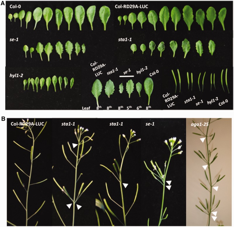
We noticed that some of the phenotypes of sta1-1 resembled those of the miRNA biogenesis mutants, se-1, hyl1-2 and ago1-25. Size, shape and vein patterning of leaves are highly regulated by several miRNAs, and therefore defects in miRNA biogenesis pathways may be easily observed by the leaf phenotype (42). Indeed, among the distinct phenotypes in se-1 are serrated leaves. The leaves of sta1-1 were also serrated but with a slightly different pattern from se-1. The degree of serration in sta1-1 is not as strong as in se-1, and sta1-1 leaves started to show serration at about the fifth to sixth leaves, while se-1 developed serrated leaves as early as the third leaf (Figure 1A). In WT, siliques were arranged in a spiral pattern. ago1-25, a weak ago1 allele defective in miRNA biogenesis, often showed an altered silique arrangement (Figure 1B). The abnormal phyllotactic arrangements were also observed in sta1-1 and se-1. Furthermore, the siliques were shorter in sta1-1, se-1 and hyl1-2 than in WT. The shaping and development of lateral organs are determined by several miRNAs. Therefore, the morphological variations strongly implied general defects of miRNA functions in sta1-1.
Figure 1.
Phenotype comparisons between sta1-1 and miRNA biogenesis defective mutants. (A) Leaf and silique morphology. sta1-1 showed serrated leaves and small siliques similar to miRNA biogenesis mutants. (B) Silique phyllotaxy defects in sta1-1 and miRNA biogenesis mutants. Arrow heads indicate altered silique phyllotaxy.
sta1-1 impairs the accumulation of mature miRNAs
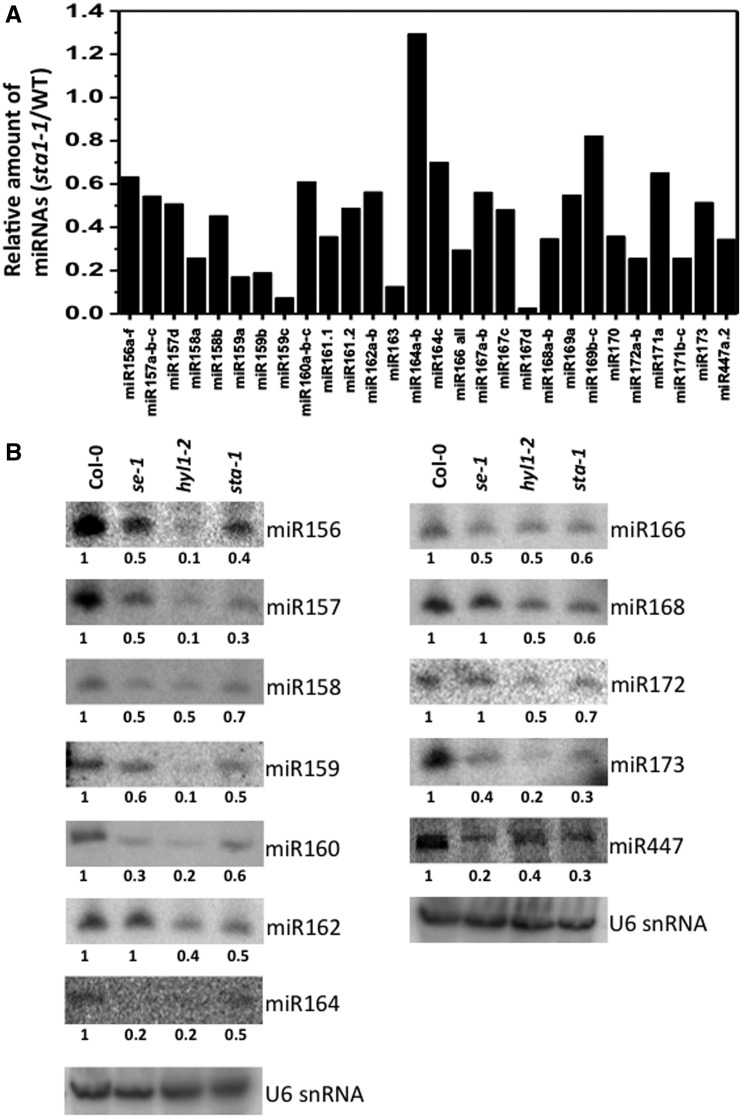
The phenotypic resemblances among sta1-1, se-1, hyl1-2 and abh1/cbp80 led us to investigate the levels of miRNAs in sta1-1. Using the Illumina platform, we generated 20.4 and 28.0 million clean reads that perfectly matched the Arabidopsis genome from the small RNA populations in sta1-1 and WT, respectively. We compared the normalized counts of mature miRNAs in sta1-1 and WT and found that the majority of miRNAs had lower expression in sta1-1 (Supplementary Table S3). Within 79 miRNAs that had total expression of at least 20 TPTM, 69 (85%) miRNAs showed reduced expression in sta1-1, including 60 (79%) miRNAs whose expression was reduced to 60% or lower compared with WT (Supplementary Table S3). We randomly chose several miRNAs to validate their altered expression in sta1-1 through small RNA blot hybridization analysis. The accumulation levels of miR156, miR157, miR158, miR159, miR160, miR162, miR166, miR168, miR171, miR172, miR173, miR393, miR398 and miR447 were reduced in sta1-1 compared with WT (Figure 2 and Supplementary Figure S2). These results were largely consistent with the results of Illumina sequencing (Figure 2, Supplementary Figure S2 and Supplementary Table S3). The only difference between the small RNA blot hybridization and Illumina sequencing was the level of miR164; the expression of miR164 was slightly increased in the sta1-1 sequencing results, but slightly decreased in the small RNA blot hybridization. Similar levels of reduction in tested miRNAs were also observed in se-1 and hyl1-2 except for miR162, miR168 and miR172 levels in se-1.
Figure 2.
Reduction of miRNA levels in sta1-1. (A) Small RNA sequencing analysis of miRNAs in WT and sta1-1. From the small RNA sequencing results, abundance of miRNAs in sta1-1 was compared with WT. (B) RNA blot hybridization of miRNAs from 4-week-old Col-0, se-1, hyl1-2 and sta1-1. The levels of U6 small nuclear RNA were shown as loading controls. Numbers below the blot images are relative intensities of the miRNA bands.
An mRNA cap-binding protein, ABH1/CBP80 was shown to act in pri-miRNA processing (31). The leaf and phyllotaxy phenotypes described above were also found in abh1/cbp80 mutants (Supplementary Figure S1). Small RNA blot hybridization analysis showed that the miRNA reductions in sta1-1 were more significant than those in abh1/cbp80 (Supplementary Figure S2). It should be noted that Col-0 was mostly used as a WT control throughout our experiments except for the tiling array and small RNA sequencing in which Col-RD29A-LUC was used as a control. sta1-1 was originally isolated from Columbia-gl1 harboring the stress-responsive RD29A promoter-driven luciferase (Col-RD29A-LUC) (34). Recent reports showed that different accumulation patterns of miRNAs can be observed in different Arabidopsis accessions (43). Thus, some selected miRNA accumulation levels were compared between Col-0 and Col-RD29A-LUC and very similar levels of miR156, miR158, miR159, miR160, miR166 and miR172 accumulation were observed (Supplementary Figure S3). In addition, the patterns of higher accumulation of pri-miRNAs in sta1-1 were still observed when compared with its background line, Col-RD29A-LUC (Supplementary Figure S4). Taken together, these results suggest that STA1 is involved in miRNA biogenesis.
Expression levels of miRNA target genes are increased in sta1-1
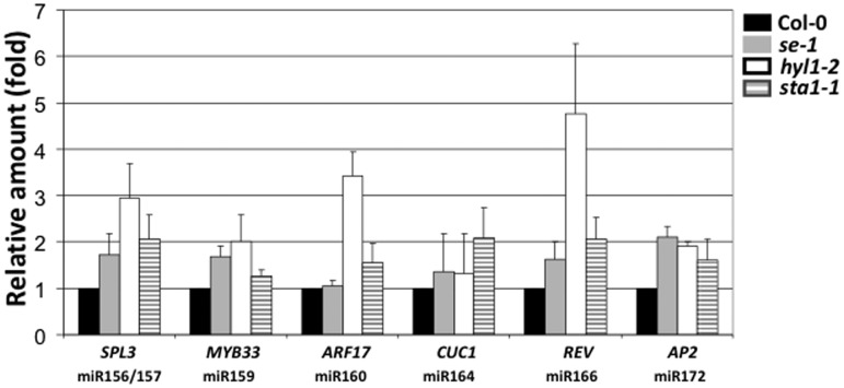
Each miRNA recognizes specific mRNA target(s) through sequence specificity and initiates degradation/translation repression of the targets (44–46). Since the amount of target mRNAs is highly inversely correlated to the amount of specific miRNAs, we determined the levels of target mRNAs for some miRNAs by qRT-PCR (Figure 3). SQUAMOSA PROMOTER-BINDING PROTEIN-LIKE (SPL) transcription factors are involved in a variety of developmental processes in flowers and leaves and the expression of the SPL gene family is directly regulated by miR156/157 (47–49). The upregulation of target SPL3 transcripts was expected due to the decrease of miR156 and miR157 in sta1-1 (Figure 2). As expected, an increase of the SPL3 transcript level was observed in sta1-1 (Figure 3). Increased SPL3 expression levels were also observed in the other two miRNA defective mutants as these mutants also generated lower levels of miR156/157 than WT (Figure 3). Plastochron, a temporal period between two successive leaves (50), is modulated by the transcript levels of SPL9/15 and eventually determines the leaf numbers at bolting (51). High levels of SPL9/15 can cause long plastochron and decreased leaf numbers at bolting. Indeed, in the miRNA biogenesis mutants examined, the reduced miR156/157 levels were linked to the higher expression levels of SPL9/15 genes and eventually the decreased leaf number (Supplementary Figure S5). Interestingly, the levels of SPL9 transcripts show a stronger correlation than those of SPL15 transcripts to the leaf numbers in the tested mutants. MYB33 encodes a MYB protein-like transcription factor that regulates many developmental processes, and the expression of MYB33 is regulated by miRNA159 (52). se-1, hyl1-2 and sta1-1 with decreased miR159 levels showed marginally increased expression levels of MYB33 (Figure 3). AUXIN RESPONSE FACTOR17 (ARF17), whose expression is controlled by miR160, is important for proper development and modulates expression of early auxin response genes (53). Higher levels of ARF17 transcripts accumulated in sta1-1, consistent with its reduced miR160 levels compared with WT (Figure 3). CUP-SHAPED COTYLEDON1 (CUC1) is essential for boundary size control of meristems and is a target of miR164 (54). REVOLUTA (REV), the target of miR166, is expressed in young leaves and controls development of the leaves (55). Compared with WT, the accumulation of CUC1 and REV transcripts was higher in sta1-1, which is linked to decreased miR164/166 levels and explains the serrated leaf phenotype of sta1-1 (Figure 3). APETALA 2 (AP2), a target of miR172 (56), showed a slight increase in se-1, hyl1-2 and sta1-1 that accumulated lower amounts of miR172 than WT (Figure 3). Overall, the expression levels of target mRNAs in se-1, hyl1-2 and sta1-1 were largely inverse-correlated to the corresponding miRNA levels in each mutant (Figures 2 and 3).
Figure 3.
qRT-PCR expression analysis of selected miRNA target transcripts in Col-0, se-1, hyl1-2 and sta1-1. Relative amounts of mRNA levels were obtained by dividing the expression level of the mutant with WT value. Three biological samples were used.
Expression levels of a subset of pri-miRNAs are increased in sta1-1
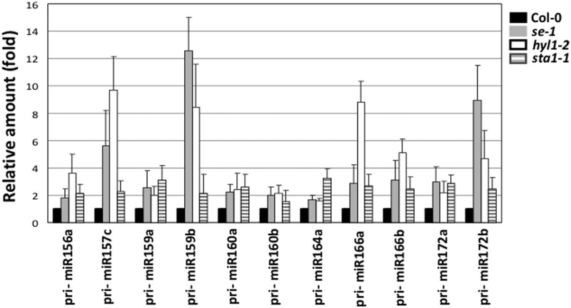
The results of small RNA blot hybridization analysis (Figure 2) suggested a possible role of STA1 in miRNA biogenesis. Mutations in HYL1 and SE generally lead to a dramatic increase in accumulation of pri-miRNAs (19,21). To investigate the involvement of STA1 in the pri-miRNA processing, we determined pri-miRNA transcript levels by qRT-PCR (Figure 4). Transcripts of all tested pri-miRNAs accumulated more in sta1-1 compared with WT. Consistent with the previous reports, most transcript levels of tested pri-miRNAs were largely increased in se-1 and hyl1-2. These data indicate that STA1 is a factor required for proper processing of pri-miRNAs.
Figure 4.
qRT-PCR expression analysis of selected pri-miRNA transcripts in Col-0, se-1, hyl1-2 and sta1-1. Relative amounts of mRNA levels were obtained by dividing the expression level of the mutant with the WT value. Three biological samples were used.
sta1-1 accumulates unspliced transcript of pri-miRNAs
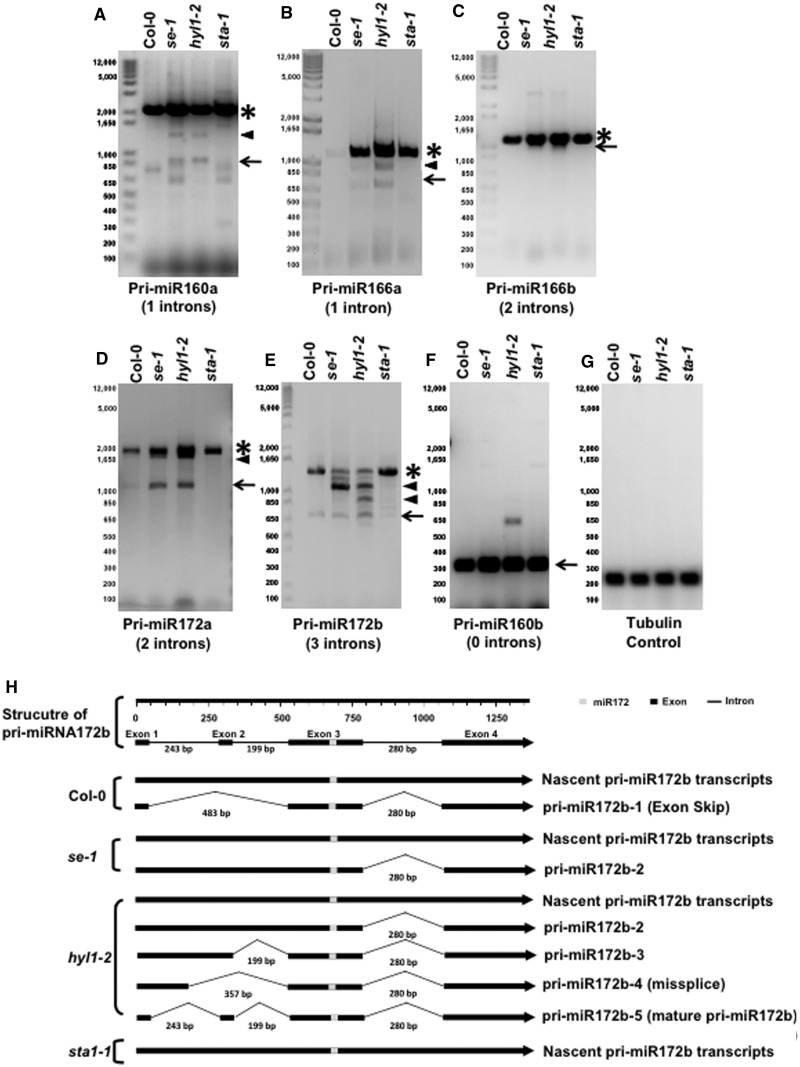
Recent studies showed that several pri-miRNAs contain introns (31,56–58). In the case of intron-containing pri-miRNAs, the miRNA biogenesis pathway can be categorized into two distinct steps: (i) splicing of intron-containing pri-miRNAs and (ii) processing of pri-miRNAs into pre-miRNAs and eventually mature miRNAs. SE has roles in splicing of introns in several pri-miRNAs and also in further steps of miRNA biogenesis from the intron-less and intron-excised pri-miRNAs (31). The STA1 protein has high similarity to human U5 snRNP-associated 102-kDa protein, PRP1p and Prp6p (34). Indeed, pre-mRNA splicing of a stress-responsive COR15A gene was defective in the sta1-1 mutants (34), and the tiling array results from this study confirmed the general role of STA1 in the splicing process (Supplementary Table S1). Based on these results, we investigated the plausible roles of STA1 in splicing of pri-miRNAs. Five intron-containing pri-miRNAs were selected for the splicing analysis by RT-PCR; pri-miR160a (one intron), pri-miR166a (one intron), pri-miR166b (two introns), pri-miR172a (two introns) and pri-miR172b (three introns). We compared the splicing variants of the pri-miRNAs from Col-0, hyl1-2, se-1 and sta1-1. Splicing variants were examined more than eight times with four different biological samples and the tests consistently produced the similar results. The unspliced pri-miR160a (∼2.0 kb) accumulated more in se-1 and sta1-1 compared with Col-0 and hyl1-2 (Figure 5A). The se-1 and hyl1-2 mutants displayed a band ∼850 bp which corresponds to the predicted size of mature pri-miR160a, while both Col-0 and sta1-1 did not show this band. The accumulation of unspliced pri-miR160a transcripts without mature pri-miR160a fragment in sta1-1 implies the roles of STA1 in splicing of pri-miR160a. The other bands around 1.5 kb and 650 bp in se-1 and hyl1-2 and 750 bp in Col-0 may be the products of mis-splicing. High levels of unspliced pri-miRNA166a transcripts accumulated in se-1, hyl1-2 and sta1-1 as compared with Col-0, indicating the similar impairment in pri-miR166a splicing process in these mutants (Figure 5B). Unlike sta1-1, se-1 and hyl1-2 showed two additional fragments with sizes of ∼650 and ∼1000 bp. The former matches to the predicted size of mature pri-miR166a and the latter might be the products of alternative splicing (Figure 5B). Similar to pri-miR166a, unspliced pri-miR166b was accumulated more in se-1, hyl1-2 and sta1-1 than in Col-0 (Figure 5C). In contrast to a previous report (57), we could not observe the spliced fragments of pri-miR166 in hyl1-2. We suspect that the splicing efficiency of intron containing pri-miRNAs may be more dependent on growth conditions or developmental stages in hyl1-2 than other mutants. Nascent transcript of pri-miR172a (Figure 5D) with the predicted size of 2 kb contains two introns. As shown in Figure 5D, unspliced pri-miRNA172a was accumulated more in sta1-1 than in Col-0. The splicing-intermediate transcript with one intron (148 bp) and mature pri-miR172a were clearly observed in se-1 and hyl1-2, but not in WT and sta1-1. The missing bands of the splicing-intermediates and fully spliced pri-miR172a in WT and sta1-1 might result from different reasons; in WT, the spliced pri-miR172a could be quickly used for further downstream steps while in sta1-1, the splicing of pri-miRNA172a was inhibited from the beginning. In fact, the missing band of 850 bp (fully spliced pri-miR160a) in pri-miR160a and of ∼650 bp in pri-miR166a in both Col-0 and sta1-1 (Figure 5A) can be explained in the same way. These results suggest that STA1 functions at very early steps in pri-miRNA processing. Unspliced pri-miR172b (Figure 5E) contains three introns with the total size of ∼1.5 kb and mature pri-miR172b was predicted to be ∼0.7 kb. Higher accumulation of unspliced pri-miR172b was also observed in sta1-1 compared with Col-0, se-1 and hyl1-2. In addition to the intron retention in sta1-1, we observed unusual accumulation of the predicted mature pri-miR172b (∼0.7 kb), which was present in Col-0, se-1 and hyl1-2 at very similar levels (Figure 5E). Normally, mature pri-miRNAs rapidly undergo further processing to pre-miRNAs that are known to be less detectable in WT than the processing defective mutants (59). To clarify the identity of the ∼0.7-kb fragment in Col-0, we cloned the fragment for sequence analysis and found that the fragment is a mis-spliced product caused by an exon-skipping at 5′-region during splicing (Figure 5B). The sequence analysis also found that the ∼0.7 kb fragment from hyl1-2 was normally spliced mature pri-miR172b but the fragment from se-1 was not. Furthermore, several bands ranging from ∼0.85 to ∼1.4 kb accumulated to high levels in se-1 and hyl1-2, but not in Col-0 and sta1-1 (Figure 5E). Interestingly, the splicing intermediates of pri-miR172b were more variable in hyl1-2 than in se-1. Indeed, the sequence analysis of these fragments showed various intermediates in hyl1-2 and only one kind of intermediate in se-1 (Figure 5B). This may be due to the involvement of SE in the splicing process in addition to the miRNA processing pathway (31). The other bands of ∼1.2 kb (the second band from the top) and ∼0.7 kb (the last band from the top) in se-1 and weak bands (the second, third and fifth from the top) in hyl1-2 could not be retrieved for sequence analysis. Apparent unspliced nascent pri-miR172b transcript in sta1-1 was confirmed by the sequence analysis. Thus, the observation that stat1-1 accumulated non-spliced nascent pri-miR172b without any splicing variants implies that STA1 has a major role in pri-miRNA splicing. In fact, of all tested pri-miRNAs, accumulation of apparent splicing intermediates or misspliced transcripts was observed in se-1 and hyl1-2, but not in sta1-1 (Figure 5), strongly suggesting a significant role of STA1 in pri-miRNA splicing steps, rather than in the cleavage of pri-miRNAs into mature miRNAs. We also quantitatively evaluate the retention of introns in pri-miR172b by comparing the levels of the introns in the WT control and sta1-1 using qRT-PCR. The retention levels of intron 1 and 2 in pri-miR172b were higher in se-1, hyl1-2 and sta1-1 than Col-0, while the retention of intron 3 was high only in sta1-1 (Supplementary Figure S6). These results are consistent with the results from RT-PCR (Figure 5E) where the major bands were the unspliced transcript (for sta1-1) and the splicing intermediate without the third intron (for se-1 and hyl1-2). Overall, the results indicate that sta1-1 causes high levels of intron retention and accumulates unspliced transcript of pri-miRNAs.
Figure 5.
Intron-retention analysis of selected pri-miRNA. (A–G) Analysis of retained intron in Col-0, se-1, hyl1-2 and sta1-1. Five intron-containing pri-miRNAs were investigated by RT-PCR. Splicing pattern of pri-miR160b (A), pri-miR166a (B), pri-miR166b (C), pri-miR172a (D), pri-miR172b (E) and pri-miR166a (F). Tubulin gene was used as a loading control (G). Asterisk, unspliced pri-miRNA; arrow-head, splicing intermediate; arrow, fully spliced or mature pri-miRNA. (H) Patterns of processing intermediates of pri-miR172b in Col-0, se-1, hyl1-2 and sta1-1. RT-PCR fragments of pri-miR172b in each genotype were cloned and sequenced to compare the retention patterns.
DCL1 transcript levels were reduced in sta1-1
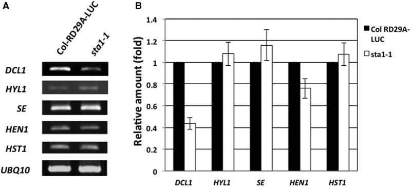
Intriguingly, the intron-less pri-miR160b transcripts were accumulated slightly more in sta1-1 than in Col-0, which was similar to those in se-1 and hyl1-2 (Figure 5F). Indeed, many intronless miRNAs accumulated less in sta1-1 than in WT in our small RNA sequencing results (Supplementary Table S3). Therefore, we examined the transcript levels of canonical miRNA biogenesis genes including DCL1, HYL1, SE, HEN1 and HST1 and found the transcript level of DCL1 is reduced in sta1-1 (Figure 6A). The decreased levels of DCL1 transcript in sta1-1 were confirmed by qRT-PCR (Figure 6B). We speculate that STA1 involvement in miRNA processing of intronless miRNA transcripts could be mediated at least in part by modulating the levels of DCL1 transcripts.
Figure 6.
Comparison of transcript levels of miRNA biogenesis genes in Col-RD29A-LUC and sta1-1. (A) RT-PCR results of miRNA biogenesis genes. DCL1 transcript levels were lower in sta1-1 than Col-RD29A-LUC. (B) qRT-PCR results of miRNA biogenesis genes. qRT-PCR confirmed that reduced levels of DCL1 transcript in sta1-1. Three replicates were used.
DISCUSSION
Recently, three research groups independently reported an important relationship between pre-mRNA maturation and pri-miRNA processing. Laubinger et al. (31) revealed the dual roles of ABH1/CBP80, CBP20 and SE in pre-mRNA splicing and pri-miRNA processing. Gregory et al. (60) also uncovered the function of ABH1/CBP80 in miRNA biogenesis. Around the same time, Kim et al. (33) showed that ABH1/CBP80 and CBP20 directly bind to pri-miRNA transcripts and have a general role in miRNA biogenesis, but other reported cap-binding proteins in plants such as CUM1 (eIF4E1) and CUM2 (eIF4G) are irrelevant (33). Compared with the canonical miRNA processing components (DCL1, SE, HYL1, etc.), CBC seems to play less essential roles in the miRNA processing pathway because most miRNAs were not significantly affected by ABH1/CBP80 and CBP20 deficiency (Supplementary Figure 2) (61). However, the additional role of CBC in miRNA biogenesis introduced a new prospect on the proteins that play roles in mRNA metabolism; cap-binding, splicing and polyadenylation can influence miRNA processing pathway. Several studies reported that some of pri-miRNA transcripts appear to be spliced, polyadenylated and capped, which implies splicing may also be an important step for further processing (56,58,62). Hence, two fundamental questions arise: how does ABH1/CBP80 or the cap-binding process affect miRNA biogenesis and are splicing and polyadenylation of pri-miRNAs also important for miRNA biogenesis? For the former question, it was suggested that ABH1/CBP80 guides mature pri-miRNAs to the miRNA processing complex by direct interaction with miRNA processing proteins (63). For the latter question, analysis of reported mutants defective in general splicing or polyadenylation could be a plausible approach. In addition, as the results from the se mutant suggested (31), examination of miRNA biogenesis gene involvement in splicing would answer how important the splicing is in miRNA biogenesis. To answer this question, Szarzynska et al. (57) performed a detailed study on HYL1-dependent processing of intron-containing pri-miRNAs and suggested the coupled roles of HYL1 in splicing and miRNA processing. In our results, the accumulation of unspliced pri-miR166a, pri-miR166b and pri-miR172a was observed in hyl1-2, which supports the possible role of HYL1 in pri-miRNA splicing (Figure 5). However, the splicing of pri-miR160a and pri-miR172b transcripts was not dramatically affected in hyl1-2 and fully spliced pri-miRNAs were detected in all tested pri-miRNAs in hyl1-2. In addition, various splicing intermediates of pri-miRNAs were highly accumulated in hyl1-2 (Figure 5). It should be noted that the splicing intermediates or abnormally spliced transcripts were more often found in hyl1-2 than in se-1. This means that defects in SE-dependent splicing might block post-splicing processes, resulting in lower accumulation of splicing variants in se-1 compared with hyl1-2. Thus, these results suggest that SE appears to be more important for splicing than HYL1. Although our data did not fully address whether HYL1 functions simultaneously in the splicing and miRNA processing steps, our observation of accumulation of the splicing intermediates and mature pri-miRNAs in hyl1-2 supports the hypothesis that HYL1 functions primarily or preferentially in the post-splicing processes to produce pre-miRNA and miRNAs and perhaps functions selectively in pri-miRNA splicing. These splicing defects are clearly contrary to the fact that the pri-miRNA splicing defective sta1-1 mutants did not show any splicing intermediates of tested pri-miRNAs (Figure 5), indicating that STA1 functions more significantly in pri-miRNA splicing than the other two genes.
Our study with the sta1-1 mutants defective in an Arabidopsis pre-mRNA processing factor 6 homolog showed that splicing is an important step for both pre-mRNA and pri-miRNA processing. The first indication of the dual functions of STA1 was the phenotype similarity between sta1-1 and the other miRNA defective mutants. Indeed, we found dramatic reduction in the majority of miRNAs, the accumulation of pri-miRNAs and target mRNAs in sta1-1 (Figures 2–4 and Supplementary Table S3). Suggested by STA1 function in pre-mRNA splicing, we tested the possible function of STA1 on pri-miRNA splicing by RT-PCR. Our results showed that STA1 also has roles in splicing of intron-containing pri-miRNAs (Figure 5). For example, all introns of pri-miR172b are retained in sta1-1 but not in se-1 and hyl1-2 (Figure 5E). Sequencing of cloned fragments showed that the unspliced pri-miR172b in sta1-1 perfectly matched to the annotated sequence of nascent pri-miR172b transcript. Moreover, accumulation of mature pri-miRNAs was undetectable in sta1-1. These different patterns of pri-miRNA processing intermediates in these mutants also suggest that STA1 has distinct functions from the other genes—particularly SE in splicing of pri-miRNAs. While HYL1 and SE have canonical roles in miRNA processing with their potential roles in splicing, STA1 seems to have major roles in splicing of nascent pri-miRNA transcripts. Furthermore, STA1-dependent splicing seems more important for further downstream processing than ABH1/CBP80 because sta1-1 showed more significantly reduced levels of many miRNAs than abh1/cbp80 (Supplementary Figure S2). Previously, Yang et al. (22) reported that hyl1-1 se-1 double mutants are embryonic lethal. Similarly, a homozygous T-DNA insertion (SALK _009304) in STA1 likely causes the embryonic lethality which also implies the vital roles of STA1 in pri-miRNA splicing and also pre-mRNA splicing for gene expression control from the early stage of development (34).
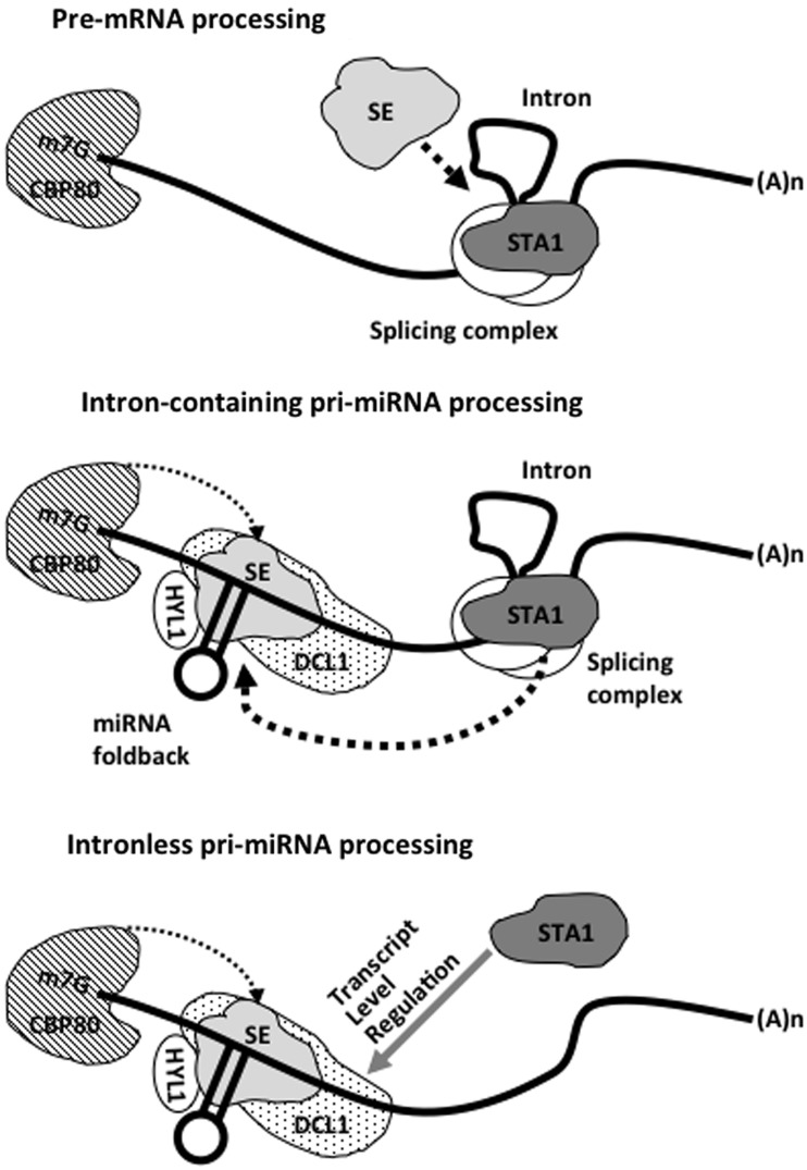
Our data also showed that the accumulation of intronless pri-miR160b in sta1-1 was slightly higher than control plants (Figure 5). In addition, small RNA sequencing results indicated the general reduction of mature miRNAs including those generated from intronless pri-miRNAs. These results cannot be interpreted solely by our suggestion on the roles of STA1 in the intron-containing pri-miRNA splicing. Interestingly, we found that the level of DCL1 transcripts was notably reduced in sta1-1 (Figure 6). This reduction of DCL1 transcript levels appears to be because of splicing defects in sta1-1 rather than regulation by miR162. DCL1 transcripts are constitutively subject to negative feedback regulation by miR162 (64). However, sta1-1 accumulates miR162 only at half the levels of WT (Figure 2). Thus, reduced DCL1 levels in sta1-1 cannot be explained by miR162 regulation. Our RT-PCR results showed reduced levels of spliced DCL1 transcripts along with an inversely correlated, high accumulation of unspliced DCL1 transcripts in sta1-1 (Supplementary Figure S7), suggesting splicing defect-caused downregulation of DCL1 in sta1-1. However, this unspliced DCL1 transcript band was not always detected in our RT-PCR results. This is also probably because of the unstable nature of unspliced DCL1 transcripts or rapid degradation of unspliced DCL1 transcripts by unknown mechanisms rather than miR162-mediated negative regulation. Taken together, these results suggested that STA1 also functions in processing of intronless pri-miRNA, probably through the modulation of the levels of DCL1 transcripts (Figure 7). However, this does not weaken our argument that STA1 has an important role in splicing of intron-containing pri-miRNAs during miRNA biogenesis. Kurihara et al. (19) showed that dcl1-9, a weak allele of dcl1, accumulates fully spliced pri-miR166a (between 1000 and 500 bp), while we observed only unspliced pri-miR166a (>1000 bp) in sta1-1 where the level of DCL1 is supposed to be low. These observations indicate that DCL1 is not involved in pri-miR166a splicing and the accumulation of unspliced pri-miR166a transcript is mainly due to the defects in splicing in sta1-1. These findings also suggest that the STA1-dependent splicing is precedent to DCL1-catalyzed miRNA processing pathway. In our analysis, fully spliced pri-miRNAs of all the tested intron-containing pri-miRNAs were not observed in sta1-1. In summary, we suggest that STA1 is a new player in miRNA biogenesis and has a major role in splicing intron-containing pri-miRNAs. STA1 also seems to have an indirect role in processing intronless pri-miRNAs probably through modulating the levels of DCL1 transcripts (Figure 7).
Figure 7.
A model for STA1 function in miRNA biogenesis. SE involvement in pre-mRNA processing was previously reported (57). In addition to mRNA splicing, STA1 is also involved in splicing of intron-containing pri-miRNAs. STA1 has a direct role in pri-miRNA splicing that affects miRNA processing and also indirectly affects intronless pri-miRNA processing by modulating the DCL1 transcript levels. It is not clear that STA1 regulation on the DCL1 transcript levels occurs through the STA1-including splicing complex or other STA1 unique functions.
We further envisage that STA1 may be the part of a large miRNA processing/splicesome complex that orchestrates pri-miRNA splicing and processing. Certainly, further investigations for detailed mechanism are required to understand the molecular linkage between upstream splicing and downstream processing in miRNA biogenesis. For instance, STA1 has domains known for protein–protein interactions that may be important for its interaction with the other components in miRNA biogenesis. DCL1, SE, HYL1 and ABH1/CBP80 will be the primary targets for protein interaction studies of STA1.
SUPPLEMENTARY DATA
Supplementary Data are available at NAR Online: Supplementary Tables 1–8, and Supplementary Figures 1–7.
FUNDING
The National Research Foundation of Korea by the Korean Government MEST [2011-0027376 to B.-h.L., 2012R1A1A2008826 to B.-h.L.]; Next-Generation BioGreen 21 Program, Rural Development Administration, Republic of Korea [PJ009104 to B.-h.L.]; Sogang University Research [201214003.01 to B.-h.L.]; UNIK research initiative of the Danish Ministry of Science, Technology and Innovation [09-065274 to S.W.Y.]; Chinese Academy of Sciences and National Institutes of Health Grants [R01GM070795 to J.-K.Z., R01GM059138 to J.-K.Z.]; USDA Grant [2008-35100-04518 to J.-K.Z.]; University of California at Riverside Initial Complement Fund and a United States Department of Agriculture hatch fund [CA-R-BPS-7754H to R.L.]. Funding for open access charge: National Research Foundation of Korea by the Korean Government MEST [2012R1A1A2008826 to B.-h.L.]
Conflict of interest statement. None declared.
Supplementary Material
ACKNOWLEDGEMENTS
The authors thank laboratory members for helpful discussion and Brian Christopher King for helpful comments on the manuscript. S.W.Y. gratefully acknowledges support from Center for Synthetic Biology at University of Copenhagen and experimental equipment support from iNtRON Biotechnology, Inc. Korea.
REFERENCES
- 1.Carrington JC, Ambros V. Role of microRNAs in plant and animal development. Science. 2003;301:336–338. doi: 10.1126/science.1085242. [DOI] [PubMed] [Google Scholar]
- 2.Chen XM. A microRNA as a translational repressor of APETALA2 in Arabidopsis flower development. Science. 2004;303:2022–2025. doi: 10.1126/science.1088060. [DOI] [PMC free article] [PubMed] [Google Scholar]
- 3.Lauter N, Kampani A, Carlson S, Goebel M, Moose SP. microRNA172 down-regulates glossy15 to promote vegetative phase change in maize. Proc. Natl Acad. Sci. USA. 2005;102:9412–9417. doi: 10.1073/pnas.0503927102. [DOI] [PMC free article] [PubMed] [Google Scholar]
- 4.Reyes JL, Chua NH. ABA induction of miR159 controls transcript levels of two MYB factors during Arabidopsis seed germination. Plant J. 2007;49:592–606. doi: 10.1111/j.1365-313X.2006.02980.x. [DOI] [PubMed] [Google Scholar]
- 5.Bari R, Pant BD, Stitt M, Scheible WR. PHO2, microRNA399, and PHR1 define a phosphate-signaling pathway in plants. Plant Physiol. 2006;141:988–999. doi: 10.1104/pp.106.079707. [DOI] [PMC free article] [PubMed] [Google Scholar]
- 6.Fujii H, Chiou TJ, Lin SI, Aung K, Zhu JK. A miRNA involved in phosphate-starvation response in Arabidopsis. Curr. Biol. 2005;15:2038–2043. doi: 10.1016/j.cub.2005.10.016. [DOI] [PubMed] [Google Scholar]
- 7.Hwang EW, Shin SJ, Yu BK, Byun MO, Kwon HB. miR171 family members are involved in drought response in Solanum tuberosum. J. Plant Biol. 2011;54:43–48. [Google Scholar]
- 8.Sunkar R, Li YF, Jagadeeswaran G. Functions of microRNAs in plant stress responses. Trends Plant Sci. 2012;17:196–203. doi: 10.1016/j.tplants.2012.01.010. [DOI] [PubMed] [Google Scholar]
- 9.Sunkar R, Zhu JK. Novel and stress-regulated microRNAs and other small RNAs from Arabidopsis. Plant Cell. 2004;16:2001–2019. doi: 10.1105/tpc.104.022830. [DOI] [PMC free article] [PubMed] [Google Scholar]
- 10.Zhao BT, Ge LF, Liang RQ, Li W, Ruan KC, Lin HX, Jin YX. Members of miR-169 family are induced by high salinity and transiently inhibit the NF-YA transcription factor. BMC Mol. Biol. 2009;10:29. doi: 10.1186/1471-2199-10-29. [DOI] [PMC free article] [PubMed] [Google Scholar]
- 11.Boutet S, Vazquez F, Liu J, Beclin C, Fagard M, Gratias A, Morel JB, Crete P, Chen XM, Vaucheret H. Arabidopsis HEN1: a genetic link between endogenous miRNA controlling development and siRNA controlling transgene silencing and virus resistance. Curr. Biol. 2003;13:843–848. doi: 10.1016/s0960-9822(03)00293-8. [DOI] [PMC free article] [PubMed] [Google Scholar]
- 12.Grigg SP, Canales C, Hay A, Tsiantis M. SERRATE coordinates shoot meristem function and leaf axial patterning in Arabidopsis. Nature. 2005;437:1022–1026. doi: 10.1038/nature04052. [DOI] [PubMed] [Google Scholar]
- 13.Lu C, Fedoroff N. A mutation in the Arabidopsis HYL1 gene encoding a dsRNA binding protein affects responses to abscisic acid, auxin, and cytokinin. Plant Cell. 2000;12:2351–2365. doi: 10.1105/tpc.12.12.2351. [DOI] [PMC free article] [PubMed] [Google Scholar]
- 14.Park MY, Wu G, Gonzalez-Sulser A, Vaucheret H, Poethig RS. Nuclear processing and export of microRNAs in Arabidopsis. Proc. Natl Acad. Sci. USA. 2005;102:3691–3696. doi: 10.1073/pnas.0405570102. [DOI] [PMC free article] [PubMed] [Google Scholar]
- 15.Park W, Li JJ, Song RT, Messing J, Chen XM. CARPEL FACTORY, a Dicer homolog, and HEN1, a novel protein, act in microRNA metabolism in Arabidopsis thaliana. Curr. Biol. 2002;12:1484–1495. doi: 10.1016/s0960-9822(02)01017-5. [DOI] [PMC free article] [PubMed] [Google Scholar]
- 16.Reinhart BJ, Weinstein EG, Rhoades MW, Bartel B, Bartel DP. MicroRNAs in plants. Genes Dev. 2002;16:1616–1626. doi: 10.1101/gad.1004402. [DOI] [PMC free article] [PubMed] [Google Scholar]
- 17.Kurihara Y, Watanabe Y. Arabidopsis micro-RNA biogenesis through Dicer-like 1 protein functions. Proc. Natl Acad. Sci. USA. 2004;101:12753–12758. doi: 10.1073/pnas.0403115101. [DOI] [PMC free article] [PubMed] [Google Scholar]
- 18.Dong Z, Han MH, Fedoroff N. The RNA-binding proteins HYL1 and SE promote accurate in vitro processing of pri-miRNA by DCL1. Proc. Natl Acad. Sci. USA. 2008;105:9970–9975. doi: 10.1073/pnas.0803356105. [DOI] [PMC free article] [PubMed] [Google Scholar]
- 19.Kurihara Y, Takashi Y, Watanabe Y. The interaction between DCL1 and HYL1 is important for efficient and precise processing of pri-miRNA in plant microRNA biogenesis. RNA. 2006;12:206–212. doi: 10.1261/rna.2146906. [DOI] [PMC free article] [PubMed] [Google Scholar]
- 20.Yang SW, Chen HY, Yang J, Machida S, Chua NH, Yuan YA. Structure of Arabidopsis HYPONASTIC LEAVES1 and its molecular implications for miRNA processing. Structure. 2010;18:594–605. doi: 10.1016/j.str.2010.02.006. [DOI] [PMC free article] [PubMed] [Google Scholar]
- 21.Lobbes D, Rallapalli G, Schmidt DD, Martin C, Clarke J. SERRATE: a new player on the plant microRNA scene. EMBO Rep. 2006;7:1052–1058. doi: 10.1038/sj.embor.7400806. [DOI] [PMC free article] [PubMed] [Google Scholar]
- 22.Yang L, Liu ZQ, Lu F, Dong AW, Huang H. SERRATE is a novel nuclear regulator in primary microRNA processing in Arabidopsis. Plant J. 2006;47:841–850. doi: 10.1111/j.1365-313X.2006.02835.x. [DOI] [PubMed] [Google Scholar]
- 23.Yu B, Yang ZY, Li JJ, Minakhina S, Yang MC, Padgett RW, Steward R, Chen XM. Methylation as a crucial step in plant microRNA biogenesis. Science. 2005;307:932–935. doi: 10.1126/science.1107130. [DOI] [PMC free article] [PubMed] [Google Scholar]
- 24.Yu B, Bi L, Zheng BL, Ji LJ, Chevalier D, Agarwal M, Ramachandran V, Li WX, Lagrange T, Walker JC, et al. The FHA domain proteins DAWDLE in Arabidopsis and SNIP1 in humans act in small RNA biogenesis. Proc. Natl Acad. Sci. USA. 2008;105:10073–10078. doi: 10.1073/pnas.0804218105. [DOI] [PMC free article] [PubMed] [Google Scholar]
- 25.Ren GD, Xie M, Dou YC, Zhang SX, Zhang C, Yu B. Regulation of miRNA abundance by RNA binding protein TOUGH in Arabidopsis. Proc. Natl Acad. Sci. USA. 2012;109:12817–12821. doi: 10.1073/pnas.1204915109. [DOI] [PMC free article] [PubMed] [Google Scholar]
- 26.Cai XZ, Hagedorn CH, Cullen BR. Human microRNAs are processed from capped, polyadenylated transcripts that can also function as mRNAs. RNA. 2004;10:1957–1966. doi: 10.1261/rna.7135204. [DOI] [PMC free article] [PubMed] [Google Scholar]
- 27.Lee Y, Kim M, Han JJ, Yeom KH, Lee S, Baek SH, Kim VN. MicroRNA genes are transcribed by RNA polymerase II. EMBO J. 2004;23:4051–4060. doi: 10.1038/sj.emboj.7600385. [DOI] [PMC free article] [PubMed] [Google Scholar]
- 28.Chang YF, Imam JS, Wilkinson ME. The nonsense-mediated decay RNA surveillance pathway. Anuu. Rev. Biochem. 2007;76:51–74. doi: 10.1146/annurev.biochem.76.050106.093909. [DOI] [PubMed] [Google Scholar]
- 29.Fang YD, Spector DL. Identification of nuclear dicing bodies containing proteins for microRNA biogenesis in living Arabidopsis plants. Curr. Biol. 2007;17:818–823. doi: 10.1016/j.cub.2007.04.005. [DOI] [PMC free article] [PubMed] [Google Scholar]
- 30.Fujioka Y, Utsumi M, Ohba Y, Watanabe Y. Location of a possible miRNA processing site in SmD3/SmB nuclear bodies in Arabidopsis. Plant Cell Physiol. 2007;48:1243–1253. doi: 10.1093/pcp/pcm099. [DOI] [PubMed] [Google Scholar]
- 31.Laubinger S, Sachsenberg T, Zeller G, Busch W, Lohmann JU, Rascht G, Weigel D. Dual roles of the nuclear cap-binding complex and SERRATE in pre-mRNA splicing and microRNA processing in Arabidopsis thaliana. Proc. Natl Acad. Sci. USA. 2008;105:8795–8800. doi: 10.1073/pnas.0802493105. [DOI] [PMC free article] [PubMed] [Google Scholar]
- 32.Izaurralde E, Lewis J, McGuigan C, Jankowska M, Darzynkiewicz E, Mattaj IW. A nuclear cap-binding protein complex involved in pre-mRNA splicing. Cell. 1994;78:657–668. doi: 10.1016/0092-8674(94)90530-4. [DOI] [PubMed] [Google Scholar]
- 33.Kim S, Yang JY, Xu J, Jang IC, Prigge MJ, Chua NH. Two cap-binding proteins CBP20 and CBP80 are involved in processing primary microRNAs. Plant Cell Physiol. 2008;49:1634–1644. doi: 10.1093/pcp/pcn146. [DOI] [PMC free article] [PubMed] [Google Scholar]
- 34.Lee B-h, Kapoor A, Zhu J, Zhu J-K. STABILIZED1, a stress-upregulated nuclear protein, is required for pre-mRNA splicing, mRNA turnover, and stress tolerance in Arabidopsis. Plant Cell. 2006;18:1736–1749. doi: 10.1105/tpc.106.042184. [DOI] [PMC free article] [PubMed] [Google Scholar]
- 35.Prigge MJ, Wagner DR. The Arabidopsis SERRATE gene encodes a zinc-finger protein required for normal shoot development. Plant Cell. 2001;13:1263–1279. doi: 10.1105/tpc.13.6.1263. [DOI] [PMC free article] [PubMed] [Google Scholar]
- 36.Livak KJ, Schmittgen TD. Analysis of relative gene expression data using real-time quantitative PCR and the 2(T)(-delta delta C) method. Methods. 2001;25:402–408. doi: 10.1006/meth.2001.1262. [DOI] [PubMed] [Google Scholar]
- 37.Griffiths-Jones S, Saini HK, van Dongen S, Enright AJ. miRBase: tools for microRNA genomics. Nucleic Acids Res. 2008;36:D154–D158. doi: 10.1093/nar/gkm952. [DOI] [PMC free article] [PubMed] [Google Scholar]
- 38.Li RQ, Yu C, Li YR, Lam TW, Yiu SM, Kristiansen K, Wang J. SOAP2: an improved ultrafast tool for short read alignment. Bioinformatics. 2009;25:1966–1967. doi: 10.1093/bioinformatics/btp336. [DOI] [PubMed] [Google Scholar]
- 39.Bengtsson H, Irizarry R, Carvalho B, Speed TP. Estimation and assessment of raw copy numbers at the single locus level. Bioinformatics. 2008;24:759–767. doi: 10.1093/bioinformatics/btn016. [DOI] [PubMed] [Google Scholar]
- 40.Tusher VG, Tibshirani R, Chu G. Significance analysis of microarrays applied to the ionizing radiation response. Proc. Natl Acad. Sci. USA. 2001;98:10515–10515. doi: 10.1073/pnas.091062498. [DOI] [PMC free article] [PubMed] [Google Scholar]
- 41.Benjamini Y, Hochberg Y. Controlling the false discovery rate - a practical and powerful approach to multiple testing. J. R. Stat. Soc. Ser. B. 1995;57:289–300. [Google Scholar]
- 42.Bezerra IC, Michaels SD, Schomburg FM, Amasino RM. Lesions in the mRNA cap-binding gene ABA HYPERSENSITIVE 1 suppress FRIGIDA-mediated delayed flowering in Arabidopsis. Plant J. 2004;40:112–119. doi: 10.1111/j.1365-313X.2004.02194.x. [DOI] [PubMed] [Google Scholar]
- 43.Todesco M, Balasubramanian S, Cao J, Ott F, Sureshkumar S, Schneeberger K, Meyer RC, Altmann T, Weigel D. Natural vVariation in biogenesis efficiency of individual Arabidopsis thaliana microRNAs. Curr. Biol. 2012;22:166–170. doi: 10.1016/j.cub.2011.11.060. [DOI] [PubMed] [Google Scholar]
- 44.Doench JG, Sharp PA. Specificity of microRNA target selection in translational repression. Genes Dev. 2004;18:504–511. doi: 10.1101/gad.1184404. [DOI] [PMC free article] [PubMed] [Google Scholar]
- 45.Elbashir SM, Harborth J, Lendeckel W, Yalcin A, Weber K, Tuschl T. Duplexes of 21-nucleotide RNAs mediate RNA interference in cultured mammalian cells. Nature. 2001;411:494–498. doi: 10.1038/35078107. [DOI] [PubMed] [Google Scholar]
- 46.Martinez J, Tuschl T. RISC is a 5′ phosphomonoester-producing RNA endonuclease. Genes Dev. 2004;18:975–980. doi: 10.1101/gad.1187904. [DOI] [PMC free article] [PubMed] [Google Scholar]
- 47.Cardon GH, Hohmann S, Nettesheim K, Saedler H, Huijser P. Functional analysis of the Arabidopsis thaliana SBP-box gene SPL3: a novel gene involved in the floral transition. Plant J. 1997;12:367–377. doi: 10.1046/j.1365-313x.1997.12020367.x. [DOI] [PubMed] [Google Scholar]
- 48.Unte US, Sorensen AM, Pesaresi P, Gandikota M, Leister D, Saedler H, Huijser P. SPL8, an SBP-box gene that affects pollen sac development in Arabidopsis. Plant Cell. 2003;15:1009–1019. doi: 10.1105/tpc.010678. [DOI] [PMC free article] [PubMed] [Google Scholar]
- 49.Stone JM, Liang X, Nekl ER, Stiers JJ. Arabidopsis AtSPL14, a plant-specific SBP-domain transcription factor, participates in plant development and sensitivity to fumonisin B1. Plant J. 2005;41:744–754. doi: 10.1111/j.1365-313X.2005.02334.x. [DOI] [PubMed] [Google Scholar]
- 50.Lee B-h, Yu SI, Jackson D. Control of plant architecture: the role of Phyllotaxy and Plastochron. J. Plant Biol. 2009;52:277–282. [Google Scholar]
- 51.Wang JW, Czech B, Weigel D. miR156-Regulated SPL Transcription factors define an endogenous flowering pathway in Arabidopsis thaliana. Cell. 2009;138:738–749. doi: 10.1016/j.cell.2009.06.014. [DOI] [PubMed] [Google Scholar]
- 52.Alonso-Peral MM, Li JY, Li YJ, Allen RS, Schnippenkoetter W, Ohms S, White RG, Millar AA. The microRNA159-regulated GAMYB-like genes inhibit growth and promote programmed cell death in Arabidopsis. Plant Physiol. 2010;154:757–771. doi: 10.1104/pp.110.160630. [DOI] [PMC free article] [PubMed] [Google Scholar]
- 53.Mallory AC, Bartel DP, Bartel B. MicroRNA-directed regulation of Arabidopsis AUXIN RESPONSE FACTOR17 is essential for proper development and modulates expression of early auxin response genes. Plant Cell. 2005;17:1360–1375. doi: 10.1105/tpc.105.031716. [DOI] [PMC free article] [PubMed] [Google Scholar]
- 54.Laufs P, Peaucelle A, Morin H, Traas J. MicroRNA regulation of the CUC genes is required for boundary size control in Arabidopsis meristems. Development. 2004;131:4311–4322. doi: 10.1242/dev.01320. [DOI] [PubMed] [Google Scholar]
- 55.Williams L, Grigg SP, Xie MT, Christensen S, Fletcher JC. Regulation of Arabidopsis shoot apical meristem and lateral organ formation by microRNA miR166g and its AtHD-ZIP target genes. Development. 2005;132:3657–3668. doi: 10.1242/dev.01942. [DOI] [PubMed] [Google Scholar]
- 56.Aukerman MJ, Sakai H. Regulation of flowering time and floral organ identity by a microRNA and its APETALA2-like target genes. Plant Cell. 2003;15:2730–2741. doi: 10.1105/tpc.016238. [DOI] [PMC free article] [PubMed] [Google Scholar]
- 57.Szarzynska B, Sobkowiak L, Pant BD, Balazadeh S, Scheible WR, Mueller-Roeber B, Jarmolowski A, Szweykowska-Kulinska Z. Gene structures and processing of Arabidopsis thaliana HYL1-dependent pri-miRNAs. Nucleic Acids Res. 2009;37:3083–3093. doi: 10.1093/nar/gkp189. [DOI] [PMC free article] [PubMed] [Google Scholar]
- 58.Xie ZX, Allen E, Fahlgren N, Calamar A, Givan SA, Carrington JC. Expression of Arabidopsis MIRNA genes. Plant Physiol. 2005;138:2145–2154. doi: 10.1104/pp.105.062943. [DOI] [PMC free article] [PubMed] [Google Scholar]
- 59.Jones-Rhoades MW, Bartel DP, Bartel B. MicroRNAs and their regulatory roles in plants. Annu. Rev. Plant Biol. 2006;57:19–53. doi: 10.1146/annurev.arplant.57.032905.105218. [DOI] [PubMed] [Google Scholar]
- 60.Gregory BD, O'Malley RC, Lister R, Urich MA, Tonti-Filippini J, Chen H, Millar AH, Ecker JR. A link between RNA metabolism and silencing affecting Arabidopsis development. Dev. Cell. 2008;14:854–866. doi: 10.1016/j.devcel.2008.04.005. [DOI] [PubMed] [Google Scholar]
- 61.Montgomery TA, Carrington JC. Splicing and dicing with a SERRATEd edge. Proc. Natl Acad. Sci. USA. 2008;105:8489–8490. doi: 10.1073/pnas.0804356105. [DOI] [PMC free article] [PubMed] [Google Scholar]
- 62.Sunkar R, Girke T, Jain PK, Zhu JK. Cloning and characterization of microRNAs from rice. Plant Cell. 2005;17:1397–1411. doi: 10.1105/tpc.105.031682. [DOI] [PMC free article] [PubMed] [Google Scholar]
- 63.Chen XM. A silencing safeguard: links between RNA silencing and mRNA processing in Arabidopsis. Dev. Cell. 2008;14:811–812. doi: 10.1016/j.devcel.2008.05.012. [DOI] [PMC free article] [PubMed] [Google Scholar]
- 64.Xie ZX, Kasschau KD, Carrington JC. Negative feedback regulation of Dicer-Like1 in Arabidopsis by microRNA-guided mRNA degradation. Curr. Biol. 2003;13:784–789. doi: 10.1016/s0960-9822(03)00281-1. [DOI] [PubMed] [Google Scholar]
Associated Data
This section collects any data citations, data availability statements, or supplementary materials included in this article.


