Abstract
Platelet factor 4 (PF4) is an angiostatic chemokine that suppresses tumor growth and metastasis. We previously revealed frequent transcriptional silencing of PF4 in multiple myeloma, but the functional roles of this chemokine are still unknown. We studied the apoptotic effects of PF4 on myeloma cell lines and primary myeloma in vitro, and investigated the involved signaling pathway. The in vivo effects were also studied using a mouse model. PF4 not only suppressed myeloma-associated angiogenesis, but also inhibited growth and induced apoptosis in myeloma cells. We found that PF4 negatively regulated STAT3 and concordantly inhibited constitutive and interleukin-6-induced phosphorylation of STAT3, and down-regulated the expression of STAT3 target genes (Mcl-1, survivin and VEGF). Overexpression of constitutively activated STAT3 could rescue PF4-induced apoptotic effects. Furthermore, we found that PF4 induced the expression of SOCS3, a STAT3 inhibitor, and gene silencing of SOCS3 abolished its ability to inhibit STAT3 activation, suggesting a critical role of SOCS3 in PF4-induced STAT3 inhibition. Knockdown of LRP1, a putative PF4 receptor, could also abolish PF4-induced apoptosis and STAT3 inhibition. Finally, the tumor growth inhibitory effect of PF4 was confirmed by in vivo mouse models. Immunostaining of rabbit bone xenografts from PF4-treated mice showed induction of apoptosis of myeloma cells and inhibition of angiogenesis, which was associated with suppression of STAT3 activity. Together, our preclinical data indicate that PF4 may be a potential new targeting agent for the treatment of myeloma.
Introduction
Human platelet factor 4 (PF4), a member of the C-X-C chemokine family, was one of the first chemokines isolated from platelets.1 Although originally developed as a heparin neutralization factor, numerous reports suggest that PF4 inhibits tumor growth and spread, by suppression of tumor-induced angiogenesis, in many types of solid tumors. First, recombinant human PF4 repressed endothelial cell proliferation and migration in vitro.2-4 Second, in mouse tumor xenograft models, recombinant human PF4 inhibited tumor angiogenesis and growth of various tumors such as colon carcinoma and melanoma through an angiogenesis-dependent mechanism.4-6 Third, adenoviral vector-mediated transduction of PF4 cDNA resulted in inhibition of intracerebral glioma growth in mice by reducing tumor-associated angiogenesis.7 In addition, it was demonstrated that PF4 exerted direct anti-proliferative activity in human erythroleukemia cells by down-regulating protein tyrosine kinase activity.8,9 This body of biological evidence paved the way for the development of PF4 as an anti-tumor agent. Indeed, anti-tumor responses have been observed in patients with Kaposi's sarcoma after intravenous administration of PF4.10,11 Previously, our group first revealed frequent allelic loss of PF4 in multiple myeloma (MM) cells from patients.12 Transcriptional inactivation was also confirmed in MM cell lines and patients' MM cells by us and others.12,13 However, the functional roles of PF4 in the pathogenesis of MM are still unclear and the mechanisms underlying the effects of PF4 on MM have not been investigated.
In this study, we examined the tumor suppressive function of PF4 by means of both in vitro and in vivo studies using MM cell lines and patients' MM cells, to provide a scientific basis and framework for clinical studies of PF4 as a new targeting agent in the treatment of MM.
Design and Methods
The design and methods of this study are described in full in the Online Supplementary Design and Methods. Briefly, we investigated the in vitro functions of PF4 using cell growth, proliferation, apoptosis and in vitro tube formation assays. Cell signaling pathways modulated by PF4 treatment were investigated by protein/DNA arrays, an electrophoretic mobility shift assay, and a luciferase reporter assay. Cells treated with PF4 or control cells were harvested for gene and protein expression assays. Finally, the in vivo effects of PF4 were studied by mouse models.14-16
Results
PF4 inhibits growth by induction of apoptosis in multiple myeloma
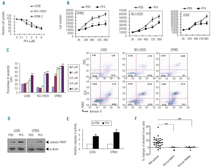
To examine the effects of PF4 on myeloma cells, we first determined its effects on the growth of U266, OPM2 and NCI-H929 cells using increasing doses over a period of 96 h. Results of WST-1 assays (Figure 1A) and trypan blue exclusion (Figure 1B) showed that PF4 markedly inhibited the growth of these cell lines in time- and dose-dependent manners. A significant decrease in cell number was observed for OPM2, NCI-H929 and U266 after 24, 72 and 96 h of incubation with PF4. The inhibitory concentration at 50% (IC50) for these three cell lines were approximately 2, 4 and 4 μM, respectively.
Figure 1.
PF4 inhibited cell growth and induced apoptosis in MM. (A) MM cell lines were treated with or without PF4 at the indicated doses for 96 h and then assessed by WST-1 assay. (B) The viability of MM cell lines after PF4 treatment was assessed using the trypan blue exclusion assay. Data represent the mean±SD of results of culture experiments in triplicate (*P<0.05, **P<0.01). (C-E) MM cell lines were cultured for 96 h with 4 μM PF4. (C) Apoptosis was determined by flow-cytometric analysis for annexin V and 7-AAD staining on MM cell lines. (D) Cells were subjected to immunoblot analysis with cleaved PARP. (E) Caspase-3 colorimetric activity assay. (F) Patients' MM cells (CD138-enriched MM cells from patients) (n=26), normal bone marrow plasma cells (BMPC) (CD138-positive) (n=3) and peripheral blood mononuclear cells (PBMNC) from healthy donors (n=7) were treated with 4 μM PF4 for 48 h. Apoptosis was determined by flow-cytometric analysis for annexin V and 7-AAD staining. The horizontal lines indicate the mean percentage of apoptotic cells.
Next, we investigated whether the observed inhibitory effects of PF4 on cell growth were due to cell cycle arrest, apoptosis, or both. The effect of PF4 on the cellular DNA content was determined using flow cytometric analysis in U266 and NCI-H929 cell lines. While changes in G0/G1, S, and G2/M phases were not distinctively different, we observed a population of cells in the sub-G1 phase indicative of increased apoptosis after PF4 treatment (data not shown). To further confirm that apoptosis was induced by PF4, we treated U266, OPM2 and NCI-H929 cells with increasing doses of PF4 and determined the percentage of apoptotic cells by flow cytometric analysis of annexin V and 7-amino-actinomycin D (7-AAD). Results showed that PF4 led to an increase in apoptotic cells (annexin V+ and/or 7AAD+) in all three of these MM cell lines (Figure 1C). Pretreatment of cells with cycloheximide, a protein synthesis inhibitor, inhibited PF4-induced apoptosis of MM cells (P=0.001) (Online Supplementary Figure S1), indicating that the induction of apoptosis by PF4 is likely dependent on up-regulation of pro-apoptotic proteins. Terminal deoxynucleotidyl transferase dUTP nick end labeling (TUNEL) assays in U266 and OPM2 cells further confirmed that PF4 induced apoptosis in MM cells, as evidenced by the observed increase in staining of nuclear DNA fragments (Online Supplementary Figure S2). In addition, treatment of OPM2 and U266 cells with PF4 triggered a marked increase in proteolytic cleavage of PARP, a signature event during apoptosis (Figure 1D). Similarly, PF4 increased caspase-3 activity, an upstream activator of PARP, by 2.6-fold in U266 cells and by 3.2-fold in OPM2 cells (Figure 1E).
We also examined the effect of PF4 on purified cells from patients with MM. CD138+ plasma cells were isolated from 26 patients diagnosed with MM as described in Online Supplementary Table S2. Cells were treated with PF4 for 48 h and the levels of apoptosis were measured by annexin V-7AAD staining. To compare the cytotoxicity of PF4 in MM and normal cells, normal plasma cells from the bone marrow of healthy donors and normal mononuclear cells from the peripheral blood of healthy donors were obtained. We found minimal changes and a significant increase of mean percentages of apoptotic cells in PF4-treated normal (bone marrow plasma cells and peripheral blood mononuclear cells) and patients' MM cells, respectively (0.01±2.78%, 0.06±1.36%, and 15.16±2.52%) (Figure 1F). Taken together, our findings suggest that PF4 inhibits growth and induces apoptosis in both MM cell lines and primary MM cells.
PF4 suppresses multiple myeloma-associated angiogenesis
Previous studies have shown that PF4 is a potent anti-angiogenic chemokine which inhibits endothelial cell proliferation and angiogenesis.2-5 The effect of PF4 on angiogenesis in MM was, therefore, examined. Endothelial cells isolated from the bone marrow of myeloma patients (MMEC) were treated with PF4 for 96 h and their cell growth was then evaluated. Our results showed that PF4 inhibited the growth of MMEC in a dose-dependent manner, with an IC50 value of approximately 8 μM (Online Supplementary Figure S3A). We next used in vitro capillary-like tube structure formation assays to further examine the anti-angiogenic activity of PF4 in MM. MMEC were seeded in 96-well culture plates pre-coated with Matrigel, treated with phosphate-buffered saline (PBS) and PF4 for 6 h, and then examined for tube formation using an inverted microscope. As shown in Online Supplementary Figures S3B and S3C, tube formation decreased by approximately 39%, compared to control cells, in the cells treated with 8 μM PF4. Taken together, these findings suggest that PF4 suppresses tumor-associated angiogenesis in MM.
PF4 inhibits STAT3 signaling in multiple myeloma
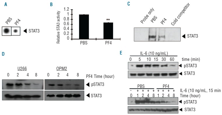
To delineate the potential pathways modulated by PF4 in the control of MM cell growth, we performed protein/DNA arrays on PBS- and PF4-treated U266 cells. Compared to the control treatment, treatment with PF4 inhibited STAT3, AP-1, Elk-1 and NF-κB in response to PF4 treatment, of which only STAT3 underwent significant reduction of transcriptional activity as confirmed using a dual-luciferase reporter assay (Figure 2A, B; other data not shown). Previous studies demonstrated that STAT3 is one of the major mediators of MM tumorigenesis,17-19 which prompted us to further investigate the effect of PF4 on the STAT3 signaling pathway. To confirm the results of the array experiment, an electrophoretic mobility shift assay was performed for STAT3 using the same nuclear extract that was used in the protein/DNA array. As shown in Figure 2C, PF4 decreased DNA binding activity of STAT3 at 8 h (a similar experiment was performed using OPM2 with similar results, data not shown). Together, these data provided the evidence that STAT3 is the potential downstream signaling pathway modulated by PF4.
Figure 2.
PF4 inhibited constitutive and IL-6-induced activation of STAT3 in MM cells. (A) U266 cells were treated with 4 μM PF4 or PBS control, and then nuclear extracts were subjected to protein/DNA arrays. PF4 resulted in decreased STAT3 transcription factor binding to target DNA by protein/DNA arrays. (B) STAT3 transcriptional activity was suppressed by 4 μM PF4 treatment as assessed by a luciferase reporter assay. (C) Nuclear extracts of PF4-treated or untreated U266 cells were subjected to electrophoretic mobility shift assay (EMSA) analysis. (D) MM cells were treated with 4 μM PF4 at indicated times and whole cell lysates were subjected to immunoblot analysis. (E) NCI-H929 cells were treated with IL-6 (10ng/mL) for indicated times, after which western blotting was done and probed for phospho-STAT3 (Tyr 705) and total STAT3 (upper panel). NCI-H929 cells were treated with or without PF4 for the indicated times and then stimulated with IL-6 (10 ng/mL) for 15 min, after which whole cell lysates were subjected to immunoblot analysis (lower panel).
PF4 inhibited constitutive and interleukin-6-induced STAT3 phosphorylation in multiple myeloma cells
Since STAT3 protein undergoes phosphorylation prior to its transcriptional activation,20 we studied the level of phosphorylated STAT3 protein in U266 and OPM2 cells using antibody which detects STAT3 protein that is phosphorylated at the Tyrosine-705 residue. As shown in Figure 2D, PF4 inhibited the phosphorylation of STAT3 in U266 and OPM2 cells in a time-dependent manner, with maximum inhibition occurring at 8 h, but had no effect on the expression of total STAT3 protein.
Interleukin-6 (IL-6) is one of the major myeloma growth factors abundant in the bone marrow microenvironment of MM, and a critical activator of STAT3.21-23 We next examined whether PF4 could inhibit IL-6-induced STAT3 activation. NCI-H929 cells were stimulated with 10 ng/mL IL-6 for different periods. We found that IL-6 rapidly induced STAT3 activation, with the maximum induction occurring at 5-15 min (Figure 2E, upper panel). Exposure of cells to PF4 for 4-8 h was sufficient to suppress IL-6-induced STAT3 phosphorylation in NCI-H929 cells (Figure 2E, lower panel). Collectively, these data further confirmed that PF4 could inhibit STAT3 signaling.
PF4 suppressed STAT3-regulated gene expression
STAT3 is known to regulate proliferation, apoptosis and angiogenesis in MM through regulating the expression of its target genes.17 We examined whether PF4 could down-regulate the expression of STAT3 target genes, including c-Myc, Bcl-XL, Bcl-2, Mcl-1, Survivin and VEGF. Real-time polymerase chain reaction results showed that PF4 down-regulated mRNA expression of STAT3 target genes involved in survival (Bcl-XL, Bcl-2, Mcl-1, Survivin) and angiogenesis (VEGF) in either or both U266 and OPM2 cells (Online Supplementary Figure S4A). Results from western blot further confirmed that PF4 could down-regulate the protein level of these genes in either or both cell lines (Online Supplementary Figure S4B). These data were consistent with our in vitro observations that PF4 could inhibit MM cell growth as well as suppress angiogenesis.
Enforced expression of constitutively active STAT3 rescued cells from PF4-induced apoptosis
To confirm whether PF4 induced apoptosis in MM cells by inactivation of STAT3, constitutively active STAT3 plasmid (Stat3 Flag pRc/CMV) was transfected into NCI-H929 cells, and PF4-induced apoptosis was examined. We confirmed the constitutive expression of STAT3 after transfection of STAT3 Flag pRC/CMV plasmid by western blot analysis (Online Supplementary Figure S4C, left). Notably, this forced expression of STAT3 significantly rescued cells from PF4-induced apoptosis, by 51% compared to cells transfected with empty vector (Online Supplementary Figure S4C, right).
Inhibition of STAT3 by PF4 involves SOCS3 induction
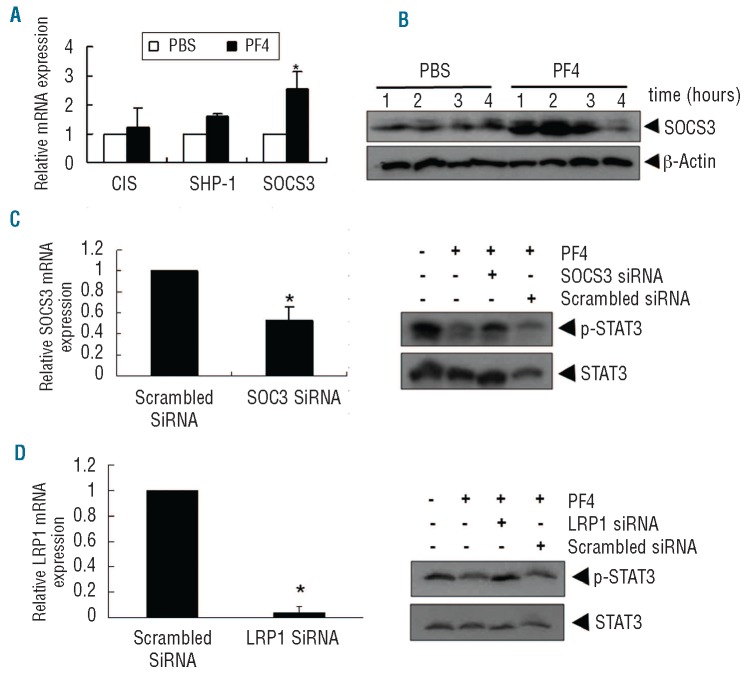
Three protein families have been reported to regulate the STAT3 pathway negatively: SHP, SOCS and PIAS.21 Both SHP and SOCS proteins can inactivate and dephosphorylate STAT3 by inhibiting JAK activity. PIAS proteins can inhibit STAT3 DNA binding and transcriptional activation.21 We next examined whether PF4-induced inhibition of STAT3 phosphorylation was due to the activation of these proteins. Since PF4 could inhibit not only STAT3 transcriptional activity but also its phosphorylation, we decided to focus on SOCS and SHP proteins, mainly the SOCS3, CIS and SHP-1, which have been widely reported to inhibit STAT3.21,22 U266 cells were treated with PF4 for 2 h and the real-time polymerase chain reaction analysis showed that PF4 strongly induced SOCS3 mRNA levels by 2.5-fold, but had little or no effects on SHP1 and CIS mRNA levels (Figure 3A). We, therefore, next examined the effects of PF4 on protein levels of SOCS3. Results from western blot analysis confirmed that PF4 induced the expression of SOCS3 protein in U266 cells, with the maximum level at approximately 2 h (Figure 3B) (the same experiments were performed in OPM2 cells with similar results, data not shown). These results suggested that the induced expression of SOCS3 by PF4 was correlated with the down-regulation of constitutive STAT3 activation in U266 cells.
Figure 3.
PF4 regulated STAT3 through inducing SOCS3. (A) Cells were treated with 4 μM PF4 for 2 h, after which RNA extracts were prepared and real-time PCR was performed. (B) U266 cells were treated with PF4 for indicated times and western blot for SOCS3 was performed. (C) PF4-induced inhibition of STAT3 activation was reversed by silencing of SOCS3. Cells were transfected with either scrambled or SOCS3-specific siRNA (50 nM). Knocking down efficacy was analyzed by real-time PCR after 24 h of transfection (Left panel). After 24 h of transfection, cells were treated with PF4 for 8 h, and whole-cell extracts were subjected to western blot analysis for p-STAT3 and total STAT3 (right panel). (D) PF4-induced STAT3 inhibition was abolished by silencing LRP1. U266 cells were transfected with either scrambled or LRP1 siRNA (50nM), and knockdown of LRP1 expression was confirmed by quantitative PCR of LRP1 after 24 h (left panel). After transfection of siRNA for 24 h, cells were treated with 4 μM PF4 for 8 h followed by western blot analysis for p-STAT3 and total STAT3 (right panel).
PF4-induced inhibition of STAT3 activation was reversed by gene silencing of SOCS3
We then determined whether suppression of SOCS3 expression by short-interfering RNA (siRNA) could abrogate the inhibition of STAT3 phosphorylation by PF4. The knockdown of SOCS3 expression by siRNA transfection in U266 cells was confirmed with real-time polymerase chain reaction analysis (Figure 3C left). Indeed, we found that PF4 failed to suppress STAT3 activation in cells transfected with SOCS3 siRNA (Figure 3C right). These results further corroborate our earlier evidence for the critical role of SOCS3 in the suppression of STAT3 phosphorylation by PF4.
The pro-apoptotic effect of PF4 is mediated by LRP1
It has been reported that PF4 is able to interact with cell surface receptors including CXCR3B and lipoprotein-related protein-1 (LRP1), and induces cascades of signaling events.24,25 We first checked the expression of these receptors and found that both CXCR3B and LRP1 were expressed in the MM cell lines U266, OPM2 and NCI-H929 (data not shown). To investigate whether PF4-induced apoptosis is mediated by these receptors, we suppressed their expression in MM cells to see whether the pro-apoptotic effect of PF4 would be abolished. The knockdown of CXCR3B and LRP1 in U266 was done by siRNA transfections, followed by treatment with PF4. Results showed that knockdown of LRP1 (P<0.001) (Online Supplementary Figure S5) but not CXCR3B (data not shown) completely abrogated PF4-induced apoptosis, indicating that PF4-induced apoptosis is dependent on an interaction with LRP1.
To determine whether the suppression of LRP1 would affect the inhibition of STAT3 signaling by PF4, we then examined the level of pSTAT3 upon LRP1 siRNA transfection and PF4 treatment. We found that PF4 lost its ability to suppress pSTAT3 after knockdown of LRP1 (Figure 3D), suggesting LRP1 could mediate the effect of PF4 to inhibit STAT3 phosphorylation in MM.
PF4 inhibits human myeloma cell growth and angiogenesis and prolongs survival in vivo
In light of the in vitro effects of PF4 on both MM cells and MMEC, we next examined the in vivo efficacy of PF4 using two distinct mouse models. In the first study, OPM2 cells mixed with Matrigel were subcutaneously xenografted into SCID mice. The tumor-bearing mice were then treated intravenously with 200 ng PF4 or PBS, three times a week for 6 weeks. As shown in Online Supplementary Figure S6A, a marked reduction (P=0.036) in tumor growth was noted in PF4-treated mice (n=5) compared to mice given only PBS (n=5). Importantly, PF4 treatment significantly prolonged the survival of the mice (Online Supplementary Figure S6B, P=0.012); the median survival in the control group was 23 days versus 42 days in the PF4-treated group.
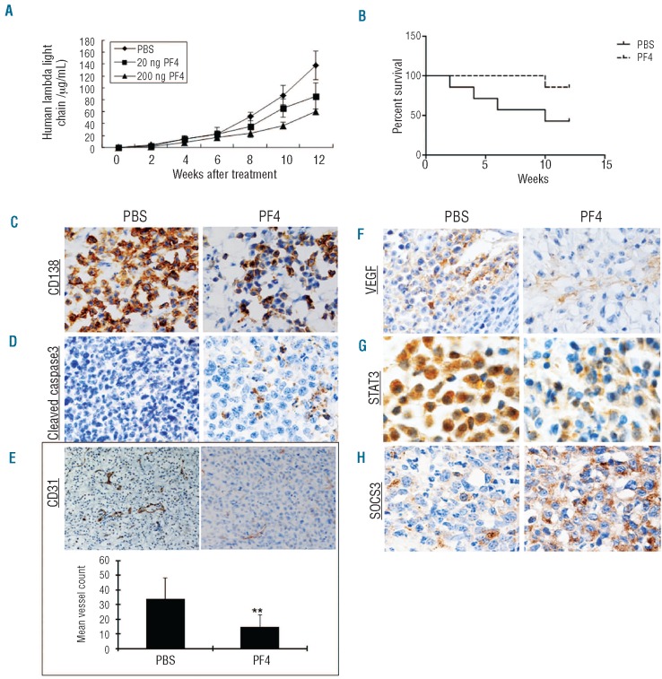
Previous studies have shown that the MM-host bone marrow microenvironment confers growth and survival advantages and drug resistance to MM cells.26 Next, we therefore used the SCID-rab mouse model which recapitulates the human bone marrow milieu in vivo to examine whether the anti-MM activity of PF4 could be substantiated in the presence of a human bone marrow microenvironment. In this model, U266 MM cells were injected directly into rabbit bones that were implanted subcutaneously into the SCID mice, and MM cell growth was assessed by serial measurements of circulating levels of human λ light chain in mouse serum. After 10 weeks of treatment, the levels of human λ light chain were reduced by 58% in the group treated with 200 ng PF4 (n=4) compared to the levels in the PBS-treated group (n=4) (P=0.04) (Figure 4A), suggesting that PF4 inhibited MM tumor growth in vivo. In addition, Kaplan-Meier analysis showed that PF4 improved the survival rate of mice. The survival rate of PBS-treated mice (n=4) was 70% after 6 weeks and less than 45% after 12 weeks, whereas the survival rate of the PF4-treated group (n=4) was 75% after 12 weeks (Figure 4B).
Figure 4.
Effects of PF4 on tumor growth, apoptosis and angiogenesis in the SCID-rab mouse model. (A) PF4 inhibited tumor growth. Upon detection of measurable human lambda light chain in mice (2 weeks after injection of 1×10⁶ U266 cells), mice were treated with either PBS alone or indicated doses of PF4 by tail vein injection (three doses per week for 12 weeks); Tumor growth was measured using enzyme-linked immunosorbent assays of human lambda light chain from mouse serum samples (mean±SD; P<0.05 for 200 ng PF4; n=4). (B) Kaplan-Meier survival plot showed a trend of an increase in survival of mice receiving PF4 (200 ng) compared with PBS-treated controls. (C-F) PF4 inhibited tumor cell growth, induced apoptosis and suppressed angiogenesis in vivo. Rabbit bones were removed from mice after the last treatment and immunostained with antibodies against CD138 (C), cleaved caspase-3 (D), anti-CD31 (E, upper panel) and VEGF (F). (E, lower panel) Blood vessels (E, left panel) (mean±SD of five separate fields) were enumerated. (G-H) Rabbit bones removed from mice were immuno -stained with antibodies against STAT3 and SOCS3.
We next examined the effects of PF4 on in vivo tumor growth and apoptosis using immunostaining of the implanted rabbit bone for CD138 and cleaved caspase-3 expression. We found that the number of CD138-positive cells in the PF4-treated group were substantially reduced compared with that in the PBS-treated group (Figure 4C), while the number of caspase-3 cleavage-positive cells was significantly increased by PF4, compared with vehicle treatment alone (Figure 4D). Importantly, we noted that microvessels were reduced significantly by 57% within tumors of PF4-treated mice compared with controls, as evidenced by CD31 staining (Figure 4E). Similarly, a marked decrease in VEGF expression was also observed in rabbit bone sections from mice injected with PF4 versus vehicle alone (Figure 4F). These in vivo immunohistochemistry data confirmed the growth inhibitory effects of PF4 and also its pro-apoptotic and anti-angiogenic activities in MM cells. Finally, we also investigated whether PF4 could inhibit STAT3 activation in vivo. By immunostaining analysis, we further found that PF4 inhibited STAT3 nuclear translocation in which STAT3 was observed preferentially in the cytoplasm after PF4 treatment, but distributed in both the cytoplasm and nucleus in the PBS-treated mice (Figure 4G), together with a marked increase in SOCS3 expression in PF4-treated mice versus those treated with vehicle alone (Figure 4H). These results indicated that PF4 also inhibited STAT3 activation and induced SOCS3 expression in vivo. Taken together, our results from the MM SCID-rab mouse model provide in vivo evidence for the ability of PF4 to inhibit human MM cell growth within the bone marrow microenvironment.
Discussion
Our and other studies revealed frequent loss of expression of PF4 in primary MM and MM cell lines, which prompted us to investigate the biological function of PF4 in MM.12,13 As an angiogenesis inhibitor, PF4 has not been shown to have direct suppressive effects on tumor cell proliferation and apoptosis in vitro in solid tumors in published studies.4 In contrast, Han et al. demonstrated that the proliferation rate of the human erythroleukemia cell line (HEL) could be inhibited by PF4.8 In this study, in addition to its effect of anti-angiogenesis, which indirectly suppresses MM cell growth, we first found that PF4 directly inhibited MM cell growth both in vitro and in vivo by induction of cell apoptosis, possibly mediated in part by inhibition of STAT3, via up-regulation of SOCS3 expression and an interaction with the cell surface receptor LRP1.
STAT3 is a transcription factor that plays essential roles in the pathogenesis of many cancers, in which constitutive activation leads to inappropriate regulation of genes important for survival and angiogenesis.17 First, constitutively active STAT3 contributes to oncogenesis by protecting cancer cells from apoptosis; thus suppression of STAT3 activation by PF4 could facilitate apoptosis. Second, the induction of resistance to apoptosis from constitutively active STAT3 is possibly effected through the expression of target genes.17 In this study, we first reported that PF4 inhibited the STAT3 signaling pathway in MM cells by inhibiting constitutive phosphorylation of STAT3 and its DNA binding activity. Constitutive STAT3 activation occurs in 50% of primary MM samples19 and STAT3 can be activated by cytokines including IL-6 and others, which are important for the survival and drug resistance of MM cells.23,27 In the bone marrow microenvironment, IL-6 is secreted by stromal cells or the MM cells themselves, and promotes the continued survival and proliferation of MM cells.23,28,29 Our findings that activation of STAT3 induced by IL-6 was suppressed by PF4 suggest that it can overcome cytokine-mediated tumor cell growth in the bone marrow milieu. More importantly, immunohistochemistry results on engrafted bone in the SCID-rab model further supported that PF4 prevented nuclear localization of STAT3. Here we also showed that PF4 effectively down-regulated STAT3 target genes, including Mcl-1 and Survivin, in MM cell lines. Mcl-1 belongs to the Bcl-2 family of proteins and is a critical survival factor for MM.30 Previous studies demonstrated that the down-regulation of Mcl-1 and Survivin precedes caspase activation.31,32 Indeed, PF4 was found, in our study, to induce apoptosis, activate caspase-3 activity and increase cleaved PARP in MM cells, which could be due to the down-regulation of anti-apoptotic genes including Mcl-1 and Survivin. The down-regulation of STAT3 target genes is, therefore, likely linked with the ability of PF4 to induce apoptosis in MM cells. Finally, we observed that overexpression of constitutively active STAT3 in NCI-H929 cells could rescue the apoptotic effects of PF4 in MM cells, suggesting that the STAT3 signaling pathway is one of the crucial mechanisms for PF4-mediated cell apoptosis. However, we cannot rule out the possibility that other pathways, especially those not assessed in the DNA/protein array, are involved in PF4-induced apoptosis.
Angiogenesis plays a crucial role in cancer development and metastasis. As an anti-angiogenic chemokine, PF4 functions to suppress endothelial cell proliferation and migration and thus inhibits tumor growth in various cancers including MM.3-6,33 However, the anti-angiogenic effects of PF4 on MMEC have not been investigated. MMEC were reported to secrete larger amounts of growth factors, including VEGF and bFGF, than healthy endothelial cells and express more adhesion molecules, including CD31, for enhanced dissemination of MM cells and are thus indicative of an angiogenic state.34 In this study, we found that PF4 exhibited direct inhibitory effects on MM angiogenesis both in vitro and in vivo. VEGF is one of the major pro-angiogenic cytokines responsible for the induction of neo-angio-genesis in MM patients.35,36 In our study, we not only found that PF4 decreased VEGF expression in MM cells, but also uncovered the potential mechanism by which PF4 exerts its effect on VEGF. Activated STAT3 plays an essential role in tumor angiogenesis and studies on the link between STAT3 and VEGF suggested that STAT3 influences the regulation of VEGF and also acts as a direct transcription factor of VEGF promoter.37,38 Thus, our observation of STAT3 suppression by PF4 in MM cells suggests that STAT3 may be a mediator between PF4 and VEGF. Our findings, together with those of a previous study, indicate that PF4 is an angiogenesis inhibitor in MM.33 Importantly, PF4 is the second reported factor in MM (the first one is BMP6) to be down-regulated, and is pro-apoptotic and anti-angiogenic.39
At present, several proteins, including SOCS3, are known to regulate the JAK/STAT pathways negatively.21 SOCS3 can bind both the cytokine receptor and JAK and is recruited to the tyrosine phosphorylated receptor, facilitating inhibition of JAK and finally resulting in the inactivation of STAT3.40 SOCS3 has been shown to be silenced in various human cancers and could be activated by cytokines.21,41,42 Tumor necrosis factor-α was found to inhibit STAT3 activation via SOCS3 induction.43 Here, we demonstrated that PF4 could increase both mRNA and protein levels of SOCS3, while knocking down SOCS3 with siRNA abolished its STAT3 inhibitory effects. These findings suggest that the inhibition of STAT3 by PF4 is mediated by SOCS3 induction.
It has been reported that PF4 could modulate cell signaling by binding to cell surface receptors including CXCR3B and LRP1 in endothelial cells and megakaryocytes, which contributed to the anti-angiogenic and antiproliferative effects on these cells.24,25 We, therefore, examined whether these receptors mediated PF4 effects on apoptosis of MM cells. Knockdown of LRP1 expression, but not of CXCR3B, abolished the pro-apoptotic effect of PF4. In addition, inhibition of STAT3 signaling by PF4 was reversed upon LRP1 knockdown. These findings suggest that PF4 could elicit its effects on MM cells by binding to LRP1 on the cell surface, then blocking the downstream signal transduction including STAT3, to cause apoptosis in MM.
In addition to our in vitro studies, we also examined the anti-MM activity of PF4 in vivo using two distinct human MM xenograft mouse models, namely the subcutaneous Matrigel xenograft and SCID-rab mouse model. We found that the dosage required for inhibition by PF4 is 200 ng, which is much lower than in previous studies.4,6 One possible reason is that PF4 targets not only angiogenesis but also acts directly on tumor cells in MM unlike in solid tumors. Recently some PF4-derived molecules or molecular targeting therapies against PF4 have been developed which exhibit stronger anti-angiogenic properties than the parent molecule, and may serve as leads for further therapeutic developments. Our in vitro and in vivo data provide the framework for future clinical trials of PF4 or its analogues as novel STAT3 inhibitors.
Acknowledgments
Funding: This study was fully supported by a grant from the NSFC/RGC Joint Research Scheme sponsored by the Research Grants Council of Hong Kong and the National Science Foundation of China (Project N. N-CUHK 431/07).
Footnotes
The online version of this article has a Supplementary Appendix.
Authorship and Disclosures: Information on authorship, contributions, and financial & other disclosures was provided by the authors and is available with the online version of this article at www.haematologica.org.
References
- 1.Hannah AL. Kinases as drug discovery targets in hematologic malignancies. Curr Mol Med. 2005;5(7):625-42 [DOI] [PubMed] [Google Scholar]
- 2.Gupta SK, Singh JP. Inhibition of endothelial cell proliferation by platelet factor-4 involves a unique action on S phase progression. J Cell Biol. 1994;127(4):1121-7 [DOI] [PMC free article] [PubMed] [Google Scholar]
- 3.Maione TE, Gray GS, Petro J, Hunt AJ, Donner AL, Bauer SI, et al. Inhibition of angiogenesis by recombinant human platelet factor-4 and related peptides. Science. 1990;247(4938):77-9 [DOI] [PubMed] [Google Scholar]
- 4.Sharpe RJ, Byers HR, Scott CF, Bauer SI, Maione TE. Growth inhibition of murine melanoma and human colon carcinoma by recombinant human platelet factor 4. J Natl Cancer Inst. 1990;82(10):848-53 [DOI] [PubMed] [Google Scholar]
- 5.Maione TE, Gray GS, Hunt AJ, Sharpe RJ. Inhibition of tumor growth in mice by an analogue of platelet factor 4 that lacks affinity for heparin and retains potent angiostatic activity. Cancer Res. 1991;51(8):2077-83 [PubMed] [Google Scholar]
- 6.Kolber DL, Knisely TL, Maione TE. Inhibition of development of murine melanoma lung metastases by systemic administration of recombinant platelet factor 4. J Natl Cancer Inst. 1995;87(4):304-9 [DOI] [PubMed] [Google Scholar]
- 7.Tanaka T, Manome Y, Wen P, Kufe DW, Fine HA. Viral vector-mediated transduction of a modified platelet factor 4 cDNA inhibits angiogenesis and tumor growth. Nat Med. 1997;3(4):437-42 [DOI] [PubMed] [Google Scholar]
- 8.Han ZC, Maurer AM, Bellucci S, Wan HY, Kroviarski Y, Bertrand O, et al. Inhibitory effect of platelet factor 4 (PF4) on the growth of human erythroleukemia cells: proposed mechanism of action of PF4. J Lab Clin Med. 1992;120(4):645-60 [PubMed] [Google Scholar]
- 9.Liu YJ, Lu SH, Han ZC. Signal transduction of chemokine platelet factor 4 in human erythroleukemia cells. Int J Hematol. 2002;75(4):401-6 [DOI] [PubMed] [Google Scholar]
- 10.Northfelt D, Robles R, Lanf W, Wagner B, Kahn J, Bonnem E. Phase I/II study of intravenous recombinant platelet factor 4 (rPF4) in AIDS related Kaposi's sarcoma (AIDS KS). American Society of Clinical Oncology Annual Meeting; 1995; 1995 p. 820 [Google Scholar]
- 11.Staddon A, Henry D, Bonnem E. A randomized dose finding study of recombinant platelet factor 4 (rPF4) in cutaneous AIDS-related Kaposi's sarcoma (KS). American Society of Clinical Oncology Annual Meeting; 1994; 1994 p. 50 [Google Scholar]
- 12.Cheng SH, Ng MH, Lau KM, Liu HS, Chan JC, Hui AB, et al. 4q loss is potentially an important genetic event in MM tumorigenesis: identification of a tumor suppressor gene regulated by promoter methylation at 4q13.3, platelet factor 4. Blood. 2007;109(5):2089-99 [DOI] [PubMed] [Google Scholar]
- 13.Hose D, Moreaux J, Meissner T, Seckinger A, Goldschmidt H, Benner A, et al. Induction of angiogenesis by normal and malignant plasma cells. Blood. 2009;114(1):128-43 [DOI] [PubMed] [Google Scholar]
- 14.Cheng Y, Geng H, Cheng SH, Liang P, Bai Y, Li J, et al. KRAB zinc finger protein ZNF382 is a proapoptotic tumor suppressor that represses multiple oncogenes and is commonly silenced in multiple carcinomas. Cancer Res. 2010;70(16):6516-26 [DOI] [PubMed] [Google Scholar]
- 15.Bromberg JF, Wrzeszczynska MH, Devgan G, Zhao Y, Pestell RG, Albanese C, et al. Stat3 as an oncogene. Cell. 1999;98(3):295-303 [DOI] [PubMed] [Google Scholar]
- 16.Yata K, Yaccoby S. The SCID-rab model: a novel in vivo system for primary human myeloma demonstrating growth of CD138-expressing malignant cells. Leukemia. 2004;18(11):1891-7 [DOI] [PubMed] [Google Scholar]
- 17.Aggarwal BB, Sethi G, Ahn KS, Sandur SK, Pandey MK, Kunnumakkara AB, et al. Targeting signal-transducer-and-activator-of-transcription-3 for prevention and therapy of cancer: modern target but ancient solution. Ann N Y Acad Sci. 2006;1091:151-69 [DOI] [PubMed] [Google Scholar]
- 18.Catlett-Falcone R, Landowski TH, Oshiro MM, Turkson J, Levitzki A, Savino R, et al. Constitutive activation of Stat3 signaling confers resistance to apoptosis in human U266 myeloma cells. Immunity. 1999;10(1):105-15 [DOI] [PubMed] [Google Scholar]
- 19.Bharti AC, Shishodia S, Reuben JM, Weber D, Alexanian R, Raj-Vadhan S, et al. Nuclear factor-kappaB and STAT3 are constitutively active in CD138+ cells derived from multiple myeloma patients, and suppression of these transcription factors leads to apoptosis. Blood. 2004;103(8):3175-84 [DOI] [PubMed] [Google Scholar]
- 20.Kisseleva T, Bhattacharya S, Braunstein J, Schindler CW. Signaling through the JAK/STAT pathway, recent advances and future challenges. Gene. 2002;285(1-2):1-24 [DOI] [PubMed] [Google Scholar]
- 21.Wormald S, Hilton DJ. Inhibitors of cytokine signal transduction. J Biol Chem. 2004;279(2):821-4 [DOI] [PubMed] [Google Scholar]
- 22.Pandey MK, Sung B, Aggarwal BB. Betulinic acid suppresses STAT3 activation pathway through induction of protein tyrosine phosphatase SHP-1 in human multiple myeloma cells. Int J Cancer. 2010;127(2):282-92 [DOI] [PMC free article] [PubMed] [Google Scholar]
- 23.Klein B, Zhang XG, Jourdan M, Content J, Houssiau F, Aarden L, et al. Paracrine rather than autocrine regulation of myeloma-cell growth and differentiation by interleukin-6. Blood. 1989;73(2):517-26 [PubMed] [Google Scholar]
- 24.Lambert MP, Wang Y, Bdeir KH, Nguyen Y, Kowalska MA, Poncz M. Platelet factor 4 regulates megakaryopoiesis through low-density lipoprotein receptor-related protein 1 (LRP1) on megakaryocytes. Blood. 2009;114(11):2290-8 [DOI] [PMC free article] [PubMed] [Google Scholar]
- 25.Lasagni L, Francalanci M, Annunziato F, Lazzeri E, Giannini S, Cosmi L, et al. An alternatively spliced variant of CXCR3 mediates the inhibition of endothelial cell growth induced by IP-10, Mig, and I-TAC, and acts as functional receptor for platelet factor 4. J Exp Med. 2003;197(11):1537-49 [DOI] [PMC free article] [PubMed] [Google Scholar]
- 26.Anderson KC. Targeted therapy of multiple myeloma based upon tumor-microenvironmental interactions. Exp Hematol. 2007;35(4 Suppl 1):155-62 [DOI] [PubMed] [Google Scholar]
- 27.Sprynski AC, Hose D, Caillot L, Réme T, Shaughnessy JD, Barlogie B, et al. The role of IGF-1 as a major growth factor for myeloma cell lines and the prognostic relevance of the expression of its receptor. Blood. 2009;113(19):4614-26 [DOI] [PMC free article] [PubMed] [Google Scholar]
- 28.van de Donk NW, Lokhorst HM, Bloem AC. Growth factors and antiapoptotic signaling pathways in multiple myeloma. Leukemia. 2005;19(12):2177-85 [DOI] [PubMed] [Google Scholar]
- 29.Hideshima T, Mitsiades C, Tonon G, Richardson PG, Anderson KC. Understanding multiple myeloma pathogenesis in the bone marrow to identify new therapeutic targets. Nat Rev Cancer. 2007;7(8):585-98 [DOI] [PubMed] [Google Scholar]
- 30.Zhang B, Gojo I, Fenton RG. Myeloid cell factor-1 is a critical survival factor for multiple myeloma. Blood. 2002;99(6):1885-93 [DOI] [PubMed] [Google Scholar]
- 31.Romagnoli M, Trichet V, David C, Clément M, Moreau P, Bataille R, et al. Significant impact of survivin on myeloma cell growth. Leukemia. 2007;21(5):1070-8 [DOI] [PubMed] [Google Scholar]
- 32.Nijhawan D, Fang M, Traer E, Zhong Q, Gao W, Du F, et al. Elimination of Mcl-1 is required for the initiation of apoptosis following ultraviolet irradiation. Genes Dev. 2003;17(12):1475-86 [DOI] [PMC free article] [PubMed] [Google Scholar]
- 33.Yang L, Du J, Hou J, Jiang H, Zou J. Platelet factor-4 and its p17-70 peptide inhibit myeloma proliferation and angiogenesis in vivo. BMC Cancer. 2011;11:261. [DOI] [PMC free article] [PubMed] [Google Scholar]
- 34.Vacca A, Ria R, Semeraro F, Merchionne F, Coluccia M, Boccarelli A, et al. Endothelial cells in the bone marrow of patients with multiple myeloma. Blood. 2003;102(9):3340-8 [DOI] [PubMed] [Google Scholar]
- 35.Kline M, Donovan K, Wellik L, Lust C, Jin W, Moon-Tasson L, et al. Cytokine and chemokine profiles in multiple myeloma; significance of stromal interaction and correlation of IL-8 production with disease progression. Leuk Res. 2007;31(5):591-8 [DOI] [PubMed] [Google Scholar]
- 36.Rajkumar SV, Mesa RA, Fonseca R, Schroeder G, Plevak MF, Dispenzieri A, et al. Bone marrow angiogenesis in 400 patients with monoclonal gammopathy of undetermined significance, multiple myeloma, and primary amyloidosis. Clin Cancer Res. 2002;8(7):2210-6 [PubMed] [Google Scholar]
- 37.Niu G, Wright KL, Huang M, Song L, Haura E, Turkson J, et al. Constitutive Stat3 activity up-regulates VEGF expression and tumor angiogenesis. Oncogene. 2002;21(13):2000-8 [DOI] [PubMed] [Google Scholar]
- 38.Chen Z, Han ZC. STAT3: a critical transcription activator in angiogenesis. Med Res Rev. 2008;28(2):185-200 [DOI] [PubMed] [Google Scholar]
- 39.Seckinger A, Meissner T, Moreaux J, Goldschmidt H, Fuhler GM, Benner A, et al. Bone morphogenic protein 6: a member of a novel class of prognostic factors expressed by normal and malignant plasma cells inhibiting proliferation and angiogenesis. Oncogene. 2009;28(44):3866-79 [DOI] [PMC free article] [PubMed] [Google Scholar]
- 40.Nicholson SE, De Souza D, Fabri LJ, Corbin J, Willson TA, Zhang JG, et al. Suppressor of cytokine signaling-3 preferentially binds to the SHP-2-binding site on the shared cytokine receptor subunit gp130. Proc Natl Acad Sci USA. 2000;97(12):6493-8 [DOI] [PMC free article] [PubMed] [Google Scholar]
- 41.He B, You L, Uematsu K, Zang K, Xu Z, Lee AY, et al. SOCS-3 is frequently silenced by hypermethylation and suppresses cell growth in human lung cancer. Proc Natl Acad Sci USA. 2003;100(24):14133-8 [DOI] [PMC free article] [PubMed] [Google Scholar]
- 42.Weber A, Hengge UR, Bardenheuer W, Tischoff I, Sommerer F, Markwarth A, et al. SOCS-3 is frequently methylated in head and neck squamous cell carcinoma and its precursor lesions and causes growth inhibition. Oncogene. 2005;24(44):6699-708 [DOI] [PubMed] [Google Scholar]
- 43.Bode JG, Nimmesgern A, Schmitz J, Schaper F, Schmitt M, Frisch W, et al. LPS and TNFalpha induce SOCS3 mRNA and inhibit IL-6-induced activation of STAT3 in macrophages. FEBS Lett. 1999;463(3):365-70 [DOI] [PubMed] [Google Scholar]


