Abstract
Oxidative stress and oxidized dopamine contribute to the degeneration of the nigrostriatal pathway in Parkinson’s disease (PD). Selenoproteins are a family of proteins containing the element selenium in the form of the amino acid selenocysteine, and many of these proteins have antioxidant functions. We recently reported changes in expression of the selenoprotein, phospholipid hydroperoxide glutathione peroxidase GPX4 and its co-localization with neuromelanin in PD brain. To further understand the changes in GPX4 in PD, we examine here the expression of the selenium transport protein selenoprotein P (Sepp1) in postmortem Parkinson’s brain tissue. Sepp1 in midbrain was expressed in neurons of the substantia nigra (SN), and expression was concentrated within the centers of Lewy bodies, the pathological hallmark of PD. As with GPX4, Sepp1 expression was significantly reduced in SN from PD subjects compared with controls, but increased relative to cell density. In putamen, Sepp1 was found in cell bodies and in dopaminergic axons and terminals, although levels of Sepp1 were not altered in PD subjects compared to controls. Expression levels of Sepp1 and GPX4 correlated strongly in the putamen of control subjects but not in the putamen of PD subjects. These findings indicate a role for Sepp1 in the nigrostriatal pathway, and suggest that local release of Sepp1 in striatum may be important for signaling and/or synthesis of other selenoproteins such as GPX4.
Keywords: Selenium, selenoproteins, selenoprotein P, GPX4, glutathione peroxidase, Parkinson’s disease, Lewy bodies, dopamine, substantia nigra, striatum, putamen, presynaptic terminals
INTRODUCTION
The periodic firing activity and high oxidizable iron content of nigrostriatal dopaminergic neurons, and the potentially toxic structure of dopamine (DA) itself, render these neurons highly vulnerable [1, 2]. The consequences of DA neuron loss are most prevalent in Parkinson’s disease (PD), one of the most common neurodegenerative disorders [3]. DA neuron fibers primarily emanate from two ventral midbrain regions, the substantia nigra (SN), and the ventral tegmental area (VTA) [2]. Cells in these two areas send axons throughout the brain, and most notably to the striatum, limbic areas and frontal cortex.
Selenoproteins have important antioxidant and redox functions, and members of the selenoprotein family are known to reduce oxidative stress [4–6]. Selenoproteins contain the micronutrient selenium (Se) incorporated as the amino acid, selenocysteine (Sec). Se deficiency is associated with developmental and neurological disorders [7]. Most of the glutathione peroxidases (GPX), which are glutathione-dependent hydroperoxidase enzymes essential for maintaining redox balance in cells, are selenoproteins. Glutathione is greatly decreased in early stages of PD [8], which contributes to decreased peroxidase activity [9]. GPX1 is found in microglia and co-localizes with Lewy bodies, the inclusion bodies characteristic of PD [10]. Synthesis of the phospholipid hydroperoxide glutathione peroxidase GPX4, is regulated by oxidation of the PD-associated gene DJ-1 [11], and is increased in cortex of PD subjects [12]. We previously found co-localizationof GPX4 with neuromelanin in SN, as wells as changes in nigral expression and an increased presence in dystrophic axons within the PD brain [13].
Selenoprotein P (Sepp1) is a selenium transport protein with antioxidant properties and is important for supply of selenium to the brain and other organs [14]. However, Sepp1 is abundant in brain and may have direct functions there as an antioxidant [15, 16]. We previously found an increase in Sepp1-positive cells in Alzheimer’s brain and an association of Sepp1 with amyloid plaques and neurofibrillary tangles [17]. This may represent a response to oxidative stress-related neurodegeneration, in which case an increase in Sepp1 might be found in other neurodegenerative disorders involving increased oxidative stress. Here we sought to determine if expression patterns of Sepp1 are altered in PD brain and if changes coincide with our previous findings for GPX4. We report the specific presence of Sepp1 in cell bodies, axons and presynaptic terminals of SN neurons. We additionally report changes in expression of Sepp1 in SN of PD subjects and its’ localization in Lewy bodies, along with a strong correlation between Sepp1 and GPX4 in putamen of control subjects but not of PD subjects.
MATERIALS AND METHODS
Subjects
Formalin-fixed human brain tissue was provided by the Honolulu-Asia Aging Study (HAAS), an ongoing project that has monitored the health and lifestyle of Japanese-American men born between 1900 and 1919 and residing on Oahu, Hawaii [18]. Sections (10 μm) of SN and putamen from 12 subjects with marked signs of PD including Lewy bodies and degeneration of dopaminergic terminals and cell bodies, as well as sections from 11 age-matched control subjects with no symptoms of PD, were used in this study.
Western blot
HEK293 cells were transfected with pcDNA3.1 empty vector or vector with human Sepp1 with Lipofectamine (Invitrogen) and media samples collected as described previously [19]. Post-mortem tissue from human parietal cortex was homogenized by sonication in CelLytic (Sigma) per manufacturer instructions, centrifuged at 8,000 × g for 10 min. HepG2 cells were grown in DMEM media with 10% FBS having a measured selenium content of 30 nM, either non-supplemented or supplemented with 70 or 170 nM Na2Se3 for final Se concentrations of 30, 100 or 200 nM. Protein was extracted from HepG2 cells using CelLytic buffer per manufacturer’s instructions, separated by electrophoresis and blotted to PVDF membranes. Blots were blocked with Odyssey blocking buffer (LiCore Biosciences) for 1 hr and then incubated in Sepp1 antibody (AbFrontier) diluted 1: 500. After washing with PBS containing 0.05% tween-20 (PBST), membranes were treated with secondary antibodies labeled with infrared fluorophores (LiCore Biosciences). After further washes in PBST, blots were imaged with the Odyssey infrared imaging system (LiCore Biosciences).
Immunolabeling
Immunolabeling was performed as previously described [13, 17]. Deparaffinized brain sections (10 μm) were heated in a pressure cooker to 95°C and 15 psi for 20 min in Trilogy alkaline solution with EDTA (Cell Marque), followed by 3 min in 90% formic acid, for antigen unmasking. Samples were blocked in PBS with 5% serum species matched to secondary antibody. Tissue was incubated in Sepp1 primary antibody (AbFrontier, 1: 100) overnight at 4°C in 3% serum. After washes, sections were incubated in biotinylated secondary antibody followed by ABC™ reagent. HRP signals were developed with 3,4-diaminobenzamidine hydrochloride (DAB, Vector Labs), with or without the addition of nickel chloride to darken color as per manufacturer’s instructions.
Double labeling
Following first primary antibody, tissue was subsequently blocked in 5% normal horse serum (NHS), followed with separate blocking steps in streptavidin and biotin solutions (from ABC kit) five minutes each before second primary antibody reaction. Additional primary antibodies used were anti-alpha synuclein (AS) (1: 1000, Chemicon) or (1: 50, Abcam) anti-tyrosine hydroxylase (TH, 1: 8000, Sigma), and anti-GPX4 (1: 250, AbFrontier). Combinations of HRP-labeled secondary antibodies detected with DAB or DAB containing nickel chloride (DAB-Ni) (Vector Laboratories), and alkaline phosphatase (AP) detected with BCIP reactions, were used to maximize contrast between the different antibodies.
Multi-spectral imaging
Bright light and fluorescent images of midbrain tissue samples were imaged using an Olympus microscope equipped with the Nuance multispectral imaging system (Cambridge Research and Instrumentation, Inc.). After obtaining spectral libraries for bright light images of unlabeled tissue and neuromelanin, and fluorescent images of fluorophores and background autofluorescence, the brightfield and fluorescent images were “unmixed” into individual signal components (i.e., neuromelanin or fluorescent probes) that were then pseudocolored for comparison.
Confocal microscopy
Tissue was prepared and incubated in primary antibodies as described above, and detected with secondary antibodies conjugated with Alexa 488 (green) and either Alexa 546 (red) or Alexa 680 (Molecular Probes). Endogenous fluorescence was reduced by treating with an autofluorescence eliminator reagent (Chemicon). Images were collected with Zeiss LSM Pascal laser confocal microscope, and analyzed with ImageJ software (http://rsbweb.nih.gov/ij/).
Stereology
Volume-density of immunolabeling was determined with a Cavalieri probe using a Zeiss Axioscope equipped with an ASI motorized stage and Zeiss camera operated by Stereologer software (Stereology Resource Center). First, the computer image of the region (SN or putamen) was outlined under 5X magnification, using the motorized stage to track the cursor. Then a computer-generated array of systematic-random loci were visited and observed under 40X. A Cavalieri probe was placed over an 800 μm2 area of the image. The probe consisted of an array of 400 (20 × 20) points (+), with each point covering 40 μm2. The number of points contacting immunolabeled cells was counted. The fraction of points contacting immunolabeled cells was used to estimate the area fraction of immunolabeling at each location. Two random 10 μm sections that were spaced 30 μm apart in the original tissue were used for each subject. The total area fraction of immunolabeling was estimated as the average area fraction of all systematic-randomly chosen sites. According to the Delesse principle, area fraction on random sections is equivalent to the volume fraction [20].
Statistical analysis
Statistical analysis was conducted using SAS Enterprise Guide and GraphPad Prism 5. Group differences were compared using Student t-test. To evaluate the relationship between GPX4 and Sepp1 expression levels, we used Pearson correlational analysis and analysis of co-variance (ANCOVA). Data are presented as mean values ± standard error of the mean (SEM). P ≤ 0.05 is considered statistically significant.
RESULTS
Tissue was obtained from the Honolulu-Asia Aging Study (HAAS). We examined Sepp1 in postmortem brain of 12 subjects that had been clinically diagnosed with PD, as well as 11 subjects without clinical or postmortem pathological features of PD. The subjects used in the present study are the same as in our previously published report on GPX4 in PD [13], and additional group and subject information is available therein. All control subjects had Braak scores of 0, while PD subjects had scores of 5 or 6. The mean age at death, range of ages, and postmortem intervals were not significantly different between groups.
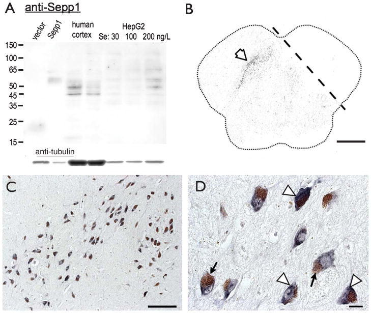
We first verified the specificity of the Sepp1 antibody. As we found previously [17], the antibody recognized two bands of ~55–60 kD in media from HEK293 cells transfected with recombinant Sepp1 but not in media from empty vector control transfected cells. These bands were also found in lysates from postmortem parietal cortex from control subjects. However, the cortex samples showed stronger bands at 46 and 52 kD. The 46 kD band corresponds to the size of full-length, unglycosylated Sepp1, which has been reported in human cultured astrocytes [21]. The 52 kD band may be partially glycosylated Sepp1 post synthesis and prior to secretion [22]. As Sepp1 expression increases in HepG2 hepatocytes with Se supplementation [23, 24], we used HepG2 cells grown with different amounts of Se to confirm if the bands shown are actually isoforms of Sepp1. We found that the 52 kD and larger bands increased with Se supplementation relative to tubulin, indicating that these are indeed different forms of Sepp1. The 46 kD band increased less with supplementation compared to other bands, possibly because the rate of glycosylation prevents accumulation of the unglycosylated form of Sepp1.
Immunolabeling of Sepp1 in midbrain from non-PD subjects shows Sepp1 expression concentrated within neurons of the SN (Fig. 1). Sepp1 expression was primarily confined within the SN. Within the large neurons of SN, Sepp1 location was cytoplasmic. As seen in Fig. 1B, Sepp1 immunoreactivity is concentrated in the medial SN. The area opposite the dotted line was sectioned from the rest of the tissue, and primary antibody was omitted as a negative control. Figure 1C shows the distribution of Sepp1-positive cells in pars compacta and pars reticulata of SN. A higher magnification (Fig. 1D) reveals the distribution of Sepp1 within SN neurons. The brown pigmentation is endogenous neuromelanin.
Fig. 1.
Sepp1 is abundant in and restricted to SN in midbrain. A. Western blot showing specificity of the Sepp1 antibody. The first two lanes are media samples from HEK293 cells transfected with empty pCDNA3.1 vector or vector containing human Sepp1. Third and fourth lanes are protein lysates of postmortem human cortex from control subjects. The next three lanes are cell lysates from cultured HepG2 cells grown in 30, 100 or 200 nM Se. Bands of 52 kD or larger are increased in HepG2 cells supplemented with Se. B. Low magnification image of a whole midbrain section showing dark Sepp1 immunolabeling specific to SN. The upper right region was sectioned from the rest of the tissue (shown by dashed line) and the Sepp1 primary antibody was omitted as a negative control. C. Low magnification of Sepp1, and D, higher magnification images of Sepp1 expression (purple BCIP marked by white arrowheads) in SN neurons. Brown pigmentation is endogenous neuromelanin (black arrows). Scale Bars: B, 5 mm; C, 100 μm, D; 20 μm.
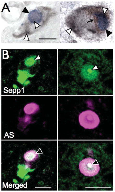
Intracellular aggregates of alpha-synuclein (AS) in brain are the pathological hallmark of PD, so we examined Sepp1 expression in relation to AS aggregates in SN from PD subjects. Sepp1 was distributed in specific loci throughout the DA neurons, and expression overlapped with AS in Lewy bodies (Fig. 2A). We used confocal microscopy to confirm if Sepp1 was co-localized with AS. As shown in Fig. 2B, Sepp1 was remarkably concentrated within the centers of Lewy bodies.
Fig. 2.
Sepp1 expression coincides with AS aggregates. A. Sepp1 expression (grey Ni-DAB, white arrowheads) coincides with Lewy bodies (blue BCIP, marked with black arrowheads in SN neurons). The black arrow indicates neuromelanin. B. Confocal images showing colocalization of Sepp1 (green, top) with AS (magenta, middle). Signal colocalization is shown in white (bottom, black arrowhead). Scale bars: 20 μm.
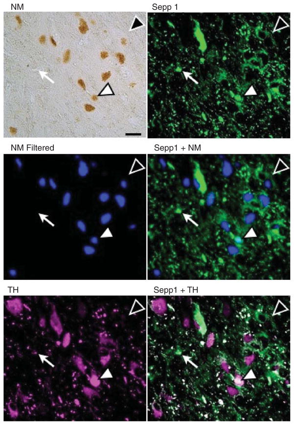
We previously reported the colocalization of GPX4 with neuromelanin(NM) in SN [13]. We thus investigated if Sepp1 had a similar association using multispectral imaging of both bright light and fluorescent microscope images. As shown in Fig. 3, Sepp1 immunoreactivity was present in cells expressing the dopamine synthesizing enzyme tyrosine hydroxylase (TH), as well as NM positive cells, and cells with both TH and NM. Thus in contrast to GPX4, Sepp1 was not specifically colocalized with either TH or NM.
Fig. 3.
Sepp1 is expressed in both TH and NM positive cells. Neuromelanin was filtered from multispectral bright light images, pseudocolored blue, and compared to Sepp1 (green) and TH (magenta) immunoreactivity. Sepp1 was found in cells positive for both TH and neuromelanin (white arrowhead), for neuromelanin alone (black arrowhead) and for TH alone (white arrow). Scale bar: 50 μm.
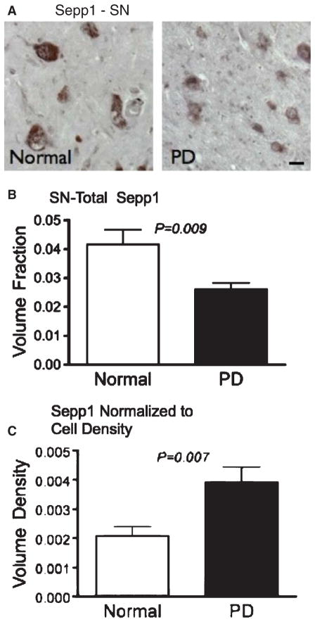
To determine and quantify if Sepp1 expression was different in PD brain, we measured the Sepp1 immunoreactivity volume fraction. This was estimated from the cumulative area of immunoreactivity estimated in multiple tissue sections using a Cavalieri probe [25]. Sepp1 in PD SN was markedly reduced from 0.042 ± 0.005 in control subjects to 0.026 ± 0.002 in PD subjects (P = 0.009) (Fig. 4B). Such a decrease could be explained by cell loss. However, this decrease was not as great as the total cell loss in the SN of PD subjects compared with controls. We calculated the volume density of labeling by dividing the volume fraction by cell densities for the subjects obtained in a previous study [26]. We found that, as with GPX4 [13], Sepp1 labeling was actually increased relative to the total cell number, from 0.00201 ± 0.0003 in control SN to 0.0039 ± 0.0005 in PD SN (P = 0.007). This could indicate an upregulation of Sepp1 within these cells, or an increased protection of cells expressing Sepp1. Both explanations suggest a potential role for Sepp1 in preventing neuronal death in PD.
Fig. 4.
Sepp1 expression in PD SN relative to controls is reduced overall but increased relative to cell density. A. Examples of Sepp1 labeling in SN from Normal and PD subjects. Scale bar: 25 μm. B. Sepp1 expression is significantly reduced in SN (P = 0.0091). C. Sepp1 is increased relative to cell density (P = 0.007).
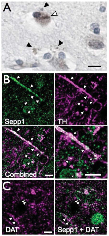
We also examined Sepp1 expression in the putamen. As we found with SN neurons, the Sepp1 antibody labeled cells throughout the cytoplasm (Fig. 5A). However, scattered Sepp1 labeling was also present between identifiable cell bodies, either as small punctate labeling or thin lines. We questioned if Sepp1 could be present within dopaminergic axons and terminals. To test this, we performed confocal microscopy on tissue immunolabeled for Sepp1 along with TH. As seen in Fig. 5B, punctate Sepp1 labeling (green, middle left panel) was present in axonal processes, within cell bodies and in surrounding areas. The area of labeling matched closely with the presence of TH (magenta, middle right). Co-localization between Sepp1 and TH can be seen as white label in the lower left panel of Fig. 5B. In the enlarged section (lower right panel), the punctate co-localized signal can be seen along axons and in terminals of the TH-positive neurons. This suggests the presence of Sepp1 in small presynaptic compartments of dopaminergic processes. To further verify the presence of Sepp1 in DA terminals, we investigated the spatial location of Sepp1 relative to the DA transporter (DAT). We did find Sepp1 co-localized with DAT (Fig. 5C), confirming that Sepp1 is present in DA terminals.
Fig. 5.
Sepp1 expression in cells and dopaminergic axons in putamen. A. Sepp1 expression (gray) was expressed in bodies of some cells (white arrowheads) as well as punctate staining in neuropil (black arrowheads). Nuclei are counterstained with hematoxylin for clarity. B. Confocal images of Sepp1 and TH labeling. Punctate Sepp1 labeling (green, top left) can be seen along axons, within cell bodies and neuropil. TH labeling (magenta, top right) marks dopaminergic axons and terminals. When images are combined, colocalization (marked in all panels by white arrowheads) of Sepp1 and TH is white (lower left). The boxed area is enlarged at the right to emphasize the minute areas of colocalized signal within axons. C. Confocal images showing DAT labeling (left) and Sepp1 with DAT (right). Co-localization of Sepp1 with DAT, shown by white arrowheads, further supports the presence of Sepp1 in dopaminergic presynaptic terminals. Scale bars: 10 μm.
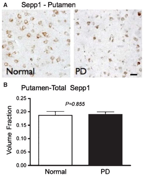
There was no significant alteration in overall Sepp1 labeling in putamen in PD subjects compared with controls (Fig. 6). The Sepp1 volume fraction was 0.187 ± 0.013 in control putamen and 0.190 ± 0.009 in PD putamen (P = 0.855).
Fig. 6.
Sepp1 levels are unchanged in PD putamen. A. Examples of Sepp1 labeling in putamen from non-PD and PD subjects. Scale bar: 25 μm. B. Total volume fraction of Sepp1.
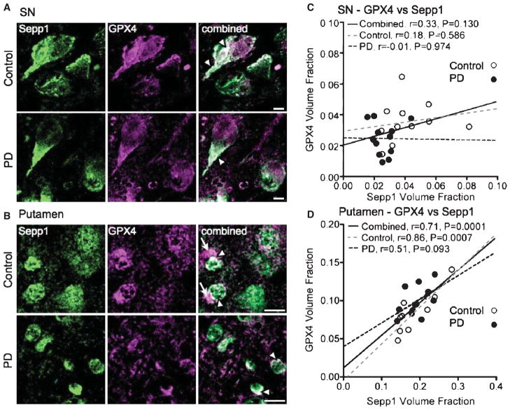
Sepp1 is thought to function primarily in transporting selenium between organs through plasma. However, the presence of Sepp1 in dopaminergic neurons and terminals indicates a local function. We hypothesized that synthesis of other selenoproteins, such as the phospholipid hydroperoxidase GPX4, may depend on local release of Sepp1. We therefore investigated whether GPX4 immunoreactivity corresponded with that of Sepp1 in SN and putamen from PD and control subjects. We used double immunolabeling with Sepp1 and GPX4 to look for co-localization in SN (Fig. 7A) and putamen (Fig. 7B). We observed some co-localization of Sepp1 and GPX4 in sub-cellular structures in SN neurons (white arrowheads), particularly at the base of the major proximal dendrites. However, this expression did not differ between control and PD tissue. In control putamen, we observed some co-localization of these proteins on what appear to be cell surfaces. Additionally, we observed a pattern of concentrated GPX4 neighboring structures with Sepp1, rather than direct co-localization (indicated by white arrows in 7B, upper right panel). This conspicuous pattern was not present in putamen sections from PD subjects.
Fig. 7.
Altered relationship between GPX4 and Sepp1 in PD subjects. A. Double labeling of Sepp1 (green) and GPX4 (magenta) in SN, comparing control tissue (above) with PD tissue (below). In merged images with both labels, co-localization of Sepp1 and GPX4 is shown by white color (examples marked by white arrowheads). Scale bar: 10 μm. B. Double labeling of Sepp1 and GPX4 in putamen. Details are the same as in A. Aggregates of GPX4 immunolabeling (white arrows) frequently neighbor structures labeled with Sepp1. C. Correlational analysis for SN shows no association between Sepp1 and GPX4 in SN for the combined (PDs and controls) or separate groups. D. Sepp1 correlates strongly with GPX4 in combined groups (solid line) in putamen. The correlation remains strong for the control subject group (white circles and grey dotted line). However, there is no significant relationship between Sepp1 and GPX4 within the PD group (black circles and black dotted line).
We compared the amount of Sepp1 signal with our previously published measurements of GPX4 [13] using correlational and ANCOVA analyses. As shown in Fig. 7C, there was no correlation between Sepp1 and GPX4 in SN, and there was no effect of Sepp1 on GPX4 expression in SN as indicated by ANCOVA analysis (P = 0.143). However, a strong positive correlation between the two proteins in putamen was found when both groups were combined (Fig. 7D, P = 0.0001) which may indicate interdependence and suggest that GPX4 synthesis may rely on the local presence, and possibly release, of Sepp1. Further, ANCOVA analysis also revealed a strong effect of Sepp1 on GPX4 expression within the putamen (P = 0.0001). The correlation was strong among control subjects (P = 0.0007); however, there was only a trend for significance between the relationship of Sepp1 and GPX4 in PD subjects (P = 0.094). Thus the interdependence of the two proteins is disrupted in PD, possibly due to the loss of DA terminals containing Sepp1.
DISCUSSION
In this study, we found that Sepp1 is abundant in SN neurons and colocalizes with Lewy bodies, particularly within the cores of the inclusion bodies. Additionally, Sepp1 is greatly reduced in the SN of PD subjects, but is actually increased relative to the total number of cells. In putamen, Sepp1 is not only present in cell bodies but also in dopaminergic axons and terminals. However, the total amount of Sepp1 protein is not changed in PD putamen. GPX4 and Sepp1 correlate strongly in control putamen, but not in PD putamen or in SN of either group. Altogether, these findings suggest that Sepp1 has a role in SN neurons and in nigrostriatal dopaminergic transmission, and may be important for survival of these neurons in PD.
The colocalization of Sepp1 with AS and its concentration suggests an interaction during the development of PD. AS is normally present in presynaptic terminals of DA neurons, and Sepp1 may associate with AS at this location early in the progression of the pathology. The localization of Sepp1 specifically within the cores is reminiscent of our previous finding of Sepp1 within the cores of amyloid beta plaques [17]. Sepp1 may specifically interact with aggregates of misfolded proteins. Alternatively, Sepp1 may be specifically bound to one or more proteins within these structures. We have also found Sepp1 colocalized in neurofibrillary tangles with tau [17], which is present in some Lewy bodies [27].
The specific localization of Sepp1 to SN neurons as well as its presence in presynaptic terminals suggests a role for this protein in the nigrostriatal pathway. As Sepp1 is a secreted protein, it is plausible that this protein is released from presynaptic DA terminals in the striatum. Sepp1 binds to the ApoER2 receptor [28, 29], which also has reelin and ApoE as ligands [30]. ApoER2 can functionally associate with NMDA receptors [31] that, in addition to their well-established postsynaptic location, might be located presynaptically on dopaminergic nerve terminals [32–34]. Thus Sepp1 may have an important signaling function in the nigrostriatal pathway. The disruptions in memory and synaptic plasticity previously described in Sepp1 knockout animals support the idea of a signaling role for Sepp1 in brain [35].
The correlation between Sepp1 and GPX4 in non-PD brain suggests that striatal expression of GPX4 is dependent upon local release of Sepp1 for supply of Se. Sepp1 present in CSF [15] could supply Se to SN, putamen and other brain regions. However, the high DA concentration putamen may require increased Se or coordination of pre- and postsynaptic selenoprotein synthesis. The immunolabeling pattern of GPX4 neighboring structures with Sepp1 could indicate that local release of Sepp1 between cells facilitates synthesis of GPX4 in neighboring cells. Although we focus in this study on correlations of Sepp1 with GPX4, other selenoproteins may be similarly dependent upon local Sepp1.
The lack of correlation between Sepp1 and GPX4 in PD putamen implies that Sepp1-mediated supply of selenium is disrupted in PD. As shown in Fig. 6, there are no increases in Sepp1 level of expression in the PD putamen. This would limit any Sepp1-dependent increases in GPX4 from reaching maximal levels, thus explaining the smaller slope of the GPX4/Sepp1 relationship in the PD brain. Wepreviously reported that GPX4 is increased specifically in dystrophic DA neurites in PD putamen [13]. Although GPX4 levels were higher in putamen, this increase did not reach statistically significant levels. A small increase of GPX4 or Sepp1 in DA terminals would be offset by the loss of these terminals in PD, preventing these changes from being detected. Similarly, any increase in local synthesis of selenoproteins dependent upon release of Sepp1 from DA terminals would be limited by loss of these terminals in PD. The loss of correlation between these two proteins in PD putamen may indicate important local changes in these proteins that could contribute further to the pathology of PD.
The increase in Sepp1 relative to surviving cells in PD SN suggests either an increase in response to pathological conditions such as oxidative stress, or that greater levels of Sepp1 prior to disease onset can improve the likelihood of cell survival. A relative increase in Sepp1 in SN neurons is consistent with our previous findings of increased Sepp1 expression in cortex of Alzheimer’s brain [17]. Thus conditions of increased oxidative stress may lead to a local upregulation of Sepp1. Alternatively, higher expression in a subset of SN neurons could promote their survival, accounting for the increase in Sepp1 relative to cell number.
These findings suggest important roles for Sepp1 in the nigrostriatal pathway. Local release of Sepp1 appears to be an important source of Se for synthesis of selenoproteins such as GPX4. Aside from being a Se supplier, Sepp1 may modulate cell signaling through the ApoER2 receptor. The disrupted relationship between Sepp1 and GPX4 in PD brain could perhaps be eradicated by increasing selenium supply through other means. Further studies into the function of Sepp1 in SN and putamen could help elucidate PD pathology and could possibly lead to better therapies for this disorder.
Acknowledgments
The authors thank Kristen Ewell for tissue sectioning, Yanling Lin and Chrislyn Andres for technical assistance, Elizabeth Nguyen Wu for manuscript comments and Linda Chang for suggestions. Supported by: NIH RO1 NS40302 (MJB), US Department of the Army grant DAMD17-98-1-8621 and the Office of Research and Development, Medical Research Service, Department of Veterans Affairs (GWR), NIH P20RR016467 to (FPB), Hawaii Community Foundation Ingeborg v.F. McKee Fund 08PR-43031 (FPB), NIH U01 AG019349 (LRW), and NIH G12 RR003061/G12 MD007601 which supports the JAB-SOM histology/imaging core facility. The information contained in this paper does not necessarily reflect the position or the policy of the US government, and no official endorsement should be inferred.
References
- 1.Chinta SJ, Andersen JK. Dopaminergic neurons. Int J Biochem Cell Biol. 2005;37:942–946. doi: 10.1016/j.biocel.2004.09.009. [DOI] [PubMed] [Google Scholar]
- 2.Iversen SD, Iversen LL. Dopamine: 50 years in perspective. Trends Neurosci. 2007;30:188–193. doi: 10.1016/j.tins.2007.03.002. [DOI] [PubMed] [Google Scholar]
- 3.Fahn S. Description of Parkinson’s disease as a clinical syndrome. Ann N Y Acad Sci. 2003;991:1–14. doi: 10.1111/j.1749-6632.2003.tb07458.x. [DOI] [PubMed] [Google Scholar]
- 4.Hatfield DL, Carlson BA, Xu XM, Mix H, Gladyshev VN. Selenocysteine incorporation machinery and the role of selenoproteins in development and health. Prog Nucleic Acid Res Mol Biol. 2006;81:97–142. doi: 10.1016/S0079-6603(06)81003-2. [DOI] [PubMed] [Google Scholar]
- 5.Labunskyy VM, Hatfield DL, Gladyshev VN. The Sep 15 protein family: Roles in disulfide bond formation and quality control in the endoplasmic reticulum. IUBMB Life. 2007;59:1–5. doi: 10.1080/15216540601126694. [DOI] [PubMed] [Google Scholar]
- 6.Kim HY, Gladyshev VN. Methionine sulfoxide reductases: Selenoprotein forms and roles in antioxidant protein repair in mammals. Biochem J. 2007;407:321–329. doi: 10.1042/BJ20070929. [DOI] [PubMed] [Google Scholar]
- 7.Chen J, Berry MJ. Selenium and selenoproteins in the brain and brain diseases. J Neurochem. 2003;86:1–12. doi: 10.1046/j.1471-4159.2003.01854.x. [DOI] [PubMed] [Google Scholar]
- 8.Zeevalk GD, Razmpour R, Bernard LP. Glutathione and Parkinson’s disease: Is this the elephant in the room? Biomed Pharmacother. 2008;62:236–249. doi: 10.1016/j.biopha.2008.01.017. [DOI] [PubMed] [Google Scholar]
- 9.Ambani LM, Van Woert MH, Murphy S. Brain peroxidase and catalase in Parkinson disease. Arch Neurol. 1975;32:114–118. doi: 10.1001/archneur.1975.00490440064010. [DOI] [PubMed] [Google Scholar]
- 10.Power JH, Blumbergs PC. Cellular glutathione peroxidase in human brain: Cellular distribution, and its potential role in the degradation of Lewy bodies in Parkinson’s disease and dementia with Lewy bodies. Acta Neuropathol. 2009;117:63–73. doi: 10.1007/s00401-008-0438-3. [DOI] [PubMed] [Google Scholar]
- 11.van der Brug MP, Blackinton J, Chandran J, Hao LY, Lal A, Mazan-Mamczarz K, Martindale J, Xie C, Ahmad R, Thomas KJ, Beilina A, Gibbs JR, Ding J, Myers AJ, Zhan M, Cai H, Bonini NM, Gorospe M, Cookson MR. RNA binding activity of the recessive parkinsonism protein DJ-1 supports involvement in multiple cellular pathways. Proc Natl Acad Sci U S A. 2008;105:10244–10249. doi: 10.1073/pnas.0708518105. [DOI] [PMC free article] [PubMed] [Google Scholar]
- 12.Blackinton J, Kumaran R, van der Brug MP, Ahmad R, Olson L, Galter D, Lees A, Bandopadhyay R, Cookson MR. Post-transcriptional regulation of mRNA associated with DJ-1 in sporadic Parkinson disease. Neurosci Lett. 2009;452:8–11. doi: 10.1016/j.neulet.2008.12.053. [DOI] [PMC free article] [PubMed] [Google Scholar]
- 13.Bellinger FP, Bellinger MT, Seale LA, Takemoto AS, Raman AV, Miki T, Manning-Bog AB, Berry MJ, White LR, Ross GW. Glutathione Peroxidase 4 is associated with Neuromelanin in Substantia Nigra and Dystrophic Axons in Putamen of Parkinson’s brain. Mol Neurodegener. 2011;6:8. doi: 10.1186/1750-1326-6-8. [DOI] [PMC free article] [PubMed] [Google Scholar]
- 14.Burk RF, Hill KE. Selenoprotein P: An extra-cellular protein with unique physical characteristics and a role in selenium homeostasis. Annu Rev Nutr. 2005;25:215–235. doi: 10.1146/annurev.nutr.24.012003.132120. [DOI] [PubMed] [Google Scholar]
- 15.Scharpf M, Schweizer U, Arzberger T, Roggendorf W, Schomburg L, Kohrle J. Neuronal and ependymal expression of selenoprotein P in the human brain. J Neural Transm. 2007;114:877–884. doi: 10.1007/s00702-006-0617-0. [DOI] [PubMed] [Google Scholar]
- 16.Schweizer U, Brauer AU, Kohrle J, Nitsch R, Savaskan NE. Selenium and brain function: A poorly recognized liaison. Brain Res Brain Res Rev. 2004;45:164–178. doi: 10.1016/j.brainresrev.2004.03.004. [DOI] [PubMed] [Google Scholar]
- 17.Bellinger FP, He QP, Bellinger MT, Lin Y, Raman AV, White LR, Berry MJ. Association of selenoprotein p with Alzheimer’s pathology in human cortex. J Alzheimers Dis. 2008;15:465–472. doi: 10.3233/jad-2008-15313. [DOI] [PMC free article] [PubMed] [Google Scholar]
- 18.White L, Petrovitch H, Ross GW, Masaki KH, Abbott RD, Teng EL, Rodriguez BL, Blanchette PL, Havlik RJ, Wergowske G, Chiu D, Foley DJ, Murdaugh C, Curb JD. Prevalence of dementia in older Japanese-American men in Hawaii: The Honolulu-Asia Aging Study. JAMA. 1996;276:955–960. [PubMed] [Google Scholar]
- 19.Squires JE, Stoytchev I, Forry EP, Berry MJ. SBP2 binding affinity is a major determinant in differential selenoprotein mRNA translation and sensitivity to nonsense-mediated decay. Mol Cell Biol. 2007;27:7848–7855. doi: 10.1128/MCB.00793-07. [DOI] [PMC free article] [PubMed] [Google Scholar]
- 20.Mouton PR, Long JM, Lei DL, Howard V, Jucker M, Calhoun ME, Ingram DK. Age and gender effects on microglia and astrocyte numbers in brains of mice. Brain Res. 2002;956:30–35. doi: 10.1016/s0006-8993(02)03475-3. [DOI] [PubMed] [Google Scholar]
- 21.Steinbrenner H, Alili L, Bilgic E, Sies H, Brenneisen P. Involvement of selenoprotein P in protection of human astrocytes from oxidative damage. Free Radic Biol Med. 2006;40:1513–1523. doi: 10.1016/j.freeradbiomed.2005.12.022. [DOI] [PubMed] [Google Scholar]
- 22.Steinbrenner H, Alili L, Stuhlmann D, Sies H, Brenneisen P. Post-translational processing of selenoprotein P: Implications of glycosylation for its utilisation by target cells. Biol Chem. 2007;388:1043–1051. doi: 10.1515/BC.2007.136. [DOI] [PubMed] [Google Scholar]
- 23.Hill KE, Chittum HS, Lyons PR, Boeglin ME, Burk RF. Effect of selenium on selenoprotein P expression in cultured liver cells. Biochim Biophys Acta. 1996;1313:29–34. doi: 10.1016/0167-4889(96)00047-x. [DOI] [PubMed] [Google Scholar]
- 24.Hoefig CS, Renko K, Kohrle J, Birringer M, Schomburg L. Comparison of different selenocompounds with respect to nutritional value vs. toxicity using liver cells in culture. J Nutr Biochem. 2011;22:945–955. doi: 10.1016/j.jnutbio.2010.08.006. [DOI] [PubMed] [Google Scholar]
- 25.Mouton PR. Principles and Practices of Unbiased Stereology: An Introduction for Bioscientists. Baltimore and London: 2002. pp. 97–99. [Google Scholar]
- 26.Ross GW, Petrovitch H, Abbott RD, Nelson J, Markesbery W, Davis D, Hardman J, Launer L, Masaki K, Tanner CM, White LR. Parkinsonian signs and substantia nigra neuron density in decendents elders without PD. Ann Neurol. 2004;56:532–539. doi: 10.1002/ana.20226. [DOI] [PubMed] [Google Scholar]
- 27.Lucking CB, Brice A. Alpha-synuclein and Parkinson’s disease. Cell Mol Life Sci. 2000;57:1894–1908. doi: 10.1007/PL00000671. [DOI] [PMC free article] [PubMed] [Google Scholar]
- 28.Burk RF, Hill KE, Olson GE, Weeber EJ, Motley AK, Winfrey VP, Austin LM. Deletion of apolipoprotein E receptor-2 in mice lowers brain selenium and causes severe neurological dysfunction and death when a low-selenium diet is fed. J Neurosci. 2007;27:6207–6211. doi: 10.1523/JNEUROSCI.1153-07.2007. [DOI] [PMC free article] [PubMed] [Google Scholar]
- 29.Olson GE, Winfrey VP, Nagdas SK, Hill KE, Burk RF. Apolipoprotein E receptor-2 (ApoER2) mediates selenium uptake from selenoprotein P by the mouse testis. J Biol Chem. 2007;282:12290–12297. doi: 10.1074/jbc.M611403200. [DOI] [PubMed] [Google Scholar]
- 30.Weeber EJ, Beffert U, Jones C, Christian JM, Forster E, Sweatt JD, Herz J. Reelin and ApoE receptors cooperate to enhance hippocampal synaptic plasticity and learning. J Biol Chem. 2002;277:39944–39952. doi: 10.1074/jbc.M205147200. [DOI] [PubMed] [Google Scholar]
- 31.Beffert U, Weeber EJ, Durudas A, Qiu S, Masiulis I, Sweatt JD, Li WP, Adelmann G, Frotscher M, Hammer RE, Herz J. Modulation of synaptic plasticity and memory by Reelin involves differential splicing of the lipoprotein receptor Apoer2. Neuron. 2005;47:567–579. doi: 10.1016/j.neuron.2005.07.007. [DOI] [PubMed] [Google Scholar]
- 32.Johnson KM, Jeng YJ. Pharmacological evidence for N-methyl-D-aspartate receptors on nigrostriatal dopaminergic nerve terminals. Can J Physiol Pharmacol. 1991;69:1416–1421. doi: 10.1139/y91-212. [DOI] [PubMed] [Google Scholar]
- 33.Krebs MO, Desce JM, Kemel ML, Gauchy C, Godeheu G, Cheramy A, Glowinski J. Glutamatergic control of dopamine release in the rat striatum: Evidence for presynaptic N-methyl-D-aspartate receptors on dopaminergic nerve terminals. J Neurochem. 1991;56:81–85. doi: 10.1111/j.1471-4159.1991.tb02565.x. [DOI] [PubMed] [Google Scholar]
- 34.Wang JK. Presynaptic glutamate receptors modulate dopamine release from striatal synaptosomes. J Neurochem. 1991;57:819–822. doi: 10.1111/j.1471-4159.1991.tb08224.x. [DOI] [PubMed] [Google Scholar]
- 35.Peters MM, Hill KE, Burk RF, Weeber EJ. Altered hippocampus synaptic function in selenoprotein P deficient mice. Mol Neurodegener. 2006;1:12. doi: 10.1186/1750-1326-1-12. [DOI] [PMC free article] [PubMed] [Google Scholar]


