Abstract
OBJECTIVE
To examine whether type 2 diabetes is associated with microstructural abnormalities in specific cerebral white matter tracts and to relate these microstructural abnormalities to cognitive functioning.
RESEARCH DESIGN AND METHODS
Thirty-five nondemented older individuals with type 2 diabetes (mean age 71 ± 5 years) and 35 age-, sex-, and education-matched control subjects underwent a 3 Tesla diffusion-weighted MRI scan and a detailed cognitive assessment. Tractography was performed to reconstruct several white matter tracts. Diffusion tensor imaging measures, including fractional anisotropy (FA) and mean diffusivity (MD), were compared between groups and related to cognitive performance.
RESULTS
MD was significantly increased in all tracts in both hemispheres in patients compared with control subjects (P < 0.05), reflecting microstructural white matter abnormalities in the diabetes group. Increased MD was associated with slowing of information-processing speed and worse memory performance in the diabetes but not in the control group after adjustment for age, sex, and estimated IQ (group × MD interaction, all P < 0.05). These associations were independent of total white matter hyperintensity load and presence of cerebral infarcts.
CONCLUSIONS
Individuals with type 2 diabetes showed microstructural abnormalities in various white matter pathways. These abnormalities were related to worse cognitive functioning.
Type 2 diabetes is associated with a twofold increased risk of dementia (1). The etiology is still largely unknown, which hampers the development of preventive treatment. Previous findings in nondemented patients with type 2 diabetes suggest that early changes in brain structure and function can contribute to the increased dementia risk (2). The first changes in cognitive functioning include slowing of information-processing speed and problems with attention, executive functioning, and verbal memory (3,4). Brain-imaging studies in patients with type 2 diabetes have demonstrated a higher prevalence of lacunar infarcts and increased white matter hyperintensity (WMH) volume compared with control subjects (5,6), but results are not consistent (7,8). Moreover, these lesions are only modestly associated with the diabetes-related cognitive decrements (9), suggesting that other, possibly more subtle brain abnormalities play a role.
Recent brain autopsy studies report subtle microscopic vascular and nonvascular white matter abnormalities in patients with type 2 diabetes (10,11). These subtle abnormalities cannot be detected with conventional structural MRI but may be detected with diffusion tensor imaging (DTI) (12). DTI is a noninvasive technique that is sensitive to subtle white matter pathology in the brain. Damage to white matter fibers, such as demyelination and axonal changes, may lead to changes in the diffusion of water molecules and therefore to a change in the DTI parameters (13). In addition, measurement of the directionality of the diffusion makes it possible to obtain maps of white matter tract anatomy and to study the connectivity between brain regions (14). Abnormalities in specific white matter tracts can lead to disruption in information transfer between brain areas resulting in deficits in cognitive functioning. Previous studies, not specifically addressing type 2 diabetes, have indeed demonstrated that DTI can provide information that is clearly complementary to the classical magnetic resonance imaging (MRI) markers of small vessel disease, such as WMH and lacunar infarcts (15,16).
The current study examined 1) whether type 2 diabetes is associated with microstructural abnormalities in specific white matter tracts and 2) whether these microstructural abnormalities are related to decrements in cognitive functioning in nondemented older individuals with type 2 diabetes.
RESEARCH DESIGN AND METHODS
Thirty-five participants with type 2 diabetes and thirty-five age-, sex-, and education-matched control subjects were recruited through their general practitioners as part of the second Utrecht Diabetic Encephalopathy Study (UDES2). The UDES2 is a population-based case-control study on microvascular MRI markers of impaired cognition in type 2 diabetes. Participants were included between April 2010 and June 2011. For inclusion, participants had to be between 65 and 80 years of age, functionally independent, and Dutch speaking. Patients had to have type 2 diabetes for at least 1 year. Participants were considered to have diabetes if they were receiving diabetes medication or if they had a fasting blood glucose ≥7.0 mmol/L. Control subjects had to have a fasting blood glucose <7.0 mmol/L. Exclusion criteria for both groups were transient ischemic attack or noninvalidating stroke in the past 2 years or any invalidating stroke, neurologic diseases (unrelated to diabetes) likely to affect cognition, known history of psychiatric disorders requiring hospitalization, indication of dementia based on a Mini-Mental State Examination (MMSE) score ≤24, and alcohol abuse. We intentionally did not exclude participants for vascular risk factors other than type 2 diabetes in order to improve the generalizability of our results. Cognitive test data and brain scans were obtained from all participants. The study was approved by the medical ethics committee of the University Medical Center Utrecht. Informed written consent was obtained from all participants. All clinical investigation was conducted according to the principles expressed in the Declaration of Helsinki.
Cognitive testing
All participants underwent a detailed standardized cognitive assessment consisting of verbal and nonverbal tasks administered in a fixed order. We based our test selection on the literature on cognitive dysfunction in type 2 diabetes, previous studies from our group, and recommendations on the assessment of cognitive dysfunction in the context of vascular disease (2,17). IQ was estimated with the Dutch version of the National Adult Reading Test, which is generally accepted to reflect the premorbid level of intellectual functioning. Possible dementia was assessed by the MMSE. Depressive symptoms were evaluated with the Dutch version of the Beck Depression Inventory; possible depression was defined as a score >13. The remaining tasks were divided into three cognitive domains to reduce the amount of neuropsychological variables in the analysis and for clinical clarity. This division was made a priori, according to standard neuropsychological practice and cognitive theory. The domain verbal memory was assessed by the immediate and delayed task of the Rey Auditory Verbal Learning Test. The domain information-processing speed was assessed by the Trail Making Test, part A; the Stroop Color-Word Test (parts I and II); and the subtest Digit Symbol of the Wechsler Adult Intelligence Scale-III. The domain attention and executive function was assessed by the Trail Making Test, part B (ratio score); the Stroop Color-Word Test (Part III; ratio score); a letter fluency test using the “N” and “A”; and category fluency (animal naming). For each domain, the raw test scores were standardized into z scores, such that the mean of the whole study sample is 0 and the SD 1.0. The z score for each cognitive domain was derived by calculating the mean of the z scores for tests comprising that domain.
Medical history and biometric measurements
Systolic and diastolic blood pressure was measured at three different time points during the day and averaged. Fasting glucose, HbA1c, and cholesterol levels were measured with standard laboratory testing. BMI was calculated as weight in kilograms divided by the square of height in meters. Medication use was assessed with a standardized questionnaire.
MRI data acquisition
MRI data were acquired on a Philips 3.0 Tesla scanner (Intera; Philips, Best, the Netherlands). Diffusion MRI data were obtained using a single-shot spin echo planar imaging sequence with the following parameters: 48 contiguous slices, reconstructed voxel size 1.72 × 1.72 × 2.50 mm2, repetition time 6,638 ms, echo time 73 ms, flip angle of 90 degrees, 45 isotropically distributed diffusion-sensitizing gradients with a b value of 1,200 s/mm2, and one b = 0 s/mm2 (three averages). The acquisition time was 5 min and 32 s. Data preprocessing, such as tensor estimation and correction of subject motion, was performed as previously described (18).
Fluid-attenuated inversion recovery scans were obtained with the following parameters: 48 continuous slices, reconstructed voxel size 0.96 × 0.95 × 3 mm3, repetition time 11,000 ms, echo time 125 ms, and inversion time 2,800 ms.
Tractography
Tractography was performed with the ExploreDTI software package (www.exploredti.com) (19). The cognitive functions that are affected in patients with type 2 diabetes depend primarily on frontal, parietal, and temporal connections (20). Therefore, we selected four major white matter tracts connecting those regions, namely, the superior longitudinal fasciculus (SLF), the uncinate fasciculus (UF), the inferior longitudinal fasciculus (ILF), and the genu and splenium of the corpus callosum (CC). Fiber tracts were reconstructed using constraint spherical deconvolution–based fiber tractography with a uniform seed point resolution of 2 × 2 × 2 mm3 and a termination threshold for the fiber orientation distribution of 0.1 (the harmonic degree of the estimated fiber orientation distribution coefficients was limited to 6). Constraint spherical deconvolution–based tractography allows fiber tracking to proceed through crossing fiber regions and is therefore one of the preferred methods for selecting white matter tracts containing voxels with multiple fiber orientations, such as the SLF, ILF, and UF (21,22).
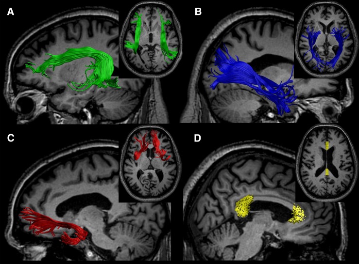
Tracts were reconstructed with a multiple region of interest (ROI) selection approach. Reconstruction was performed in each hemisphere and was based on a standardized atlas of white matter tracts (23) (Fig. 1). Previously defined anatomical landmarks for ROI slice selection and placement were used to reduce subjectivity in fiber tracking. For reconstruction of the SLF, three “AND” ROIs were placed: two on a coronal slice in the fronto-parietal lobe and one on a sagittal slice just after the curvature to the temporal lobe. Only those fiber trajectories that penetrated all “AND” ROIs were selected. For reconstruction of the UF, one “AND” ROI was placed on a coronal slice in the frontal lobe and one on an axial slice after the curvature to the temporal lobe. The ILF was reconstructed by placement of two “AND” regions on a coronal slice: one in the temporal and one in the occipital lobe.
Figure 1.
White matter tracts were reconstructed from each hemisphere based on a standardized atlas (23), the SLF (A), the ILF (B), the UF (C), and the genu and splenium of the medial segment of the CC (D).
The CC was reconstructed as previously described (24). In summary, only the midsagittal segment of the CC was selected to exclude regions of crossing fibers from the more laterally projecting pathways of the CC. Subsequently, the genu and splenium of the CC were automatically segmented according to the division described by Hofer and Frahm (25).
Diffusion parameters that were used to quantify microstructural white matter abnormalities (fractional anisotropy [FA], mean diffusivity [MD], radial diffusivity, and axial diffusivity) were obtained for each tract. We additionally calculated the mean planar diffusion coefficient of each tract. The planar diffusion coefficient ranges from zero to one and is relatively high in voxels with a higher degree of fiber complexity such as in crossing fiber regions (26).
Classical markers of small vessel disease
Quantitative assessment of WMH was performed on the fluid-attenuated inversion recovery images using the age-related white matter changes scale (27) by two raters (Y.D.R. and M.B.) who were blinded for clinical data and group allocation. Five different regions were rated in the right and left hemispheres separately. In addition, cerebral large vessel infarcts and lacunar infarcts were identified. In case of disagreement, consensus was obtained in a consensus meeting.
Statistical analyses
The cognitive domain scores and diffusion parameters were all normally distributed. Demographic variables, cognitive performance, diffusion parameters, and classical markers of small vessel disease (WMH and lacunar infarcts) were compared between participants with and without type 2 diabetes with an independent-samples t test for continuous variables, a Mann-Whitney U test for nonparametric data, and a χ2 test for proportions.
DTI parameters from tracts that showed significant between-group differences were selected to evaluate the relation between these parameters and cognitive performance with linear regression analyses. To test whether the relation between DTI parameters and cognitive performance in the diabetes group was different from the control group, we assessed group × DTI interactions. In case of a significant interaction effect, we stratified the analyses for group. Because the between-group differences in DTI parameters were similar for tracts in the left and right hemisphere, we averaged diffusion measures from both hemispheres to obtain one value per tract. For significant group × DTI parameter interactions, post hoc analyses were performed on the left and right hemisphere separately. All linear regression analyses were adjusted for age, sex, and estimated IQ. Because crossing fibers have been shown to confound the relation between diffusion measures and cognition (22), we also adjusted for the degree of crossing fibers by entering the planar diffusion coefficient as a covariate in the model (28).
To examine whether the relation between DTI parameters and cognition was mediated by classical markers of small vessel disease, we adjusted significant group × DTI parameter interactions on cognition for the presence of cerebral infarcts and total WMH load. To exclude the possibility that the relation between diffusion parameters and memory performance is affected by tract volume, we ran a separate model with tract volume as covariate. Finally, we explored the relation between DTI parameters, cognition and vascular risk factors (hypertension, BMI, and hypercholesterolemia), and diabetes-related factors (fasting glucose and HbA1c levels) with linear regression analyses adjusted for age, sex, and group.
RESULTS
Between-group differences
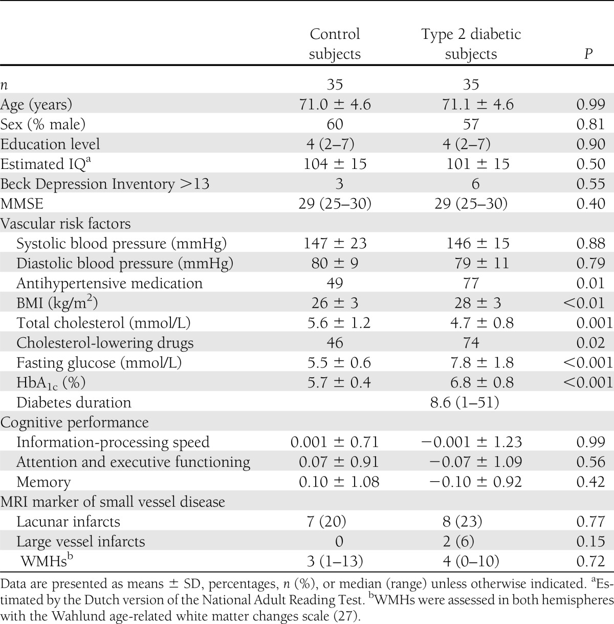
Group characteristics are shown in Table 1. Groups did not differ in age, sex, or estimated IQ. Patients with type 2 diabetes performed slightly worse on the cognitive domains memory and attention and executive functioning compared with control subjects, but differences were nonsignificant (Supplementary Table 1). Raw test scores and corresponding z scores of each individual test are presented in Table 3 (Supplementary Data). There were also no significant differences in the presence of large vessel infarcts, lacunar infarcts, or WMH load. In two patients with diabetes, a large vessel infarct was observed on MRI. White matter tracts traversing through the affected regions were excluded from the analyses (in one patient the left UF and in the other patient the right ILF and right UF). The other tracts from these patients were included in the analyses.
Table 1.
Group characteristics
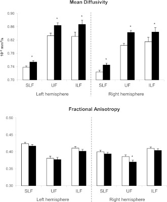
Significant between-group differences in MD values were observed in the SLF, UF, and ILF in both the left and right hemisphere and in the splenium of the CC demonstrating microstructural white matter abnormalities in patients compared with control subjects (Fig. 2). A between-group difference in FA was found in the right UF (P = 0.046). The between-group differences in MD were driven by increased diffusivity along both the axial direction (parallel to the tract) and radial direction (perpendicular to the tract) for the left and right SLF, left and right UF, left ILF (all P < 0.05), and right ILF (trend P = 0.09; data not shown). Between-group difference in the splenium of the CC was explained by increased axial diffusivity (P = 0.02). Important to note is that the tract volume did not differ between the diabetes and control groups (P > 0.05).
Figure 2.
Differences in MD and FA between the control group (□) and the diabetes group (■) ±SEM. High MD values and low FA values indicate reduced white matter tract integrity. *P < 0.05.
Association between DTI measures and cognitive performance
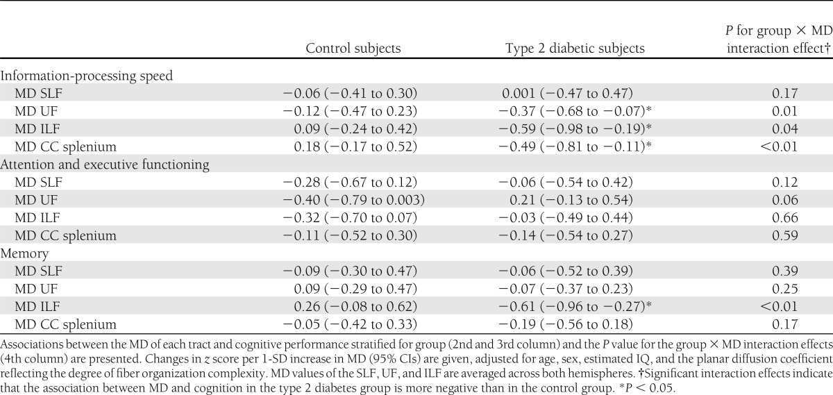
The relation between white matter abnormalities and cognitive performance was evaluated for those DTI measures that showed a significant group difference between patients and control subjects. We found significant group × MD interaction effects, showing a negative association in the diabetes group between MD of the UF, ILF, and splenium of the CC and information-processing speed (standardized β [95% CI] −0.37 to −0.59) (Table 2) and between MD of the ILF and memory (−0.61 [−0.96 to −0.27]), while in the control group no significant relation between MD and cognition was observed. The group × FA interaction effect for the UF was also significant on the domain information-processing speed, indicating a stronger positive association between FA and cognitive performance in the diabetes group (0.37 [3.28–25.01], P = 0.012) than in the control group (0.02 [−7.15 to 7.92], P = 0.918). We did not observe a significant interaction effect for the SLF on any of the three cognitive domains.
Table 2.
Association between MD and cognitive functioning stratified for group and group × MD interaction effects
Post hoc analyses on the significant group × MD interaction effects showed similar interaction coefficients for the left and right hemisphere: group × MD UF and information-processing speed (left, −3.36; right, −3.40), group × MD ILF and information-processing speed (left, −2.68; right, −2.92), and group × MD ILF and memory (left, −3.50; right, −4.30).
Adjustment for total WMH load and presence of cerebral infarcts did not modulate the significant associations (data not shown). Also, adjustment for tract volume did not change the results.
Relation with vascular risk factors
The group difference in MD between patients and control subjects was not modulated after adjustment for hypertension, BMI, or hypercholesterolemia (data not shown). Across the groups, hypertension was significantly associated with worse memory performance (β −0.24 [−1.19 to −0.04]) after adjustment of age, sex, estimated IQ, and group. No relation between BMI, hypercholesterolemia, and cognitive performance was found. Multivariate models showed that the relation between MD and memory within the diabetes group remained significant after adjustment for hypertension (data not shown).
With respect to diabetes-related measures, elevated HbA1c was associated with worse executive functioning within the T2DM group (−0.42 [−0.89 to −0.15], P = 0.01). A trend was observed between HbA1c levels and memory (−0.28 [−0.68 to 0.002], P = 0.05) and higher MD in the ILF (0.28 [0.00–0.56], P = 0.05). No relation between the duration of diabetes and cognition or DTI parameters was observed.
CONCLUSIONS
The current study demonstrated microstructural abnormalities in several major white matter tracts in older nondemented patients with type 2 diabetes compared with control subjects. These microstructural abnormalities were related to worse cognitive performance independent of classical MRI markers of small vessel disease (WMH and lacunar infarcts). Interestingly, we observed dissociations between abnormalities in specific white matter tracts and different aspects of cognitive functioning.
Previous studies have reported modest cognitive decrements in patients with type 2 diabetes on tests measuring information-processing speed, attention and executive functioning, and memory with effect sizes between −0.2 and −0.8 in the majority of studies (29). The effect sizes observed in this study are on the lower end of this spectrum (maximum −0.2). One factor that may have contributed to attenuation of the effect sizes is the fact that vascular and metabolic risk factors were relatively well controlled in the diabetes group (Table 1). This reflects current clinical practice guidelines, which are more stringent on cardiovascular control for primary and secondary prevention in type 2 diabetes (30), and is similar to other reports from Dutch population-based cohorts (9,31). The distribution of vascular risk factors in the control group was in line with previous observations in Dutch populations of similar age (32).
Despite the small differences in cognitive performance, we observed consistent group differences in MD values in the majority of tracts in both hemispheres, indicating microstructural white matter abnormalities in individuals with type 2 diabetes. These results are in line with recent reports from a DTI study on type 2 diabetes (33). By contrast, changes in FA were less pronounced. This can be explained by the fact that diffusion was increased parallel (axial) and perpendicular (radial) to the white matter fibers, which has a larger impact on the total MD than on the ratio between the axial and radial diffusion (FA). Based on the cognitive profile and results from previous neuroimaging studies, we expected that white matter tracts in patients with type 2 diabetes would be affected in frontal, temporal, and parietal regions. Indeed, white matter abnormalities were found in the UF, SLF, and ILF. In addition, we observed group differences in the splenium of the CC, indicating that the microstructural white matter abnormalities may also extend to more posterior areas.
Importantly, increased MD was associated with worse cognitive performance in the diabetes group after adjustment for age, sex, and estimated IQ but not in the control group, suggesting that microstructural white matter alterations underlie the cognitive decrements in older individuals with type 2 diabetes. Widespread deterioration of the brain network has previously been shown to affect age-related reductions in information-processing speed (34,35). We now demonstrate that in patients with diabetes, disruption of white matter tracts connecting frontal, parietal, and temporal regions is related to slowing of information-processing speed independent of age. The observed relationships between different aspects of cognition and white matter integrity are in line with current theories about the localization of higher cerebral function. Verbal memory performance was specifically related to microstructural abnormalities in the ILF, a large white matter tract crossing through the temporal lobe. In contrast, information-processing speed, an aspect of cognitive functioning commonly regarded as not specifically located in a particular brain region, was found to be related to more widespread changes in white matter integrity. These region-specific structure-function relationships support the hypothesis that disruption of white matter connections plays an important role in the pathogenesis of diabetes-related cognitive deficits.
Microstructural white matter abnormalities have also been observed in younger adults with type 1 diabetes (average age 45 years) (36) and in adolescents with type 2 diabetes (37). Our observations extend these previous findings by evaluation of the microstructural integrity of specific white matter tracts using fiber tractography in older participants in relation to individual cognitive domains. The finding that diabetes-associated microstructural white matter abnormalities can already be observed at a much younger age suggests that they should not be regarded as an early stage of a dementia process. Instead, this (vascular) white matter pathology makes the brain more vulnerable to the effects of Alzheimer pathology and other dementia-related processes later in life. In our view, the combined burden of these subtle microstructural white matter abnormalities, with other pathological changes that are common at an older age, such as global atrophy, WMH, and lacunar infarcts (9), impacts on the reserve capacity of the brain and is thus likely to contribute to the increased dementia risk in type 2 diabetes.
The association between DTI measures, type 2 diabetes, and cognition was independent of classical markers of small vessel disease (WMH and lacunar infarcts) indicating that DTI is a more sensitive marker for the subtle diabetes-related white matter abnormalities. White matter alterations in patients with diabetes may involve microvascular lesions such as microbleeds and microinfarcts (10,11) or endothelial dysfunction (38,39), which may in turn affect cerebral blood flow resulting in hypoperfusion and impaired brain function (38). Nevertheless, the exact pathological basis of these DTI changes in patients with diabetes remains to be established.
Strengths of our study are the detailed analyses of both high-resolution brain-imaging scans and cognitive functioning in a well-defined population-based cohort. This allowed us to accurately assess the relation between these parameters. We used fiber tractography to assess the white matter microstructure in patients with type 2 diabetes. This method is preferred for examining microstructural correlates of cognitive functions such as information-processing speed, attention, and memory because those functions depend on the transfer and integration of information between brain regions via those fiber tracts. The advantage over automated voxel-based analyses is that it is not sensitive to imperfect registration and smoothing errors (40). Moreover, averaging across voxels from one tract reduces the variance in diffusion measures and thereby increases the power to detect more subtle changes in white matter structure. Our study has some limitations. First, selection bias may have led to a relatively healthy study sample and thus an underestimation of the effect. The more intensive cardiovascular treatment regimen in the diabetes group relative to the control subjects reflects the current clinical practice guidelines (30). This does make our results generalizable to the population of well-controlled patients but is likely to underestimate the effect in less controlled patient populations. Second, although the cognitive tests used in our study have previously been shown to be sensitive to diabetes-related changes in cognitive performance in middle-aged individuals (2), our test selection was not exhaustive and inclusion of other tests would possibly have increased the sensitivity to detect modest cognitive decrements in patients with type 2 diabetes. Finally, to limit the number of comparisons we focused on a selection of white matter tracts, but future studies could demonstrate whether these findings extend to other fiber pathways or specific segments thereof.
This study demonstrated microstructural abnormalities in specific white matter tracts in nondemented older individuals with type 2 diabetes. Microstructural changes, assessed with DTI, are a potential marker of early white matter abnormalities in type 2 diabetes and may be more sensitive than classical MRI markers of small vessel disease. Furthermore, disruption of white matter connections is a potential correlate of worse cognitive functioning in patients with diabetes.
Acknowledgments
The research of G.J.B. is supported by Vidi Grant 91711384 from ZonMw, the Netherlands Organisation for Health Research and Development, and Hersenstichting Nederland Grant 2011(1)-87. The research of A.L. is supported by the project Care4Me (Cooperative Advanced REsearch for Medical Efficiency) in the framework of the EU research program Information Technology for European Advancement.
No potential conflicts of interest relevant to this article were reported.
Y.D.R. acquired data, wrote the manuscript, and performed the analysis. M.B. acquired data and made critical revisions to the manuscript. J.d.B., L.J.K., A.L., and G.J.B. made substantial contributions to the conception and design of the study and critically revised the manuscript for important intellectual content. Y.D.R. is the guarantor of this work and, as such, had full access to all the data in the study and takes responsibility for the integrity of the data and the accuracy of the data analysis.
The authors express their special thanks to the primary care practices Ametisthof, Glennhof, De Poort, 't Steyn, and De Weegbree of Huisartsenzorg IJsselstein, IJsselstein, the Netherlands (mentor Ph.L. Salomé), and G. Visser, Nieuwegein, the Netherlands, for their supportive role in the recruitment process. The authors also thank C. Buvens and A.W. Kingma, Utrecht University, for assisting with the data collection and S.B. Vos, the Images Sciences Institute, University Medical Center Utrecht, the Netherlands, for his assistance with data processing.
Appendix
Members of the Utrecht Vascular Cognitive Impairment Study Group involved in the Utrecht Diabetic Encephalopathy Study are as follows (in alphabetical order by department): the Department of Neurology, E. van den Berg, G.J. Biessels, M. Brundel, W. Bouvy, S. Heringa, and L.J. Kappelle; the Department of Radiology/Image Sciences Institute, J. de Bresser, H.J. Kuijf, A. Leemans, P.R. Luijten, W.P.Th.M. Mali, M.A. Viergever, K.L. Vincken, and J. Zwanenburg; and Julius Center for Health Sciences and Primary Care, A. Algra and G.E.H.M. Rutten.
Footnotes
This article contains Supplementary Data online at http://care.diabetesjournals.org/lookup/suppl/doi:10.2337/dc12-0493/-/DC1.
A slide set summarizing this article is available online.
A complete list of members of the Utrecht Vascular Cognitive Impairment Study Group involved in the Utrecht Diabetic Encephalopathy Study can be found in the appendix.
References
- 1.Biessels GJ, Staekenborg S, Brunner E, Brayne C, Scheltens P. Risk of dementia in diabetes mellitus: a systematic review. Lancet Neurol 2006;5:64–74 [DOI] [PubMed] [Google Scholar]
- 2.Reijmer YD, van den Berg E, Ruis C, Kappelle LJ, Biessels GJ. Cognitive dysfunction in patients with type 2 diabetes. Diabetes Metab Res Rev 2010;26:507–519 [DOI] [PubMed] [Google Scholar]
- 3.Ryan CM, Geckle MO. Circumscribed cognitive dysfunction in middle-aged adults with type 2 diabetes. Diabetes Care 2000;23:1486–1493 [DOI] [PubMed] [Google Scholar]
- 4.Brands AMA, Van den Berg E, Manschot SM, et al. A detailed profile of cognitive dysfunction and its relation to psychological distress in patients with type 2 diabetes mellitus. J Int Neuropsychol Soc 2007;13:288–297 [DOI] [PubMed] [Google Scholar]
- 5.Korf ES, van Straaten EC, de Leeuw FE, et al. LADIS Study Group Diabetes mellitus, hypertension and medial temporal lobe atrophy: the LADIS study. Diabet Med 2007;24:166–171 [DOI] [PubMed] [Google Scholar]
- 6.van Harten B, Oosterman JM, Potter van Loon BJ, Scheltens P, Weinstein HC. Brain lesions on MRI in elderly patients with type 2 diabetes mellitus. Eur Neurol 2007;57:70–74 [DOI] [PubMed] [Google Scholar]
- 7.Schmidt R, Launer LJ, Nilsson LG, et al. CASCADE Consortium Magnetic resonance imaging of the brain in diabetes: the Cardiovascular Determinants of Dementia (CASCADE) Study. Diabetes 2004;53:687–692 [DOI] [PubMed] [Google Scholar]
- 8.Longstreth WT, Jr, Manolio TA, Arnold A, et al. Clinical correlates of white matter findings on cranial magnetic resonance imaging of 3301 elderly people. The Cardiovascular Health Study. Stroke 1996;27:1274–1282 [DOI] [PubMed] [Google Scholar]
- 9.Manschot SM, Brands AM, van der Grond J, et al. Utrecht Diabetic Encephalopathy Study Group Brain magnetic resonance imaging correlates of impaired cognition in patients with type 2 diabetes. Diabetes 2006;55:1106–1113 [DOI] [PubMed] [Google Scholar]
- 10.Arvanitakis Z, Schneider JA, Wilson RS, et al. Diabetes is related to cerebral infarction but not to AD pathology in older persons. Neurology 2006;67:1960–1965 [DOI] [PubMed] [Google Scholar]
- 11.Nelson PT, Smith CD, Abner EA, et al. Human cerebral neuropathology of type 2 diabetes mellitus. Biochim Biophys Acta 2009;1792:454–469 [DOI] [PMC free article] [PubMed] [Google Scholar]
- 12.Basser PJ, Mattiello J, LeBihan D. MR diffusion tensor spectroscopy and imaging. Biophys J 1994;66:259–267 [DOI] [PMC free article] [PubMed] [Google Scholar]
- 13.Beaulieu C. The basis of anisotropic water diffusion in the nervous system - a technical review. NMR Biomed 2002;15:435–455 [DOI] [PubMed] [Google Scholar]
- 14.Jones DK. Studying connections in the living human brain with diffusion MRI. Cortex 2008;44:936–952 [DOI] [PubMed] [Google Scholar]
- 15.O’Sullivan M, Summers PE, Jones DK, Jarosz JM, Williams SC, Markus HS. Normal-appearing white matter in ischemic leukoaraiosis: a diffusion tensor MRI study. Neurology 2001;57:2307–2310 [DOI] [PubMed] [Google Scholar]
- 16.van Norden AGW, de Laat KF, van Dijk EJ, et al. Diffusion tensor imaging and cognition in cerebral small vessel disease. The RUN DMC study. Biochim Biophys Acta 2012;1822:401–407 [DOI] [PubMed] [Google Scholar]
- 17.Gorelick PB, Scuteri A, Black SE, et al. American Heart Association Stroke Council, Council on Epidemiology and Prevention, Council on Cardiovascular Nursing, Council on Cardiovascular Radiology and Intervention, and Council on Cardiovascular Surgery and Anesthesia Vascular contributions to cognitive impairment and dementia: a statement for healthcare professionals from the american heart association/american stroke association. Stroke 2011;42:2672–2713 [DOI] [PMC free article] [PubMed] [Google Scholar]
- 18.Leemans A, Jones DK. The B-matrix must be rotated when correcting for subject motion in DTI data. Magn Reson Med 2009;61:1336–1349 [DOI] [PubMed] [Google Scholar]
- 19.Leemans A, Jeurissen B, Sijbers J, Jones DK. ExploreDTI: a graphical toolbox for processing, analyzing, and visualizing diffusion MR data. In International Society for Magnetic Resonance in Medicine: 17th Scientific Meeting . Honolulu, HI, 2009. , p. 3537 [Google Scholar]
- 20.Voineskos AN, Rajji TK, Lobaugh NJ, et al. Age-related decline in white matter tract integrity and cognitive performance: a DTI tractography and structural equation modeling study. Neurobiol Aging 2012;33:21–34 [DOI] [PMC free article] [PubMed] [Google Scholar]
- 21.Tournier JD, Calamante F, Connelly A. Robust determination of the fibre orientation distribution in diffusion MRI: non-negativity constrained super-resolved spherical deconvolution. Neuroimage 2007;35:1459–1472 [DOI] [PubMed] [Google Scholar]
- 22.Vos SB, Jones DK, Jeurissen B, Viergever MA, Leemans A. The influence of complex white matter architecture on the mean diffusivity in diffusion tensor MRI of the human brain. Neuroimage 2012;59:2208–2216 [DOI] [PMC free article] [PubMed] [Google Scholar]
- 23.Catani M, Thiebaut de Schotten M. A diffusion tensor imaging tractography atlas for virtual in vivo dissections. Cortex 2008;44:1105–1132 [DOI] [PubMed] [Google Scholar]
- 24.Caeyenberghs K, Leemans A, Coxon J, et al. Bimanual coordination and corpus callosum microstructure in young adults with traumatic brain injury: a diffusion tensor imaging study. J Neurotrauma 2011;28:897–913 [DOI] [PubMed] [Google Scholar]
- 25.Hofer S, Frahm J. Topography of the human corpus callosum revisited—comprehensive fiber tractography using diffusion tensor magnetic resonance imaging. Neuroimage 2006;32:989–994 [DOI] [PubMed] [Google Scholar]
- 26.Ennis DB, Kindlmann G. Orthogonal tensor invariants and the analysis of diffusion tensor magnetic resonance images. Magn Reson Med 2006;55:136–146 [DOI] [PubMed] [Google Scholar]
- 27.Wahlund LO, Barkhof F, Fazekas F, et al. European Task Force on Age-Related White Matter Changes A new rating scale for age-related white matter changes applicable to MRI and CT. Stroke 2001;32:1318–1322 [DOI] [PubMed] [Google Scholar]
- 28.Reijmer YD, Leemans A, Heringa SM, et al. Improved sensitivity to cerebral white matter abnormalities in Alzheimer's disease with spherical deconvolution based tractography. PLoS One. In press [DOI] [PMC free article] [PubMed] [Google Scholar]
- 29.van den Berg E, Kloppenborg RP, Kessels RP, Kappelle LJ, Biessels GJ. Type 2 diabetes mellitus, hypertension, dyslipidemia and obesity: a systematic comparison of their impact on cognition. Biochim Biophys Acta 2009;1792:470–481 [DOI] [PubMed]
- 30.Buse JB, Ginsberg HN, Bakris GL, et al. American Heart Association. American Diabetes Association Primary prevention of cardiovascular diseases in people with diabetes mellitus: a scientific statement from the American Heart Association and the American Diabetes Association. Circulation 2007;115:114–126 [DOI] [PubMed] [Google Scholar]
- 31.van ’t Riet E, Dekker JM, Sun Q, Nijpels G, Hu FB, van Dam RM. Role of adiposity and lifestyle in the relationship between family history of diabetes and 20-year incidence of type 2 diabetes in U.S. women. Diabetes Care 2010;33:763–767 [DOI] [PMC free article] [PubMed] [Google Scholar]
- 32.Newson RS, Hek K, Luijendijk HJ, Hofman A, Witteman JCM, Tiemeier H. Atherosclerosis and incident depression in late life. Arch Gen Psychiatry 2010;67:1144–1151 [DOI] [PubMed] [Google Scholar]
- 33.Hsu JL, Chen YL, Leu JG, et al. Microstructural white matter abnormalities in type 2 diabetes mellitus: a diffusion tensor imaging study. Neuroimage 2011;59:1098–1105 [DOI] [PubMed] [Google Scholar]
- 34.Sullivan EV, Pfefferbaum A. Diffusion tensor imaging and aging. Neurosci Biobehav Rev 2006;30:749–761 [DOI] [PubMed] [Google Scholar]
- 35.Kennedy KM, Raz N. Aging white matter and cognition: differential effects of regional variations in diffusion properties on memory, executive functions, and speed. Neuropsychologia 2009;47:916–927 [DOI] [PMC free article] [PubMed] [Google Scholar]
- 36.Kodl CT, Seaquist ER. Cognitive dysfunction and diabetes mellitus. Endocr Rev 2008;29:494–511 [DOI] [PMC free article] [PubMed] [Google Scholar]
- 37.Yau PL, Javier DC, Ryan CM, et al. Preliminary evidence for brain complications in obese adolescents with type 2 diabetes mellitus. Diabetologia 2010;53:2298–2306 [DOI] [PMC free article] [PubMed] [Google Scholar]
- 38.Cosentino F, Battista R, Scuteri A, et al. Impact of fasting glycemia and regional cerebral perfusion in diabetic subjects: a study with technetium-99m-ethyl cysteinate dimer single photon emission computed tomography. Stroke 2009;40:306–308 [DOI] [PubMed] [Google Scholar]
- 39.Yang Y, Rosenberg GA. Blood-brain barrier breakdown in acute and chronic cerebrovascular disease. Stroke 2011;42:3323–3328 [DOI] [PMC free article] [PubMed] [Google Scholar]
- 40.Van Hecke W, Leemans A, De Backer S, Jeurissen B, Parizel PM, Sijbers J. Comparing isotropic and anisotropic smoothing for voxel-based DTI analyses: a simulation study. Hum Brain Mapp 2010;31:98–114 [DOI] [PMC free article] [PubMed] [Google Scholar]


