Abstract
Background
Multiple myeloma is characterized by the accumulation of tumor plasma cells in the bone marrow. Despite therapeutic improvements brought by proteasome inhibitors such as bortezomib, myeloma remains an incurable disease. In a variety of human cancers, human immunodeficiency virus protease inhibitors (e.g. nelfinavir) effectively inhibit tumor progression, but their impact on myeloma is unknown. We assessed the in vitro and in vivo effects of nelfinavir on multiple myeloma.
Design and Methods
The effects of nelfinavir (1–10 μM) on proteasome activity, proliferation and viability of myeloma cell lines and plasma cells from patients were assessed by measuring PERK, AKT, STAT3 and ERK1/2 phosphorylation and CHOP expression with immunoblotting or flow cytometry. The in vivo effect was assessed in NOD/SCID mice injected with luciferase expressing human myeloma cell lines and treated with nelfinavir at a dose of 75 mg/kg/day. Tumor progression was evaluated using a bioluminescent system.
Results
Nelfinavir inhibited 26S chymotrypsin-like proteasome activity, impaired proliferation and triggered apoptosis of the myeloma cell lines and fresh plasma cells. It activated the pro-apoptotic unfolded protein response pathway by inducing PERK phosphorylation and CHOP expression. Cell death triggered by nelfinavir treatment correlated with decreased phosphorylation of AKT, STAT3 and ERK1/2. Nelfinavir enhanced the anti-proliferative activity of bortezomib, dexamethasone and histone deacetylase inhibitors and delayed tumor growth in a myeloma mouse model.
Conclusions
These results suggest that nelfinavir, used at a pharmacological dosage, alone or in combination, may be useful in the treatment of myeloma. Our data provide a preclinical basis for clinical trials using nelfinavir in patients with myeloma.
Keywords: HIV protease inhibitor, nelfinavir, proteasome, multiple myeloma, apoptosis, PI3K/AKT
Introduction
Multiple myeloma (MM) is characterized by the proliferation of clonal plasma cells in the bone marrow and accounts for approximately 10% of all hematologic cancers.1 Despite recent therapeutic advances, including the use of “novel agents” such as immunomodulatory drugs and proteasome inhibitors, MM remains an incurable disease with a median survival of approximately 5 to 6 years.2–5
Bortezomib (Velcade®) is a selective inhibitor of the 26S proteasome that has led to improved survival of patients.6–8 The three enzymatic activities of the 26S proteasome –chymotrypsin (CT), trypsin (T) and caspase-like (C) – are located on the β5, β2 and β1 subunits, respectively.9 This complex is constitutively activated in plasma cells and is involved in multiple biological functions, including degradation of damaged proteins, regulation of the cell cycle, transformation and apoptosis.7,8 Thus, the 26S proteasome represents an interesting therapeutic target in cancer.10–12
Bortezomib-mediated proteasome inhibition entails accumulation of unfolded protein leading to induction of pro-apoptotic genes of the unfolded protein response (UPR). The UPR is a multi-branch system located in the endoplasmic reticulum (ER) which regulates the production and secretion of immunoglobulins, allowing either the survival or apoptosis of the cell during ER stress.2,13,14 Excessive accumulation of misfolded proteins leads to their preferential binding to BiP and its dissociation from the ER membrane, thereby rendering active the three pathways via three transmembrane protein (IRE-1α, ATF6 and PERK). During this ER stress, IRE1-α and ATF6 induction leads to XBP1 activation and synthesis of the molecular chaperone proteins including BiP/GRP78 and Hsp90. In contrast, the PERK pathway is thought to decrease protein synthesis via eIF2α phosphorylation as well as generate a pro-apoptotic signal mediated by the up-regulation of activating factor transcription 4 (ATF4) transcription factor.15,16 ATF4 induces the expression of C/EBP homologous protein (CHOP/GADD153) transcription factor, known to activate apoptosis in the case of overwhelming ER stress. Both the effect on proteasome and on the UPR system may explain how proteasome inhibition induces apoptosis in tumor plasma cells.17
In clinical practice, the use of bortezomib is limited by the occurrence of severe side effects (including peripheral neuropathy) and resistance.3 In this regard, a new generation of proteasome inhibitors is needed to further improve the outcome of patients with MM.15,16
Human immunodeficiency virus (HIV) protease inhibitors (PI) were developed in the early 1990s to treat chronically HIV-infected patients. In combination with nucleoside analogs as part of highly active antiretroviral therapy, they have led to a reduction in HIV-related morbidity and mortality.18,19 HIV PI are peptidomimetic analogs of the peptide bond between phenylalanine 167 and pro-line 168 of the gag-pol polyprotein, which is the target of the HIV aspartyl-protease. The cleavage sites of HIV-I pro-tease were initially thought to be unique and distinct from those of mammalian proteases.20 However, it has been suggested that the 26S proteasome is able to cleave the same sites and may, therefore, be a target of HIV PI.21 In addition, HIV PI have been successfully used for treating HIV-related Kaposi’s sarcoma indicating anti-tumor properties.22 Recent studies have shown that these drugs are able to inhibit cell proliferation and induce apoptosis in numerous cancer cells including malignant glioma, melanoma, prostate tumor and carcinomas.20,23,24 Various mechanisms of action, depending on the HIV PI and the cell type, have been hypothesized. For example, it has been suggested that the HIV PI saquinavir and ritonavir could induce apoptosis of fibroblast and prostate cancer cell lines through inhibition of proteasome activity.25,26
In this study, we investigated the effect of the HIV PI nelfinavir on 26S proteasome activity and on the proliferation and viability of MM plasma cells both in vitro and in vivo.
Design and Methods
Cell lines
U266, MM1S, RPMI, OPM2 and LP1 human myeloma cells lines and 293T were respectively grown in RPMI 1640, Iscove’s modified Dulbecco’s medium and GIBCO® Dulbecco’s modified Eagle medium (DMEM) (Gibco BRL, Invitrogen, Paris, France) supplemented with 2 mM L-glutamine (l-Glu), 100 U/mL penicillin, 100 g/mL streptomycin and maintained routinely in a humidified chamber at 37°C with 5% carbon dioxide.
Human multiple myeloma plasma cells
Bone marrow aspirates or peripheral blood samples were harvested and mononuclear cells were isolated by Ficoll-Hypaque centrifugation and washed twice in phosphate-buffered saline (PBS) containing 1% bovine serum albumin (BSA). CD138+ plasma cells were purified using the Direct CD138 Progenitor Isolation kit with immunomagnetic beads conjugated to monoclonal mouse anti-human CD138 antibody (Miltenyi Biotech, Paris, France). The purity of sorted cells analyzed by flow cytometry was up to 90%. All patients gave written informed consent.
Drug treatment
Nelfinavir (Viracept® Roche) and the synthetic PI MG132 (SIGMA Z-Leu-Leu-Leu-al) were solubilized in dimethyl sulfoxide (DMSO, Sigma). Bortezomib (Velcade®, Janssen) was solubilized in saline buffer (NaCl 9%). Fibroblast growth factor (rh-FGF acidic) and insulin growth factor 1 (rh-IGF-1) were obtained from R&D System. Interleukin-6 (rh-IL6) was from PeproTech. The histone deacetylase-like (HDAC) inhibitor, valproic acid, was provided by Sanofi Aventis and dexamethasone was obtained from Mylan.
26S Proteasome activity
MM cells were lysed in RIPA buffer (Santa Cruz sc.24948) supplemented with PI and sodium orthovanadate (100 mM). For each cell line, 30 μg of protein were collected in a Tris buffer [Tris 50 mM (pH 7.5), DTT (1 mM), MgCl2 (10 mM), ATP (2 mM)] and incubated with nelfinavir (5 μM), bortezomib (10 nM) or MG132 (1 μM) for 2 h at 37°C. Then 1 mM of the fluorogenic substrate was added and the enzymatic activities were measured by a FLUOstar OPTIMA (BMG Labtech) (λexc: 380nM and λem: 460nM). The fluorogenic substrates were Z-Leu-Leu-Val-Tyr-AMC (Calbiochem, San Diego, CA, USA) for the chymotrypsin-like activity and Bz-Val-Gly-Arg-AMC (BIOMOL) for the trypsin-like activity. All experiments were performed in triplicate.
Cell proliferation and viability assay
The proliferation of plasma cell lines was measured by tritiated thymidine uptake ([6-3H] thymidine). In brief, 3×10⁴ cells were plated in a 96-well plate and incubated for 48 h with the drugs. Then [6-3H] thymidine (Amersham Biosciences, UK) was added (1 μCi/well) for a further 16 h. The [6-3H] thymidine incorporation was analyzed using a liquid scintillation counter (Wallace, PerkinElmer).
The viability of the plasma cells from patients (5×10⁴ cells/well) was assessed using the WST-1 kit (Roche®) according to the manufacturer’s instructions. All experiments were performed in triplicate.
Flow cytometry
Apoptosis and AKT phosphorylation were analyzed by flow cytometry (Becton Dickinson FACS Calibur / BD Bioscience) as described in the Online Supplementary Design and Methods.
Western blot and antibodies
Details on the western blot techniques are described in the Online Supplementary Design and Methods. The antibodies for CHOP, ubiquitin, AKT, P-AKT(ser473) and caspase-3 were from Cell Signaling, for ATF4 (H-290), and actin from Santa Cruz. The rabbit polyclonal antibody for P-PERK was from Biolegend.
Generation of U226-luc cells
The U266-luc cells were obtained as described in the Online Supplementary Design and Methods.27
Human multiple myeloma xenograft model
Immunodeficient non-obese diabetic/severe combined immunodeficiency (NOD/SCID) mice (10–12 weeks old) were obtained from the Institut André Lwoff-Villejuif, France. The mice were maintained in a specific pathogen-free breeding facility and, in line with the recommendations of the Institut Universitaire d'Hématologie (IUH) Animal Care and User Committee, were sacrificed 25 days after inoculation of the U266-luc cells. The mice were handled and housed in compliance with guidelines from the IUH Animal Care and User Committee.
Fourteen NOD/SCID mice received whole-body irradiation with 2.5 Gy on the day of the subcutaneous injection of 8×10⁶ U266-luc cells in 0.2 mL in PBS into the flank. Treatments started 24 h after cell inoculation. The mice were separated into two groups of seven. One group was treated daily with an intraperitoneal infusion of nelfinavir (75 mg/kg) dissolved in a solution of PBS containing 50% PEG- 10% DMSO and Tween® 80% (Sigma) in a final volume of 300 μL, for 20 days. The control group received an intraperitoneal infusion of the same solution without nelfinavir (vehicle). On designated days post-inoculation, U266-luc cells were detected using the IVIS imaging system (Xenogen Corporation, Alameda, CA, USA) described in the Online Supplementary Design and Methods.
Experimental values were expressed as the mean ± SEM. In all experiments, statistical comparisons of mean values were done using the Student’s t-test. The study was approved by the local IUH Ethics Committee and written informed consent was obtained from each subject.
Results
Nelfinavir inhibits 26S proteasome activity
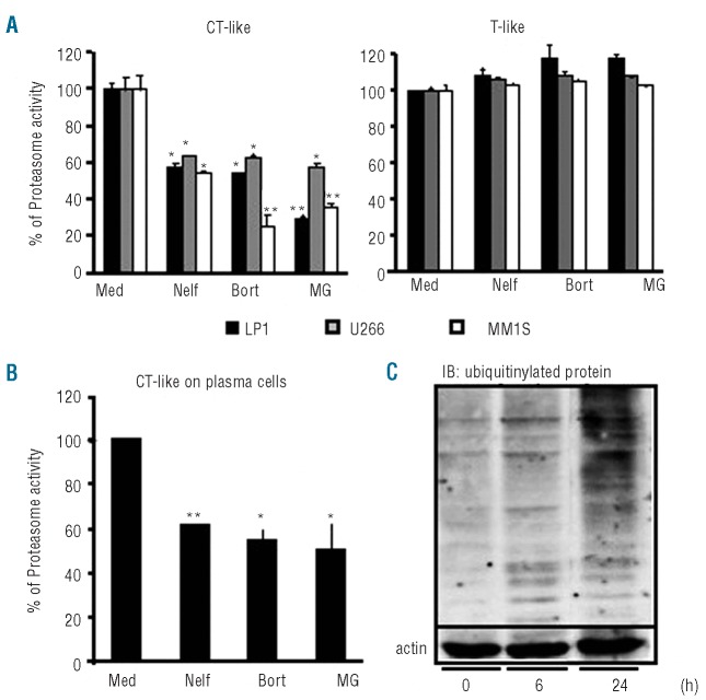
To investigate the effect of nelfinavir on the 26S proteasome in MM cells, we measured the enzymatic proteasomal activity in the presence or absence of nelfinavir, bortezomib or MG 132. As expected, bortezomib and MG132 readily inhibited CT-like activity in LP1, U266 and MM1S cells. Similarly, the 26S CT-like proteasome activity was reduced by 60% when using a pharmacological concentration of nelfinavir (5 μM) (Figure 1A). As previously described for bortezomib, this inhibitory effect of nelfinavir was not restricted to MM cells and was also observed on peripheral blood lymphocytes from healthy donors (data not shown). Neither nelfinavir nor bortezomib had an effect on T-like activity. Nelfinavir inhibited the CT-like activity of the 26S proteasome of CD138-selected tumor plasma cells from MM patients as well as bortezomib (Figure 1B). As shown in Figure 1C, nelfinavir induced a time-dependent accumulation of ubiquitinylated protein after 6 h of treatment in MM cell lines. Next, we investigated the activities of saquinavir and tipranavir and found that both PI reduced CT-like activity in MM cell lines (Online Supplementary Figure S1A). Thus, nelfinavir (like saquinavir and tipranavir) is an effective inhibitor of the 26S proteasome CT-like activity in MM cell lines and fresh plasma cells from patients.
Figure 1.
Nelfinavir inhibits 26S proteasome activity. Histograms show the chymotrypsin-like (CT-like) and trypsin-like (T-like) activities of the 26S proteasome (A) Cellular lysates of LP1, U266 or MM1S cell lines were incubated with 5 μM nelfinavir (Nelf), 10 nM bortezomib (Bort) or 1 μM MG132 (MG) or cultured in medium alone (Med). (B) Cellular lysates of plasma cells from patients were incubated with nelfinavir (Nelf) 10 μM, bortezomib (Bort) 10 nM and MG132 (MG) 1 μM or cultured in medium alone (Med). (C) U266 cells were incubated with nelfinavir (5 μM) for the indicated times and the level of the ubiquitinylated proteins was determined by western blot. Error bars correspond to the standard deviation. The P value was calculated with the Student’s t-test, (*P≤0.05, **P≤0.01).
Nelfinavir inhibits the proliferation of multiple myeloma cells in vitro even in the presence of pro-survival cytokines
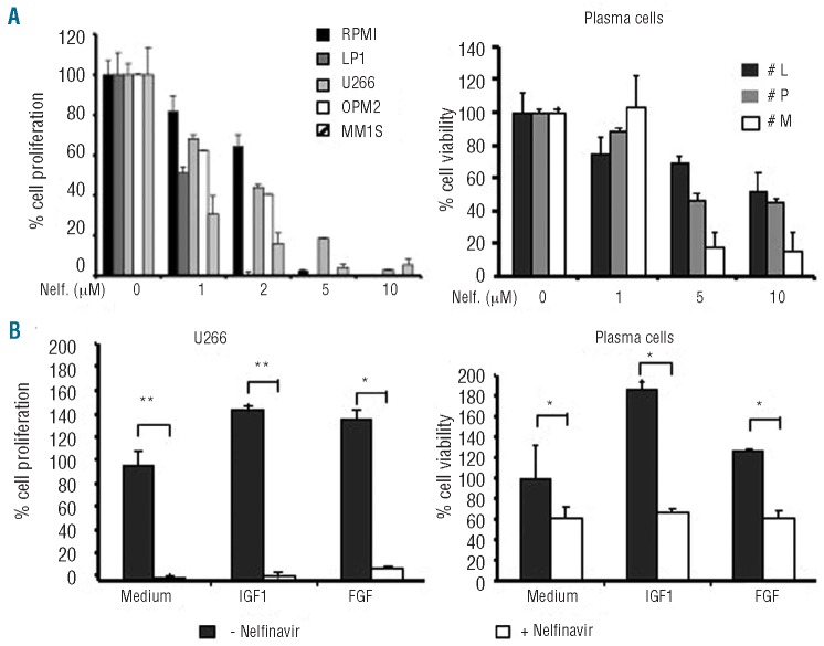
To investigate the impact of nelfinavir-induced proteasome inhibition on the proliferation of MM cells, RPMI, LP1, U266, OPM2 and MM1S cell lines were grown in the presence or absence of increasing concentration of nelfinavir. Nelfinavir inhibited the proliferation of RPMI, LP1, U266, OPM2 and MM1S cell lines in a dose-dependent manner with an IC50 of 1 – 5 μM. Similar results were observed with saquinavir and tipranavir (Online Supplementary Figure S1B). Interestingly, in U266 cells treated with nelfinavir, the kinetics of CT-like inhibition correlated with the inhibition of cell proliferation (Online Supplementary Figure S2). In addition, nelfinavir (1 to 5 μM) also decreased the viability of CD138+ plasma cells purified from three patients (Figure 2A).
Figure 2.
Nelfinavir inhibits proliferation of MM cell lines in vitro even in the presence of pro-survival cytokines. (A) (Right) Histograms show the proliferation of RPMI, LP1, U266, OPM2 and MM1S cell lines treated with nelfinavir (1 μM to 10 μM). A. (Left) Viability of plasma cells from three patients who received bortezomib (“P” and “M” were not responsive to bortezomib) the cells were cultured with the indicated concentrations of nelfinavir. (B) (Right) Histograms show the proliferation of U266 cells treated (filled) or not (open) with 5 μM nelfinavir in the presence of IGF-1 (100 ng/mL) or FGF (100 ng/mL) + heparin (100 μg/mL). (B) (Left) Histograms show the viability of the plasma cells from a patient treated (filled) or not (open) with 10 μM of nelfinavir in the presence of IGF-1 (100 ng/mL) or FGF (100 ng/mL) + heparin (100 μg/mL). Error bars correspond to the standard deviation. The P value was calculated with Student’s t-test, (*P≤0.05, **P≤0.01).
The effect of nelfinavir was then tested on the survival of U266 cells and plasma cells from an MM patient in the presence of the pro-survival cytokine known to be secreted in vivo in the plasma cell microenvironment. U266 cells treated with or without nelfinavir were grown in the presence of IGF-1 or FGF. As illustrated in Figure 2B, while FGF and IGF-1 markedly increased cell proliferation, neither of these cytokines could counteract the inhibitory effect of nelfinavir. The survival of CD138+ purified plasma cells from an MM patient was doubled in the presence of IGF-1 whereas the inhibitory effect of nelfinavir (5 μM) was not counteracted by the addition of either of these cytokines. Similar results were obtained with IL-6-treated MM cells (Online Supplementary Figure S3). These data show that nelfinavir inhibits the proliferation not only of MM cell lines, but also of plasma cells from patients. Moreover, this activity is not reversed by pro-survival signals delivered by several soluble factors such as IGF-1 and FGF.
Nelfinavir enhances the anti-proliferative activity of anti-myeloma agents
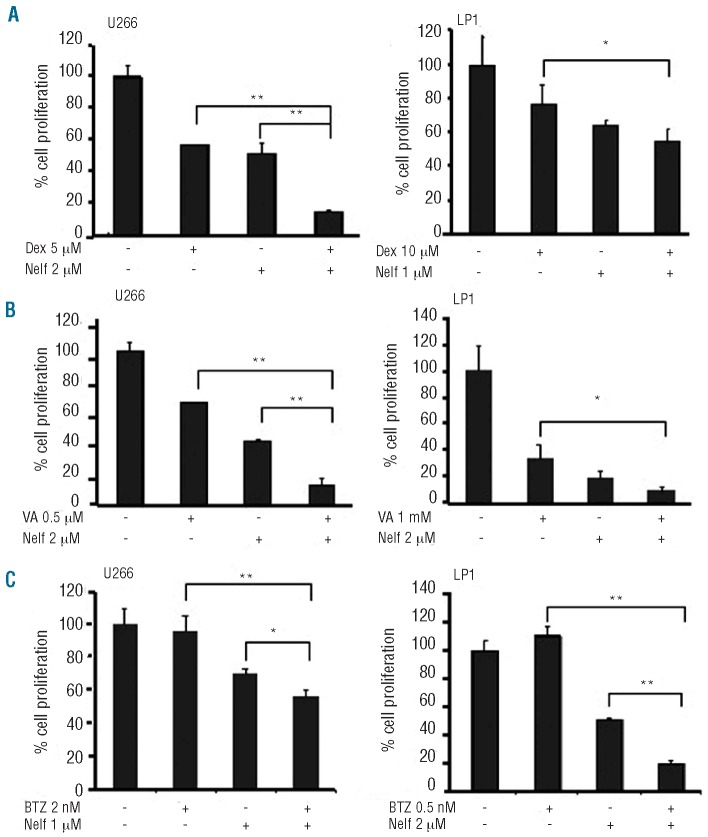
Next we investigated whether nelfinavir could enhance anti-proliferative activities on MM cells in combination with other anti-myeloma drugs such as the HDAC inhibitor valproic acid or dexamethasone. In this experiment, the drugs were used at their optimal concentrations in order to unmask potential cooperative effects. Used separately, dexamethasone (5 μM) and nelfinavir (2 μM) each induced a 40% inhibition of U266 cell proliferation. Combined in the culture medium, they induced an 80% reduction in the proliferation of U266 (Figure 3A). The cooperative effect was also observed on the LP1 cell proliferation using dexamethasone (10 μM) and nelfinavir (1 μM). Similarly, an enhanced effect was found between valproic acid (0.5 mM) and nelfinavir (2 μM) with a 90% inhibition of U266 cell proliferation when these drugs were used together (Figure 3B) as compared to 50 to 60% when used separately. This combination also inhibited the proliferation of LP1 cells although, in this cell line, the cooperative effect of nelfinavir was achieved with valproic acid, 1 mM. Moreover, the two PI, bortezomib and nelfinavir, had an enhanced anti-proliferative effect on the U266 and LP1 cell lines (Figure 3C). These data demonstrate cooperative cytotoxic effects between nefinavir and dexamethasone, bortezomib and valproic acid on MM cell lines.
Figure 3.
Nelfinavir cooperates with anti-myeloma agents to inhibit the proliferation of multiple myeloma cells. Bar graphs show the proliferation of U266 and LP1 cells cultured in the presence of nelfinavir (nelf) and (A) dexamethasone (Dex), (B) valproic acid (VA), and (C) bortezomib (BTZ). The concentrations at which each drug was used are indicated. Error bars correspond to the standard deviation. The P value was calculated with Student’s t-test, (*P≤0.05, **P≤0.01).
Nelfinavir induces apoptosis of multiple myeloma cell lines and activates the cleavage of caspase-3
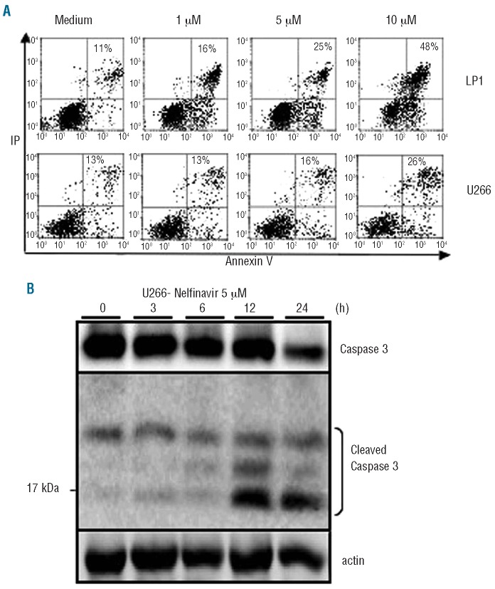
To obtain further insights into the mechanism of the nelfinavir-induced anti-proliferative effect, we investigated the impact of HIV PI on the induction of apoptosis of LP1 and U266 cells (Figure 4A). After 17 h of incubation with increasing concentrations of nelfinavir (1–10 μM), the percentage of annexin V+/propidium iodide+ apoptotic cells was analyzed by flow cytometry. Nelfinavir induced a dose-dependent increase in the percentage of annexin V+/propidium iodide+ cells. The effect of nelfinavir on the activation of caspase-3 was then studied. Nelfinavir induced pro-caspase-3 cleavage in a time-dependent manner (Figure 4B) in the U266 cell line. These results suggest that nelfinavir inhibits proliferation of MM cells lines by inducing apoptosis.
Figure 4.
Nelfinavir induces the cleavage of pro-caspase 3 and apoptosis in MM cell lines. (A) Dot plots show apoptosis of LP1 and U266 cells cultured with nelfinavir at the indicated concentrations. The percentages of dead cells, measured by annexin V and propidium iodide staining, are indicated in each quadrant. (B). Western blot analysis of the kinetics of pro-caspase-3 cleavage in U266 cells cultured with nelfinavir (5 μM). Cleaved and native forms of the caspase-3 are indicated as is the actin control.
Nelfinavir decreases the phosphorylation of AKT, STAT-3, ERK1/2 and activates the pro-apoptotic pathway of the unfolded protein response system
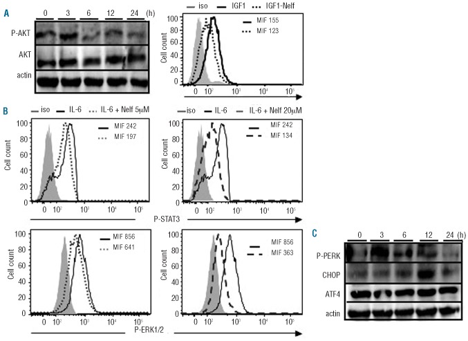
Knowing the importance of the PI3K/AKT pathway for cell survival, we studied the impact of nelfinavir on AKT phosphorylation. Western blot analysis revealed that the level of AKT phosphorylation in U266 cells decreased 6 h after nelfinavir (5 μM) treatment (Figure 5A. left panel). In addition, intracellular FACS analysis revealed that, at this concentration, nelfinavir also reduced the mean intensity of fluorescence (MIF) corresponding to IGF1-mediated AKT phosphorylation (Figure 5A, right panel).
Figure 5.
Nelfinavir decreases the phosphorylation of AKT, STAT3, ERK, and activates the pro-apoptotic pathway of the UPR system. (A) Right. U266 cells were starved for 4 h and incubated at the indicated times with 5 μM nelfinavir. The levels of AKT, phosphorylated AKT (P-AKT) and the control actin were detected by western blotting. Left. Histograms show the level of phosphorylated AKT in IGF1-stimulated U266 cells in the presence of nelfinavir (5 μM). (B) Histogram showing the intracellular FACS analysis of phosphorylated STAT3 (P-STAT3) and ERK1/2 (P-ERK1/2) in IL-6-stimulated U266 cells. Nelfinavir treated cells are indicated by the dotted lines (5 μM) or dashed line (20 μM), the bold line corresponds to non-treated cells and the isotype control is represented in filled gray. The mean intensity of fluorescence (MIF) of each treatment is indicated on the histograms. (C) Western blot analysis of the kinetics of P-PERK, ATF4 and CHOP expression in U266 cells treated with nelfinavir (5 μM). Actin is used as a loading control.
We next used FACS analysis to investigate the impact of nelfinavir on the phosphorylation of STAT3 and ERK1/2 following IL-6 stimulation. As previously described,29 our results show that, upon IL-6 treatment, high concentrations of nelfinavir (20 μM) dramatically decreased the MIF corresponding to phosphorylated STAT3 and ERK1/2 in U266 cells (Figure 5B, right panel). At 5 μM, nelfinavir induced smaller but significant reductions of phosphorylated STAT3 and ERK1/2 (Figure 5B left panel).
Next we investigated the impact of nelfinavir on the UPR system. No effect was observed on the level of chaperone protein BiP and Hsp90 (data not shown). We then studied the activation of the pro-apoptotic pathway of the UPR system. Nelfinavir induced a transient phosphorylation of PERK after 3 h of treatment (Figure 5C). The expression of CHOP protein was up-regulated after 6 h of treatment but no variation of ATF4 protein level was observed. These results suggest that nelfinavir inhibits the phosphorylation of AKT, STAT3 and ERK1/2 and induces the pro-apoptotic pathway of the UPR system.
Nelfinavir decreases multiple myeloma cell growth in NOD/SCID mice
To establish the anti-proliferative effect of HIV PI on MM cells in vivo, a xenograft model was set up by injecting U266-luc cells into NOD/SCID mice. No toxicity or drug-related death was observed in the mice treated with nelfinavir (75 mg/kg) administered intraperitoneally 5 days a week for 21 days. To monitor MM growth serially in vivo, we examined whole-body photon emission from the inoculated mice once a week starting 7 days after injection. Luciferase activity was detected in all mice at day 7 following cell inoculations (Figure 6A) indicating the presence of similar amounts of tumor cells in both nelfinavir- and vehicle-treated cohorts. At day 14, low levels of luciferase activity were detected in some nelfinavir-treated animals even if the average light emission remained similar in both groups (Figure 6B). In contrast, at day 21 after inoculation, there was significantly lower photon emission in mice treated with nelfinavir than in vehicle-treated mice (82 versus 33 mean photon emission, P=0.026). These data indicate that nelfinavir delays MM cell growth in vivo. After day 21 the vehicle-treated mice were killed because of tumor burden. Thus, nelfinavir was shown to significantly delay growth of MM cells in vivo.
Figure 6.
Nelfinavir decreases MM cell growth in NOD/SCID mice. U266-luc cells (8×10⁶) were injected subcutaneously into the flank of NOD/SCID mice. Intraperitoneal administration of nelfinavir (75 mg/kg for 5 days/week) was initiated 24 h after cell inoculation and continued all through to the end of the experiment. The tumor burden was measured based on photon emission on the indicated day. (A) Representative bioluminescence images of mice on days 7, 14 and 21 are shown (days after the inoculation). (B) The mean photon emission readout indicated for each group (n=7).
Discussion
Despite recent therapeutic advances, MM remains an incurable disease. A growing number of reports suggest that HIV PI may act as anti-cancer agents as they are able to inhibit growth of solid tumors in part through the impairment of activity of the proteasome.20,28 However, to date, most of the anti-neoplastic effects of HIV PI have been tested on solid tumors and very little is known about their effect in MM plasma cells.
In this study we show that nelfinavir induces a dose-dependent inhibition of proliferation and induces cell death through apoptosis of MM cells lines and tumor plasma cell derived from patients. It has been suggested that ritonavir, saquinavir and nelfinavir induce growth arrest but that high concentrations (50 μM) are needed to trigger a significant inhibitory effect.29 Our results show that nelfinavir is effective at relatively low doses (between 1 and 10 μM) that are achievable in patients. In vivo, it could either trigger apoptosis of MM cells at high concentrations or inhibit their growth at lower dosages. We also found that the addition of IL-6, IGF-1 and FGF, secreted in the bone marrow microenvironment, did not reverse the inhibitory effect of nelfinavir suggesting that human growth factors may not counteract HIV PI activity in vivo. Furthermore, our data show that nelfinavir was able to inhibit IL-6-induced phosphorylation of STAT3 and ERK1/2. Considering the critical role of both pathways in vitro and in vivo, their inhibition might contribute to repressing the growth of MM cells. Indeed, using a human MM xenograft model in NOD/SCID mice, we showed that nelfinavir (75 mg/kg 5 days/week) was able to delay the growth of engrafted MM cell lines. Given the short half-life and the well-characterized pharmacokinetics of nelfinavir in mice, the efficacy could well be improved by more frequent dosing.24,30
Proteasome activity is crucial for the survival of MM cells.31 Our results show that both nelfinavir and bortezomib selectively inhibit the CT-like activity of the 26S proteasome in MM cells. It has been suggested that the HIV PI saquinavir and ritonavir cause in vitro inhibition of proteasome activity in several types of cancer including human leukemic cells and lymphomas.21,23 However, in all the studies, the inhibition of cell proliferation and the induction of apoptosis were only shown for concentrations over 50 μM, a dosage which is not achievable in patients. Here, instead, we show that 5 μM of nelfinavir reduces CT-like proteasome activity by 40% in MM cells. This level of inhibition is achieved with bortezomib at a concentration of 10 nM. Altogether, our pre-clinical findings show that nelfinavir, used at a pharmacological dosage, exhibits anti-myeloma activity.
Bortezomib-induced proteasome inhibition is thought to entail the accumulation of misfolded proteins thereby leading to ER stress, induction of terminal UPR activation and apoptosis of MM plasma cells. During ER stress, induction of IRE1-α and ATF6 trigger XBP1 activation and the synthesis of molecular chaperone proteins including GRP78/BIP and Hsp90. In addition, the PERK/ATF4/CHOP pathway reduces the level of protein synthesis and may induce apoptosis through an unclear mechanism.32 Bortezomib is thought to induce the UPR pro-apoptotic pathway by triggering PERK auto-phophorylation and subsequent activation of the PERK/ATF4/CHOP cascade. We show here that nelfinavir activates the UPR pro-apoptotic pathway as revealed by PERK phosphorylation and increased CHOP expression. Although an intricate link between ER stress and apoptosis has been suggested,32–35 the precise mechanisms inducing apoptosis through ER stress in MM cells are still unclear.36 Whatever the mechanism, we show that apoptosis induced by nelfinavir, as well as by bortezomib, correlates with activation of the PERK/ATF4/CHOP pro-apoptotic pathway of the UPR.
The PI3K/AKT pathway has been shown to play a major role in malignant growth and survival of MM cells and is involved in cell-cycle and apoptosis regulation in MM cell lines and primary tumor samples.37–39 In addition, AKT has been shown to be frequently activated in primary MM plasma cells and its pharmacological inhibition has been associated with cell death induction.40 We found that nelfinavir also inhibits growth factor induced AKT phosphorylation in MM cells. Interestingly, nelfinavir-induced inhibition of proteasome activity in carcinoma cell lines may prevent degradation of the phosphatase PP1, thereby leading to an increase in AKT dephosphorylation.41 This inhibition of the PI3K/AKT pathway may be a mechanism by which nelfinavir induces apoptosis. Indeed, AKT is in part involved in maintaining mitochondrial integrity by phosphorylating BAD (Bcl2 Antagonist of Death) and inhibiting its binding to the Bcl2 protein. AKT also decreases the expression of FoxO transcription factors that regulate the transcription of pro-apoptotic genes.42 Furthermore, a proteasome-independent AKT inactivation may induce CHOP expression.43 During the UPR stress triggered by bortezomib treatment, up-regulation of CHOP is mainly due to the rapid induction of ATF4 as a consequence of PERK activation. Although our results show that PERK is phosphorylated in response to nelfinavir treatment and that CHOP is up-regulated, we were unable to monitor ATF4 modulation. This could indicate that CHOP is up-regulated in a PERK-independent manner. This hypothesis is strengthened by the kinetics of PERK phosphorylation and CHOP accumulation, which seems to be much slower during nelfinavir treatment than during bortezomib treatment.14 These results suggest that nelfinavir-induced AKT inhibition and subsequent CHOP up-regulation may represent an UPR-independent mechanism of cell death in MM cells.
In our study, the time course of the nelfinavir-induced PERK/ATF4/CHOP pathway is different from that reported for bortezomib.14 Bortezomib has been shown to induce more rapid PERK activation (as early as 30 minutes) and subsequent CHOP up-regulation after as little as 4 hours of treatment. These results suggest a potential additive effect between nelfinavir and bortezomib which we did indeed observe in MM cells. Of note, it has been reported that the HIV PI ritonavir, which is ineffective when used alone, can sensitize sarcoma cells to bortezomib.44 In patients with MM, combinations including HIV PI could also be particularly useful in cases of peripheral neuropathy, which frequently complicates bortezomib or thalidomide therapy. Indeed, the well-known side effects of most HIV PI do not include neurotoxicity.
Importantly, all in vivo and in vitro results in our study were obtained at pharmacological concentrations that are usually found in patients with chronic HIV infection treated with HIV PI.18 We also showed that nelfinavir significantly enhanced the anti-proliferative effect of drugs currently used or recently developed for the treatment of MM including dexamethasone and the HDAC inhibitor valproic acid. These combinations could also benefit from the low hematologic toxicity of HIV PI, promoting their combination with potentially synergistic conventional chemotherapeutic drugs or novel agents. Indeed, numerous clinical trials currently evaluating the safety and efficacy of nelfinavir in combination with other drugs in patients with solid cancer indicate an acceptable toxicity with promising anti-tumor activity (http://clinicaltrials.gov).
In conclusion, our results show for the first time the effect of nelfinavir on MM tumor growth both in vitro and in vivo. They provide a rationale for the therapeutic evaluation of HIV PI, alone or in combination, in patients with relapsed/refractory MM, especially with bortezomib-induced severe side effects such as peripheral neuropathy.
Acknowledgments
We thank M. Bargis for helpful technical assistance, and M. Chopin for assistance with the animal studies.
Footnotes
Funding: this work was supported by grants from the Association pour la Recherche Contre le Cancer N. 3539 and 3750 and a grant from the Fondation de France N. 2004004116. C. Bono is a recipient of a Convention Industrielle de Formation par la REcherche (CIFRE) fellowship and a grant from the Association pour la Recherche Contre le Cancer. L. Karlin was supported by the Association pour la Recherche Contre le Cancer.
Authorship and Disclosures
The information provided by the authors about contributions from persons listed as authors and in acknowledgments is available with the full text of this paper at www.haematologica.org.
Financial and other disclosures provided by the authors using the ICMJE (www.icmje.org) Uniform Format for Disclosure of Competing Interests are also available at www.haematologica.org.
References
- 1.Bruno B, Giaccone L, Rotta M, Anderson K, Boccadoro M. Novel targeted drugs for the treatment of multiple myeloma: from bench to bedside. Leukemia. 2005;19(10):1729–38. doi: 10.1038/sj.leu.2403905. [DOI] [PubMed] [Google Scholar]
- 2.Brenner H, Gondos A, Pulte D. Recent major improvement in long-term survival of younger patients with multiple myeloma. Blood. 2008;111(5):2521–6. doi: 10.1182/blood-2007-08-104984. [DOI] [PubMed] [Google Scholar]
- 3.Kumar SK, Rajkumar SV, Dispenzieri A, Lacy MQ, Hayman SR, Buadi FK, et al. Improved survival in multiple myeloma and the impact of novel therapies. Blood. 2008;111(5):2516–20. doi: 10.1182/blood-2007-10-116129. [DOI] [PMC free article] [PubMed] [Google Scholar]
- 4.Mitsiades CS, Hayden PJ, Anderson KC, Richardson PG. From the bench to the bedside: emerging new treatments in multiple myeloma. Best Pract Res Clin Haematol. 2007;20(4):797–816. doi: 10.1016/j.beha.2007.09.008. [DOI] [PMC free article] [PubMed] [Google Scholar]
- 5.Richardson PG, Mitsiades C, Schlossman R, Munshi N, Anderson K. New drugs for myeloma. Oncologist. 2007;12(6):664–89. doi: 10.1634/theoncologist.12-6-664. [DOI] [PubMed] [Google Scholar]
- 6.Hideshima T, Richardson P, Chauhan D, Palombella VJ, Elliott PJ, Adams J, et al. The proteasome inhibitor PS-341 inhibits growth, induces apoptosis, and overcomes drug resistance in human multiple myeloma cells. Cancer Res. 2001;61(7):3071–6. [PubMed] [Google Scholar]
- 7.Richardson PG, Hideshima T, Anderson KC. Bortezomib (PS-341): a novel, first-in-class proteasome inhibitor for the treatment of multiple myeloma and other cancers. Cancer Control. 2003;10(5):361–9. doi: 10.1177/107327480301000502. [DOI] [PubMed] [Google Scholar]
- 8.Richardson PG, Sonneveld P, Schuster MW, Irwin D, Stadtmauer EA, Facon T, et al. Bortezomib or high-dose dexamethasone for relapsed multiple myeloma. N Engl J Med. 2005;352(24):2487–98. doi: 10.1056/NEJMoa043445. [DOI] [PubMed] [Google Scholar]
- 9.Almond JB, Cohen GM. The proteasome: a novel target for cancer chemotherapy. Leukemia. 2002;16(4):433–43. doi: 10.1038/sj.leu.2402417. [DOI] [PubMed] [Google Scholar]
- 10.Ma Y, Hendershot LM. The role of the unfolded protein response in tumour development: friend or foe? Nat Rev Cancer. 2004;4(12):966–77. doi: 10.1038/nrc1505. [DOI] [PubMed] [Google Scholar]
- 11.Rajkumar SV, Richardson PG, Hideshima T, Anderson KC. Proteasome inhibition as a novel therapeutic target in human cancer. J Clin Oncol. 2005;23(3):630–9. doi: 10.1200/JCO.2005.11.030. [DOI] [PubMed] [Google Scholar]
- 12.Ron D, Walter P. Signal integration in the endoplasmic reticulum unfolded protein response. Nat Rev Mol Cell Biol. 2007;8(7):519–29. doi: 10.1038/nrm2199. [DOI] [PubMed] [Google Scholar]
- 13.Kim I, Xu W, Reed JC. Cell death and endoplasmic reticulum stress: disease relevance and therapeutic opportunities. Nat Rev Drug Discov. 2008;7(12):1013–30. doi: 10.1038/nrd2755. [DOI] [PubMed] [Google Scholar]
- 14.Obeng EA, Carlson LM, Gutman DM, Harrington WJ, Jr, Lee KP, Boise LH. Proteasome inhibitors induce a terminal unfolded protein response in multiple myeloma cells. Blood. 2006;107(12):4907–16. doi: 10.1182/blood-2005-08-3531. [DOI] [PMC free article] [PubMed] [Google Scholar]
- 15.Chauhan D, Singh A, Brahmandam M, Podar K, Hideshima T, Richardson P, et al. Combination of proteasome inhibitors bortezomib and NPI-0052 trigger in vivo synergistic cytotoxicity in multiple myeloma. Blood. 2008;111(3):1654–64. doi: 10.1182/blood-2007-08-105601. [DOI] [PMC free article] [PubMed] [Google Scholar]
- 16.Parlati F, Lee SJ, Aujay M, Suzuki E, Levitsky K, Lorens JB, et al. Carfilzomib can induce tumor cell death through selective inhibition of the chymotrypsin-like activity of the proteasome. Blood. 2009;114(16):3439–47. doi: 10.1182/blood-2009-05-223677. [DOI] [PubMed] [Google Scholar]
- 17.LeBlanc R, Catley LP, Hideshima T, Lentzsch S, Mitsiades CS, Mitsiades N, et al. Proteasome inhibitor PS-341 inhibits human myeloma cell growth in vivo and prolongs survival in a murine model. Cancer Res. 2002;62(17):4996–5000. [PubMed] [Google Scholar]
- 18.Palella FJ, Jr, Delaney KM, Moorman AC, Loveless MO, Fuhrer J, Satten GA, et al. Declining morbidity and mortality among patients with advanced human immunodeficiency virus infection. HIV Outpatient Study Investigators. N Engl J Med. 1998;338(13):853–60. doi: 10.1056/NEJM199803263381301. [DOI] [PubMed] [Google Scholar]
- 19.Perrin L, Telenti A. HIV treatment failure: testing for HIV resistance in clinical practice. Science. 1998;280(5371):1871–3. doi: 10.1126/science.280.5371.1871. [DOI] [PubMed] [Google Scholar]
- 20.Chow WA, Jiang C, Guan M. Anti-HIV drugs for cancer therapeutics: back to the future? Lancet Oncol. 2009;10(1):61–71. doi: 10.1016/S1470-2045(08)70334-6. [DOI] [PubMed] [Google Scholar]
- 21.Schmidtke G, Holzhutter HG, Bogyo M, Kairies N, Groll M, de Giuli R, et al. How an inhibitor of the HIV-I protease modulates proteasome activity. J Biol Chem. 1999;274(50):35734–40. doi: 10.1074/jbc.274.50.35734. [DOI] [PubMed] [Google Scholar]
- 22.Sgadari C, Barillari G, Toschi E, Carlei D, Bacigalupo I, Baccarini S, et al. HIV protease inhibitors are potent anti-angiogenic molecules and promote regression of Kaposi sarcoma. Nat Med. 2002;8(3):225–32. doi: 10.1038/nm0302-225. [DOI] [PubMed] [Google Scholar]
- 23.Pajonk F, Himmelsbach J, Riess K, Sommer A, McBride WH. The human immunodeficiency virus (HIV)-1 protease inhibitor saquinavir inhibits proteasome function and causes apoptosis and radiosensitization in non-HIV-associated human cancer cells. Cancer Res. 2002;62(18):5230–5. [PubMed] [Google Scholar]
- 24.Pyrko P, Kardosh A, Wang W, Xiong W, Schonthal AH, Chen TC. HIV-1 protease inhibitors nelfinavir and atazanavir induce malignant glioma death by triggering endoplasmic reticulum stress. Cancer Res. 2007;67(22):10920–8. doi: 10.1158/0008-5472.CAN-07-0796. [DOI] [PubMed] [Google Scholar]
- 25.Monini P, Sgadari C, Toschi E, Barillari G, Ensoli B. Antitumour effects of antiretroviral therapy. Nat Rev Cancer. 2004;4(11):861–75. doi: 10.1038/nrc1479. [DOI] [PubMed] [Google Scholar]
- 26.Yang Y, Ikezoe T, Takeuchi T, Adachi Y, Ohtsuki Y, Takeuchi S, et al. HIV-1 pro-tease inhibitor induces growth arrest and apoptosis of human prostate cancer LNCaP cells in vitro and in vivo in conjunction with blockade of androgen receptor STAT3 and AKT signaling. Cancer Sci. 2005;96(7):425–33. doi: 10.1111/j.1349-7006.2005.00063.x. [DOI] [PMC free article] [PubMed] [Google Scholar]
- 27.Nasr R, Guillemin MC, Ferhi O, Soilihi H, Peres L, Berthier C, et al. Eradication of acute promyelocytic leukemia-initiating cells through PML-RARA degradation. Nat Med. 2008;14(12):1333–42. doi: 10.1038/nm.1891. [DOI] [PubMed] [Google Scholar]
- 28.Dewan MZ, Tomita M, Katano H, Yamamoto N, Ahmed S, Yamamoto M, et al. An HIV protease inhibitor, ritonavir targets the nuclear factor-kappaB and inhibits the tumor growth and infiltration of EBV-positive lymphoblastoid B cells. Int J Cancer. 2009;124(3):622–9. doi: 10.1002/ijc.23993. [DOI] [PubMed] [Google Scholar]
- 29.Ikezoe T, Saito T, Bandobashi K, Yang Y, Koeffler HP, Taguchi H. HIV-1 protease inhibitor induces growth arrest and apoptosis of human multiple myeloma cells via inactivation of signal transducer and activator of transcription 3 and extracellular signal-regulated kinase 1/2. Mol Cancer Ther. 2004;3(4):473–9. [PubMed] [Google Scholar]
- 30.Gills JJ, Lopiccolo J, Tsurutani J, Shoemaker RH, Best CJ, Abu-Asab MS, et al. Nelfinavir, A lead HIV protease inhibitor, is a broad-spectrum, anticancer agent that induces endoplasmic reticulum stress, autophagy, and apoptosis in vitro and in vivo. Clin Cancer Res. 2007;13(17):5183–94. doi: 10.1158/1078-0432.CCR-07-0161. [DOI] [PubMed] [Google Scholar]
- 31.Richardson PG, Anderson KC. Bortezomib: a novel therapy approved for multiple myeloma. Clin Adv Hematol Oncol. 2003;1(10):596–600. [PubMed] [Google Scholar]
- 32.Marciniak SJ, Yun CY, Oyadomari S, Novoa I, Zhang Y, Jungreis R, et al. CHOP induces death by promoting protein synthesis and oxidation in the stressed endoplasmic reticulum. Genes Dev. 2004;18(24):3066–77. doi: 10.1101/gad.1250704. [DOI] [PMC free article] [PubMed] [Google Scholar]
- 33.Gotoh T, Terada K, Oyadomari S, Mori M. hsp70-DnaJ chaperone pair prevents nitric oxide- and CHOP-induced apoptosis by inhibiting translocation of Bax to mitochondria. Cell Death Differ. 2004;11(4):390–402. doi: 10.1038/sj.cdd.4401369. [DOI] [PubMed] [Google Scholar]
- 34.McCullough KD, Martindale JL, Klotz LO, Aw TY, Holbrook NJ. Gadd153 sensitizes cells to endoplasmic reticulum stress by down-regulating Bcl2 and perturbing the cellular redox state. Mol Cell Biol. 2001;21(4):1249–59. doi: 10.1128/MCB.21.4.1249-1259.2001. [DOI] [PMC free article] [PubMed] [Google Scholar]
- 35.Oyadomari S, Mori M. Roles of CHOP/GADD153 in endoplasmic reticulum stress. Cell Death Differ. 2004;11(4):381–9. doi: 10.1038/sj.cdd.4401373. [DOI] [PubMed] [Google Scholar]
- 36.Lisbona F, Rojas-Rivera D, Thielen P, Zamorano S, Todd D, Martinon F, et al. BAX inhibitor-1 is a negative regulator of the ER stress sensor IRE1alpha. Mol Cell. 2009;33(6):679–91. doi: 10.1016/j.molcel.2009.02.017. [DOI] [PMC free article] [PubMed] [Google Scholar]
- 37.Hideshima T, Nakamura N, Chauhan D, Anderson KC. Biologic sequelae of inter-leukin-6 induced PI3-K/Akt signaling in multiple myeloma. Oncogene. 2001;20(42):5991–6000. doi: 10.1038/sj.onc.1204833. [DOI] [PubMed] [Google Scholar]
- 38.Peterson TR, Laplante M, Thoreen CC, Sancak Y, Kang SA, Kuehl WM, et al. DEP-TOR is an mTOR inhibitor frequently over-expressed in multiple myeloma cells and required for their survival. Cell. 2009;137(5):873–86. doi: 10.1016/j.cell.2009.03.046. [DOI] [PMC free article] [PubMed] [Google Scholar]
- 39.Tu Y, Gardner A, Lichtenstein A. The phosphatidylinositol 3-kinase/AKT kinase pathway in multiple myeloma plasma cells: roles in cytokine-dependent survival and proliferative responses. Cancer Res. 2000;60(23):6763–70. [PubMed] [Google Scholar]
- 40.Barnett SF, Defeo-Jones D, Fu S, Hancock PJ, Haskell KM, Jones RE, et al. Identification and characterization of pleckstrin-homology-domain-dependent and isoenzyme-specific Akt inhibitors. Biochem J. 2005;385(Pt 2):399–408. doi: 10.1042/BJ20041140. [DOI] [PMC free article] [PubMed] [Google Scholar]
- 41.Gupta AKLB, Cerniglia GJ, Ahmed MS, Hahn SM, Maity A. The HIV protease inhibitor nelfinavir downregulates Akt phosphorylation by inhibiting proteasomal activity and inducing the unfolded protein response. Neoplasia. 2007;9(4):271–8. doi: 10.1593/neo.07124. [DOI] [PMC free article] [PubMed] [Google Scholar]
- 42.Franke TF. PI3K/Akt: getting it right matters. Oncogene. 2008;27(50):6473–88. doi: 10.1038/onc.2008.313. [DOI] [PubMed] [Google Scholar]
- 43.Feng W, Wang Y, Cai L, Kang YJ. Metallothionein rescues hypoxia-inducible factor-1 transcriptional activity in cardiomyocytes under diabetic conditions. Biochem Biophys Res Commun. 2007;360(1):286–9. doi: 10.1016/j.bbrc.2007.06.057. [DOI] [PMC free article] [PubMed] [Google Scholar]
- 44.Kraus M, Malenke E, Gogel J, Muller H, Ruckrich T, Overkleeft H, et al. Ritonavir induces endoplasmic reticulum stress and sensitizes sarcoma cells toward bortezomib-induced apoptosis. Mol Cancer Ther. 2008;7(7):1940–8. doi: 10.1158/1535-7163.MCT-07-2375. [DOI] [PubMed] [Google Scholar]


