Abstract
A reverse transcriptase-polymerase chain reaction experiment was done to synthesize a homologous polyphenol oxidase (PPO) probe from apricot (Prunus armeniaca var Bergeron) fruit. This probe was further used to isolate a full-length PPO cDNA, PA-PPO (accession no. AF020786), from an immature-green fruit cDNA library. PA-PPO is 2070 bp long and contains a single open reading frame encoding a PPO precursor peptide of 597 amino acids with a calculated molecular mass of 67.1 kD and an isoelectric point of 6.84. The mature protein has a predicted molecular mass of 56.2 kD and an isoelectric point of 5.84. PA-PPO belongs to a multigene family. The gene is highly expressed in young, immature-green fruit and is turned off early in the ripening process. The ratio of PPO protein to total proteins per fruit apparently remains stable regardless of the stage of development, whereas PPO specific activity peaks at the breaker stage. These results suggest that, in addition to a transcriptional control of PPO expression, other regulation factors such as translational and posttranslational controls also occur.
PPOs (EC 1.14.18.1), also referred to as catecholoxidases, are copper-containing enzymes widely distributed in the plant kingdom. PPOs are known as the main class of enzymes involved in the browning of damaged fruits or vegetables. PPOs catalyze the oxidation of phenols into o-quinones with a concomitant O2 reduction. The so-formed o-quinones undergo subsequent reactions leading to dark-colored pigments (Nicolas et al., 1994). Although PPOs are localized in plastids (Vaughn et al., 1988), their phenolic substrates are mainly located in the vacuole so that enzymatic browning occurs only when this subcellular compartmentation is lost. Because of the considerable economic and nutritional loss induced by enzymatic browning in the commercial production of fruits and vegetables, numerous studies have been devoted to the biochemical and catalytic properties of PPOs (Mayer and Harel, 1991; Zawistowski et al., 1991). However, the physiological function of PPOs in plants remains unclear. Three main arguments have supported the assumption that PPOs are involved in disease resistance: (a) the impervious scab of melanin generated by the o-quinones' secondary reactions, which prevents the spread of infection (Zawistowski et al., 1991); (b) the ability of o-quinones to covalently bind plant proteins, thereby decreasing the nutritive availability of nucleophilic amino acids (Duffey and Felton, 1991); and (c) the bacteriostatic effect of o-quinones (Mayer and Harel, 1979). Because of their plastidic location, PPOs were also supposed to play a role in the photosynthetic reaction of chloroplasts, more precisely in the mediation of pseudocyclic photophosphorylation (Vaughn and Duke, 1984; Trebst and Depka, 1995).
Investigators have recently succeeded in cloning and characterizing the multigene families encoding leaf PPOs from different plant species, including faba bean (Cary et al., 1992), potato (Hunt et al., 1993), tomato (Newmannn et al., 1993), and Virginian pokeweed (Joy et al., 1995). These studies have revealed a high degree of sequence conservation among the investigated PPOs. Leaf PPO sequences can be roughly divided into three domains, with a central domain containing the copper-binding sites (Van Gelder et al., 1997). All genes encode mature proteins of 52 to 62 kD and 8- to 12-kD transit peptides responsible for the transport of the enzyme into the thylakoid lumen. Research on fruit PPOs is not yet quite as advanced, although two reports showing high sequence homology with leaf PPOs have recently been published. In 1994, Dry and Robinson described the molecular cloning and characterization of grape berry PPO. Southern analysis suggested the presence of only one gene in the grapevine. Moreover, high levels of gene expression were found in young, developing berries, whereas expression in mature tissues was low. More recently, Boss et al. (1995) isolated a full-length cDNA clone encoding apple PPO and described it as a multigene family.
As another step to more fully understand the structure, regulation, and function of fruit PPO, we report here the isolation and characterization of an apricot (Prunus armeniaca) PPO cDNA. PPO sequence conservation was exploited in an RT-PCR strategy to generate an apricot PPO probe. We then used the probe to isolate a full-length cDNA clone, PA-PPO, from a ripe fruit cDNA library and studied its differential expression during ripening. To elucidate the PPO maturation process, we purified the enzyme and compared its sequence with that of the immature protein.
MATERIALS AND METHODS
Plant Material
The apricot (Prunus armeniaca var Bergeron) was used in all of the experiments. Fruits were harvested at the immature-green-1 and -2, mature-green, straw-yellow (breaker), light-orange-1 (breaker +), light-orange-2 (half-ripe), deep-orange-1 (fully ripe), and deep-orange-2 (overripe) stages (89, 96, 103, 110, 117, 124, 128, and 131 d respectively, after anthesis) at Gotheron, near Valence, France. At each harvest date, we formed random lots, each with 30 fruits. Immediately after picking, fruits were cut into small pieces, frozen in liquid nitrogen, and stored at −80°C for subsequent protein and RNA analysis. Fruits used for PPO isolation and characterization were harvested at the deep-orange-1 (fully ripe) stage. Immediately after picking, the unseeded cortex was cut into small pieces, frozen in liquid nitrogen, lyophilized, and stored at −20°C until use.
Leaves and stems were harvested from the same trees and immediately frozen in liquid nitrogen before storage at −80°C. For wounding experiments, leaves were scarified on the tree with a sterile scalpel blade, mixed 24 h later, and immediately frozen in liquid nitrogen before storage at −80°C.
PPO Purification and Characterization
PPO Assay
We used the procedure of Janovitz-Klapp et al. (1989) to polarographically assay the PPO.
Extraction and Purification Procedures
A 12-g sample of lyophilized apricot was homogenized in 120 mL of cold McIlvaine's buffer at pH 7.0, containing 30 mm ascorbic acid, 1% Triton X-100, and 3 g of PVPP. The homogenate was centrifuged at 40,000g for 40 min and the supernatant was used as crude extract. Inactive proteins were partially removed by (NH4)2SO4 precipitation (30% saturation on a molar basis). The resulting supernatant was dialyzed and loaded onto a Phenyl Sepharose CL4B column (Pharmacia), according to the protocol developed by Gauillard and Richard-Forget (1997). Active fractions were combined, dialyzed overnight against 10 mm sodium acetate buffer, pH 5.0, and applied to a DEAE-Sepharose CL6B column (10 × 1.6 cm i.d., 20 mL bed volume [Pharmacia]), pre-equilibrated with the same buffer. The column was eluted with the equilibration buffer and the eluted protein was monitored by measuring the A280. After the absorbance returned to the baseline, we carried out a further elution, with a linear salt gradient from 0 to 0.1 m (NH4)2SO4 in 10 mm acetate buffer, pH 5.0. Proteins still bound to the gel were removed with 40 mL of equilibration buffer successively supplemented with 0.1 and 0.25 m (NH4)2SO4. The flow rate was 60 mL h−1 and the absorbance and PPO activity were determined in each 5-mL fraction.
Protein Characterization
IEF in liquid medium of purified PPO was done with a 110-mL column (type 8101, LKB, Bromma, Sweden) in the pH range of 3.5 to 5.0, as described by Fils et al. (1985). To check the PPO-preparation purity, an SDS-PAGE electrophoresis experiment was performed according to the procedure of Gauillard and Richard-Forget (1997). For molecular mass determination, we used the calibration kit of SDS-PAGE standards (low range of molecular mass) from Bio-Rad. A protocol adapted from that described by Fils-Lycaon et al. (1996) was used to determine the N-terminal amino acid sequence of the purified apricot PPO.
Protein Extraction and Electrophoretic Experiments
We extracted total proteins from the frozen fruit pericarp and mesocarp tissues as described by Lelièvre et al. (1995) and determined the protein concentration by the Bradford method (1976), using BSA as a standard.
SDS-PAGE was performed in a minigel apparatus (Bio-Rad) as described by Laemmli (1970). Ten micrograms of total denatured proteins was separated in a 12% polyacrylamide denaturing gel. Separated polypeptides were electroblotted to a 0.45-μm nitrocellulose membrane (Hybond-C-Pure, Amersham), using a minitrans-blot cell (Bio-Rad) and a Towbin transfer buffer (Towbin et al., 1979).
Western Immunoblotting
We detected the apricot PPO using the immunoblot method described by Fraignier et al. (1995), with a serum/anti-apple PPO (a gift from L. Marques, Université des Sciences et Techniques du Languedoc, Montpellier, France). The second antibody reaction was carried out using goat anti-rabbit IgG, conjugated with alkaline phosphatase (Sigma). First and second antibodies were diluted 1000- and 7500-fold, respectively. Prestained molecular mass standards were obtained from Bio-Rad.
Total RNA Extraction and Purification and Poly(A+)-Rich RNA Preparation
We extracted and purified total RNAs from frozen intact leaves, wounded leaves, and stems, according to the method of Fils-Lycaon et al. (1996).
Total RNAs were extracted from fruit by modifying the method of Wan and Wilkins (1994). Seven grams of frozen tissue was powdered in liquid nitrogen with a blender (Waring) and mixed with 0.46 g of diethyldithiocarbamic acid. The mixture was then homogenized (10 min at 65°C) in 12 mL of preheated extraction buffer (0.2 m Gly, 0.2 m boric acid, and 0.6 m sodium chloride, pH 9.6), supplemented with 5% SDS, 1% Nonidet P-40, 1 g of PVPP (preswollen in the extraction buffer), and 0.65 mL of 2-mercaptoethanol. Twelve milliliters of phenol (pH 8.0, equilibrated with Tris-HCl at room temperature) was then added to the extract, which was shaken at 65°C for 10 additional minutes. After the sample was centrifuged the aqueous phase was brought to a final concentration of 160 mm potassium chloride and left to precipitate for 1 h on ice. The nucleic acids in the aqueous phase were precipitated overnight with ethanol at −20°C, recovered by centrifugation, and purified on cellulose CC 41 (Whatman), according to the method of Fils-Lycaon et al. (1996).
We used a polyATtract mRNA isolation system (Promega) to obtain poly(A+)-rich RNAs. Total RNAs were passed through an oligo(dT)-cellulose column, and poly(A+)-rich RNAs were eluted as described by the manufacturer.
Reverse Transcription, PCR Amplifications, and Cloning
Consensus sequences of the thylakoid-transfer domain (in the transit peptide) and the copper-binding domain-B of previously published PPOs were used to design the following degenerate oligonucleotide primers: (I, downstream) 5′-AGGAGAAA(CT)(AG)TICT(CT)IT(AT)GG(GC)(CT)T(AT)GG-3′ for 5′-RRN(VM)L(IL)G(LI)G-3′; and (II, upstream) 5′-CATCC(GT)(AG)TC(GC)AC(AG)(AT)TIG(AC)(AG)TGGTG-3′ for 5′-HH(AS)NVDRM-3′.
We performed reverse transcription and PCR (Saiki et al., 1985) in the same 0.5-mL tube, using an Access RT-PCR system kit (Promega), as described by the manufacturer. Degenerate primers (1 μm each), 1 μg of total RNA from immature-green-1 or mature-green fruit tissue, 5 units of AMV (avian myeloblastosis virus) RT, and 5 units of Tfl (Thermus flavus) DNA polymerase were used in 50 μL of a standard reaction mixture made of supplied buffer, 1 mm magnesium sulfate, and 0.2 mm of each dNTP.
First-strand cDNA was synthesized by incubation at 50°C for 1 h. The reaction mixture was then heated at 94°C for 2 min to denature the RNA/cDNA hybrid and inactivate the AMV RT.
The second-strand cDNA was then produced and amplified in the following PCR conditions: 45 cycles of template denaturation at 94°C for 30 s, primer annealing at 51°C for 1 min, and primer extension at 68°C for 2 min.
The cDNA fragment produced by the PCR amplification was then purified from the electrophoresis gel by digestion of agarose with AgarACE (Promega) as described by the manufacturer. The purified cDNA fragment was amplified again using the same PCR protocol, except that Tfl DNA polymerase was used alone with its appropriate, supplied buffer.
For cloning, the reamplified cDNA fragment was treated with Pfu (Pyrococcus furiosus) DNA polymerase (Stratagene) to generate blunt ends and then ligated to the SmaI-digested pBS SK− plasmid vector (Stratagene), using a T4 DNA ligase (Life Technologies, GIBCO-BRL). Positive clones of transformed Easy-Pore Electro-Competent cells (Eurogentec, Seraing, Belgium) were isolated on an X-GAL-, isopropyl-β-thiogalactopyranoside-supplemented Luria broth agar-ampicillin medium. Eurogentec sequenced the cloned cDNA fragment.
Construction of the cDNA Library of Immature-Green Fruit
A UNI-ZAP XR (Stratagene) cDNA library was prepared from 5 μg of immature-green-1 apricot poly(A+), containing RNAs as described by the manufacturer. The number of independent recombinants generated was 2.3 × 10⁷. We estimated the average size of the cloned cDNA at 1.3 kb using PCR analysis of individual plaques.
UNI-ZAP XR cDNA Library DNA Screening and Clone Analysis
The cDNA previously generated by RT-PCR and coding for a putative PPO was randomly labeled to high activity with [α-32P]dCTP, using a Ready-to-Go labeling kit (Pharmacia) as described by the manufacturer. We used this probe to screen 2.5 × 10⁵ recombinant plaques of our cDNA library by in situ plaque hybridization (Sambrook et al., 1989). Duplicate plaque lifts were made with a Nytran+ membrane (Schleicher & Schuell, Cera-Labo, Aubervilliers, France), and the DNA was fixed by baking at 80°C for 2 h and treatment with UV. The membranes were prehybridized at 40°C for 2 h in a prehybridization buffer solution containing 40% formamide, 5× SSC (20× SSC = 3 m sodium chloride and 0.3 m sodium citrate, pH 7.0), 2× Denhardt's reagent (0.2 g of Ficoll, 0.2 g of PVPP, and 0.2 g of BSA), 0.5% SDS, and 100 μg μL−1 denatured salmon-sperm DNA. Hybridization was performed overnight at 40°C with fresh prehybridization solution, supplemented with the labeled probe. Hybridized membranes were washed in 2× SSC-1% SDS, twice at 40°C for 20 min, and twice at 45°C for 20 min, before being autoradiographed overnight at −80°C, using Kodak X-AR film and an intensifying screen.
Two positive clones were plaque purified and subcloned by the Zap procedure (Stratagene). Eurogentec then fully sequenced both DNA strands of each clone.
Sequence Analysis
We used the advanced BLAST program (Altschul et al., 1997) to search the nonredundant peptide sequence database on the National Center for Biotechnology Information BLAST E-mail server (National Library of Medicine, Bethesda, MD).
We determined the molecular mass, pI, potential glycosylation sites, and hydrophilic/hydrophobic profiles using the method of Kyte and Doolittle (1982) and the Genetics Computer Group software of the University of Wisconsin (Devereux et al., 1984). The sequences were aligned with the MultAlin program of Corpet (1988).
Southern-Blot Analysis
We prepared genomic DNA from apricot leaves by modifying the method of Bernatzky and Tanksley (1986). Approximately 20 μg of DNA was digested with EcoRI and/or HindIII restriction enzymes. DNA fragments were then separated on a 0.8% agarose gel, depurinated in 0.25 n HCl for 1 h, denatured in 0.4 n NaOH for 1 h, and blotted to a Nytran+ membrane (Schleicher & Schuell, Cera-Labo) by overnight capillary transfer in 0.4 n NaOH. Prehybridization, hybridization, and autoradiography of blots were performed as described above for cDNA library screening. Hybridized membranes were washed in 2× SSC-1% SDS, once at room temperature for 20 min, twice at 40°C for 20 min, and once at 45°C for 10 min.
Northern-Blot Analysis
Fifteen micrograms of total RNA of intact leaves, wounded leaves, stem, and fruit tissues taken at different ripening stages was separated on a 1.2% agarose denaturing gel containing 10% formaldehyde. After electrophoresis, the RNA was transferred to a Nytran+ membrane (Schleicher & Schuell, Cera-Labo) by overnight capillary transfer in 20× SSC. Prehybridization, hybridization, and autoradiography of the membranes were done as described above for cDNA library screening. Hybridized membranes were washed in 2× SSC-1% SDS, twice at 40°C for 20 min, once at 45°C for 20 min, and once at 50°C for 20 min.
RESULTS AND DISCUSSION
PPO Purification and Characterization
Full extraction of apricot PPO required the use of Triton X-100. No PPO activation (in the presence of SDS, trypsin, or fatty acids) was observed to take place in our crude extract. This suggests either that apricot fruit does not contain latent PPO forms or that full activation was achieved during extraction. Fraignier et al. (1995) have also reported the absence of latent forms in PPO crude extracts from several Prunus sp., including apricot. Moreover, if latent PPO forms have been frequently described in plant leaves, latency of fruit PPO was seldom reported. To our knowledge, the existence of latent PPO forms has been shown in only four species: avocado (Kahn, 1977), grape (Rathjen and Robinson, 1992), mango (Robinson et al., 1993), and pear (Gauillard and Richard-Forget, 1997). Several factors may be involved in activation: (a) the existence of a proenzyme (Rathjen and Robinson, 1992; Söderhall, 1995), (b) the removal of a PPO-bound inhibitor (Sanchez-Ferrer et al., 1993), and (c) a conformational change (Gauillard and Richard-Forget, 1997).
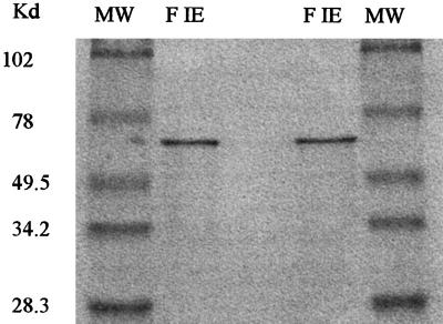
The results of the PPO purification procedure are summarized in Table I. Precipitation with ammonium sulfate resulted in more than 90% recovery of activity with a 4.2-fold purification. All PPO activity was eluted from the Phenyl Sepharose CL4B column in a single peak, representing more than 80% of the loaded activity with less than 30% of the applied protein, indicating an overall purification factor greater than 13. Further purification was done using ion-exchange chromatography on a DEAE-Sepharose CL6B (Pharmacia). The eluted activity representing 92% of the loaded activity was recovered in only one peak. The purification factor of the most active fraction was close to 38. The homogeneity and purity of the former fraction were checked by SDS-PAGE analysis (Fig. 1). A single band at 60 kD was revealed by Coomassie brilliant blue staining. Antibodies raised against an apple PPO interacted strongly with this 60-kD protein after its transfer on nitrocellulose (data not shown). Antibodies did not react with any fractions eluted from ion-exchange chromatography other than that containing the PPO activity. Mature and active apricot PPO was therefore characterized by a 60-kD molecular mass. This value is higher than that previously reported (43 kD) by Fraignier et al. (1995) for different Prunus sp., including apricot. This difference may be due to the occurrence during the extraction and purification procedure of a proteolytic cleavage at the C-terminal end of the protein, as suggested by Robinson and Dry (1992).
Table I.
Summary of apricot PPO purification
| Purification Step | Volume | Activity | Proteins | Yield | Specific Activity | Purification Factor |
|---|---|---|---|---|---|---|
| mL | μkat | mg | % | μkat mg−1 | ||
| Crude extract (NH4)2SO4 precipitation | 85 | 116 | 77 | 100 | 1.5 | 1 |
| Supernatant 30%a | 88 | 108 | 17.2 | 93 | 6.3 | 4.2 |
| Supernatant 30%adialyzed | 102 | 95 | 16.1 | 82 | 5.9 | 3.9 |
| Hydrophobicity | ||||||
| Active fractions | 50 | 78.5 | 3.8 | 67.7 | 20.6 | 13.8 |
| Ion exchange | ||||||
| Active fractions | 16 | 54 | 126 | 47 | 42.8 | 28.6 |
| Most active fraction | 4 | 20 | 0.35 | 17 | 57.2 | 38.1 |
Saturation 30% on a molar basis.
Figure 1.
SDS-PAGE of purified apricot PPO. MW, Molecular size markers; F IE, most active fraction eluted from the DEAE-Sepharose CL6B ionic exchange column.
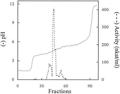
A second hypothesis may be an inadequate reduction of the protein sample before electrophoresis, leaving presumed intramolecular disulfide bridges intact and thereby preventing an accurate electrophoretic molecular mass estimation. Thus, Cary et al. (1992), who observed a 45-kD faba bean PPO form under partially denatured conditions, reported that this form was converted to a 63-kD one with full denaturation. Isoenzyme composition of the purified PPO extract was determined by IEF in a liquid medium with a 3.5 to 5.0 pH gradient. The profile obtained (Fig. 2) shows the presence of a main peak with a maximum at pH 4.6 and three minor peaks at pH 3.8, 4.3, and 4.9. The former pI values are consistent with data available in the literature for other PPO species (Zawistowski et al., 1991). At this step of our investigation it is difficult to conclude whether we are in the presence of isoforms and/or forms resulting from interactions of the protein with phenolic compounds, as reported by Smith and Montgomery (1985). The 18 N-terminal residues of the purified PPO were N-Asp-Pro-Ile-Ala-Pro-Pro-Asp-Leu-Thr-Thr-Cys-Lys-Pro-Ala-Glu-Ile-Thr-Pro.
Figure 2.
IEF in liquid medium of apricot PPO. IEF was performed at 4°C for 3 d in a pH gradient (3.5–5.0). On each fraction of 1.5 mL collected, pH was measured and PPO activity was assayed.
Isolation of a PPO-Related cDNA
PCR amplification of an apricot fruit cDNA generated by RT-PCR led to the production of one cDNA fragment of 915 bp, whose amino acid sequence presented a very high homology with that of Malus domestica PPO cDNA (Boss et al., 1995). This clone was therefore used to screen the cDNA library. Two apricot cDNAs that strongly hybridized were isolated and found to be identical after sequencing. The longer cDNA clone was 2070 bp long and presented a complete coding sequence. It was deposited in GenBank database, given the accession no. AF020786, and labeled PA-PPO (for Prunus armeniaca polyphenol oxidase). This cDNA contained 3 bp of 5′-untranslated region, an open reading frame of 1794 nucleotides encoding for 597 amino acids, and 273 nucleotides of the 3′-untranslated region. The 3′-untranslated region contained three AATAA polyadenylation signals (Joshi, 1987).
Analysis of the Amino Acid Sequence Deduced from the Isolated cDNA
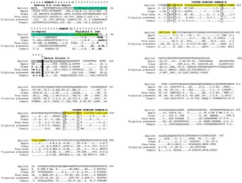
A search of the nonredundant peptide sequence database on the National Center for Biotechnology Information BLAST server using the BLAST program has pointed out a high homology of the isolated apricot cDNA with PPOs from various sources. The deduced amino acid sequence of the isolated clone was compared with the sequences of PPOs from other plant species (Fig. 3). A 67.7% homology with the PPO of apple (Boss et al., 1995) was found, which was the highest homology among the aligned sequence. This high homology is in accordance with the crossed reaction of apple PPO antibodies with apricot PPO. The identity and similarity scores shared by apricot PPO with the proteins of tomato (Newmann et al., 1993), Virginian pokeweed (Joy et al., 1995), faba bean (Cary et al., 1992), and grape berry (Dry and Robinson, 1994) ranged from 43.0% to 53.3% and 57.1% to 65.7%. These data confirm the high level of PPO conservation in higher plants.
Figure 3.
Optimal alignment of PPOs from several plant species. Accession nos.: apricot, no. AF020786; apple, no. P43309 (Boss et al., 1995); grape, no. P43311 (Dry and Robinson, 1994); fava bean, no. 418754 (Cary et al., 1992); Virginian pokeweed, no. D45385 (Joy et al., 1995); tomato, no. Q08296 (Newmann et al., 1993). A dot refers to identity with apricot. A space denotes a gap introduced for improved alignment. Single underlined amino acid residues correspond to the transit peptide. Domain I of transit peptide is marked by •. Domain II of transit peptide is marked by ⋄. The “n-region” of domain II of transit peptide is shaded in blue. The thylakoid transfer domain of the domain II of transit peptide is shaded in green. Shown as bold letters are the hydrophobic amino acids of thylakoid transfer domain and the precleavage site. The first amino acid residue of the mature protein is shaded in black. Double underlined amino acid residues correspond to the sequence obtained from N-terminal sequencing of the purified protein. Copper domains A and B of the mature protein are shaded in yellow. His residues predicted to be copper-binding ligands are boxed.
Comparison of the sequence deduced from the isolated PA-PPO cDNA with that of the purified protein showed a 100% homology, which started with the Asp in position 102. This result suggests that the apricot PPO is synthesized as a precursor protein. The predicted molecular mass of the preprotein was 67.1 kD and its pI was 6.84. A long sequence of 101 amino acids (single underlined residues in Fig. 3), with a predicted molecular mass of 10.9 kD and a pI of 10.91 preceded the mature protein and presented the structure of a chloroplast transit peptide. Following the model given by De Boer and Weisbeek (1991) and recently completed by Joy et al. (1995), transit peptides of lumen-targeted proteins contain two main domains for a two-step process to a mature protein (Sommer et al., 1994). Domain I targets the protein to the translocation complex, where it enters the stroma and is subsequently cleaved. Domain II targets the thylakoid membrane or lumen. This second domain preceded the cleavage site where the mature protein is processed from the transit peptide. In the PA-PPO sequence a putative cleavage site can be predicted between Ala-101 and Asp-102, which is in accordance with results obtained from the N-terminal sequencing of the purified protein.
The mature PPO protein was composed of 496 residues, with a predicted molecular mass of 56.2 kD and a pI of 5.84. The apricot cDNA clone contains the two copper-binding domains typical of PPOs (Fig. 3). Both domains are highly conserved through aligned PPO sequences, have high homology among different species (Shahar et al., 1992), and contain typical His residues thought to be involved in copper binding. The amino acid sequence deduced from the PA-PPO clone does not contain the His-rich region present in the C-terminal part of other aligned sequences and described as a putative third copper-binding domain by Hunt et al. (1993).
The predicted values of molecular mass and pIs of the precursor, the peptide signal, and the mature protein are very close to those calculated from the cDNA sequences of apple (Boss et al., 1995), grape (Dry and Robinson, 1994), faba bean (Cary et al., 1992), Virginian pokeweed (Joy et al., 1995), potato (Hunt et al., 1993), and tomato (Newmann et al., 1993) PPOs.
The slight difference of 1.24 pH unit, observed between the pI calculated from the cDNA and the value obtained for the major PPO peak in the IEF experiment of the purified protein, may be ascribed to phenolic compounds bound to the protein leading to a modification of its net charge. However, charge masking may also result from the final three-dimensional conformation of the protein, which is not entirely taken into account in the pI prediction program.
Southern-Blot Analysis
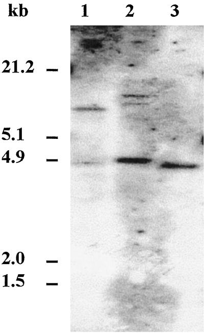
Previous works have shown that PPO genes isolated from faba bean (Cary et al., 1992), potato (Hunt et al., 1993), tomato (Newmann et al., 1993), Virginian pokeweed (Joy et al., 1995), and apple (Boss et al., 1995) belong to a multigene family. In contrast, Southern analysis has demonstrated the presence of only one gene in grapevine (Dry and Robinson, 1994). To determine whether more than one gene was related to PA-PPO, the labeled PPO RT-PCR fragment was hybridized to apricot genomic DNA cut with EcoRI and/or HindIII restriction endonucleases (Fig. 4). Digestion of apricot DNA with EcoRI (Fig. 4, lane 1) produced one major band and one faint band. Digestion of DNA with HindIII (Fig. 4, lane 2) produced one major band and two faint bands. Digestion of DNA with EcoRI and HindIII (Fig. 4, lane 3) produced one major band. Because PA-PPO does not contain an EcoRI or a HindIII restriction site, and PPO genes do not have introns (Newmann et al., 1993; Dry and Robinson, 1994), the number and size of the hybridizing genomic fragments indicate that, as in other fruits, there may be more than one closely related PPO gene in the apricot genome. However, the cloned PA-PPO seems to be present in a single copy.
Figure 4.
Genomic DNA analysis of PA-PPO. Genomic DNA (20 μg per lane) was digested with EcoRI (lane 1), HindIII (lane 2), and EcoRI and HindIII (lane 3), hybridized with an RT-PCR PPO fragment from apricot and washed at low stringency. λDNA codigested with EcoRI and HindIII (Promega) was used as a molecular mass marker.
Northern-Blot analysis: Expression of the PA-PPO Gene during Apricot Ripening
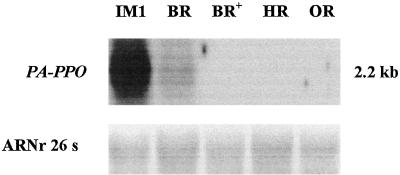
The expression of the PA-PPO gene was examined during fruit development and in vegetative tissues by northern analysis. A labeled PPO RT-PCR fragment was used as a probe in hybridization of equal amounts of total RNAs of each tissue. A single 2.2-kb RNA transcript accumulated at the immature-green stage of fruit development (Fig. 5). The gene was further transcriptionally down regulated with fruit aging (breaker stage) and totally turned off early in the ripening process. Autoradiography exposure for 1 week confirmed the lack of expression after the breaker stage in fruit and also in leaf, wounded leaf, and stem (data not shown). The early expression of PA-PPO in fruit is consistent with results obtained in grape berry (Dry and Robinson, 1994) and apple fruit (Boss et al., 1995) and is more generally in accordance with a higher expression of PPO genes in young tissues (Rathjen and Robinson, 1992; Shahar et al., 1992; Thygesen et al., 1995). In contrast, the expression of Virginian pokeweed PPO specifically localized in ripened fruits (Joy et al., 1995) may be highly related to the accumulation of betalains.
Figure 5.
Expression of PA-PPO gene during ripening of apricot fruit. Fifteen micrograms of total RNA from apricot fruit was used at five stages of development: IM1 (immature green 1), BR (breaker), BR+ (breaker +), HR (half-ripe), and OR (overripe). The blot was hybridized to an RT-PCR PPO fragment from apricot and washed at high stringency.
Activity Assay and Protein Content
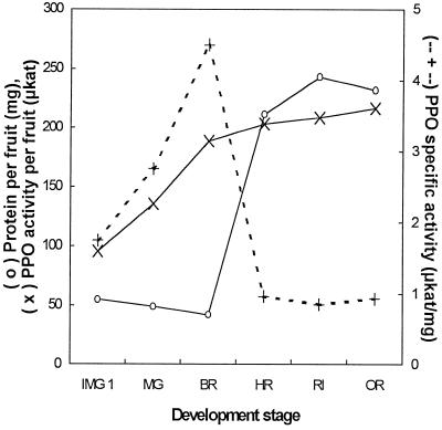
A significant PPO activity (close to 200 nkat g−1 fresh weight) was found in intact and wounded leaves and in stems. In fruit, when expressed per gram of fresh weight, PPO activity slightly decreased from 3.1 μkat at the immature-green stage to 2.2 μkat at the overripe stage (data not shown). Similar results have already been reported for apple fruit (Janovitz-Klapp et al., 1989). The amount of fresh weight of apricot fruit was, however, increased from 30.8 g at the immature-green stage to 98.5 g at the overripe stage (data not shown). Such an effect of fruit growth has been taken into account in Figure 6 by expressing PPO activity per fruit. PPO activity per fruit was found to increase from 96 μkat at the immature-green stage to 188 μkat at the breaker stage. A slight increase was then observed as PPO activity reached a value close to 220 μkat at the overripe stage of fruit development.
Figure 6.
Changes in PPO activity per fruit, protein per fruit, and PPO specific activity during ripening of var Bergeron apricot. Six stages of development were considered: IMG 1 (immature green 1), MG (mature green), BR (breaker), HR (half-ripe), RI (fully ripe), and OR (overripe).
In Figure 6 the amount of total extracted proteins is also expressed per fruit. A value close to 55 mg was determined for the immature-green stage. The protein amount slightly decreased until the breaker stage and then sharply increased to reach 210 mg at the half-ripe stage and 243 mg at the ripe stage.
We also report in Figure 6 the PPO specific activity during fruit development. Starting from 1.75 μkat mg−1 at the immature-green stage, specific activity reached a peak (close to 4.5 μkat mg−1) at the breaker stage. A sharp decrease was then observed; a value close to 0.95 μkat mg−1 was determined for the half-ripe stage. Specific activity remained stable during the following development stages.
Western-Blot Analysis
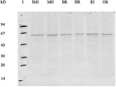
An immunoassay analysis was also performed on a crude protein extract of fruit with antibodies raised against apple PPO. The specificity of the antibodies was examined using total proteins of fruit taken from the immature-green to the overripe stages. One band was visible on the immunoblot (Fig. 7) at approximately 63 kD, whatever the fruit age, which is in accordance with the measurable activity during the same period. Moreover, the apparent constant intensity of the band suggested that the ratio of PPO protein to total proteins remained stable whatever the development stage. The size of the band was similar to that deduced from SDS-PAGE analysis of the purified protein. The slight difference between molecular mass calculated from the PA-PPO cDNA (56.2 kD) and the experimental molecular mass of apricot PPO under denaturing conditions in electrophoresis experiments is thus confirmed. Such a difference, already reported for faba bean PPO by Cary et al. (1992), and in a less extensive report by Robinson and Dry (1992), was ascribed to the glycosylation of the protein by the former authors. The mature apricot PA-PPO clone contains effectively one putative glycosylation site. The PPO may also be artifactually bound by carbohydrates such as phenolic glucosides, which could partially explain its higher-than-56.2-kD size. The results obtained from this western-blot experiment performed with proteins from a crude extract have also indicated that no inactive PPO form putatively different from 63 kD had been lost during the purification procedure.
Figure 7.
Amounts of PPO protein during ripening of apricot fruit. Total proteins from apricot fruit at six stages of development: IM1 (immature green 1), MG (mature green), BR (Breaker), HR (half-ripe), RI (fully ripe), OR (overripe) were used. Proteins (10 μg per lane) of each stage were separated on SDS-PAGE, transferred and immunoblotted with anti-apple PPO crude serum. Molecular size markers are in lane 1.
CONCLUSIONS
We have demonstrated that apricot PPO is still active and present at an advanced stage of the fruit development, whereas its mRNA cannot be detected. PPO appears therefore as a rather stable protein. A similar stability of the PPO protein has also been described in potato tissues (Hunt et al., 1993) and in the young, developing tissues of grape berries (Dry and Robinson, 1994).
There was no transcript of PA-PPO in leaves, wounded leaves, and stems that have been characterized by a significant PPO activity. These data may be explained by the expression of other forms of PPO in these tissues, as reported by Newmann et al. (1993) for tomato tissues. However, since the RT-PCR probe used for northern hybridization contains the thylakoid and copper A- and B-binding domains, which are highly conserved in all PPOs, this probe should have detected other forms of PPO if they were present. This led us to conclude that, whatever the PPO gene expressed, the leaves and stems may have been harvested at a stage too advanced to present a detectable amount of transcript, even after stimulation by wounding (Boss et al., 1995).
Results obtained from western-blot analysis (Fig. 7) have suggested that the ratio of PPO protein to total protein remained stable throughout ripening. We have also demonstrated that specific activity reached a peak at the breaker stage (Fig. 6). These two different trends led us to conclude that, in addition to the transcriptional control of PPO expression demonstrated in this work, other regulation factors such as posttranslational controls also occur in the appearance and amount of apricot PPO activity.
This work has allowed real progress in the characterization of a new fruit PPO. However, the physiological role of the apricot fruit protein remains to be elucidated. The presence of the protein at all stages of fruit development may argue for an active role in disease resistance.
ACKNOWLEDGMENTS
The authors express their gratitude to Dr. Laurence Marquès from Université de Montpellier II for providing them with PPO antibodies and to Dr. P. Sautière from Centre National de la Recherche Scientifique, Institut Pasteur, for the N-terminal sequencing of the purified PPO protein.
Abbreviations:
- PPO
polyphenol oxidase
- PVPP
polyvinylpolypyrrolidone
- RT
reverse transcriptase
LITERATURE CITED
- Altschul SF, Madden TL, Schaffer AA, Zhang J, Zhang Z, Miller W, Lipman DJ. Gapped BLAST and PSI-BLAST: a new generation of protein database search programs. Nucleic Acids Res. 1997;25:3389–3402. doi: 10.1093/nar/25.17.3389. [DOI] [PMC free article] [PubMed] [Google Scholar]
- Bernatzky R, Tanksley SD. Genetics of actin-related sequences in tomato. Theor Appl Genet. 1986;72:314–321. doi: 10.1007/BF00288567. [DOI] [PubMed] [Google Scholar]
- Boss PK, Gardner RC, Janssen B-J, Ross GS. An apple polyphenol oxidase cDNA is up-regulated in wounded tissues. Plant Mol Biol. 1995;27:429–433. doi: 10.1007/BF00020197. [DOI] [PubMed] [Google Scholar]
- Bradford MM. A rapid and sensitive method for the quantitation of microgram quantities of protein utilizing the principle of protein-dye binding. Anal Biochem. 1976;72:248–254. doi: 10.1016/0003-2697(76)90527-3. [DOI] [PubMed] [Google Scholar]
- Cary JW, Lax AR, Flurkey WH. Cloning and characterization of a cDNA coding for Vicia faba polyphenoloxidase. Plant Mol Biol. 1992;20:245–253. doi: 10.1007/BF00014492. [DOI] [PubMed] [Google Scholar]
- Corpet F. Multiple sequence alignment with hierarchical clustering. Nucleic Acids Res. 1988;16 (22)::10881–10890. doi: 10.1093/nar/16.22.10881. [DOI] [PMC free article] [PubMed] [Google Scholar]
- De Boer AD, Weisbeek PJ. Chloroplast protein topogenesis: import, sorting, and assembly. Biochim Biophys Acta. 1991;1071:221–253. doi: 10.1016/0304-4157(91)90015-o. [DOI] [PubMed] [Google Scholar]
- Devereux J, Haeberli P, Smithies O. A comprehensive set of sequence analysis programs for the VAX. Nucleic Acids Res. 1984;12:387–395. doi: 10.1093/nar/12.1part1.387. [DOI] [PMC free article] [PubMed] [Google Scholar]
- Dry IB, Robinson SP. Molecular cloning and characterisation of grape berry polyphenoloxidase. Plant Mol Biol. 1994;26:495–502. doi: 10.1007/BF00039560. [DOI] [PubMed] [Google Scholar]
- Duffey S, Felton G. Enzymatic antinutritive defenses of the tomato plant against insects. In: Hedin P, editor. Naturally Occurring Pest Bioregulators. Washington, DC: American Chemical Society; 1991. pp. 166–197. [Google Scholar]
- Fils B, Sauvage X, Nicolas J. Tomato peroxidases. Purification and some properties. Sci Aliments. 1985;5:217–232. [Google Scholar]
- Fils-Lycaon BR, Wiersma PA, Eastwell KC, Sautiere P. A cherry protein and its gene, abundantly expressed in ripening fruit, have been identified as thaumatin-like. Plant Physiol. 1996;111:269–273. doi: 10.1104/pp.111.1.269. [DOI] [PMC free article] [PubMed] [Google Scholar]
- Fraignier MP, Marques L, Fleuriet A, Macheix JJ. Biochemical and immunochemical characteristics of polyphenol oxidases from different fruits of Prunus. J Agric Food Chem. 1995;43:2375–2380. [Google Scholar]
- Gauillard F, Richard-Forget F. Polyphenoloxidases from Williams pear (Pyrus communis L. cv Williams): activation, purification and some properties. J Sci Food Agric. 1997;74:49–56. [Google Scholar]
- Hunt MD, Eannetta NT, Yu H, Newmann SM, Steffens JC. cDNA cloning and expression of potato polyphenol oxidase. Plant Mol Biol. 1993;21:59–68. doi: 10.1007/BF00039618. [DOI] [PubMed] [Google Scholar]
- Janovitz-Klapp A, Richard F, Nicolas J. Polyphenoloxidase from apple, partial purification, and some properties. Phytochemistry. 1989;28:2903–2907. [Google Scholar]
- Joshi CP. Putative polyadenylation signals in nuclear genes of higher plants: a compilation and analysis. Nucleic Acids Res. 1987;15:9627–9641. doi: 10.1093/nar/15.23.9627. [DOI] [PMC free article] [PubMed] [Google Scholar]
- Joy RW, Sugiyama M, Fukuda H, Komamine A. Cloning and characterization of polyphenol oxidase cDNAs of Phytolacca americana. Plant Physiol. 1995;107:1083–1089. doi: 10.1104/pp.107.4.1083. [DOI] [PMC free article] [PubMed] [Google Scholar]
- Kahn V. Effects of proteins, protein hydrolyzates and amino acids on O-dihydroxyphenolase activity of polyphenol oxidase of mushroom, avocado and banana. J Food Sci. 1977;50:111–119. [Google Scholar]
- Kyte M, Doolittle RF. A simple method for displaying the hydropathic character of protein. J Mol Biol. 1982;157:105–132. doi: 10.1016/0022-2836(82)90515-0. [DOI] [PubMed] [Google Scholar]
- Laemmli UK. Cleavage of structural proteins during the assembly of the head of bacteriophage T4. Nature. 1970;227:680–685. doi: 10.1038/227680a0. [DOI] [PubMed] [Google Scholar]
- Lelièvre J-M, Tichit L, Fillion L, Larrigaudière C, Vendrell M, Pech J-C. Cold-induced accumulation of 1-aminocyclopropane-1-carboxylate oxidase protein in Granny Smith apples. Postharvest Biol Technol. 1995;5:11–17. [Google Scholar]
- Mayer AM, Harel E. Polyphenol oxidases in plants. Phytochemistry. 1979;18:193–215. [Google Scholar]
- Mayer AM, Harel E (1991) Phenoloxidases and their significance in fruit and vegetables. In FP Fox, ed, Food Enzymology. Elsevier Science Publishers, New York, pp 373–398
- Newmann SM, Eannetta NT, Yu H, Prince JP, de Vicente CM, Tanksley SD, Steffens JC. Organization of the tomato polyphenol oxidase gene family. Plant Mol Biol. 1993;21:1035–1051. doi: 10.1007/BF00023601. [DOI] [PubMed] [Google Scholar]
- Nicolas J, Richard-Forget FC, Goupy PM, Amiot MJ, Aubert SY. Enzymatic browning reactions in apple and apple products. Crit Rev Food Sci Nutr. 1994;34:109–157. doi: 10.1080/10408399409527653. [DOI] [PubMed] [Google Scholar]
- Rathjen AH, Robinson SP. Aberrant processing of polyphenol oxidase in a variegated grapevine mutant. Plant Physiol. 1992;99:1619–1625. doi: 10.1104/pp.99.4.1619. [DOI] [PMC free article] [PubMed] [Google Scholar]
- Robinson SP, Dry IB. Broad bean leaf polyphenol oxidase is a 60-kD protein susceptible to proteolytic cleavage. Plant Physiol. 1992;99:317–323. doi: 10.1104/pp.99.1.317. [DOI] [PMC free article] [PubMed] [Google Scholar]
- Robinson SP, Loveys BR, Chacko EK. Polyphenol oxidase enzymes in the sap and skin of mango fruit. J Plant Physiol. 1993;20:99–107. [Google Scholar]
- Saiki RK, Scharf S, Faloona F, Mullis KB, Horn GT, Erlich H, Arnheim N. Enzymatic amplification of β-globin genomic sequences and restriction site analysis for diagnosis of sickle cell anemia. Science. 1985;230:1350–1354. doi: 10.1126/science.2999980. [DOI] [PubMed] [Google Scholar]
- Sambrook J, Fritsch EF, Maniatis T. Molecular Cloning: A Laboratory Manual, Ed 2. Cold Spring Harbor, NY: Cold Spring Harbor Laboratory Press; 1989. [Google Scholar]
- Sanchez-Ferrer A, Laveda F, Garcia-Carmona F. Substrate-dependent activation of latent potato leaf polyphenol oxidase by anionic surfactants. J Agric Food Chem. 1993;41:1583–1586. [Google Scholar]
- Shahar T, Henning N, Gutfinger T, Hareven D, Lifschitz E. The tomato 66.3-kD polyphenoloxidase gene: molecular identification and developmental expression. Plant Cell. 1992;4:135–147. doi: 10.1105/tpc.4.2.135. [DOI] [PMC free article] [PubMed] [Google Scholar]
- Smith DM, Montgomery MW. Improved methods for the extraction of polyphenol oxidase from d'Anjou pears. Phytochemistry. 1985;24:901–904. [Google Scholar]
- Söderhall I. Properties of carrot polyphenoloxidase. Phytochemistry. 1995;39:33–38. [Google Scholar]
- Sommer A, Nemann E, Steffens JC. Import, targeting, and processing of a plant polyphenol oxidase. Plant Physiol. 1994;105:1301–1311. doi: 10.1104/pp.105.4.1301. [DOI] [PMC free article] [PubMed] [Google Scholar]
- Thygesen PW, Dry IB, Robinson SP. Polyphenol oxidase in potato. A multigene family that exhibits differential expression patterns. Plant Physiol. 1995;109:525–531. doi: 10.1104/pp.109.2.525. [DOI] [PMC free article] [PubMed] [Google Scholar]
- Towbin H, Staehelin T, Gordon J. Electrophoretic transfer of protein from polyacrylamide gel to nitrocellulose sheets: procedure and some applications. Proc Natl Acad Sci USA. 1979;76:4350–4354. doi: 10.1073/pnas.76.9.4350. [DOI] [PMC free article] [PubMed] [Google Scholar]
- Trebst A, Depka B. Polyphenol oxidase and photosynthesis research. Photosynth Res. 1995;46:41–44. doi: 10.1007/BF00020414. [DOI] [PubMed] [Google Scholar]
- Van Gelder CWG, Flurkey WH, Wichers HJ. Sequence and structural features of plant and fungal tyrosinases. Phytochemistry. 1997;45:1309–1323. doi: 10.1016/s0031-9422(97)00186-6. [DOI] [PubMed] [Google Scholar]
- Vaughn KC, Duke SO. Tentoxin stops the processing of polyphenol oxidase into an active protein. Physiol Plant. 1984;60:257–261. [Google Scholar]
- Vaughn KC, Lax AR, Duke SO. Polyphenol oxidase: the chloroplast oxidase with no established function. Physiol Plant. 1988;72:659–665. [Google Scholar]
- Wan C-Y, Wilkins TA. A modified hot borate method significantly enhances the yield of high-quality RNA from cotton (Gossypium hirsutum L.) Anal Biochem. 1994;223:7–12. doi: 10.1006/abio.1994.1538. [DOI] [PubMed] [Google Scholar]
- Zawistowski J, Biliaderis CG, Eskin NAM. Polyphenol oxidase. In: Robinson DS, Eskin NAM, editors. Oxidative Enzymes in Foods. London: Elsevier Science Publishers; 1991. pp. 217–273. [Google Scholar]


