Abstract
The rapid yet transient transcriptional activation of heat shock genes is mediated by the reversible conversion of HSF1 from an inert negatively regulated monomer to a transcriptionally active DNA-binding trimer. During attenuation of the heat shock response, transcription of heat shock genes returns to basal levels and HSF1 reverts to an inert monomer. These events coincide with elevated levels of Hsp70 and other heat shock proteins (molecular chaperones). Here, we show that the molecular chaperone Hsp70 and the cochaperone Hdj1 interact directly with the transactivation domain of HSF1 and repress heat shock gene transcription. Overexpression of either chaperone represses the transcriptional activity of a transfected GAL4–HSF1 activation domain fusion protein and endogenous HSF1. As neither the activation of HSF1 DNA binding nor inducible phosphorylation of HSF1 was affected, the primary autoregulatory role of Hsp70 is to negatively regulate HSF1 transcriptional activity. These results reveal that the repression of heat shock gene transcription, which occurs during attenuation, is due to the association of Hsp70 with the HSF1 transactivation domain, thus providing a plausible explanation for the role of molecular chaperones in at least one key step in the autoregulation of the heat shock response.
Keywords: Transcriptional control, autoregulation, heat shock proteins, Hsp70, activation domain
The cellular response to diverse forms of environmental and physiological stress, including heat shock, heavy metals, oxidants, UV, cytokines, or hormones, involves the rapid transcriptional induction of target genes whose activity is regulated by different stress-specific transactivators (Baeuerle 1995). The molecular response to each of these stresses serves to protect the cell against lethal exposures to the same and other potentially deleterious forms of stress. The transcription of heat shock genes, for example, is rapidly induced yet persists at maximal levels transiently and attenuates either proportionally to the intensity of the stress or upon return to control conditions (DiDomenico et al. 1982a,b; Mosser et al. 1988; Straus et al. 1990; Abravaya et al. 1991). In higher eukaryotes, the stress-induced component(s) of the heat shock transcriptional response are principally heat shock factors (HSFs), which are ubiquitously expressed and maintained in unstressed cells in an inert non-DNA-binding state. Upon exposure of cells to seemingly diverse stress conditions, HSFs become activated to a DNA-binding, transcriptionally active state, which results in the preferential transcription of heat shock genes (Mosser et al. 1988; Morimoto et al. 1990; Lis and Wu 1993; Morimoto 1993; Rabindran et al. 1993; Sarge et al. 1993; Westwood and Wu 1993; Wu 1995).
The heat shock response provides the cell with a mechanism to reestablish protein homeostasis, the balance between protein synthesis, protein folding and assembly, and protein degradation. Whereas exposure to elevated temperatures results in a response that attenuates upon prolonged heat shock or recovery, other inducers of the heat shock response, such as amino acid analogs, activate heat shock gene expression when the analogs are incorporated into nascent polypeptides. Under the latter conditions, heat shock genes are constitutively up-regulated and attenuation does not occur (DiDomenico et al. 1982b; Mosser et al. 1988). These observations, among others, have led to a widely held proposal that the heat shock response is autoregulated by heat shock proteins. This is additionally supported by genetic evidence that mutations in yeast Hsp70 result in the overexpression of heat shock genes (Craig and Jakobsen 1984; Craig and Gross 1991) and are dependent on the HSF binding site to DNA (Boorstein and Craig 1990), and biochemical evidence that Hsp70 interacts with HSFs (Abravaya et al. 1992; Baler et al. 1992; Mosser et al. 1993; Rabindran et al. 1994). It has been unclear, however, which of the many events in HSF regulation—including trimerization, DNA binding, inducible phosphorylation, or stress-induced transcriptional activation—were affected by Hsp70. As yeast HSF is constitutively trimeric and does not convert to non-DNA-binding monomer during attenuation, the principal form of regulation is likely to be at the level of transcriptional activation (Sorger et al. 1987; Jakobsen and Pelham 1988; Sorger and Nelson 1989; Boorstein and Craig 1990). A role for molecular chaperones in the regulation of the heat shock response has also been shown in Escherichia coli. The E. coli DnaK chaperone machine comprised of DnaK, DnaJ, and GrpE negatively regulates the transcription of heat shock genes by direct interaction with σ32 (Tilly et al. 1983; Grossman et al. 1987; Straus et al. 1990; Liberek et al. 1992; Blaszczak et al. 1995; Gamer et al. 1996).
In this study we demonstrate that Hsp70 stably associates in vivo and in vitro with the transactivation domain of HSF1. The consequence is to negatively regulate the transcriptional activity of HSF1 with little effect on the DNA-binding or inducibly phosphorylated state of HSF1, thus indicating that molecular chaperone Hsp70 functions as a repressor of transcriptional activity of the heat shock-specific transactivator.
Results
Hsp70 associates with the HSF1 transactivation domain
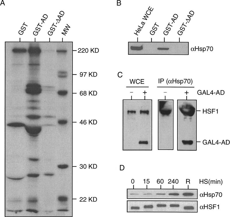
Attenuation of the heat shock transcriptional response occurs during continuous exposure to intermediate heat shock conditions or upon recovery from stress (Abravaya et al. 1991). A characteristic feature of attenuation is the rapid repression of heat shock gene transcription, which precedes the conversion of HSF1 trimers to monomers, and the loss of HSF1 DNA-binding activity (Abravaya et al. 1991; Kline and Morimoto 1997). The kinetics of these complex events have suggested a role for other proteins that could act directly on the HSF1 activation domain, perhaps to repress its transcriptional activity. Therefore, we screened for potential regulatory proteins that interact with the HSF1 transactivation domain (amino acids 395–503) using a direct protein–protein interaction assay. From an extract of HeLa 35S-labeled proteins, four proteins ranging in size from 30 to 70 kD bound specifically with the wild-type HSF1 activation domain fused to GST and not with GST alone or with a transcriptionally inert HSF1 activation domain deletion mutant (amino acids 451–503) (Fig. 1A). Hsp70 was identified as one of the proteins that associated with the HSF1 activation domain (Fig. 1B). The interaction with Hsp70 appears selective, as Hsp90 was not detected in the collection of activation domain-binding proteins (data not shown).
Figure 1.
Hsp70 binds to the HSF1 activation domain in vitro and in vivo. (A) Identification of proteins binding to the HSF1 activation domain. GST alone, HSF1 activation domain (amino acids 395–503) GST fusion protein (GST–AD), or deletion mutant of HSF1 activation domain (amino acids 451–503) GST fusion protein (GST–ΔAD) were incubated with 35S-labeled HeLa whole cell extracts. Bound proteins were analyzed by SDS-PAGE and fluorography. The sizes of the molecular weight markers (MW) are indicated. (B) Hsp70 is among the HSF1 activation domain interactive proteins. Proteins bound to GST, GST–AD, or GST–ΔAD were analyzed by Western blot analysis with Hsp70-specific antibody 3A3. HeLa whole cell extract (WCE) input was included as a positive control. (C) Hsp70 interacts with the HSF1 activation domain as determined by immunoprecipitation assay. COS-7 cells were transfected with the β-actin hsp70 construct (S.P. Murphy, unpubl.) alone (−) or together with the GAL4–HSF1 activation domain (GAL4–AD) construct (+). Cell lysates were immunoprecipitated with Hsp70-specific antibody 3A3 and immunoblotted with HSF1-specific polyclonal antibody. Endogenous HSF1 and transfected GAL4–AD are indicated (right). An aliquot of the cell lysates (WCE) was analyzed directly by Western blot as a control. (D) Increased Hsp70 is associated with HSF1 during attenuation or recovery from heat shock. HeLa cells were heat-shocked at 42°C for 0, 15, 60, and 240 min, or at 42°C for 120 min and then allowed to recover at 37°C for 240 min (R). Cell lysates were immunoprecipitated with HSF1-specific polyclonal antibody and immunoblotted with Hsp70-specific monoclonal antibody C92 or with HSF1-specific monoclonal antibody 4B4.
To further examine the interaction of the HSF1 transactivation domain with Hsp70, cells transiently overexpressing GAL4–HSF1 and Hsp70 were assayed by immunoprecipitation analysis using antibodies specific to Hsp70. The Hsp70-containing immunocomplexes were analyzed by Western blot analysis for the presence of HSF1. Both the transfected GAL4–HSF1 activation domain fusion protein and endogenous HSF1 were among the Hsp70-associated proteins (Fig. 1C). This confirms previous results that Hsp70 interacts with HSF1 (Abravaya et al. 1992; Baler et al. 1992; Rabindran et al. 1994) and demonstrates that a specific site for Hsp70 interaction is the HSF1 activation domain. The association of Hsp70 with HSF1 was also detected by coimmunoprecipitation with HSF1-specific antibody during a 4-hr heat shock time course. During attenuation and recovery from heat shock, increased levels of Hsp70 were associated in complexes with HSF1 (Fig. 1D).
HSF1 activation domain interacts directly with Hsp70 and Hdj1
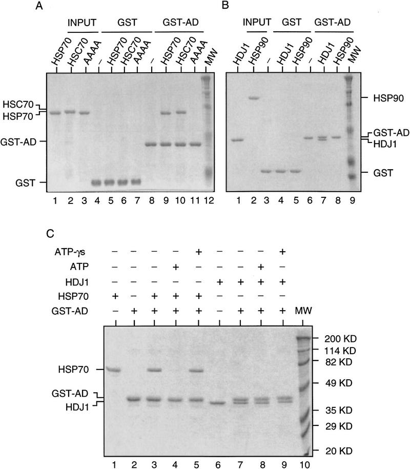
To examine whether the interaction of Hsp70 with the HSF1 activation domain is direct or involves other proteins, purified recombinant human Hsp70 was incubated with the GST–HSF1 activation domain and examined for complex formation. Hsp70, in the absence of other proteins, interacts directly with the HSF1 activation domain (Fig. 2A). The HSF1 activation domain also binds to the Hsp70 homologs, Hsc70 (Fig. 2A) and DnaK (data not shown), but not with an Hsp70 AAAA mutant (Fig. 2A) that has the last four amino acids of Hsp70 mutated from EEVD to AAAA and is deficient in chaperone function (Freeman et al. 1995). To further examine the specificity of the interaction, other chaperones including Hdj1 and Hsp90 were studied in the binding assay. Hdj1 also interacts directly with the HSF1 activation domain, but Hsp90 does not (Fig. 2B). A characteristic feature of Hsp70–substrate binding is ATP-mediated substrate release (Pelham 1986; Clarke et al. 1988; Flynn et al. 1989; Kost et al. 1989; Beckmann et al. 1990; Palleros et al. 1991). To examine the effect of ATP on Hsp70–HSF1 activation domain interaction, a similar binding assay was performed in the absence or presence of ATP, or its nonhydrolyzable analog ATP-γs. The HSF1–Hsp70 complexes were dissociated upon addition of ATP and unaffected by ATP-γs (Fig. 2C), similar to the Hsp70–substrate interaction. The HSF1 activation domain also interacts with Hdj-1, however the HSF1–Hdj1 complexes were insensitive to nucleotide (Fig. 2C), consistent with other observations on the ATP-insensitivity of DnaJ–substrate interaction (Wawrzynow and Zylicz 1995).
Figure 2.
Hsp70 and Hdj1 interact directly with the HSF1 activation domain. (A) Reconstitution of the interaction of Hsp70 and the HSF1 activation domain in vitro. Purified recombinant Hsp70, Hsc70, and the Hsp70 AAAA mutant were incubated with purified GST or GST–HSF1 activation domain fusion protein (GST–AD) on glutathione beads. The proteins bound to GST (lanes 5–7) or GST–AD (lanes 9–11) were analyzed by SDS-PAGE and Coomassie blue staining. Hsp70, Hsc70, and the Hsp70 AAAA mutant are included in lanes 1, 2, and 3, respectively; GST is in lane 4; GST–AD is in lane 8; molecular mass markers (MW) are in lane 12. (B) Reconstitution of the interaction of Hdj1 and the HSF1 activation domain in vitro. Purified recombinant Hdj1 and Hsp90 were incubated with GST or GST–AD on glutathione beads. The proteins bound to GST are in lanes 4 and 5; proteins bound to GST–AD in lanes 7 and 8; Hdj1 in lane 1; Hsp90 in lane 2; GST in lane 3; GST–AD in lane 6; and MW in lane 9. (C) The effect of nucleotides on the interaction of Hsp70 or Hdj1 with the HSF1 activation domain. Hsp70 or Hdj1 was incubated with GST–HSF1 activation domain in the absence of nucleotide (lanes 3,7), in the presence of 1 mm ATP (lanes 4,8), or 1 mm ATP-γs (lanes 5,9). Hsp70 input was included in lane 1; GST–AD in lane 2; Hdj1 in lane 6; MW in lane 10 (sizes at right).
The domain of Hsp70 that interacts with HSF1 was subsequently identified in binding assays with purified full-length Hsp70, the amino-terminal ATPase domain, or the carboxyl-terminal substrate binding domain (Fig. 3A). Only full-length Hsp70 and the substrate binding domain interact with the HSF1 activation domain; no binding of the Hsp70 ATPase domain to the HSF1 activation domain was detected (Fig. 3B). These results, together with the ATP sensitivity of the HSF1–Hsp70 complexes (Fig. 2C), indicate that the interaction between HSF1 activation domain and Hsp70 has the features of a substrate–chaperone complex and is distinct from cochaperone–Hsp70 interactions that predominantly occur through the ATPase domain of Hsp70 (Hohfeld et al. 1995; Tsai and Douglas 1996).
Figure 3.

The carboxyl-terminal substrate binding domain of Hsp70 interacts with the activation domain of HSF1. (A) A schematic diagram of full-length human Hsp70 (amino acids 1–640), deletion mutant Hsp70 N (amino acids 1–436 and 618–640), and Hsp70 C (amino acids 386–640) is shown. The ATP-binding domain (amino acids 1–386) is indicated as a hatched box; the substrate binding domain (amino acids 386–640) as a solid box. The epitopes for anti-Hsp70 monoclonal antibodies 3A3 or 5A5 are indicated. (B) The HSF1 activation domain interacts with the substrate binding domain of Hsp70. Purified full-length recombinant Hsp70, Hsp70 N (with a degradation product), and Hsp70 C (with some full-length Hsp70) were incubated with purified GST (lanes 5–7) or GST–HSF1 activation domain (GST–AD) (lanes 9–11), the bound proteins were washed extensively and analyzed by SDS-PAGE and immunoblotted with either 3A3 (top) or 5A5 (bottom) antibody (Ab). Inputs of full-length Hsp70, Hsp70 N, and Hsp70 C are shown in lanes 1, 2, and 3 individually; GST in lane 4; GST–AD in lane 8.
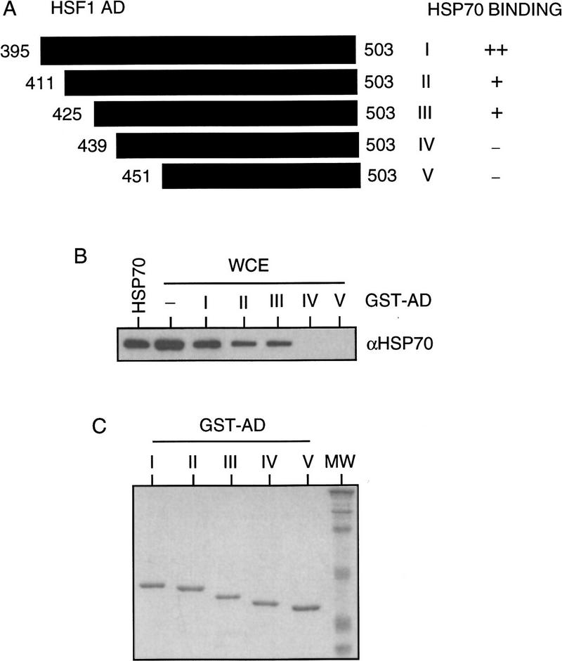
The region of the HSF1 activation domain that interacts with Hsp70 was identified using in vitro binding assays. A collection of GST–HSF1 activation domain fusion proteins, including the wild-type activation domain I (amino acids 395–503) and deletion mutants II (amino acids 411–503), III (amino acids 425–503), IV (amino acids 439–503), and V (amino acids 451–503) (Fig. 4A), were purified as recombinant proteins (Fig. 4C) and incubated with HeLa whole cell extracts containing Hsp70 or separately with recombinant Hsp70. The wild-type activation domain I and deletion mutants II and III were associated with Hsp70, whereas deletion mutants IV and V did not bind to Hsp70 (Fig. 4B; data not shown). These results delimit the Hsp70-binding site to amino acid residues 425–439 of the HSF1 activation domain.
Figure 4.
Mapping the Hsp70 binding site within the HSF1 activation domain. (A) Schematic diagram of wild-type and deletion mutants of HSF1 activation domain. Constructs I (wild type), and II–V (deletion mutants) are indicated as the regions of HSF1 activation domain fused to GST. The boundaries of each construct and the levels of Hsp70-binding activity are indicated. (B) A potential Hsp70 binding site is located from amino acid residue 425 to 439 of the HSF1 activation domain. HSF1–GST fusion proteins were incubated with HeLa whole cell extracts (WCE). The presence of Hsp70 as bound protein was detected by Western blot analysis with Hsp70-specific antibody 3A3. Purified recombinant Hsp70 and an aliquot of HeLa whole cell extracts were included as positive controls. (C) SDS-PAGE of each purified GST fusion protein visualized by Coomassie blue staining. Molecular weight markers (MW) are included.
Hsp70 and Hdj1 negatively regulate the transcriptional activation property of HSF1
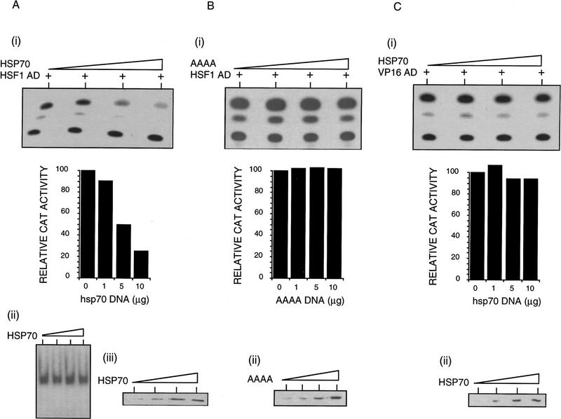
Having demonstrated that Hsp70 and Hdj1 interact directly with HSF1, we addressed whether these interactions affect HSF1 transcriptional activity. Overexpression of either chaperone by transfection was employed to examine the effect of elevated expression of the chaperone independent of other consequences of the heat shock response. The transcriptional activity of the HSF1 activation domain fused to the GAL4 DNA-binding domain, as measured by CAT activity from the G5BCAT reporter, was repressed four- to fivefold when coexpressed with a vector that expresses high levels of Hsp70 (Fig. 5A, i and iii). As neither the level of GAL4–HSF1 fusion protein (data not shown) nor its DNA-binding activity (Fig. 5A, ii) was affected, we conclude that Hsp70 functions principally as a negative regulator of the transactivation domain of HSF1.
Figure 5.
Analysis of HSF1 transcriptional activity in cells transiently overexpressing Hsp70. (A) Coexpression of the HSF1 activation domain and Hsp70. (i) The transcriptional activity of GAL4–HSF1 activation domain in the presence of β-actin hsp70 plasmid DNA (0, 1, 5, and 10 μg) was measured as relative CAT activity from the reporter plasmid G5BCAT. The CAT activity was quantified with a PhosphorImager, standardized by cotransfected internal control β-gal activity, and plotted against the amount of transfected β-actin hsp70 plasmid DNA. (ii) The DNA-binding activity of GAL4–HSF1. Only the top part of the gel corresponding to the GAL4–HSF1 DNA binding complex is shown. The gel shift assay was done in probe excess. (iii) The levels of overexpressed Hsp70 were detected by Western blot analysis with Hsp70-specific monoclonal antibody 4G4. (B) Coexpression of the HSF1 activation domain and the Hsp70 AAAA mutant. (i) The transcriptional activity of GAL4–HSF1 activation domain in the presence of β-actin hsp70 AAAA plasmid DNA (0, 1, 5, and 10 μg). (ii) The levels of overexpressed Hsp70 AAAA mutant. A similar ECL exposure was obtained to that in A (iii). (C) Coexpression of the VP16 activation domain and Hsp70. (i) The transcriptional activity of GAL4–VP16 activation domain in the presence of β-actin hsp70 plasmid DNA (0, 1, 5, and 10 μg). (ii) The levels of overexpressed Hsp70. A similar ECL exposure was obtained to that in A (iii).
The negative effect of Hsp70 on HSF1 transcriptional activity requires that the chaperone is functional. The Hsp70 AAAA mutant, which lacks chaperone activity, does not bind to the HSF1 activation domain (Fig. 2A) and consequently does not repress HSF1 transcriptional activity (Fig. 5B). Overexpression of Hsp70 neither represses the expression of a cotransfected RSV–β-gal control (data not shown) nor the transcriptional activity of the GAL4–VP16 activation domain (Fig. 5C) and other GAL4–activation domains (data not shown). These results indicate that the negative effects of Hsp70 are specific to the HSF1 transactivation domain.
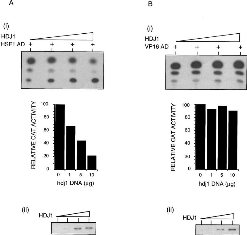
In parallel experiments, overexpression of Hdj1 also resulted in the negative regulation of HSF1 transcriptional activity. Coexpression of Hdj1 with GAL4–HSF1 activation domain resulted in a fourfold repression of the transcriptional activity of GAL4–HSF1 (Fig. 6A). As observed for Hsp70, Hdj1 did not repress transcriptional activity of the GAL4–VP16 activation domain (Fig. 6B).
Figure 6.
Analysis of HSF1 transcriptional activity in cells transiently overexpressing Hdj1. The GAL4–HSF1 activation domain (A) or the GAL4–VP16 activation domain (B) was transfected into COS-7 cells with increasing amount of β-actin hdj1 plasmid DNA (0, 1, 5, and 10 μg). (i) The transcriptional activity of GAL4–activation domain fusion was measured as relative CAT activity and plotted against the amount of transfected β-actin hdj1 plasmid DNA as in Fig. 5. (ii) The levels of overexpressed Hdj1 were examined by Western blot analysis with Hdj1-specific antibody. A similar exposure for ECL was obtained for the Western blots shown in A and B.
Autoregulation of hsp90 and hsp70 gene transcription in cells conditionally expressing Hsp70
If Hsp70 negatively regulates HSF1 transcriptional activity, it should be possible to control the levels of Hsp70 and examine the effects of increased levels of Hsp70 on the transcriptional activity of HSF1. To accomplish this, we used a stably transfected cell line (PETA70) conditionally expressing human Hsp70 under the control of a tetracycline-regulated system (Mosser et al. 1997). PETA70 cells were cotransfected with the GAL4–HSF1 activation domain, G5BCAT reporter, and RSV–β-gal internal control. Upon induction and accumulation of Hsp70, GAL4–HSF1 activity, measured by the expression of a CAT reporter, was repressed fourfold (Fig. 7A,C). No detectable changes in the levels of GAL4–HSF1 DNA-binding activity were observed (Fig. 7B); these results demonstrate that the effect of Hsp70 is on the transactivation activity of HSF1.
Figure 7.
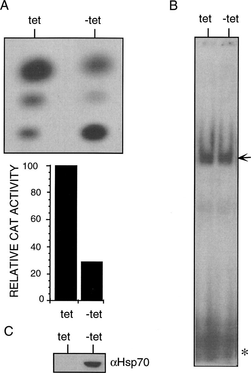
Analysis of HSF1 transcriptional activity in tetracycline-regulated Hsp70-overexpressing cells. (A) Transcriptional activity of the GAL4–HSF1. PETA70 cells transfected with the GAL4–HSF1 activation domain construct were maintained in media with or without anhydrotetracycline (tet or −tet) for 48 hr. GAL4–HSF1 transcriptional activity was measured as relative CAT activity, standardized by β-gal activity, and plotted accordingly. (B) DNA-binding activity of GAL4–HSF1. The gel shift assay was performed in probe excess. Both the GAL4–HSF1 DNA-binding complex (top, indicated by arrow) and excess free probe (bottom, indicated by asterisk) are shown. (C) Hsp70 level induced by withdrawal of anhydrotetracycline (−tet). Western blot analysis was performed with Hsp70-specific antibody 4G4.
To address whether the effects of elevated levels of chaperone on HSF1 transcriptional activity were physiologically relevant to that achieved during heat shock, we examined the effects of increased levels of Hsp70 on inducible transcription of the endogenous heat shock genes. In PETA70 cells expressing elevated levels of Hsp70 (Fig. 8E), the transcription of endogenous heat shock genes was not induced upon heat shock; this result was in striking contrast to the typical pattern of heat shock-induced transcription of the hsp70 and hsp90α genes in uninduced cells. For example, in Hsp70-overexpressing cells, transcription of the hsp90α gene was not induced following heat shock, whereas in a parallel experiment using cells uninduced for Hsp70, there was a dramatic heat shock induction of hsp90α gene transcription (Fig. 8A). These results clearly establish that Hsp70 negatively regulates heat shock gene transcription. Assessing this for the hsp70 gene was not possible as the PETA70 cells contain multiple copies of the hsp70 gene under the tetracycline-inducible promoter, thus accounting for high basal hsp70 gene transcription. However, consistent with the results obtained for the hsp90α gene, heat shock did not enhance the transcription of the hsp70 gene and noticeably reduced the signal, presumbly because of the general repressive effects of heat shock on transcription (Fig. 8A). Nevertheless, in the absence or presence of Hsp70 overexpression, HSF1 acquired high DNA-binding activity and was inducibly phosphorylated (Fig. 8B,D), suggesting that Hsp70 primarily affects HSF1 transcriptional activity.
Figure 8.
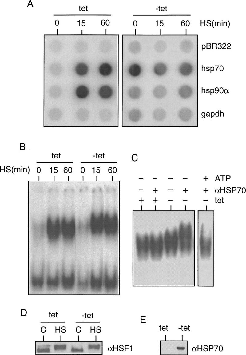
Analysis of heat shock gene transcription, HSF1 DNA-binding, and inducible phosphorylation in Hsp70-overexpressing cells. PETA70 cells were maintained in media with or without anhydrotetracycline (tet or −tet) for 48 hr before 42°C heat shock treatment for 0, 15, and 60 min. (A) The transcription rate of hsp70 and hsp90α genes in control (tet) or Hsp70-overexpressed (−tet) cells was measured by run-on transcription analysis. (B) The DNA-binding activity of endogenous HSF1. (C) Antibody supershift of HSF1 DNA-binding activity in the absence (−) or presence (+) of 5 mm exogenous ATP. Only the top part of the gel corresponding to the HSF1 DNA-binding complex is shown. (D) The inducible phosphorylation state of HSF1 was examined by Western blot analysis with HSF1-specific polyclonal antibody. (C) Non-heat-shocked control; (HS) 60-min heat shock. (E) Hsp70 level upon anhydrotetracycline withdrawal (−tet) was measured by Western blot analysis with Hsp70-specific antibody 4G4.
Previously, we had established that Hsp70 was detected in a complex with HSF1 (Fig. 1). To determine whether Hsp70 overexpressed in the PETA70 cells was associated with HSF1, we performed gel mobility-shift assays using whole cell extracts. HSF–HSE (heat shock element) complexes in extracts from heat-shocked cells expressing high levels of Hsp70 exhibited a slower electrophoretic mobility compared to that of heat-shocked cells without Hsp70 overexpression (Fig. 8B). Antibody supershift of HSF–HSE complexes from heat-shocked cells overexpressing Hsp70 detected a supershifted ternary complex (Hsp70:HSF1–HSE) with Hsp70-specific antibody, whereas no such complex was detected in extracts from heat-shocked cells without Hsp70 overexpression (Fig. 8C). The HSF1–Hsp70 complexes were disrupted upon addition of ATP and resulted in a faster migrating HSF–HSE complex, which could not be supershifted by Hsp70-specific antibody (Fig. 8C), corroborating the in vitro data that HSF–Hsp70 complexes are ATP sensitive (Fig. 2C).
To further establish that these chaperone–HSF1 interactions and subsequent effects on HSF1 activity are specific, similar experiments were performed using a stably transfected human cell line (PERTA70–AAAA) conditionally overexpressing a nonfunctional Hsp70 AAAA mutant (Fig. 9A). Upon overexpression of the Hsp70 AAAA mutant to a level similar to that obtained for wild-type Hsp70 (Fig. 9E), neither heat-shock induced transcription of the hsp90α gene (Fig. 9B) nor HSF1 DNA-binding activity (Fig. 9C) was affected. Antibody supershift of HSF–HSE complexes with Hsp70-specific antibody did not detect any HSF–HSP70 complexes in either uninduced or AAAA-expressing cells (Fig. 9D), in contrast to that from wild-type Hsp70-expressing cells (Fig. 8C). These results further indicate that the transcriptional regulation by Hsp70 requires its chaperone function for direct binding to HSF1.
Figure 9.
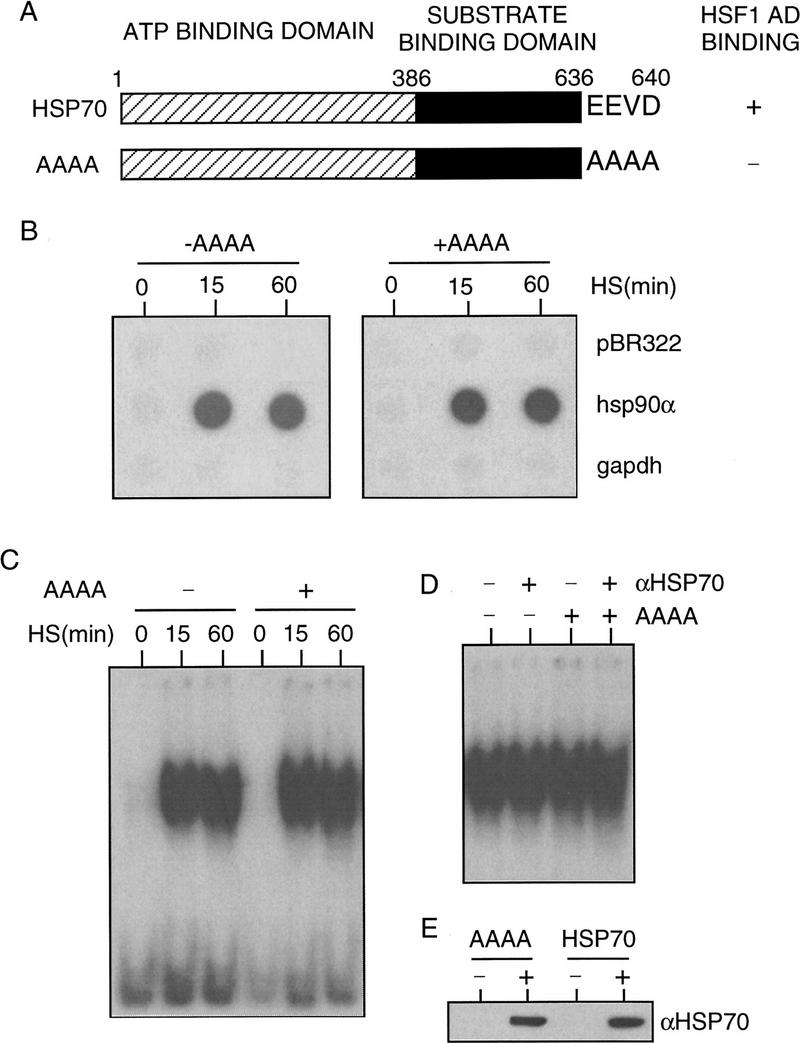
Analysis of the transcription rate of hsp70 and hsp90α genes in Hsp70 AAAA mutant-overexpressing cells. (A) Schematic diagram of wild-type Hsp70 and the AAAA mutant. The binding activity to HSF1 activation domain (Fig. 2A) and the mutated residues are indicated. (B) The transcription rate of the hsp90α gene was measured by run-on transcription analysis. PERTA70–AAAA cells were maintained in media with or without doxycycline (+AAAA or −AAAA) for 24 hr before 42°C heat shock treatment for 0, 15, and 60 min. (C) DNA-binding activity as measured by gel mobility shift assay. (D) Antibody supershift of HSF1 DNA-binding activity. Only the top part of the gel corresponding to the HSF1 DNA-binding complex is shown. (E) The level of Hsp70 AAAA mutant induced by doxycycline for 24 hr was measured by Western blot analysis with Hsp70-specific antibody 4G4 and compared to the level of wild-type Hsp70 induced by withdrawal of anhydrotetracycline from the media for 48 hr.
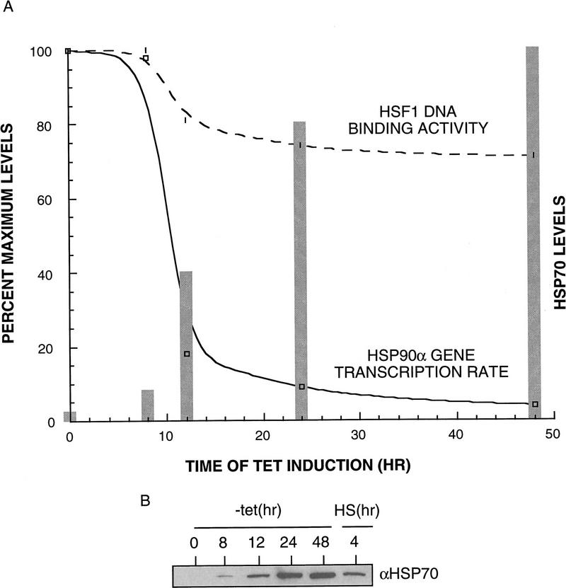
Is the transcriptional repression by Hsp70 dependent on a specific level of Hsp70? To assess the levels of Hsp70 necessary to block heat shock gene transcription, the PETA70 cells were induced over a period of 48 hr to accumulate Hsp70. During this period, aliquots of cells were removed at various time points, exposed to heat shock, and examined for the levels of Hsp70, transcription of endogenous heat shock genes, and HSF1 DNA-binding activity. Relative to the typical pattern of heat shock transcription observed in the nonoverexpressing cells (maintained in the presence of tetracycline), the transcription of the endogenous hsp90α gene was reduced 6-fold after 12 hr of Hsp70 induction, an 11-fold repression was observed after 24 hr, and a 25-fold repression after 48 hr (Fig. 10A). During this period, the levels of HSF1 DNA binding were initially unaffected and reduced by 25% at later time points (Fig. 10A). The levels of Hsp70 induced over this period were measured by Western blot analysis and compared to the levels of Hsp70 accumulated during attenuation of the heat shock response (4-hr heat shock). The level of Hsp70 achieved during attenuation was equivalent to that achieved upon 12-hr tetracycline induction (Fig. 10B) and corresponds to a 300-fold excess of Hsp70 to HSF1. These results reveal that Hsp70 primarily affects the transcriptional activity of HSF1 with only modest effects on HSF1 DNA-binding activity.
Figure 10.
Analysis of heat shock gene transcription in cells expressing different levels of Hsp70. Hsp70 was induced to various levels by withdrawal of anhydrotetracycline from media for 0, 8, 12, 24, and 48 hr. (A) The transcription rate of the hsp90α gene upon 1-hr heat shock treatment at 42°C was examined by run-on transcription analysis, quantified by a PhosphorImager, normalized to the signal obtained for pBR322 and gapdh genes, and plotted against time of anhydrotetracycline withdrawal (hr) as a solid line. HSF1 DNA-binding activity following the same treatment was measured by gel shift assay, quantified with a PhosphorImager, and plotted as a broken line. The levels of induced Hsp70 at different time points were measured by Western blot analysis, quantified with densitometry, and plotted as bars. (B) Hsp70 levels induced by anhydrotetracycline withdrawal were compared with that accumulated during 4-hr heat shock treatment at 41°C (intermediate heat shock temperature for PETA70 cells ranges from 39°C to 42°C, with the highest Hsp70 accumulation at 41°C, data not shown). Western blot analysis was performed with Hsp70-specific antibody 4G4. The relative Hsp70 levels were quantified by densitometry.
In summary, the results presented here indicate that the attenuation of the heat shock transcriptional response is a multistep event in which the elevated synthesis and accumulation of Hsp70 lead directly to the binding of Hsp70 to the HSF1 activation domain, and result in the repression of heat shock-induced transcription. Subsequent to transcriptional arrest is the conversion of HSF1 trimers to monomers and the loss of HSF1 DNA binding.
Discussion
The transcription of heat shock genes is induced transiently upon continuous exposure to elevated temperatures. Attenuation of the inducible response reveals that even in the presence of the stress signal, the elevated expression of heat shock genes either dampens the signal or negatively regulates components of the activation pathway. Conditional overexpression of Hsp70 in human cells is by itself sufficient to repress heat shock-induced transcription. These results, together with the in vivo and in vitro demonstration that chaperones associate directly with the HSF1 transactivation domain, establish a role for Hsp70 (and other chaperones) as transcriptional repressors.
A role for molecular chaperones in the autoregulation of the heat shock response is consistent with observations in Drosophila and other organisms in which exposure to amino acid analogs leads to continuous activation of heat shock gene expression (DiDomenico et al. 1982b; Mosser et al. 1988). The incorporation of amino acid analogs into nascent polypeptides that consequently do not fold to the native state sequesters the preexisting Hsp70 into futile cycles of chaperone–substrate interactions rather than the transient interactions between early folding intermediates and Hsp70 (Beckmann et al. 1990). Furthermore, as the Hsp70 synthesized during amino acid analog-induced stress is itself misfolded due to incorporation of amino acid analogs, the newly synthesized chaperone is nonfunctional and cannot repress HSF activity. Consequently, HSF, which is a stable protein and not dependent on continuous protein synthesis, remains in a transcriptionally active state (Zimarino and Wu 1987; Amici et al. 1992).
The consequence of elevated levels of Hsp70 appears specific to transcriptional repression of heat shock genes with little or no immediate effect on other features of HSF1 such as DNA-binding or stress-induced phosphorylation. This reveals that the attenuation phase of the heat shock response is comprised of distinct events in which the rate of heat shock gene transcription can be uncoupled from the DNA-binding and inducibly phosphorylated state of HSF1. These conclusions are independently supported by a detailed kinetic analysis of heat shock gene transcription, HSF1 DNA binding, and inducible phosphorylation, which showed that the rate of heat shock gene transcription declined prior to the loss of HSF1 DNA-binding activity (Kline and Morimoto 1997). Likewise, upon exposure to transient heat shock (42°C) and recovery at 37°C, heat shock gene transcription was rapidly repressed, yet HSF1 DNA-binding activity was sustained as measured by in vitro gel mobility shift assay and by in vivo footprinting assay (Abravaya et al. 1991). The uncoupling of HSF1 DNA binding from transcriptional activity noted here during the attenuation of the heat shock response has also been observed during activation of HSF1 by anti-inflammatory drugs. Under these conditions activation of HSF1 leads to a DNA-binding-competent form that lacks transcriptional activity (Jurivich et al. 1992; Giardina and Lis 1995; Cotto et al. 1996).
Although our data are consistent with a role for Hsp70 as a transcriptional repressor, we are not proposing a novel activity for Hsp70 that is distinct from its properties in other chaperone–substrate complexes. The demonstration that overexpression of Hsp70 does not directly affect the acquisition of HSF1 DNA-binding activity is consistent with previous studies in rat M21 and TTRat1 cells and in Drosophila TTSL2 cells in which the constitutive overexpression of Hsp70 did not interfere with the activation of HSF1 DNA-binding activity (Rabindran et al. 1994). Increased levels of Hsp70 had a slight effect on the kinetics of deactivation of HSF1 DNA-binding activity (Mosser et al. 1993; Rabindran et al. 1994). The regulation of heat shock genes in the yeast Saccharomyces cerevisiae involves repression of the transcriptional activity of HSFs independent of effects on the constitutively trimeric form of HSFs (Boorstein and Craig 1990). The convergence of genetic and biochemical observations, from yeast to humans, reveals that the transcriptional activity of HSF is intimately linked to autoregulatory aspects of Hsp70.
Although much of the emphasis on the role of heat shock proteins in the regulation of the heat shock response has centered on Hsp70, our data also reveal a role for other molecular chaperones such as Hdj1. The interaction of Hdj1 with HSF1 in higher eukaryotes and its repressive effect on HSF1 transcriptional activity is supported in yeast by the observation that the S. cerevisiae DnaJ homolog SIS1 negatively regulates its own expression. However, SIS1 autoregulation requires the HSE and other sequences, suggesting that additional regulatory molecules might be involved (Zhong et al. 1996). HSF may also associate with Hsp90; however this result seems variable as such associations have been detected with yeast, rat, and rabbit Hsp90 (Nadeau et al. 1993; Nair et al. 1996) but not with human Hsp90 (this study; Baler et al. 1992; Rabindran et al. 1994). A related issue is whether autoregulation requires elevated levels of specific chaperones or that the concentration of one or more chaperones increases substantially over a relatively short period of time to signal attenuation. Although the data presented here provide a clear demonstration that transcriptional repression occurs when a certain level of Hsp70 is attained, the constitutively expressed member of the Hsp70 family, Hsc70, is typically expressed at high levels in human cells. However, because Hsc70 is normally engaged in a variety of cellular activities, including protein synthesis, protein assembly and translocation, and protein degradation, presumably it is not available to negatively regulate HSF1.
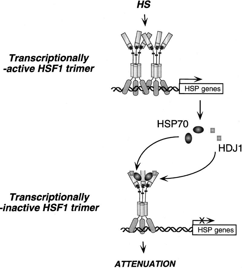
Regardless of how chaperones lead to inaccessibility of the HSF1 activation domain, Hsp70 and other molecular chaperones protect the cell against the deleterious effects of protein aggregates by stabilizing and refolding stress-induced folding intermediates. This suggests that an equilibrium must exist between protecting proteins against stress-induced damage and the activation and attenuation of the heat shock transcriptional response. Although it is unclear which component(s) of the basal transcriptional machinery contacts HSF1 to effect the high level of inducible transcription, these interactions are also likely to be transient. As the levels of Hsp70 increase in the nuclear compartment during heat shock (Velazquez et al. 1980; Welch and Feramisco 1984) or by conditional overexpression (data not shown), HSF1 is provided with an alternative interaction leading to the Hsp70-dependent repression of heat shock gene transcription. A model of the regulation of HSF1 transcriptional activity by molecular chaperones is presented in Figure 11. It is striking that the effects of overexpression of Hsp70 are specific to heat shock transcription and that the formation of stress-induced phosphorylated HSF1 trimers are unaffected. Presumably other stress-induced events, perhaps working in conjunction with Hsp70, are important for dephosphorylation and conversion of HSF1 trimers to monomers.
Figure 11.
Model for regulation of the heat shock transcriptional response by Hsp70 and Hdj1. Upon heat shock, HSF1 is rapidly converted to a transcriptionally active trimer, which binds to HSEs upstream of heat shock genes. This leads to the elevated synthesis of heat shock proteins, such as Hsp70 and Hdj1. Hsp70 and Hdj1 bind to the HSF1 activation domain, repressing its transcriptional activity, and leading to attenuation of the heat shock response.
Although our data do not reveal how the interaction between a chaperone and the activation domain of HSF1 leads to transcriptional repression, the presence of a chaperone-binding site within the activation domain could either competitively sequester the contact site(s) between HSF1 and the transcriptional apparatus or lead to a chaperone-dependent change in the conformation of HSF1. In either case, the activation domain is rendered inaccessible to the transcriptional machinery. This shares certain parallels with the role of the DnaK and DnaJ chaperones in the regulation of RepA (Wickner et al. 1991) and in phage λ replication (Georgopoulos 1990). In the former case, chaperones function as a conformational activator of RepA, and in the latter case, the chaperones interfere with the interaction of λ P protein to dnaB, thus allowing unwinding of the phage chromosome. A role for chaperones as regulators of transcriptional activators has also been described for the family of steroid aporeceptors, although in this case multiple chaperones are recruited to maintain the activator in a repressed state by formation of a stable chaperone–substrate complex (Bohen and Yamamoto 1994). It is tempting to consider that the interactions observed between p53 and Hsp70 (Hsc70) could also reflect a form of chaperone-dependent regulation of a transcriptional activator (Hupp et al. 1992).
Materials and methods
Plasmid constructions and protein purification
The GST–HSF1 activation domain (amino acids 395–503) fusion construct was created by EcoRI digestion of the corresponding GAL4–HSF1 fusion construct X (Shi et al. 1995), which has two EcoRI sites—one located in the polylinker region between the GAL4 DNA-binding domain and the HSF1 activation domain coding sequence, and the other located beyond the HSF1 stop codons—to generate a 640-bp fragment containing the HSF1 activation domain-coding region. The 640-bp EcoRI fragment was ligated into EcoRI-digested pGEX-5X-1 vector (Pharmacia–LKB). Constructs of the HSF1 activation domain–GST fusion deletion mutants II, III, IV, and V were created similarly from corresponding GAL4–HSF1 fusion constructs XI, XII, XIII, and XIV (Shi et al. 1995). All constructs were sequenced across the junction between the GST and HSF1 sequences to ensure that the HSF1 reading frame was maintained and to confirm the boundaries of the deletion mutants. β-Actin hsp70 AAAA was constructed by PCR mutating the last 4 amino acids of Hsp70 from EEVD to AAAA with plasmid pH2.3 (Wu et al. 1985) as a PCR template and creating a BamHI site at the 5′ end and an EcoRI site, followed by a BamHI site at the 3′ end of the PCR product. The PCR product was digested with BamHI and ligated into BamHI-digested pHβ-actin-1–neo vector (Gunning et al. 1987) so that the amplified hsp70 AAAA was under the control of the human β-actin promoter. Mutation of the residues was confirmed by sequencing. β-Actin hdj1 was created by PCR with pGEX–hdj1 (B.C. Freeman, unpubl.) as a template to create a BamHI site at each end of the amplified human hdj1 DNA fragment, and the BamHI-digested PCR product was ligated into BamHI-digested pHβ-actin-1–neo vector to be downstream of the human β-actin promoter. All DNA fragments that were amplified by PCR were sequenced in their entirety to ensure that mutations had not occurred randomly. pTR-DC/hsp70–gfp was created as described (Mosser et al. 1997). pTR-DC/hsp70 AAAA–gfp was created by ClaI and EcoRI digestion of β-actin hsp70 AAAA, and the 490-bp ClaI–EcoRI fragment containing the AAAA mutation was ligated into ClaI and EcoRI partially digested pTR-DC/hsp70–gfp to replace the corresponding wild-type fragment of hsp70. The purification of GST fusion proteins, wild-type or mutant Hsp70, Hsc70, Hdj1, and Hsp90, has been described (Freeman et al. 1995; Freeman and Morimoto 1996).
GST in vitro binding assays
HeLa whole cell extracts or purified recombinant proteins were incubated with purified GST or GST–HSF1 fusions fixed on glutathione-agarose beads. The beads were washed four times in 100 bed volumes of NETN buffer (0.5% NP-40, 0.1 mm EDTA, 20 mm Tris at pH 7.4, 300 mm NaCl) for HeLa whole cell extracts, and TEN buffer (20 mm Tris at pH 7.4, 0.1 mm EDTA, 100 mm NaCl) for purified proteins. The bound proteins were eluted by boiling in SDS–sample buffer and analyzed by SDS-PAGE and Coomassie blue staining or Western blot analysis. To examine the nucleotide effect on the interaction, purified recombinant Hsp70/Hdj1 proteins were incubated with GST–HSF1 activation domain in the absence or presence of 1 mm ATP, or 1 mm ATP-γs in TEK–Mg buffer (20 mm Tris at pH 7.4, 0.1 mm EDTA, 50 mm KCl, 5 mm MgCl2).
Cell culture, metabolic labeling, and transient and stable transfection
HeLa cells were grown in Dulbecco’s modified Eagle medium (DMEM) supplemented with 5% calf serum (GIBCO BRL) in a humidified 5% CO2 incubator at 37°C. For labeling, cells were washed with methionine-deficient DMEM and labeled for 12 hr in 1 ml of the same medium supplemented with 100 μCi of Tran35S-label (ICN), then washed with 1× PBS and harvested. COS-7 cells were grown in DMEM supplemented with 10% calf serum in a 37°C incubator with 5% CO2. Cells at a density of 40%–50% confluence were transfected by the calcium phosphate precipitation method (Shi et al. 1995). In all of the transient transfection experiments, cells were cotransfected with 1 μg of GAL4–HSF1 activation domain plasmid, 2 μg of G5BCAT reporter plasmid, 1 μg of RSV–β-gal internal control plasmid, and various amounts of Hsp70- or Hdj1-expressing plasmid as indicated in the legends to Figures 5 and 6. PETA70 cells (Mosser et al. 1997) were grown in RPMI 1640 supplemented with 10% fetal calf serum (Sigma), 2 mm l-glutamine (GIBCO BRL), 200 μg/ml of G418 (GIBCO BRL), 100 μg/ml of hygromycin (Sigma), and 10 ng/ml of anhydrotetracycline (Acros Organics). Anhydrotetracycline was withdrawn from the media to induce Hsp70 expression. PETA70 cells were transfected by electroporation (Mosser et al. 1997). The PERTA70–AAAA cell line was generated by cotransfection of pTR-DC/hsp70 AAAA–gfp plasmid with the plasmid ptk/hygro into PEER cells expressing the reverse tetracycline-controlled transactivator (rtTA) (Gossen et al. 1995). Hygromycin-resistant cells (200 μg/ml) were selected in batch culture, and cells with tetracycline-regulatable expression of the Hsp70 AAAA mutant protein were selected by adding the tetracycline derivative doxycycline (Sigma) into media followed by flow cytometric cell sorting of the GFP-positive cells as described (Mosser et al. 1997). PERTA70-AAAA cells were grown in RPMI 1640 supplemented with 10% fetal calf serum, 2 mm l-glutamine, 200 μg/ml of G418, and 100 μg/ml of hygromycin. Doxycycline (1 μg/ml) was added to the media to induce the expression of the Hsp70 AAAA mutant.
CAT, gel mobility shift, Western blot, and immunoprecipitation assays
Conditions for the CAT assay and GAL4 gel mobility shift assay were as described previously (Shi et al. 1995). The HSF1 gel mobility shift assay was as described (Mosser et al. 1988). The antibody supershift assay was performed with Hsp70-specific antibody C92 (Amersham) as described (Abravaya et al. 1992). Western blot analysis for HSF1 was performed with HSF1-specific polyclonal antibody (Sarge et al. 1993) or monoclonal antibody 4B4 (Cotto et al. 1997). Western blot analysis for Hsp70 was performed with Hsp70-specific monoclonal antibody 3A3, 5A5, 4G4 (S.P. Murphy, unpubl.), or C92 (Amersham). Western blot analysis for Hdj1 was performed with Hsp40-specific polyclonal antibody (Hattori et al. 1992). The condition for Western blot analysis was as described (Sarge et al. 1993). Monoclonal antibodies 3A3 and 5A5 recognize both Hsp70 and Hsc70. Monoclonal antibodies 4G4 and C92 specifically recognize Hsp70 but not the other members of the 70-kD heat shock proteins. Immunoprecipitations were carried out with either monoclonal antibody specific to Hsp70 (C92) or polyclonal antibody specific to HSF1 as described (Kline and Morimoto 1997).
Transcriptional run-on analysis
Run-on transcription reactions were performed with isolated cell nuclei in the presence of 50 μCi of [α-32P]UTP (Amersham) as described previously (Banerji et al. 1984). Radioactive RNA was hybridized to DNA probes for the human hsp70 gene (pH 2.3) (Wu et al. 1985), the human hsp90α gene (pUC801) (Hickey et al. 1989), pBR322 (Promega) as a control for nonspecific hybridization, and the rat gapdh gene (Fort et al. 1985) as a normalization control for transcription. The intensities of radioactive signals were quantitated by using a PhosphorImager (Molecular Dynamics).
Acknowledgments
We thank Brian Freeman and David Bimston for providing purified chaperone proteins and valuable discussion, Sue Fox for excellent technical assistance and comments, Jose Cotto for advice, and Sameer Mathur and Anu Mathew for comments. Antoine Caron provided assistance in the selection of stable cell lines, Steven Triezenberg (Michigan State University) provided the GAL4–VP16 (pSGVP) construct, James Douglas Engel (Northwestern University) provided the GAL4–GATA3 construct, and Kenzo Ohtsuka (Aichi Cancer Research Institute, Nagoya, Japan) provided the Hsp40 specific antibody. These studies were supported by a grant to R.M. from the National Institutes of Health. Y.S. is supported in part by a Gramm Travel Fellowship Award from the Lurie Cancer Center of Northwestern University.
The publication costs of this article were defrayed in part by payment of page charges. This article must therefore be hereby marked “advertisement” in accordance with 18 USC section 1734 solely to indicate this fact.
Footnotes
E-MAIL r-morimoto@nwu.edu; FAX (847) 491-4461.
References
- Abravaya K, Phillips B, Morimoto RI. Attenuation of the heat shock response in HeLa cells is mediated by the release of bound heat shock transcription factor and is modulated by changes in growth and in heat shock temperatures. Genes & Dev. 1991;5:2117–2127. doi: 10.1101/gad.5.11.2117. [DOI] [PubMed] [Google Scholar]
- Abravaya K, Myers MP, Murphy SP, Morimoto RI. The human heat shock protein Hsp70 interacts with HSF, the transcription factor that regulates heat shock gene expression. Genes & Dev. 1992;6:1153–1164. doi: 10.1101/gad.6.7.1153. [DOI] [PubMed] [Google Scholar]
- Amici C, Sistonen L, Santoro MG, Morimoto RI. Antiproliferative prostaglandins activate heat shock transcription factor. Proc Natl Acad Sci. 1992;89:6227–6231. doi: 10.1073/pnas.89.14.6227. [DOI] [PMC free article] [PubMed] [Google Scholar]
- Baeuerle PA. Inducible gene expression. Cambridge, MA: Birkhauser Boston; 1995. [Google Scholar]
- Baler R, Welch WJ, Voellmy R. Heat shock gene regulation by nascent polypeptides and denatured proteins: Hsp70 as a potential autoregulatory factor. J Cell Biol. 1992;117:1151–1159. doi: 10.1083/jcb.117.6.1151. [DOI] [PMC free article] [PubMed] [Google Scholar]
- Banerji SS, Laing K, Morimoto RI. Erythroid lineage-specific expression and inducibility of the major heat shock protein Hsp70 during avian embryogenesis. Genes & Dev. 1984;1:946–953. doi: 10.1101/gad.1.9.946. [DOI] [PubMed] [Google Scholar]
- Beckmann RP, Mizzen LA, Welch WJ. Interaction of Hsp70 with newly synthesized proteins: Implications for protein folding and assembly. Science. 1990;248:850–854. doi: 10.1126/science.2188360. [DOI] [PubMed] [Google Scholar]
- Blaszczak A, Zylicz M, Georgopoulos C, Liberek K. Both ambient temperature and the DnaK chaperone machine modulate the heat shock response in Escherichia coli by regulating the switch between σ70 and σ32 factors assembled with RNA polymerase. EMBO J. 1995;14:5085–5093. doi: 10.1002/j.1460-2075.1995.tb00190.x. [DOI] [PMC free article] [PubMed] [Google Scholar]
- Bohen SP, Yamamoto KR. Modulation of steroid receptor signal transduction by heat shock proteins. In: Morimoto RI, Tissieres A, Georgopoulos C, editors. The biology of heat shock proteins and molecular chaperones. Cold Spring Harbor, NY: Cold Spring Harbor Laboratory Press; 1994. pp. 313–334. [Google Scholar]
- Boorstein WR, Craig EA. Transcriptional regulation of ssa3, an hsp70 gene from Saccharomyces cerevisiae. Mol Cell Biol. 1990;10:3262–3267. doi: 10.1128/mcb.10.6.3262. [DOI] [PMC free article] [PubMed] [Google Scholar]
- Clarke CF, Cheng K, Frey AB, Stein R, Hinds PH, Levine AJ. Purification of complexes of nuclear oncogene p53 with rat and Escherichia coli heat shock proteins: In vitro dissociation of Hsc70 and DnaK from murine p53 by ATP. Mol Cell Biol. 1988;8:1206–1215. doi: 10.1128/mcb.8.3.1206. [DOI] [PMC free article] [PubMed] [Google Scholar]
- Cotto JJ, Kline M, Morimoto RI. Activation of heat shock factor 1 DNA binding precedes stress-induced serine phosphorylation. J Biol Chem. 1996;271:3355–3358. doi: 10.1074/jbc.271.7.3355. [DOI] [PubMed] [Google Scholar]
- Cotto JJ, Fox SG, Morimoto RI. HSF1 granules: I. A novel stress-induced nuclear compartment of human cells. J Cell Sci. 1997;110:2925–2934. doi: 10.1242/jcs.110.23.2925. [DOI] [PubMed] [Google Scholar]
- Craig EA, Gross CA. Is Hsp70 the cellular thermometer? Trends Biochem Sci. 1991;16:135–140. doi: 10.1016/0968-0004(91)90055-z. [DOI] [PubMed] [Google Scholar]
- Craig EA, Jakobsen K. Mutations of the heat inducible 70 kilodalton genes of yeast confer temperature sensitive growth. Cell. 1984;38:841–849. doi: 10.1016/0092-8674(84)90279-4. [DOI] [PubMed] [Google Scholar]
- DiDomenico BJ, Bugaisky GE, Lindquist S. Heat shock and recovery are mediated by different translational mechanisms. Proc Natl Acad Sci. 1982a;79:6181–6185. doi: 10.1073/pnas.79.20.6181. [DOI] [PMC free article] [PubMed] [Google Scholar]
- ————— The heat shock response is self-regulated at both the transcriptional and posttranscriptional levels. Cell. 1982b;31:593–603. doi: 10.1016/0092-8674(82)90315-4. [DOI] [PubMed] [Google Scholar]
- Flynn GC, Chappell TG, Rothman JE. Peptide binding and release by proteins implicated as catalysts of protein assembly. Science. 1989;245:385–390. doi: 10.1126/science.2756425. [DOI] [PubMed] [Google Scholar]
- Fort P, Marty L, Piechaczyk M, El Sabrouty S, Dani C, Jeanteur P, Blanchard JM. Various rat adult tissues express only one major mRNA species from the glyceraldehyde-3-phosphate dehydrogenase multigenic family. Nucleic Acids Res. 1985;13:1431–1442. doi: 10.1093/nar/13.5.1431. [DOI] [PMC free article] [PubMed] [Google Scholar]
- Freeman BC, Morimoto RI. The human cytosolic molecular chaperones Hsp90, Hsp70(Hsc70) and Hdj1 have distinct roles in recognition of a non-native protein and protein refolding. EMBO J. 1996;15:2969–2979. [PMC free article] [PubMed] [Google Scholar]
- Freeman BC, Myers MP, Schumacher R, Morimoto RI. Identification of a regulatory motif in Hsp70 that affects ATPase activity, substrate binding and interaction with Hdj1. EMBO J. 1995;14:2281–2292. doi: 10.1002/j.1460-2075.1995.tb07222.x. [DOI] [PMC free article] [PubMed] [Google Scholar]
- Gamer J, Multhaup G, Tomoyasu T, McCarty JS, Rudiger S, Schonfeld H-J, Schirra C, Bujard H, Bukau B. A cycle of binding and release of the DnaK, DnaJ and GrpE chaperones regulates activity of the Escherichia coli heat shock transcription factor σ32. EMBO J. 1996;15:607–617. [PMC free article] [PubMed] [Google Scholar]
- Georgopoulos C, Ang D, Liberek K, Zylicz M. Properties of the Escherichia coli heat shock proteins and their role in bacteriophage λ growth. In: Morimoto RI, Tissieres A, Georgopoulos C, editors. Stress proteins in biology and medicine. Cold Spring Harbor, NY: Cold Spring Harbor Laboratory Press; 1990. pp. 191–221. [Google Scholar]
- Giardina C, Lis JT. Sodium salicylate and yeast heat shock gene transcription. J Biol Chem. 1995;270:10369–10372. doi: 10.1074/jbc.270.18.10369. [DOI] [PubMed] [Google Scholar]
- Gossen M, Freundlieb S, Bender G, Muller G, Hillen W, Bujard H. Transcriptional activation by tetracyclines in mammalian cells. Science. 1995;268:1766–1769. doi: 10.1126/science.7792603. [DOI] [PubMed] [Google Scholar]
- Grossman AD, Straus DB, Walter WA, Gross CA. σ32 synthesis can regulate the synthesis of heat shock proteins in Escherichia coli. Genes & Dev. 1987;1:179–184. doi: 10.1101/gad.1.2.179. [DOI] [PubMed] [Google Scholar]
- Gunning P, Leavitt J, Muscat G, Ng S, Kedes L. A human β-actin expression vector system directs high-level accumulation of antisense transcripts. Proc Natl Acad Sci. 1987;84:4831–4835. doi: 10.1073/pnas.84.14.4831. [DOI] [PMC free article] [PubMed] [Google Scholar]
- Hattori H, Liu YC, Tohnai I, Ueda M, Kaneda T, Kobayashi T, Tanabe K, Ohtsuka K. Intracellular localization and partial amino acid sequence of a stress-inducible 40-kDa protein in HeLa cells. Cell Struct Func. 1992;17:77–86. doi: 10.1247/csf.17.77. [DOI] [PubMed] [Google Scholar]
- Hickey E, Brandon SE, Smale G, Lloyd D, Weber LA. Sequence and regulation of a gene encoding a human 89-kilodalton heat shock protein. Mol Cell Biol. 1989;9:2615–2626. doi: 10.1128/mcb.9.6.2615. [DOI] [PMC free article] [PubMed] [Google Scholar]
- Hohfeld J, Minami Y, Hartl F-U. Hip, a novel co-chaperone involved in the eukaryotic Hsc70/Hsp40 reaction cycle. Cell. 1995;83:589–598. doi: 10.1016/0092-8674(95)90099-3. [DOI] [PubMed] [Google Scholar]
- Hupp TR, Meek DW, Midgley CA, Lane DP. Regulation of the specific DNA binding function of p53. Cell. 1992;71:875–886. doi: 10.1016/0092-8674(92)90562-q. [DOI] [PubMed] [Google Scholar]
- Jakobsen BK, Pelham HRB. Constitutive binding of yeast heat shock factor to DNA in vivo. Mol Cell Biol. 1988;8:5040–5042. doi: 10.1128/mcb.8.11.5040. [DOI] [PMC free article] [PubMed] [Google Scholar]
- Jurivich DA, Sistonen L, Kroes RA, Morimoto RI. Effect of sodium salicylate on the human heat shock response. Science. 1992;255:1243–1245. doi: 10.1126/science.1546322. [DOI] [PubMed] [Google Scholar]
- Kline MP, Morimoto RI. Repression of the heat shock factor 1 transcriptional activation domain is modulated by constitutive phosphorylation. Mol Cell Biol. 1997;17:2107–2115. doi: 10.1128/mcb.17.4.2107. [DOI] [PMC free article] [PubMed] [Google Scholar]
- Kost JL, Smith DF, Sullivan WP, Welch WJ, Toft DO. Binding of heat shock proteins to the avian progesterone receptor. Mol Cell Biol. 1989;9:3829–3838. doi: 10.1128/mcb.9.9.3829. [DOI] [PMC free article] [PubMed] [Google Scholar]
- Liberek K, Galitski TP, Zylicz M, Georgopoulos C. The DnaK chaperone modulates the heat shock response of Escherichia coli by binding to the σ32 transcription factor. Proc Natl Acad Sci. 1992;89:3516–3520. doi: 10.1073/pnas.89.8.3516. [DOI] [PMC free article] [PubMed] [Google Scholar]
- Lis JT, Wu C. Protein traffic on the heat shock promoter: Parking, stalling and trucking along. Cell. 1993;74:1–20. doi: 10.1016/0092-8674(93)90286-y. [DOI] [PubMed] [Google Scholar]
- Morimoto RI. Cells in stress: Transcriptional activation of heat shock genes. Science. 1993;269:1409–1410. doi: 10.1126/science.8451637. [DOI] [PubMed] [Google Scholar]
- Morimoto RI, Tissieres A, Georgopoulos C. The stress response, function of the proteins, and perspectives. In: Morimoto RI, Tissieres A, Georgopoulos C, editors. Stress proteins in biology and medicine. Cold Spring Harbor, NY: Cold Spring Harbor Laboratory Press; 1990. pp. 1–36. [Google Scholar]
- Mosser DD, Theodorakis NG, Morimoto RI. Coordinate changes in heat shock element binding activity and hsp70 gene transcription rates in human cells. Mol Cell Biol. 1988;8:4736–4744. doi: 10.1128/mcb.8.11.4736. [DOI] [PMC free article] [PubMed] [Google Scholar]
- Mosser DD, Duchaine J, Massie B. The DNA-binding activity of the human heat shock transcription factor is regulated in vivo by Hsp70. Mol Cell Biol. 1993;13:5427–5438. doi: 10.1128/mcb.13.9.5427. [DOI] [PMC free article] [PubMed] [Google Scholar]
- Mosser DD, Caron AW, Bourget L, Denis-Larose C, Massie B. Role of the human heat shock protein Hsp70 in protection against stress-induced apoptosis. Mol Cell Biol. 1997;17:5317–5327. doi: 10.1128/mcb.17.9.5317. [DOI] [PMC free article] [PubMed] [Google Scholar]
- Nadeau K, Das A, Walsh CT. Hsp90 chaperonins possess ATPase activity and bind heat shock transcription factors and peptidyl prolyl isomerase. J Biol Chem. 1993;268:1479–1487. [PubMed] [Google Scholar]
- Nair SC, Toran EJ, Rimerman RA, Hjermstad S, Smithgall TE, Smith DF. A pathway of multi-chaperone interactions common to diverse regulatory proteins: estrogen receptor, Fes tyrosine kinase, heat shock transcription factor HSF1, and the aryl hydrocarbon receptor. Cell Stress & Chaperones. 1996;1:237–250. doi: 10.1379/1466-1268(1996)001<0237:apomci>2.3.co;2. [DOI] [PMC free article] [PubMed] [Google Scholar]
- Palleros DR, Welch WJ, Fink AL. Interaction of Hsp70 with unfolded proteins: Effects of temperature and nucleotides on the kinetics of binding. Proc Natl Acad Sci. 1991;88:5719–5723. doi: 10.1073/pnas.88.13.5719. [DOI] [PMC free article] [PubMed] [Google Scholar]
- Pelham HRB. Speculations on the functions of the major heat shock and glucose regulated proteins. Cell. 1986;46:959–961. doi: 10.1016/0092-8674(86)90693-8. [DOI] [PubMed] [Google Scholar]
- Rabindran SK, Haroun RI, Clos J, Wisniewski J, Wu C. Regulation of heat shock factor trimer formation: Role of a conserved leucine zipper. Science. 1993;259:230–234. doi: 10.1126/science.8421783. [DOI] [PubMed] [Google Scholar]
- Rabindran SK, Wisniewski J, Li L, Li GC, Wu C. Interaction between heat shock factor and Hsp70 is insufficient to suppress induction of DNA-binding activity in vivo. Mol Cell Biol. 1994;14:6552–6560. doi: 10.1128/mcb.14.10.6552. [DOI] [PMC free article] [PubMed] [Google Scholar]
- Sarge K, Murphy SP, Morimoto RI. Activation of heat shock transcription by HSF1 involves oligomerization, acquisition of DNA binding activity, and nuclear localization and can occur in the absence of stress. Mol Cell Biol. 1993;13:1392–1407. doi: 10.1128/mcb.13.3.1392. [DOI] [PMC free article] [PubMed] [Google Scholar]
- Shi Y, Kroeger PE, Morimoto RI. The carboxyl-terminal transactivation domain of heat shock factor 1 is negatively regulated and stress responsive. Mol Cell Biol. 1995;15:4309–4318. doi: 10.1128/mcb.15.8.4309. [DOI] [PMC free article] [PubMed] [Google Scholar]
- Sorger PK, Nelson HCM. Trimerization of a yeast transcriptional activator via a coiled-coil motif. Cell. 1989;59:807–813. doi: 10.1016/0092-8674(89)90604-1. [DOI] [PubMed] [Google Scholar]
- Sorger PK, Lewis MJ, Pelham HRB. Heat shock factor is regulated differently in yeast and HeLa cells. Nature. 1987;329:81–84. doi: 10.1038/329081a0. [DOI] [PubMed] [Google Scholar]
- Straus DB, Walter WA, Gross CA. Dnak, DnaJ, and GrpE heat shock proteins negatively regulate heat shock gene expression by controlling the synthesis and stability of σ32. Genes & Dev. 1990;4:2202–2209. doi: 10.1101/gad.4.12a.2202. [DOI] [PubMed] [Google Scholar]
- Tilly K, McKittrick N, Zylicz M, Georgopoulos C. The DnaK protein modulates the heat shock response of Escherichia coli. Cell. 1983;34:641–646. doi: 10.1016/0092-8674(83)90396-3. [DOI] [PubMed] [Google Scholar]
- Tsai J, Douglas MG. A conserved HPD sequence of the J-domain is necessary for YDJ1 stimulation of Hsp70 ATPase activity at a site distinct from substrate binding. J Biol Chem. 1996;271:9347–9354. doi: 10.1074/jbc.271.16.9347. [DOI] [PubMed] [Google Scholar]
- Velazquez JM, DiDomenico BJ, Lindquist S. Intracellular localization of heat shock proteins in Drosophila. Cell. 1980;20:679–689. doi: 10.1016/0092-8674(80)90314-1. [DOI] [PubMed] [Google Scholar]
- Wawrzynow A, Zylicz M. Divergent effects of ATP on the binding of the DnaK and DnaJ chaperones to each other, or to their various native and denatured protein substrates. J Biol Chem. 1995;270:19300–19306. doi: 10.1074/jbc.270.33.19300. [DOI] [PubMed] [Google Scholar]
- Welch WJ, Feramisco JR. Nuclear and nucleolar localization of the 72,000-dalton heat shock protein in heat shocked mammalian cells. J Biol Chem. 1984;259:4501–4513. [PubMed] [Google Scholar]
- Westwood JT, Wu C. Activation of Drosophila heat shock factor: Conformational change associated with a monomer-to-trimer transition. Mol Cell Biol. 1993;13:3481–3486. doi: 10.1128/mcb.13.6.3481. [DOI] [PMC free article] [PubMed] [Google Scholar]
- Wickner S, Hoskins J, McKenney K. Monomerization of RepA dimers by heat shock proteins activates binding to DNA replication origin. Proc Natl Acad Sci. 1991;88:7903–7907. doi: 10.1073/pnas.88.18.7903. [DOI] [PMC free article] [PubMed] [Google Scholar]
- Wu C. Heat shock transcription factors: structure and regulation. Annu Rev Cell Dev Biol. 1995;11:441–469. doi: 10.1146/annurev.cb.11.110195.002301. [DOI] [PubMed] [Google Scholar]
- Wu B, Hunt C, Morimoto RI. Structure and expression of the human gene encoding major heat shock protein Hsp70. Mol Cell Biol. 1985;5:330–341. doi: 10.1128/mcb.5.2.330. [DOI] [PMC free article] [PubMed] [Google Scholar]
- Zhong T, Luke MM, Arndt KT. Tanscriptional regulation of the yeast DnaJ homolog SIS1. J Biol Chem. 1996;271:1349–1356. doi: 10.1074/jbc.271.3.1349. [DOI] [PubMed] [Google Scholar]
- Zimarino V, Wu C. Induction of sequence-specific binding of Drosophila heat shock activator proteins without protein synthesis. Nature. 1987;327:727–730. doi: 10.1038/327727a0. [DOI] [PubMed] [Google Scholar]


