Abstract
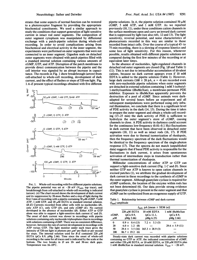
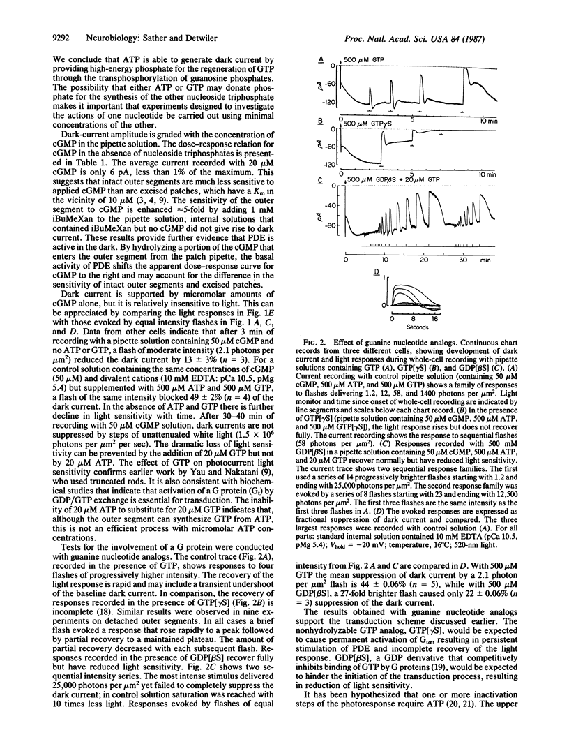
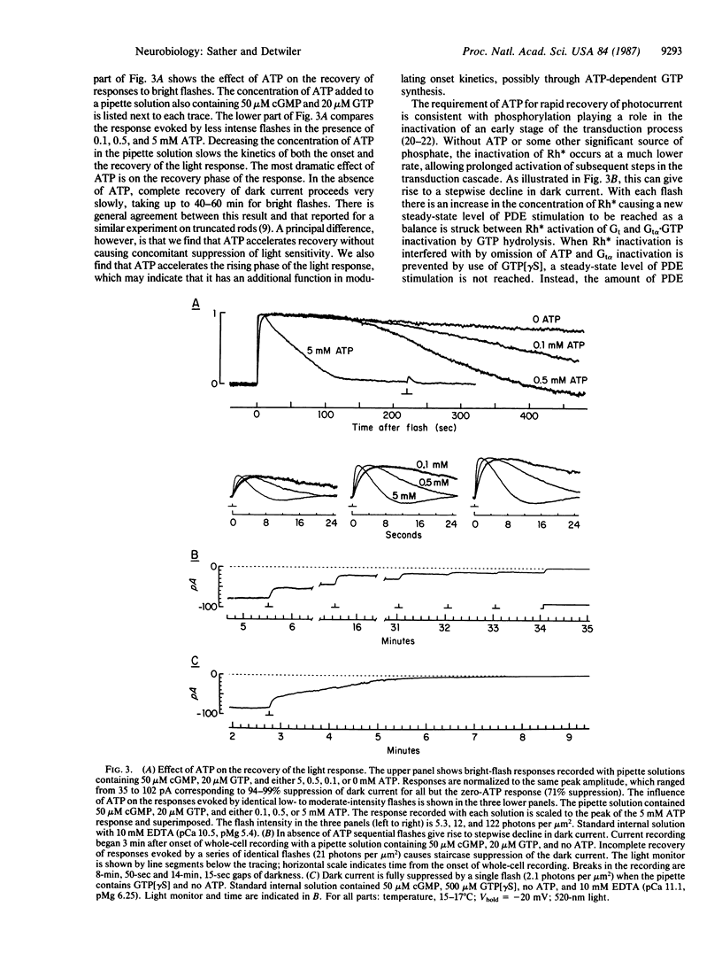
Recent progress in understanding phototransduction has come primarily from studies on cell-free systems. To investigate the transduction process under physiological conditions, a fully functional preparation of retinal rod outer segments without attached inner segments was developed that allows electrical recording of light-sensitive current during intracellular dialysis with defined solutions. No light-sensitive current is recorded from detached outer segments dialyzed with nucleotide-free solutions, whereas cells detached from the retina into Ringer's solution containing 3-isobutyl-1-methyl-xanthine (a phosphodiesterase inhibitor) develop a light-sensitive inward dark current. This indicates that there is a basal level of cGMP-specific phosphodiesterase activity in the dark. Detached outer segments dialyzed with greater than or equal to 20 microM cGMP rapidly develop a light-suppressible current. A current of similar magnitude is generated more slowly during dialysis with a 50-fold greater concentration of GTP. Apparently, cGMP can be synthesized from GTP by guanylate cyclase in the outer segment. Cells dialyzed with cGMP alone show a reduced light sensitivity that is restored to normal by addition of 20 microM GTP. This action of GTP is antagonized by guanosine 5'-[beta-thio]diphosphate. These findings are in good agreement with biochemical evidence indicating that a GTP-binding protein (transducin) plays a pivotal role in the generation of responses to light. The recovery of photocurrent following a brief flash is delayed or abolished by dialysis with solutions that lack ATP or contain guanosine 5'-[gamma-thio]triphosphate, a nonhydrolyzable GTP analog. These results support the view that both GTP hydrolysis by activated transducin and ATP-dependent phosphorylation of a rhodopsin photoproduct are necessary for termination of the transduction process.
Full text
PDF
Selected References
These references are in PubMed. This may not be the complete list of references from this article.
- Ames A., 3rd, Walseth T. F., Heyman R. A., Barad M., Graeff R. M., Goldberg N. D. Light-induced increases in cGMP metabolic flux correspond with electrical responses of photoreceptors. J Biol Chem. 1986 Oct 5;261(28):13034–13042. [PubMed] [Google Scholar]
- Baylor D. A., Matthews G., Yau K. W. Two components of electrical dark noise in toad retinal rod outer segments. J Physiol. 1980 Dec;309:591–621. doi: 10.1113/jphysiol.1980.sp013529. [DOI] [PMC free article] [PubMed] [Google Scholar]
- Bensinger R. E., Fletcher R. T., Chader G. J. Guanylate cyclase: inhibition by light in retinal photoreceptors. Science. 1974 Jan 11;183(4120):86–87. doi: 10.1126/science.183.4120.86. [DOI] [PubMed] [Google Scholar]
- Bodoia R. D., Detwiler P. B. Patch-clamp recordings of the light-sensitive dark noise in retinal rods from the lizard and frog. J Physiol. 1985 Oct;367:183–216. doi: 10.1113/jphysiol.1985.sp015820. [DOI] [PMC free article] [PubMed] [Google Scholar]
- Eckstein F., Cassel D., Levkovitz H., Lowe M., Selinger Z. Guanosine 5'-O-(2-thiodiphosphate). An inhibitor of adenylate cyclase stimulation by guanine nucleotides and fluoride ions. J Biol Chem. 1979 Oct 10;254(19):9829–9834. [PubMed] [Google Scholar]
- Fesenko E. E., Kolesnikov S. S., Lyubarsky A. L. Induction by cyclic GMP of cationic conductance in plasma membrane of retinal rod outer segment. Nature. 1985 Jan 24;313(6000):310–313. doi: 10.1038/313310a0. [DOI] [PubMed] [Google Scholar]
- Goldberg N. D., Ames A. A., 3rd, Gander J. E., Walseth T. F. Magnitude of increase in retinal cGMP metabolic flux determined by 18O incorporation into nucleotide alpha-phosphoryls corresponds with intensity of photic stimulation. J Biol Chem. 1983 Aug 10;258(15):9213–9219. [PubMed] [Google Scholar]
- Hagins W. A., Yoshikami S. Proceedings: A role for Ca2+ in excitation of retinal rods and cones. Exp Eye Res. 1974 Mar;18(3):299–305. doi: 10.1016/0014-4835(74)90157-2. [DOI] [PubMed] [Google Scholar]
- Haynes L. W., Kay A. R., Yau K. W. Single cyclic GMP-activated channel activity in excised patches of rod outer segment membrane. Nature. 1986 May 1;321(6065):66–70. doi: 10.1038/321066a0. [DOI] [PubMed] [Google Scholar]
- Liebman P. A., Pugh E. N., Jr ATP mediates rapid reversal of cyclic GMP phosphodiesterase activation in visual receptor membranes. Nature. 1980 Oct 23;287(5784):734–736. doi: 10.1038/287734a0. [DOI] [PubMed] [Google Scholar]
- Matthews G. Single-channel recordings demonstrate that cGMP opens the light-sensitive ion channel of the rod photoreceptor. Proc Natl Acad Sci U S A. 1987 Jan;84(1):299–302. doi: 10.1073/pnas.84.1.299. [DOI] [PMC free article] [PubMed] [Google Scholar]
- Matthews H. R., Torre V., Lamb T. D. Effects on the photoresponse of calcium buffers and cyclic GMP incorporated into the cytoplasm of retinal rods. Nature. 1985 Feb 14;313(6003):582–585. doi: 10.1038/313582a0. [DOI] [PubMed] [Google Scholar]
- Pannbacker R. G. Control of guanylate cyclase activity in the rod outer segment. Science. 1973 Dec 14;182(4117):1138–1140. doi: 10.1126/science.182.4117.1138. [DOI] [PubMed] [Google Scholar]
- Pugh E. N., Jr, Cobbs W. H. Visual transduction in vertebrate rods and cones: a tale of two transmitters, calcium and cyclic GMP. Vision Res. 1986;26(10):1613–1643. doi: 10.1016/0042-6989(86)90051-9. [DOI] [PubMed] [Google Scholar]
- Sitaramayya A., Liebman P. A. Phosphorylation of rhodopsin and quenching of cyclic GMP phosphodiesterase activation by ATP at weak bleaches. J Biol Chem. 1983 Oct 25;258(20):12106–12109. [PubMed] [Google Scholar]
- Stryer L. Cyclic GMP cascade of vision. Annu Rev Neurosci. 1986;9:87–119. doi: 10.1146/annurev.ne.09.030186.000511. [DOI] [PubMed] [Google Scholar]
- Torre V., Matthews H. R., Lamb T. D. Role of calcium in regulating the cyclic GMP cascade of phototransduction in retinal rods. Proc Natl Acad Sci U S A. 1986 Sep;83(18):7109–7113. doi: 10.1073/pnas.83.18.7109. [DOI] [PMC free article] [PubMed] [Google Scholar]
- Wilden U., Hall S. W., Kühn H. Phosphodiesterase activation by photoexcited rhodopsin is quenched when rhodopsin is phosphorylated and binds the intrinsic 48-kDa protein of rod outer segments. Proc Natl Acad Sci U S A. 1986 Mar;83(5):1174–1178. doi: 10.1073/pnas.83.5.1174. [DOI] [PMC free article] [PubMed] [Google Scholar]
- Yau K. W., Nakatani K. Light-suppressible, cyclic GMP-sensitive conductance in the plasma membrane of a truncated rod outer segment. Nature. 1985 Sep 19;317(6034):252–255. doi: 10.1038/317252a0. [DOI] [PubMed] [Google Scholar]
- Zimmerman A. L., Baylor D. A. Cyclic GMP-sensitive conductance of retinal rods consists of aqueous pores. Nature. 1986 May 1;321(6065):70–72. doi: 10.1038/321070a0. [DOI] [PubMed] [Google Scholar]
- de Azeredo F. A., Lust W. D., Passonneau J. V. Light-induced changes in energy metabolites, guanine nucleotides, and guanylate cyclase within frog retinal layers. J Biol Chem. 1981 Mar 25;256(6):2731–2735. [PubMed] [Google Scholar]


