Full text
PDF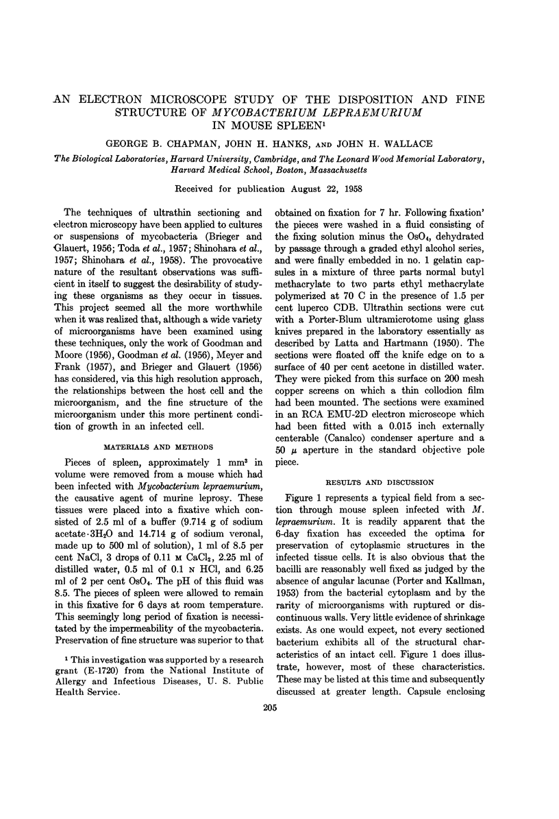
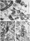
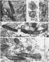
Images in this article
Selected References
These references are in PubMed. This may not be the complete list of references from this article.
- BAKER R. F., GOODMAN J. R., MOORE R. E. Electron microscopic study of phagocytosis of staphylococcus by human leukocytes. II. Virulent and non-virulent staphylococci. J Bacteriol. 1956 Dec;72(6):736–745. doi: 10.1128/jb.72.6.736-745.1956. [DOI] [PMC free article] [PubMed] [Google Scholar]
- BRIEGER E. M., GLAUERT A. M. Electron microscopy of the leprosy bacillus: a study of submicroscopical structure. Tubercle. 1956 Jun;37(3):195–206. doi: 10.1016/s0041-3879(56)80041-x. [DOI] [PubMed] [Google Scholar]
- CARO L. G., VAN TUBERGEN R. P., FORRO F., Jr The localization of deoxyribonucleic acid in Escherichia coli. J Biophys Biochem Cytol. 1958 Jul 25;4(4):491–494. doi: 10.1083/jcb.4.4.491. [DOI] [PMC free article] [PubMed] [Google Scholar]
- CHAPMAN G. B., HILLIER J. Electron microscopy of ultra-thin sections of bacteria I. Cellular division in Bacillus cereus. J Bacteriol. 1953 Sep;66(3):362–373. doi: 10.1128/jb.66.3.362-373.1953. [DOI] [PMC free article] [PubMed] [Google Scholar]
- GOODMAN J. R., MOORE R. E. Electron microscopic study of phagocytosis of Staphylococcus by human leukocytes. J Bacteriol. 1956 May;71(5):547–556. doi: 10.1128/jb.71.5.547-556.1956. [DOI] [PMC free article] [PubMed] [Google Scholar]
- HENDERSON H. J., MUDD S., TAKEYA K. Electron-scattering granules and reducing sites in mycobacteria. J Bacteriol. 1956 Dec;72(6):767–783. doi: 10.1128/jb.72.6.767-783.1956. [DOI] [PMC free article] [PubMed] [Google Scholar]
- KELLENBERGER E., RYTER A. Cell wall and cytoplasmic membrane of Escherichia coli. J Biophys Biochem Cytol. 1958 May 25;4(3):323–326. doi: 10.1083/jcb.4.3.323. [DOI] [PMC free article] [PubMed] [Google Scholar]
- KELLENBERGER E., RYTER A. Contribution à l'étude du noyau bactérien. Schweiz Z Pathol Bakteriol. 1955;18(5):1122–1137. [PubMed] [Google Scholar]
- LATTA H., HARTMANN J. F. Use of a glass edge in thin sectioning for electron microscopy. Proc Soc Exp Biol Med. 1950 Jun;74(2):436–439. doi: 10.3181/00379727-74-17931. [DOI] [PubMed] [Google Scholar]
- MEYER G. F., FRANK W. Elektronenmikroskopische Studlen zur intracellulären Symbiose verschiedener Insekten. I. Untersuchungen des Fettkörpers und der symbiontischen Bakterien der Kuchenschabe (Blatta orientalis L.). Z Zellforsch Mikrosk Anat. 1957;47(1):29–42. [PubMed] [Google Scholar]
- SHINOHARA C., FUKUSHI K., SUZUKI J. Mitochondria-like structures in ultrathin sections of Mycobacterium avium. J Bacteriol. 1957 Sep;74(3):413–415. doi: 10.1128/jb.74.3.413-415.1957. [DOI] [PMC free article] [PubMed] [Google Scholar]
- TODA T., KOIKE M., HIRAKI N., TAKEYA K. The intracellular structures of a mycobacterium. J Bacteriol. 1957 Mar;73(3):442–443. doi: 10.1128/jb.73.3.442-443.1957. [DOI] [PMC free article] [PubMed] [Google Scholar]


