Abstract
The bone morphogenetic proteins (BMPs), TGFβ superfamily members, play diverse roles in embryogenesis, but how the BMPs exert their action is unclear and how different BMP receptors (BMPRs) contribute to this process is not known. Here we demonstrate that the two type I BMPRs, BMPR-IA and BMPR-IB, regulate distinct processes during chick limb development. BmpR-IB expression in the embryonic limb prefigures the future cartilage primordium, and its activity is necessary for the initial steps of chondrogenesis. During later chondrogenesis, BmpR-IA is specifically expressed in prehypertrophic chondrocytes. BMPR-IA regulates chondrocyte differentiation, serving as a downstream mediator of Indian Hedgehog (IHH) function in both a local signaling loop and a longer-range relay system to PTHrP. BMPR-IB also regulates apoptosis: Expression of activated BMPR-IB results in increased cell death, and we showed previously that dominant-negative BMPR-IB inhibits apoptosis. Our studies indicate that in TGFβ signaling systems, different type I receptor isoforms are dedicated to specific functions during embryogenesis.
Keywords: Bone morphogenetic proteins, BMP receptor, cartilage differentiation, cell death, chondrogenesis, limb development
Several families of signaling molecules have been identified that act at multiple points in development to mediate cell–cell communication. The secreted bone morphogenetic proteins (BMPs) have been proposed to control many aspects of vertebrate development (for review, see Hogan 1996). With respect to the development of the vertebrate limb, BMPs have been implicated in the control of pattern formation, interdigital cell death, and bone morphogenesis (see references in Hogan 1996). The question of how BMP signaling mediates these disparate functions remains open.
BMPs can induce ectopic endochondral bone when introduced at intramuscular sites in adult rats (Wozney et al. 1988). Prior to the formation of cartilage elements, Bmp2, Bmp4, and Bmp7 are expressed in spatially and temporally dynamic patterns (Francis et al. 1994; Francis-West et al. 1995). Within the developing limb cartilage elements, Bmp2, Bmp4, and Bmp7 are expressed in the perichondrium (Lyons et al. 1990; Jones et al. 1991; Wozney et al. 1993; Macias et al. 1997), and Bmp6 is expressed in prehypertrophic and hypertrophic chondrocytes (Lyons et al. 1989; Wozney et al. 1993; Vortkamp et al. 1996). Gdf5, a BMP-related gene, is expressed in the developing joints and perichondrium (Chang et al. 1994; Storm et al. 1994). However, little is known about the cellular and molecular mechanisms underlying normal or ectopic bone morphogenesis by BMPs.
During endochondral bone development, a cartilage template forms when mesenchymal cells condense and differentiate into chondrocytes. These chondrocytes then undergo a program of proliferation, maturation, hypertrophy, calcification, and cell death (see Fig. 4I below; for review, see Erlebacher et al. 1995). Later, osteoblasts replace the cartilage matrix with bone matrix. This ossification initiates in the center of the cartilage shaft and then expands toward the ends of the cartilage element. Around the cartilage element is a sheath of cells, the perichondrium, which contributes to cartilage growth and regulates the rate of cartilage differentiation.
Figure 4.
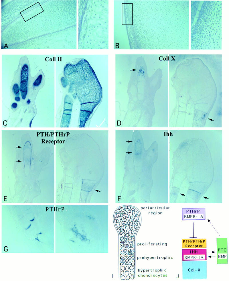
caBMPR-IA delays chondrocyte differentiation and induces PTHrP expression. Histological sections of day 9 embryonic wings (A) uninfected and (B) caBMPR-IA-infected at stage 14. The infected cartilage element lacked hypertrophic chondrocytes (HC) that were clearly present in the contralateral control. The boxed areas are enlarged on right. (C–F) Serial sections of day 8 uninfected and caBMPR-IA-infected wings (left and right, respectively). Infection was targeted to posterior distal limb mesenchyme at stage 21 resulting in expansion and fusion of the posterior digits. RNA in situ hybridization revealed that (C) Col-II was expressed in both the infected and uninfected limbs, indicating the cells were viable; (D) Col-X was expressed in HC in the uninfected limb and in the proximal part of the infected limb (arrows); however, it could not be detected in the infected digit region; (F) Ihh was expressed in prehypertrophic chondrocytes (preHC) in the uninfected limb and in the proximal part of the infected limb (arrows), but could not be detected in the infected digit region; (E) PTHrP Receptor was expressed in the uninfected chondrocytes (arrows) overlapping the expression domain of Ihh (F) and in the perichondrium; it was also readily detected in the proximal part of the infected limb (arrow), but was not highly localized in the infected digit region. (G) RNA section in situ hybridization of stage 30 tarsal region of uninfected and caBMPR-IA-infected hindlimb (left and right, respectively). PTHrP expression was induced in a broader region of the periarticular cartilage by misexpression of caBMPR-IA. (H) Histological section through an embryonic day 10.3 joint region of a limb infected with caBMPR-IA at stage 14. Ectopic vascularization was scattered throughout the cartilage element, including the joint area (shown here), which normally is not invaded by blood vessels. (I) Schematic drawing of the cartilage differentiation program. Chondrocytes undergo a program of proliferation, maturation from preHC to HC, and, eventually (not shown), calcification and cell death. (J) Model of regulation of chondrocyte differentiation through BMPR-IA-mediated signaling. We propose that there exists a local signal relay loop (depicted by two arrows). Proliferating chondrocytes (expressing PTH/PTHrP Receptor; yellow) exit the cell cycle and begin their maturation from preHC (expressing Ihh and BmpR-IA; red) to HC (expressing Col-X; blue). IHH, produced by preHC, signals to the adjacent perichondrial cells (which express ptc; green; Vortkamp et al. 1996). The perichondrium responds to IHH by expressing Bmps (green) and the BMP proteins signal back to BMPR-IA in the preHC. We also suggest that BMP from the perichondrium is an important relay signal in the proposed regulatory loop between IHH and PTHrP. Perichondrial BMPs could act on (dotted arrow) BMPR-IA in the periarticular region (purple) to regulate PTHrP production. PTHrP then signals to chondrocytes expressing PTH/PTHrP receptor (yellow) to regulate the rate of differentiation. Thus, we suggest that BmpR-IA expression in both the preHC and periarticular chondrocytes is important in regulating the progression of chondrocytes through the differentiation pathway. Model adapted from Vortkamp et al. (1996).
Because of the relative lack of knowledge of Bmp receptor (BmpR) localization in the developing cartilage (Yamaguchi et al. 1991; Dewulf et al. 1995; Ishidou et al. 1995; Kawakami et al. 1996), it is not known how BMPs regulate chondrocyte formation and differentiation. However, progress has been made in identifying other molecules involved in this process. Recently, Indian hedgehog (Ihh) has been proposed to regulate the rate of cartilage differentiation (Vortkamp et al. 1996). Ihh is expressed in prehypertrophic chondrocytes and its misexpression results in a delay in the maturation to hypertrophic chondrocytes, presumably via the parathyroid hormone-related protein (PTHrP) pathway. Misexpression of Ihh induces PTHrP, and PTHrP-defective mice are resistant to the effects of Hedgehog protein (Lanske et al. 1996; Vortkamp et al. 1996). PTHrP (−/−) or PTH/PTHrP receptor (−/−) mutant mice exhibit accelerated differentiation of chondrocytes (Karaplis et al. 1994; Lanske et al. 1996). Conversely, ectopic expression of PTHrP delays chondrocyte differentiation (Weir et al. 1996). The IHH signal has been proposed to act on the perichondrium adjacent to the prehypertrophic zone where the Hedgehog receptor patched (ptc) is expressed, and then directly or indirectly on the more distant periarticular region to induce PTHrP expression (see Fig. 4J, below). It is not clear whether IHH acts as a long-range signal or whether signaling is mediated by a relay system that ultimately regulates the PTH/PTHrP pathway.
Signaling by BMP involves two types of transmembrane serine/threonine kinases called the type I and type II receptors (for review, see Massagué 1996). Ligand binding to both receptors results in the phosphorylation of the type I receptor by the type II receptor. The type I receptor then transduces the signal by phosphorylating intracellular targets, including members of the Smad family (Hoodless et al. 1996; Liu et al. 1996). Constitutively active mutant forms of the TGFβ type I receptor signal in the absence of ligand or type II receptor (Wieser et al. 1995). In vertebrates, two type I BMPRs, BMPR-IA (ALK3, BRK1) and BMPR-IB (ALK6, BRK-II, RPK-1) have been identified that, in combination with the type II receptors BMPR-II or ACTR-II, bind BMP2, BMP4, and BMP7 (Koenig et al. 1994; ten Dijke et al. 1994b; Liu et al. 1995; Nohno et al. 1995; Rosenzweig et al. 1995). Additionally, BMPs can interact with ACTR-I, a type I receptor that is shared with the TGFβ-related factor activin (see references in Massagué 1996).
Signaling through type I receptors for different TGFβ-related ligands has been shown to result in distinct outcomes (Massagué 1996, and references therein). However, separate roles for different type I receptors within a specific subclass have not been observed previously. For the two type I BMPRs, it is not known whether they have specific roles. In vitro cell culture studies have not identified differences in the signaling function of BMPR-IA and BMPR-IB, despite significant differences in the primary structure of the receptor kinase domains (Liu et al. 1995; Rosenzweig et al. 1995; Hoodless et al. 1996; Kretzschmar et al. 1997). In Drosophila, two type I receptors, Thickveins (Tkv) and Saxophone (Sax), both mediate the signaling activity of the BMP2/BMP4 homolog, Decapentaplegic (Dpp) (for review, see Massagué and Weis-Garcia 1996). However, studies to date indicate these receptors are functionally indistinguishable (Brummel et al. 1994; Singer et al. 1997). In contrast, by examining their role in the development of the chicken limb, we provide the first demonstration that the two type I BMPRs control different developmental responses.
Here we examine the expression pattern of BmpR-IA and BmpR-IB in the developing limb. We then created point mutations in the two vertebrate type I BMPRs to block or constitutively activate BMP signaling pathways. We demonstrate that BMPR-IA and BMPR-IB play distinct roles in multiple aspects of BMP signaling in the developing limb. BMPR-IB is necessary for the early steps of mesenchyme condensation and cartilage formation. BMPR-IB also regulates programmed cell death. In contrast, BMPR-IA is essential for proper regulation of the later chondrocyte differentiation program. Moreover, our studies indicate the convergence of three signaling pathways in endochondral bone morphogenesis: IHH acts upstream of BMPs, which signal to BMPR-IA in two regions of the cartilage element, locally to influence the prehypertrophic chondrocytes and at a distance to regulate PTHrP production, which feeds back on less mature chondrocytes to regulate the rate of chondrocyte differentiation.
Results
Generation of BMPR mutations
The studies described here have investigated the role of BMP signaling during three developmental processes. Two of these roles are related to development of the cartilage elements: the early process of its formation and the later process of chondrocyte differentiation. In the third process, BMP signaling regulates apoptosis during limb development.
Mutant forms of the BMPRs were generated. A single amino acid substitution (Q to D) within the GS activation domain was made to generate constitutively active forms (Wieser et al. 1995). The caBMPRs have elevated kinase activity and can signal in the absence of ligand or type II receptor (Hoodless et al. 1996; Kretzschmar et al. 1997; data not shown). Dominant negative mutant forms were generated by a single amino acid substitution (K to R) within the adenosine triphosphate binding site, which reduced kinase activity dramatically (Zou and Niswander 1996; data not shown). Each of the mutant receptor constructs was cloned into an avian replication-competent retroviral vector (RCAS; Hughes et al. 1987), and high titer retroviral stocks were generated and used to infect either the developing chick limb in ovo or limb mesenchyme cultures.
BmpR-IB expression prefigures the cartilage primordia
In the early step of cartilage formation, undifferentiated mesenchymal cells condense and thereafter assume a chondrocytic lineage. As BMPs are thought to be involved in chondrogenesis, we isolated cDNAs encoding chick full-length BmpR-IA and BmpR-IB and examined their mRNA expression patterns during this process. BmpR-IB is strongly expressed in precartilaginous condensation zones in the limb beginning at Hamburger–Hamilton stage 24 (Fig. 1A; data not shown; Hamburger and Hamilton 1992). BmpR-IB expression precedes the early cartilage marker Collagen type II (Col-II; Fig. 1B). Following initial chondrocyte differentiation, when Col-II is strongly expressed, BmpR-IB expression decreases in the core region of the cartilage element but persists in the peripheral, less mature regions (Fig. 1, cf. A and B). From stage 27 to 32, BmpR-IB is strongly expressed in the developing digits, particularly in the distal phalangeal region where condensation is still actively occurring (Fig. 1C). A similar expression pattern at this later period has also been reported in chick and mouse limbs (Yamaguchi et al. 1991; Dewulf et al. 1995; Ishidou et al. 1995; Kawakami et al. 1996). Thus, BmpR-IB is expressed in all of the cartilage condensations of the limb and is also expressed in other regions of endochondral bone formation including the vertebrae (data not shown). In contrast, BmpR-IA mRNA was detected at low levels throughout the limb bud mesenchyme (from stage 18 to stage 29), with highest levels of expression in the distal progress zone mesenchyme (Fig. 1D; data not shown), where cell fate specification is thought to occur.
Figure 1.
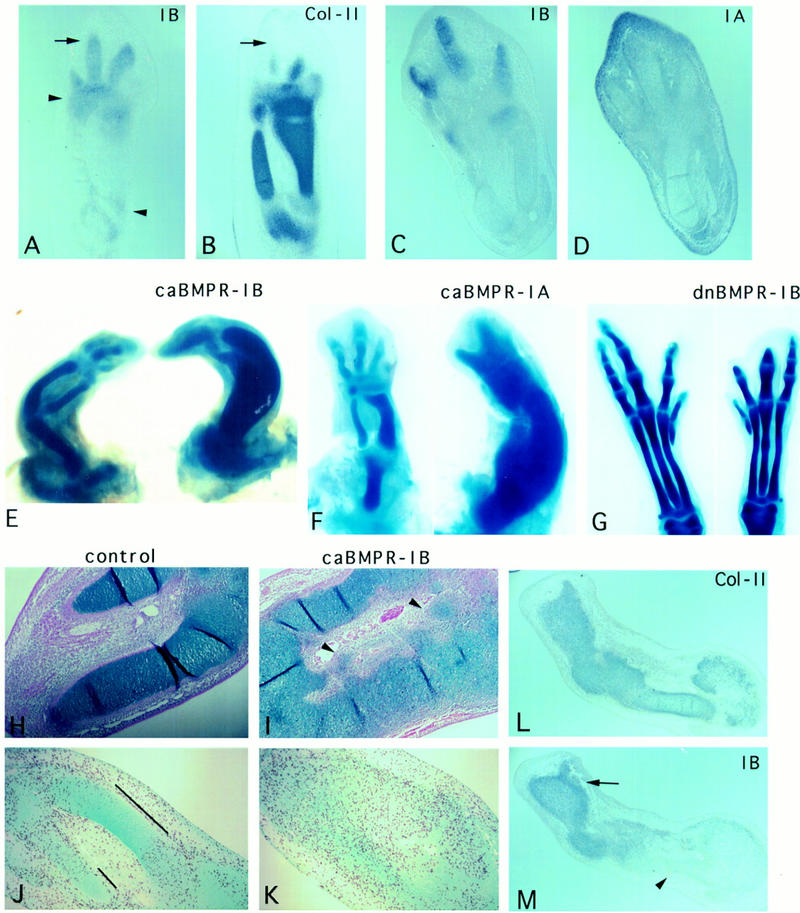
BMPR-IB expression prefigures the future cartilage and its activity is necessary and sufficient for in vivo chondrogenesis. (A–D) Section RNA in situ hybridization with digoxygenin-labeled probes. Stage 27 hindlimb (A) and stage 30 forelimb (C) show that BmpR-IB RNA (purple stain) was strongly expressed in precartilaginous condensation zones, at these stages most strongly in the phalangeal region (arrow). Expression of BmpR-IB preceded that of Col-II (B), an early marker of chondrocytes. BmpR-IB expression decreased in the core region of the cartilage element where Col-II was strongly expressed, but persisted in regions of less mature chondrocytes (arrowheads). (D) BmpR-IA RNA was detected at low levels throughout the limb bud mesenchyme, with highest levels in the distal mesenchyme. (E–G) Day 10 embryos stained with alcian blue to reveal cartilage. (E) Infection of stage 20 forelimb with caBMPR-IB virus or (F) stage 14 presumptive hind-limb field with caBMPR-IA resulted in expanded and fused cartilage elements (right), compared with the contralateral control limb (left). (G) Infection of a stage 21 hindlimb bud with dnBMPR-IB resulted in a loss of distal phalanges (right). Metatarsals were shorter and thinner than in the contralateral limb (left). Regression of the interdigital webbing was inhibited (see also Zou and Niswander 1996). (H,I) Histological section of stage 30 limbs stained with alcian blue. Uninfected left wing has a smooth perichondrium (H). In the contralateral right wing (I) infected at stage 16 with ∼3 times less caBMPR-IB virus than in (E,F), the perichondrium was indistinct and swirls of ectopic mesenchymal condensations were observed in the region of soft tissue (arrowheads). (J,K) BrdU labeling studies. Very few cells incorporated BrdU within the core regions of the cartilage elements of a stage 30 uninfected left wing (J; highlighted by lines), whereas significantly more cells incorporated BrdU throughout the cartilage of the caBMPR-IB-infected contralateral wing (K). High levels of BrdU incorporation in the cartilage were also observed following caBMPR-IA infection (not shown). (L,M) caBMPR-IA results in expansion of the chondrogenic region as distinguished by Col-II expression (L) and induces the ectopic expression of endogenous BmpR-IB (M; arrow). BmpR-IB expression is down-regulated in more mature chondrocytes (arrowheads), similar to normal chondrogenesis.
BMPR-IB activity promotes cartilage formation
Infection of a stage 20 fore- or hindlimb bud with caBMPR-IB, as well as caBMPR-IA, led to a dramatic expansion of the cartilaginous elements (n > 100). The humerus, radius, and ulna, as well as the femur, tibia, and fibula, were often fused, suggesting a disruption of joint formation (Fig. 1E,F). In the most dramatic cases, little or no muscle or other soft tissues was observed in the infected limb. The length of most skeletal elements was comparable to those of the contralateral control limb. The number of digits was often reduced by one (similar reduction was observed following caBMPR-IA infection; see Fig. 1F). Because of the aberrant morphology and lack of molecular probes that distinguish specific digits, we cannot distinguish between a patterning defect or an alteration in the distribution of mesenchymal cells into the expanded condensations such that fewer mesenchymal cells may be left to contribute to subsequent condensations.
To analyze the effect of caBMPR-IB on early cartilage condensation, the limbs were examined histologically following infection with a lesser amount of virus for more subtle effects. Cartilage condensation still occurred over a much broader region than in the contralateral limb (Fig. 1, cf. H and I). Whereas the normal perichondrium formed a smooth sheath of spindle-shaped cells around the cartilage (Fig. 1H), the perichondrium of the infected elements was severely disrupted histologically and as evidenced by the lack of expression of perichondrial molecular markers, for example, PTH/PTHrP receptor and Gli (data not shown). Ectopic mesenchymal condensations were observed and the cells were classified as chondrocytes by alcian blue staining (Fig. 1I) and Col-II RNA expression (not shown). Thus, misexpression of activated BMPR-IB in the undifferentiated limb mesenchyme results in an expansion of the chondrogenic region.
This expansion may result from an increase in the rate of chondrocyte proliferation and/or recruitment of muscle/soft tissue into a chondrocytic lineage. To study the former possibility, bromodeoxyuridine (BrdU) incorporation analyses were performed (n = 3 each for caBMPR-IB and for caBMPR-IA). In control limbs (Fig. 1J), very few cells incorporated BrdU in the core region of the cartilage elements, but higher levels of proliferation were noted in the flanking chondrogenic regions, the perichondrium, and soft tissues. In caBMPR-IB infected limbs (Fig. 1K), significantly more cells incorporated BrdU in the core region of the cartilage, such that the number of BrdU-labeled cells was similar along the entire cartilage shaft. Similar results were noted for caBMPR-IA infected limbs. Therefore, increased chondrocyte proliferation can account at least in part for the dramatic expansion of the chondrogenic region.
We further confirmed that BMPR-IB-mediated activity promotes the early steps of chondrogenesis using limb mesenchyme micromass cultures. Uninfected micromass cultures differentiate after 4 days of culture, giving rise to multiple cartilage nodules (Fig. 2A–C). Micromass cultures infected with caBmpR-IB RCAS virus gave rise to cartilage nodules after 2 days, almost two days earlier than control cultures (Fig. 2, cf. D with A and B). There was also a marked increase in the number and size of cartilage nodules (Fig. 2E,F), similar to that observed in micromass cultures treated with BMP protein (Duprez et al. 1996b; S. Pizette and L. Niswander, unpubl.) and with caBMPR-IA virus (see Fig. 2G–I).
Figure 2.
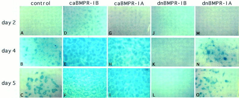
BMPR-IB activity is necessary for in vitro cartilage formation. High-density micromass cultures derived from stage 22–24 limb buds stained with alcian blue to reveal cartilage nodules after culture for 2 days (top row); 4 days (middle row), or 5 days (bottom row) following (A–C) no viral infection, or infection with (D–F) caBMPR-IB; (G–I) caBMPR-IA; (J–L) dnBMPR-IB; (M–O) dnBMPR-IA virus.
BMPR-IB is necessary for cartilage formation
As BmpR-IB is strongly expressed in the precartilaginous mesenchymal condensations and ectopic expression of caBMPR-IB elicits extensive mesenchymal condensation, we sought to determine whether BMPR-IB is essential for cartilage formation. Ectopic expression of dominant-negative BMPR-IB (dnBMPR-IB) in vivo disrupted cartilage formation and resulted in the loss of the distal phalanges (n > 30; Zou and Niswander 1996; Fig. 1G). In addition, the metatarsals were usually shorter and thinner than normal. In more severe cases in the hindlimb, the fibula was missing and the pelvis was disrupted (data not shown).
In limb micromass cultures, dnBMPR-IB completely blocked cartilage formation (Fig. 2J–L). Interestingly, infection of the micromass culture with dnBMPR-IA had no effect on nodule formation (Fig. 2M–O; Kawakami et al. 1996). Although both caBMPR-IB and caBMPR-IA can promote chondrogenesis, BMPR-IB appears to be a more direct effector of chondrogenesis. Examination of endogenous BmpR-IB transcripts after infection with caBMPR-IA both in vitro and in vivo revealed that BmpR-IB mRNA was induced by caBMPR-IA but not vice versa (Fig. 1M; data not shown). This suggests that there may exist a hierarchy of receptor action such that BMPR-IB is the mediator of chondrogenesis and the promotion activity of caBMPR-IA on chondrogenesis is a result of induction of endogenous BmpR-IB (H. Zou et al., in prep.). Taken together, our results indicate that BMPR-IB transduces BMP signals necessary for the early steps of cartilage formation.
BmpR-IA is expressed in prehypertrophic chondrocytes overlapping the Ihh domain
During endochondral bone formation and later in the growth plate, chondrocytes differentiate progressively, passing sequentially through proliferative, hypertrophic, and degenerative stages (see Fig. 4I, below). Several Bmps are expressed in the perichondrium, including Bmp2, Bmp4, and Bmp7 (shown in Fig. 3A–C; Kingsley 1994), and Bmp6 is expressed in prehypertrophic and hypertrophic cells (Kingsley 1994; Vortkamp et al. 1996). However, it is not known which chondrocytes receive and respond to BMP signaling, or what role this signaling plays in the regulation of chondrocyte function and differentiation.
Figure 3.
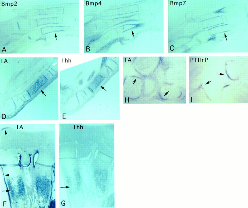
Expression patterns of Bmp, BmpR-IA, and Ihh during cartilage differentiation. (A–C) Serial sections of a stage 31 metatarsal region processed for RNA in situ hybridization with (A) Bmp2, (B) Bmp4, and (C) Bmp7. All three Bmps are expressed in the perichondrium that surrounds the cartilage elements. Serial sections through a stage 32 ulna (D,E) or through the growth plate of an embryonic day 15 distal tibia (F,G) hybridized with (D,F) BmpR-IA or (E,G) Ihh. BmpR-IA and Ihh are largely coexpressed in the prehypertrophic chondrocytes (arrows). BmpR-IA RNA is also detected in the periarticular region, as well as in the inner layer of the perichondrium (arrowheads in F). (H,I) Sections through the periarticular region of a stage 34 carpal region hybridized with (H) BmpR-IA and (I) PTHrP. Both BmpR-IA and PTHrP are expressed in other joints (not shown). Arrows in (A–I) highlight the expression domains.
We first examined the expression patterns of BmpR-IA and BmpR-IB in the limb cartilage elements. BmpR-IB appears to be expressed at a low level in the cartilage elements after their initial formation (Fig. 1A,C; data not shown). BmpR-IA, in contrast to its earlier ubiquitous expression, became restricted and was highly expressed in prehypertrophic chondrocytes, as defined histologically and by overlapping expression with the Ihh domain (Fig. 3D,E). The onset of expression of Ihh in prehypertrophic chondrocytes precedes that of BmpR-IA by ∼1 day. As chondrocyte differentiation proceeded, BmpR-IA expression faded in the central hypertrophic region [as defined by expression of collagen type-X (Col-X)] but continued to be expressed in prehypertrophic chondrocytes that now flank the hypertrophic region (data not shown), again similar to that of Ihh. This pattern of overlapping BmpR-IA and Ihh expression continued in the fetal growth plate (Fig. 3F,G). BmpR-IA was also expressed in the joint region similar to that of PTHrP (Fig. 3H,I; Vortkamp et al. 1996), and in the inner layer of the perichondrium (Fig. 3F). The highly localized expression pattern of BmpR-IA within prehypertrophic chondrocytes suggests that these cells are the direct target of BMP signals secreted from the adjacent perichondrium. The prehypertrophic chondrocytes themselves, as well as hypertrophic chondrocytes, which express Bmp6, may also signal to BMPR-IA. However, the affinity of BMP6 for BMPR-IA is less well characterized. In addition, BMPs in the perichondrium may signal to the periarticular joint region, which expresses BmpR-IA.
caBMPR-IA delays chondrocyte differentiation
To investigate the role of BMPR-IA-mediated signaling during chondrogenesis, caBMPR-IA infected wings were examined histologically. At embryonic day 9, the midsection of the contralateral uninfected radius and ulna had undergone hypertrophic cartilage differentiation (Fig. 4A). In the majority of infected limbs, however, the cartilage elements lacked hypertrophic cartilage, and the chondrocytes appeared immature (Fig. 4B).
This apparent delay in differentiation was investigated further using molecular markers. In Figure 4C–F, infection was targeted to the posterior distal region of a stage 21 limb (note expansion, fusion, and developmental delay of the posterior digits). Col-II, a general marker of all chondrocytes except hypertrophic chondrocytes, was expressed throughout the infected posterior region, indicating the chondrocytes were viable and produced cartilage matrix (Fig. 4C). In contrast, Col-X could not be detected in the infected region, even though it was expressed in hypertrophic cartilage in uninfected regions and in the contralateral limb (Fig. 4D). Ihh was expressed in uninfected prehypertrophic chondrocytes but was not detected in the infected region (Fig. 4F). ptc and Gli, presumed molecular markers of Hedgehog signaling, were also not detected in the infected regions (data not shown). PTH/PTHrP receptor expression normally overlaps slightly with the Ihh domain, extending into a region of less mature chondrocytes and in the perichondrium. In the infected elements, PTH/PTHrP receptor was expressed at low levels and not highly localized (Fig. 4E). Therefore, our histological and molecular analysis indicates that the delay in differentiation resulting from misexpression of caBMPR-IA is at a step prior to the formation of prehypertrophic chondrocytes. Interestingly, the ectopic caBMPR-IA phenotype is similar to that observed following ectopic expression of Ihh or Bmp in the limb (Duprez et al. 1996a; Vortkamp et al. 1996).
In the studies of Vortkamp et al. (1996), misexpression of Ihh-induced PTHrP in the periarticular joint region. We found that misexpression of caBMPR-IA also induced PTHrP expression in a similar manner (Fig. 4G). However, as indicated above, ectopic caBMPR-IA does not induce Ihh expression.
caBMPR-IB does not delay chondrocyte differentiation
We also examined the consequences of misexpression of caBMPR-IB on chondrocyte differentiation. Although there was extensive cartilage formation following ectopic caBMPR-IB (shown in Figs. 1 and 5B), there was no apparent delay in chondrocyte differentiation as shown by Col-X, Ihh, and PTH/PTHrP receptor expression. Interestingly, the proximodistal (i.e., longitudinal) differentiation pattern within each infected cartilage element was affected. Col-X and PTH/PTHrP receptor are normally expressed in discrete proximodistal regions of each cartilage element in a complementary pattern, with Col-X expression in the center and PTH/PTHrP receptor expression flanking (Fig. 5C,E). In the infected limb where phalanges were fused and joints missing, these mRNAs were expressed along the entire proximodistal axis, although they still displayed a complementary pattern (Fig. 5D,F).
Figure 5.
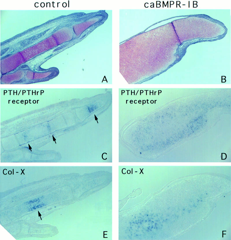
caBMPR-IB does not delay chondrocyte differentiation. Serial sections through the digit region of a day 8.5 uninfected limb (A,C,E) and limb infected with caBMPR-IA at stage 20 (B,D,F). (A,B) Weigert-Safranin stain revealed extensive cartilage formation, fusion of the phalanges, and lack of joint formation in the infected limb. (C–F) RNA section in situ hybridization with PTH/PTHrP receptor (C,D) or Col-X (E,F). In the infected fused phalanges (D,F), PTHrP receptor and Col-X mRNAs displayed complementary expression patterns but were detected along the entire proximodistal axis; in contrast to the discreet proximodistal localization of these RNAs in the contralateral limb (C,E; arrows).
Irregular differentiation and ossification following caBMPR-IA misexpression
By day 13 of incubation, in caBMPR-IA infected limbs, we noted a disarray in the pattern of ossification. During endochondral development, blood vessel invasion and ossification normally start in the midsection of the cartilage element. Strikingly, however, in the caBMPR-IA-infected limbs, this occurred at multiple sites scattered randomly throughout the cartilage shaft, sometimes even including the joint regions (Fig. 4H). This ectopic vascularization and irregular ossification is similar to that following misexpression of Ihh in the chick limb, and of PTHrP in transgenic mice (Vortkamp et al. 1996; Weir et al. 1996).
Near the ectopic ossification sites, we also observed an apparent reversal of polarity in the pattern of chondrocyte differentiation: Histologically chondrocytes became hypertrophic at the circumference of the long bone rather than the middle. Chondrocytes in the middle were separated by large amounts of matrix that was not stained by cartilage and bone-specific dyes (Alcian blue or Safranin O), suggesting that those cells were not viable. At later embryonic stages, irregular areas of cartilage persisted in the bone marrow cavity. The bones were abnormal in shape and lacked normal growth plates. The reversal in chondrocyte differentiation pattern is similar to that observed in PTHrP transgenic mice (Weir et al. 1996). Consistent with this, misexpression of caBMPR-IA in the limb induced expression of PTHrP (described above). Thus, this phenotype is likely to occur through the PTH/PTHrP pathway.
BMPR-IB is involved in programmed cell death in the embryonic limb
We showed previously that blocking BMP signaling by dnBMPR-IB in the embryonic limb results in a reduction in apoptosis leading to soft tissue syndactyly (webbing) (Zou and Niswander 1996). Here we have used caBMPR-IB and found the opposite effect: caBMPR-IB infection of the developing limb led to increased cell death. Early caBMPR-IB infection throughout the limb field (before stage 17) resulted in a very thin limb, with the anteroposterior diameter about half the size of a control limb (Fig. 6A,B). The proximodistal length was also often affected, resulting in distal truncations. The cartilage that did form was expanded to fill the limb, as described above. In the most dramatic cases (∼10%), the infected limb was severely truncated or absent (Fig. 6C). Examination 2 days following extensive infection revealed that the apical ectodermal ridge (AER) was disrupted, with some sections apparently degenerating. The distal portion of the limb was often split into several small outgrowths, presumably the consequence of degeneration of the AER and underlying mesenchymal tissue. caBMPR-IB infected limbs stained with a vital dye showed a marked increase in the number of nonviable mesenchymal and AER cells (Fig. 6D,E).
Figure 6.
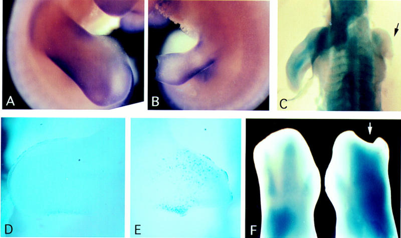
caBMPR-IB regulates cell death in the embryonic limb. (A) Day 7 control uninfected hindlimb. (B) caBMPR-IB infection of the contralateral limb at stage 15 resulted in a very thin limb that also displayed a degenerating AER and distal shortening. (C) In the most dramatic cases, caBMPR-IB infection resulted in extreme truncation of the limb (arrow). (D,E) Nile blue staining to detect nonviable cells. Forty-eight hours after caBMPR-IB infection, the infected forelimb (E) displayed a marked increase in the number of Nile blue-stained mesenchymal and AER cells compared with the contralateral limb (D). (F) Posteriorly localized caBMPR-IB infection of a stage 22 forelimb resulted in an accelerated regression of interdigital soft tissue at stage 30 (right) compared with the contralateral limb (left). The posterior phalanges were expanded and fused.
Later and more localized caBMPR-IB infection of stage 22 posterior mesenchyme resulted in accelerated regression of the interdigital soft tissue (Fig. 6F), opposite to the dnBMPR-IB-induced webbing phenotype. Massive cell death was not observed in these later infected limbs. In contrast, caBMPR-IA did not result in increased cell death, nor did dnBMPR-IA give rise to webbing phenotype (infection performed between stage 14 and 24).
Discussion
The type I BMPRs control distinct processes during vertebrate limb development
A major conclusion from this study is that the two type I BMPRs mediate distinct responses during limb development (summarized in Fig. 7). Several lines of evidence support this conclusion. First, the expression pattern of the two type I receptors differ markedly. BmpR-IB expression is not detectable in early limb mesenchyme but is detected in early condensations where expression prefigures the cartilage primordia (this paper; Kawakami et al. 1996). BmpR-IA is expressed at low levels throughout the limb mesenchyme with highest expression in the distal mesenchyme, and later becomes localized to the prehypertrophic chondrocytes. Second, BMPR-IB activity is necessary and sufficient for cartilage condensation, both in vivo and in vitro, as tested by misexpression of both gain-of-function caBMPR-IB and loss-of-function dnBMPR-IB. BMPR-IA does not appear to be a direct effector of cartilage condensation. Instead, our data suggest that BMPR-IA acts through BMPR-IB to elicit mesenchyme condensation (Fig. 1M; H. Zou et al., in prep.). Third, BMPR-IA is involved in the regulation of chondrocyte differentiation during late stages of cartilage development. BmpR-IA is expressed in prehypertrophic chondrocytes, and misexpression of caBMPR-IA results in a delay in chondrocyte differentiation. BmpR-IB expression is down-regulated during chondrogenesis and misexpression of caBMPR-IB does not inhibit chondrocyte differentiation. Finally, BMPR-IB is involved in BMP-mediated programmed cell death. Use of the dnBMPR-IB to block BMP signaling results in decreased apoptosis in the interdigital soft tissue, leading to a webbed phenotype, whereas caBMPR-IB causes a marked increase of cell death in soft tissues. In contrast, our dnBMPR-IA and caBMPR-IA point mutations do not affect cell death.
Figure 7.
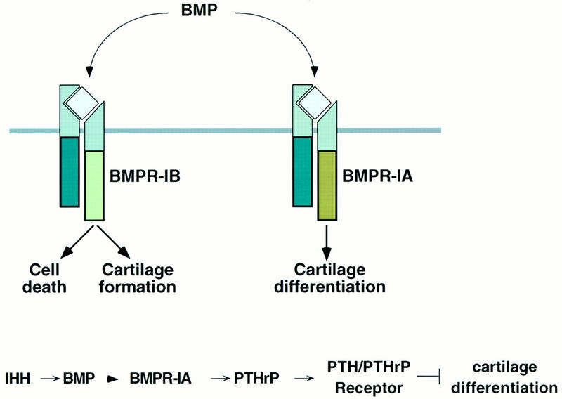
BMPR-specific functions in limb development and molecular pathways elicited by IHH signaling. (Top) We propose that BMPR-IB specifically regulates the intracellular pathways that lead to cell death and formation of the initial cartilaginous skeleton in response to BMP, whereas BMPR-IA regulates the rate of chondrocyte differentiation. Schematic adapted from Massagué and Weis-Garcia (1996). (Bottom) Proposed molecular pathway downstream of IHH. See Discussion for description. Direct interaction is indicated by an arrowhead. Steps indicated by an arrow may be direct or indirect.
Our studies thus provide the first demonstration that different type I BMPRs can elicit distinct developmental outcomes when expressed in the same cells. In vitro cell culture studies have not separated type I BMPR action, although this may reflect the paucity of biochemical and molecular criteria to evaluate receptor signaling. Two Drosophila type I BMPRs, Tkv and Sax, appear to be functionally equivalent, as loss-of function Tkv and Sax clones in the embryo and wing disc produce similar phenotypes, and Tkv can functionally substitute for Sax (Brummel et al. 1994; Nellen et al. 1994; Singer et al. 1997).
The vertebrate BMPR-IA and BMPR-IB have similar but not identical binding affinities in vitro for the BMP ligands that are expressed in the developing limb (Francis et al. 1994; Koenig et al. 1994; ten Dijke et al. 1994b; Francis-West et al. 1995; Liu et al. 1995; Nohno et al. 1995; Rosenzweig et al. 1995). It has been suggested that the temporally and spatially regulated expression of BMPs controls the formation of particular skeletal elements (Kingsley 1994). Further refinement may be regulated by the temporal and spatial expression and activity of the type I and type II BMPRs. Additional specificity may also be controlled by the differential intracellular response to BMPR-mediated signal transduction. Future studies should help distinguish how different combinations of TGFβ-family ligands, their receptors, and intracellular targets exquisitely regulate the complex formation of skeletal elements in the proper number, position, shape, and size.
Regulation of chondrocyte differentiation through BMPR-IA mediated signaling
The delay in chondrocyte differentiation caused by misexpression of caBMPR-IA phenocopies misexpression of Ihh. Expression of BmpR-IA in prehypertrophic chondrocytes overlaps that of Ihh. It has been suggested that IHH signals to the perichondrium, which expresses ptc, a proposed Hedgehog target that is also involved in reception of the HH signal (Nusse 1996; Vortkamp et al. 1996, and references therein). Studies in Drosophila and vertebrates suggest that signaling by the Hh/Ptc pathway may regulate BMP expression (references in Laufer et al. 1994; Blair 1995). Consistent with this, Bmp2, Bmp4, and Bmp7 are expressed in the perichondrium (Fig. 3A–C; Kingsley 1994). These ligands bind BMPR-IA, which is expressed in prehypertrophic chondrocytes (Fig. 3D,F). This suggests the existence of a local signal relay loop, as illustrated in Figures 4J and 7, which could provide a means to regulate the rate of chondrocyte differentiation and balance the relative populations of chondrocytes among the different zones. In our model, as proliferating chondrocytes exit the cell cycle and start to hypertrophy, they transiently express high levels of Ihh. IHH acts on the adjacent perichondrium, which expresses ptc. These cells respond to IHH by expressing BMPs, which then signal back to BMPR-IA in the prehypertrophic chondrocytes. Our misexpression studies indicate that the down-regulation of BMPR-IA activity is necessary for the initiation of the hypertrophic differentiation process. Thus, we propose another role for BMP signaling in the limb: the regulation of the rate of chondrocyte differentiation through BMPR-IA. These studies also imply that the perichondrium acts as an intermediate in the regulation of cartilage differentiation. Thus, as maturing chondrocytes produce IHH, they set off a signaling cascade mediated through BMPs to inhibit the differentiation of neighboring cells.
BMP signaling from the perichondrium might also be an important relay signal in the proposed regulatory loop between IHH and PTHrP (see Figs. 4J and 7). Misexpression of caBMPR-IA or BMPs delays chondrocyte differentiation, similar to the misexpression phenotypes of IHH or PTHrP (this paper; Duprez et al. 1996a; Vortkamp et al. 1996; Weir et al. 1996). Moreover, ectopic caBMPR-IA or IHH results in increased PTHrP expression (this paper; Vortkamp et al. 1996). PTHrP is localized to the periarticular region, both normally and in limbs misexpressing caBMPR-IA or IHH, quite distant from the endogenous Ihh domain. In the model proposed by Vortkamp et al. (1996), IHH regulates PTHrP expression in the periarticular region, most likely indirectly based on lack of ptc expression in the periarticular region. One possible mechanism for this longer-range effect involves relay signals. BMPs expressed in the perichondrium could act on the periarticular joint region, where BmpR-IA is expressed, to relay the IHH signal to PTHrP. Thus, it is possible that BMP in the cartilaginous skeleton mediates both local effects on the prehypertrophic chondrocytes, as well as longer-range effects to regulate PTHrP in the periarticular cartilage.
Our work, and that by Vortkamp et al. (1996), suggest that the sequential progression of chondrogenic differentiation may require appropriate activation as well as down-regulation of a network of signaling molecules and their receptors. Our data indicate that BMP signals mediate, and are functionally downstream of, the effects of IHH on chondrocyte differentiation. Future studies should cast additional light on the details of the network of regulatory interactions linking Ihh, ptc, Bmp, BmpR-IA, PTHrP, and PTH/PTHrP receptor to the control of the sequential steps of chondrocyte differentiation.
BMPs and programmed cell death
BMP signaling has been implicated in the regulation of embryonic programmed cell death (Graham et al. 1994; Yokouchi et al. 1996; Zou and Niswander 1996; Macias et al. 1997). Our studies support a distinct role for BMPR-IB in the regulation of BMP-mediated cell death. We have found that early caBMPR-IB infection greatly increases cell death in the developing limb. Later infection results in premature regression of the interdigital soft tissue. Moreover, dnBMPR-IB results in decreased apoptosis in the interdigital region (Zou and Niswander 1996). In contrast, neither our activated nor our dominant-negative forms of BMPR-IA affected cell death. However, Yokouchi et al. (1996) observed a decrease in apoptosis in the limb following overexpression of a different dominant-negative BMPR-IA construct. This construct lacks the entire intracellular portion of the polypeptide, including the kinase and the GS activation domains, whereas our dnBMPR-IB construct creates a single amino acid change in the ATP-binding site. A truncated BMPR-IB construct blocks chondrogenesis in limb mesenchyme cultures (Kawakami et al. 1996), as does our point-mutant dnBMPR-IB (this paper). However, the effect of the truncated dnBMPR-IB construct on cell death has not been reported.
Overexpression of a truncated or point mutant type I receptor may cause a general dominant-negative effect by sequestering ligands and type II receptors that are shared with other type I receptors. The dominant-negative effect of a point mutant type I receptor might be more specific because this construct could also sequester intracellular proteins such as Smads, which associate with type I receptors (Macias-Silva et al. 1996; Zhang et al. 1996; Kretzschmar et al. 1997). Nonetheless, in a combinatorial system of multifunctional receptors, it is not possible to determine the role of a specific receptor in a particular process through the use of dominant-negative constructs. In contrast, the ability of the activated BMPR-IB, but not the activated BMPR-IA, to elicit cell death provides evidence that the apoptotic pathway is triggered specifically through BMPR-IB-mediated signal transduction.
Our studies show that caBMPR-IB causes extensive cell death when expressed in young limb buds. However, the only obvious effect on cell death following infection of later stage limbs is an accelerated regression of interdigital soft tissue. Interdigital soft tissue seems to be the primary target tissue of BMP-mediated apoptosis, whereas the condensed digital mesenchyme cells appear to be resistant to this apoptotic signal. This difference in response of young versus old, interdigital versus digital cells to BMP-mediated apoptosis could be a result of differences in their developmental history or their state of differentiation.
Conclusion
Our studies reveal a mechanism by which specific intracellular effects can be generated in response to the same or closely related extracellular BMP signals. During embryogenesis, multiple BMPs and BMPRs are often coexpressed or display a close spatial–temporal association. Our work demonstrates that the type I BMPRs provide specificity by triggering distinct phenotypic responses. This study also presents evidence for the integration of the BMP signaling pathway with the IHH/PTHrP pathway implicated in regulation of bone morphogenesis: IHH acts upstream of BMPs, which then work through BMPR-IA in prehypertrophic chondrocytes and in the periarticular region to regulate PTHrP production, thus serving to control chondrocyte differentiation. The mutant forms of the type I BMPRs provide powerful tools to experimentally manipulate BMP signaling in developing tissues that are dependent on BMP such as the kidney, eye, mammary gland, and nervous system, as well as to elucidate the role of BMPs in triggering cell death. In the future, it will be of interest to determine the identity of the intracellular molecules that specifically interpret the signals from the different BMPRs.
Materials and methods
Cloning of chicken type I BMPRs
Full-length chicken BmpR-IA and BmpR-IB cDNA clones were isolated from a stage 12-15 chick cDNA library (kind gift of D. Wilkinson, Imperial Cancer Research Fund, London, UK) using human BmpR-IA and mouse BmpR-IB as probes (kindly provided by K. Miyazono and P. ten Dijke, Ludwig Institute, Uppsala, Sweden) under low-stringency hybridization conditions. DNA sequence was compared with the reported sequences for human and chick BmpR-IA (ten Dijke et al. 1993, 1994a; Kawakami et al. 1996) and chick BmpR-IB (Sumitomo et al. 1993). The coding regions of the two chick BMPRs were amplified by polymerase chain reaction, subcloned into pBluescript (Stratagene), and used to generate antisense dioxygenin probes. The 1.6-kb BmpR-IA was cloned into EcoRI–SalI sites, digested with EcoRI, and transcribed with T7 polymerase. The 1.5-kb BmpR-IB was cloned into BamHI–EcoRI, digested with BamHI, and transcribed with T7 polymerase.
Generation of mutant type I BMPRs and in ovo infection
cDNAs containing the coding region of chick BmpR-IB and human BmpR-IA cloned into pBluescript were subjected to oligonucleotide-mediated site-directed mutagenesis (Clontech) to change Lys-231 to Arg for dnBMPR-IB (similar phenotypes were noted with mouse dnBMPR-IB), Lys-261 to Arg for dnBMPR-IA, Gln-203 to Asp for caBMPR-IB, and Gln-233 to Asp for caBMPR-IA. The mutant constructs were cloned into Cla12–Nco shuttle vector and then into the replication-competent avian retroviral vector RCAS(A) (Hughes et al. 1987). Viruses were generated as described (Morgan and Fekete 1996). In all cases, similar results were obtained from multiple viral preparations of a given construct. Concentrated RCAS(A) virus encoding the various mutant type I BMPRs was injected into the presumptive limb field or developing limb bud.
Histological analysis
Limbs from various stages were fixed in 4% paraformaldehyde (PFA) overnight at 4°C. Limbs from day 7 or older embryos were decalcified in 5.5% EDTA /4% PFA for up to a week at 4°C, with the solution changed every 24 hr. Limbs were then dehydrated, embedded in paraffin, and sectioned at 8 μm. Sections were stained with hematoxylin and eosin or alcian blue (pH 2.5) and counter-stained with Nuclear Fast Red to localize sulfated proteoglycans, or by Weigert–Safranin staining (Prophet et al. 1994).
RNA in situ hybridization
For serial sections, two consecutive sections were collected on seven alternating slides. Section RNA in situ hybridization with digoxygenin-labeled probes was done according to Neubuser and Balling (Neubuser et al. 1995 and pers. comm.). Briefly, after rehydration, the tissues were refixed in 4% PFA, subjected to 10 μg/ml of proteinase K treatment at room temperature for 5–7 min, and fixed again in 4% PFA. Hybridization was performed at 65°C in 40% formamide, and posthybridization washes were carried out at a final stringency of 20% formamide/0.5× SSC at 60°C. Detection was performed using BM-purple substrate (Boehringer) from 12 hr to 1 week, depending on the probe. Whole-mount RNA in situ hybridization was performed essentially as described (Henrique et al. 1995). Probes were kindly provided by P. Brickell (Bmp2 and Bmp4; Francis et al. 1994), B. Houston (Bmp7; Houston et al. 1994), C. Tabin (Ihh, ptc, Gli, PTHrP receptor; references in Vortkamp et al. 1996), W. Upholt (Col-II, Hyun-Duck et al. 1988), B. Olsen (collagen type X; Ninomiya et al. 1986), and G. Strewler (PTHrP; Schermer et al. 1991).
BrdU incorporation
Undiluted BrdU labeling reagent (250 μl) (Amersham Life Science) was injected into the amniotic cavity of day 6 chick embryos (controls or caBMPR-IA/-IB infected at stage 19–20 into the limb bud). The embryos were returned to the incubator for 2.5 hr, then sacrificed and placed in Carnoy’s Fix. Paraffin sections (8 μm) were collected and processed using Amersham’s Cell Proliferation Kit. The DAB color reactions were stopped after 2 min and sections counterstained briefly in 1% methyl green.
High-density micromass cultures
Stage 22–24 limb buds were isolated; the ectoderm was removed by trypsin treatment; and the mesenchyme dissociated as described (Ahrens et al. 1977; Swalla and Solursh 1986). Mesenchyme was resuspended at a density of 2 × 10⁷ cells/ml; 12 μl aliquots were infected with 1 μl concentrated RCAS virus encoding the different BMPR mutants and plated on 35-mm tissue culture dishes. One hour later, Medium 199 (GIBCO) containing 10% fetal calf serum and 2% chick serum was added. Each 35-mm culture dish contained three similarly treated micromasses, and multiple dishes were plated allowing analysis at 24-hr intervals. The cultures were stained with alcian blue (pH 2.5) to visualize chondrogenic nodule formation. These experiments were repeated three times with similar results.
Nile blue detection of cell death
Vital staining by Nile blue sulfate (Sigma N-5632) was performed according to Tone et al. (1983). Briefly, embryos were dissected in cold PBS, transferred to prewarmed Medium 199 containing 0.001% Nile blue, incubated at 37°C for 30–45 min, washed in cold PBS for 5 hr, and photographed.
Acknowledgments
We are grateful to K. Manova of Memorial Sloan-Kettering Cancer Center (MSKCC) Molecular Cytology Facility for assistance, to A. Neubuser for the section in situ protocol, and to K. Miyazono, P. ten Dijke, C. Tabin, P. Brickell, B. Houston, B. Olsen, W. Upholt, and G. Strewler for probes. We also thank our laboratory members, in particular C. Wang, for critical reading of the manuscript. This work was supported by a Horsfall Fellowship to H.Z., a Pew Scholars award to L.N., National Institutes of Health awards to L.N. and J.M., and by the MSKCC Support Grant. R.W. and J.M. are, respectively, a Research Associate and an Investigator of the Howard Hughes Medical Institute.
The publication costs of this article were defrayed in part by payment of page charges. This article must therefore be hereby marked “advertisement” in accordance with 18 USC section 1734 solely to indicate this fact.
Footnotes
E-MAIL l-niswander@ski.mskcc.org; FAX (212) 717-3623.
References
- Ahrens PB, Solursh M, Reiter RS. Stage-related capacity for limb chondrogenesis in cell culture. Dev Biol. 1977;60:69–82. doi: 10.1016/0012-1606(77)90110-5. [DOI] [PubMed] [Google Scholar]
- Blair SS. Compartments and appendage development in Drosophila. BioEssays. 1995;17:299–309. doi: 10.1002/bies.950170406. [DOI] [PubMed] [Google Scholar]
- Brummel TJ, Twombly V, Marqués G, Wrana JL, Newfeld SJ, O’Connor MB, Gelbart WM. Characterization and relationship of Dpp receptors encoded by the saxophone and thick veins genes in Drosophila. Cell. 1994;78:251–261. doi: 10.1016/0092-8674(94)90295-x. [DOI] [PubMed] [Google Scholar]
- Chang SC, Hoang B, Thomas JT, Vukicevi S, Luyten FP, Ryba NJP, Kazak CA, Reddi AH, Moos M. Cartilage-derived morphogenetic proteins. New members of the transforming growth factor-beta superfamily predominantly expressed in long bones during human embryonic development. J Biol Chem. 1994;269:28227–28234. [PubMed] [Google Scholar]
- Dewulf N, Verschueren K, Lonnoy O, Morén A, Grimsby S, Vande Spiegle K, Miyazono K, Huylebroeck D, ten Dijke P. Distinct spatial and temporal expression patterns of two type I receptors for Bone Morphogenetic Proteins during mouse embryogenesis. Endocrinology. 1995;136:2652–2663. doi: 10.1210/endo.136.6.7750489. [DOI] [PubMed] [Google Scholar]
- Duprez D, Bell EJ, Richardson MK, Archer CW, Wolpert L, Brickell PM, Francis-West PH. Overexpression of BMP-2 and BMP-4 alters the size and shape of developing skeletal elements in the chick limb. Mech Dev. 1996a;57:145–157. doi: 10.1016/0925-4773(96)00540-0. [DOI] [PubMed] [Google Scholar]
- Duprez DM, Coltey M, Amthor H, Brickell PM, Tickle C. Bone morphogenetic protein-2 (BMP-2) inhibits muscle development and promotes cartilage formation in chick limb bud cultures. Dev Biol. 1996b;174:448–452. doi: 10.1006/dbio.1996.0087. [DOI] [PubMed] [Google Scholar]
- Erlebacher Q, Filvaroff EH, Gitelman SE, Derynck R. Toward a molecular understanding of skeletal development. Cell. 1995;80:371–378. doi: 10.1016/0092-8674(95)90487-5. [DOI] [PubMed] [Google Scholar]
- Francis PH, Richardson MK, Brickell PM, Tickle C. Bone morphogenetic proteins and a signalling pathway that controls patterning in the developing chick limb. Development. 1994;120:209–218. doi: 10.1242/dev.120.1.209. [DOI] [PubMed] [Google Scholar]
- Francis-West PH, Robertson K, Ede DA, Rodriguez C, Izpisua-Belmonte J-C, Houston B, Burt DW, Gribbin C, Brickell PM, Tickle C. Expression of genes encoding Bone Morphogenetic Proteins and Sonic Hedgehog in talpid (ta3) limb buds: Their relationships in the signalling cascade involved in limb patterning. Dev Dynam. 1995;203:187–197. doi: 10.1002/aja.1002030207. [DOI] [PubMed] [Google Scholar]
- Graham A, Francis-West P, Brickell P, Lumsden A. The signalling molecule BMP4 mediates apoptosis in the rhombencephalic neural crest. Nature. 1994;372:684–686. doi: 10.1038/372684a0. [DOI] [PubMed] [Google Scholar]
- Hamburger V, Hamilton HL. A series of normal stages in the development of the chick embryo. Dev Dynam. 1992;195:231–272. doi: 10.1002/aja.1001950404. [DOI] [PubMed] [Google Scholar]
- Henrique D, Adam J, Myat A, Chitnis A, Lewis J, Ish-Horowicz D. Expression of a Delta homologue in prospective neurons in the chick. Nature. 1995;375:787–790. doi: 10.1038/375787a0. [DOI] [PubMed] [Google Scholar]
- Hogan BLM. Bone morphogenetic proteins: Multifunctional regulators of vertebrate development. Genes & Dev. 1996;10:1580–1594. doi: 10.1101/gad.10.13.1580. [DOI] [PubMed] [Google Scholar]
- Hoodless PA, Haerry T, Abdollah S, Stapleton M, O’Connor MB, Attisano L, Wrana JL. MADR1, a MAD-related protein that functions in BMP2 signaling pathways. Cell. 1996;85:489–500. doi: 10.1016/s0092-8674(00)81250-7. [DOI] [PubMed] [Google Scholar]
- Houston B, Thorp BH, Burt DW. Molecular cloning and expression of bone morphogenetic protein-7 in the chick epiphyseal growth plate. J Mol Endocrinol. 1994;13:289–301. doi: 10.1677/jme.0.0130289. [DOI] [PubMed] [Google Scholar]
- Hughes SH, Greenhouse JJ, Petropoulos CJ, Sutrave P. Adaptor plasmids simplify the insertion of foreign DNA into helper-independent retroviral vectors. J Virol. 1987;61:3004–3012. doi: 10.1128/jvi.61.10.3004-3012.1987. [DOI] [PMC free article] [PubMed] [Google Scholar]
- Hyun-Duck N, Rodgers BJ, Kulyk WM, Kream BE, Kosher RA, Upholt WB. In situ hybridization analysis of the expression of the type II collagen gene in the developing chicken limb bud. Collagen Rel Res. 1988;8:277–294. doi: 10.1016/s0174-173x(88)80001-3. [DOI] [PubMed] [Google Scholar]
- Ishidou Y, Kitajima I, Obama H, Maruyama I, Murata F, Imamura T, Yamada N, ten Dijke P, Miyazono K, Sakou T. Enhanced expression of type I receptors for bone morphogenetic proteins during bone formation. J Bone Mineral Res. 1995;10:1651–1659. doi: 10.1002/jbmr.5650101107. [DOI] [PubMed] [Google Scholar]
- Jones CM, Lyons KM, Hogan BLM. Involvement of Bone Morphogenetic Protein-4 (BMP-4) and Vgr-1 in morphogenesis and neurogenesis in the mouse. Development. 1991;111:531–542. doi: 10.1242/dev.111.2.531. [DOI] [PubMed] [Google Scholar]
- Karaplis AC, Luz A, Glowacki J, Bronson RT, Tybulewicz VLJ, Kronenberg HM, Mulligan RC. Lethal skeletal dysplasia from targeted disruption of the parathyroid hormone-related peptide gene. Genes & Dev. 1994;8:277–289. doi: 10.1101/gad.8.3.277. [DOI] [PubMed] [Google Scholar]
- Kawakami Y, Ishikawa T, Shimabara M, Tanda N, Enomoto-Iwamoto M, Iwamoto M, Kuwana T, Ueki A, Noji S, Nohno T. BMP signaling during bone pattern determination in the developing limb. Development. 1996;122:3557–3566. doi: 10.1242/dev.122.11.3557. [DOI] [PubMed] [Google Scholar]
- Kingsley DM. What do BMPs do in mammals? Clues from the mouse short-ear mutation. Trends Genet. 1994;10:16–21. doi: 10.1016/0168-9525(94)90014-0. [DOI] [PubMed] [Google Scholar]
- Koenig BB, Cook JS, Wolsing DH, Ting J, Tiesman JP, Correa PE, Olson CA, Pecquet AL, Ventura F, Grant RA, Chen G, Wrana JL, Massagué J, Rosenbaum JS. Characterization and cloning of a receptor for BMP-2 and BMP-4 from NIH 3T3 cells. Mol Cell Biol. 1994;14:5961–5974. doi: 10.1128/mcb.14.9.5961. [DOI] [PMC free article] [PubMed] [Google Scholar]
- Kretzschmar M, Liu F, Hata A, Doody J, Massagué J. The TGF-β family mediator Smad1 is phosphorylated directly and activated functionally by the BMP receptor kinase. Genes & Dev. 1997;11:984–995. doi: 10.1101/gad.11.8.984. [DOI] [PubMed] [Google Scholar]
- Lanske B, Karaplis AC, Lee K, Luz A, Vortkamp A, Pirro A, Karperien M, Defize LHK, Ho C, Mulligan RC, Abou-Samra A-B, Jüppner H, Segre GV, Kronenberg HM. PTH/PTHrP receptor in early development and Indian Hedgehog-regulated bone growth. Science. 1996;273:663–666. doi: 10.1126/science.273.5275.663. [DOI] [PubMed] [Google Scholar]
- Laufer E, Nelson C, Johnson RL, Morgan BA, Tabin C. Sonic hedgehog and Fgf-4 act through a signaling cascade and feedback loop to integrate growth and patterning of the developing limb bud. Cell. 1994;79:993–1003. doi: 10.1016/0092-8674(94)90030-2. [DOI] [PubMed] [Google Scholar]
- Liu F, Ventura F, Doody J, Massagué J. Human type II receptor for bone morphogenetic proteins (BMPs): Extension of the two-kinase receptor model to BMPs. Mol Cell Biol. 1995;15:3479–3486. doi: 10.1128/mcb.15.7.3479. [DOI] [PMC free article] [PubMed] [Google Scholar]
- Liu F, Hata A, Baker JC, Doody J, Carcamo J, Harland RM, Massagué J. A human Mad protein acting as a BMP-regulated transcriptional activator. Nature. 1996;381:620–623. doi: 10.1038/381620a0. [DOI] [PubMed] [Google Scholar]
- Lyons KM, Pelton RW, Hogan BLM. Patterns of expression of murine Vgr-1 and BMP-2a RNA suggest that transforming growth factor-β-like genes coordinately regulate aspects of embryonic development. Genes & Dev. 1989;3:1657–1668. doi: 10.1101/gad.3.11.1657. [DOI] [PubMed] [Google Scholar]
- ————— Organogenesis and pattern formation in the mouse: RNA distribution patterns suggest a role for Bone Morphogenetic Protein-2A (BMP-2A). Development. 1990;109:833–844. doi: 10.1242/dev.109.4.833. [DOI] [PubMed] [Google Scholar]
- Macias D, Ganan Y, Sampath TK, Peidra ME, Ros MA, Hurle JM. Role of BMP-2 and OP-1 (BMP-7) in programmed cell death and skeletogenesis during chick limb development. Development. 1997;124:1109–1117. doi: 10.1242/dev.124.6.1109. [DOI] [PubMed] [Google Scholar]
- Macias-Silva M, Abdollah S, Hoodless PA, Pirone R, Attisano L, Wrana JL. MADR2 is a substrate of the TGFβ receptor and its phosphorylation is required for nuclear accumulation and signaling. Cell. 1996;87:1215–1224. doi: 10.1016/s0092-8674(00)81817-6. [DOI] [PubMed] [Google Scholar]
- Massagué J. TGFβ signaling: Receptors, tranducers, and Mad proteins. Cell. 1996;85:947–950. doi: 10.1016/s0092-8674(00)81296-9. [DOI] [PubMed] [Google Scholar]
- Massagué J, Weis-Garcia F. Serine/threonine kinase receptors: Mediators of TGF-β family signals. Cancer Surv. 1996;27:41–64. [PubMed] [Google Scholar]
- Morgan BA, Fekete DM. Manipulating gene expression with replication-competent retroviruses. Methods Cell Biol. 1996;51:185–218. doi: 10.1016/s0091-679x(08)60629-9. [DOI] [PubMed] [Google Scholar]
- Nellen D, Affolter M, Basler K. Receptor serine/threonine kinases implicated in the control of Drosophila body pattern by decapentaplegic. Cell. 1994;78:225–237. doi: 10.1016/0092-8674(94)90293-3. [DOI] [PubMed] [Google Scholar]
- Neubuser A, Koseki H, Balling R. Characterization and developmental expression of Pax9, a paired-box-containing gene related to Pax1. Dev Biol. 1995;170:701–716. doi: 10.1006/dbio.1995.1248. [DOI] [PubMed] [Google Scholar]
- Ninomiya Y, Gordon M, van der Rest M, Schmid T, Linsenmayer T, Olsen BR. The developmentally regulated type X collagen gene contains a long open reading frame without introns. J Biol Chem. 1986;261:5041–5050. [PubMed] [Google Scholar]
- Nohno T, Ishikawa T, Saito T, Hosokawa K, Noji S, Wolsing DH, Rosenbaum J. Identification of a human type II receptor for Bone Morphogenetic Protein-4 that forms differential heteromeric complexes with Bone Morphogenetic Protein type I receptors. J Biol Chem. 1995;270:22522–22526. doi: 10.1074/jbc.270.38.22522. [DOI] [PubMed] [Google Scholar]
- Nusse R. Patching up Hedgehog. Nature. 1996;384:119–120. doi: 10.1038/384119a0. [DOI] [PubMed] [Google Scholar]
- Prophet, E.B., B. Mills, J.B. Arrington, and L.H. Sobin. 1994. In Laboratory methods in histotechnology, pp. 156, 167. American Registry of Pathology, Washington, DC.
- Rosenzweig BL, Imamura T, Okadome T, Cox GN, Yamashita H, ten Dijke P, Heldin C-H, Miyazono K. Cloning and characterization of a human type II receptor for bone morphogenetic proteins. Proc Natl Acad Sci. 1995;92:7632–7636. doi: 10.1073/pnas.92.17.7632. [DOI] [PMC free article] [PubMed] [Google Scholar]
- Schermer DT, Chan SD, Bruce R, Nissenson RA, Wood WI, Strewler GJ. Chicken parathyroid hormone-related protein and its expression during embryologic development. J Bone Mineral Res. 1991;6:149–155. doi: 10.1002/jbmr.5650060208. [DOI] [PubMed] [Google Scholar]
- Singer MA, Penton A, Twombly V, Hoffmann FM, Gelbart WM. Signaling through both type I DPP receptors is required for anterior–posterior patterning of the entire Drosophila wing. Development. 1997;124:79–89. doi: 10.1242/dev.124.1.79. [DOI] [PubMed] [Google Scholar]
- Storm EE, Huynh TV, Copeland NG, Jenkins NA, Kingsley DM, Lee S-J. Limb alterations in brachypodism mice due to mutations in a new member of the TGFβ-superfamily. Nature. 1994;368:639–643. doi: 10.1038/368639a0. [DOI] [PubMed] [Google Scholar]
- Sumitomo S, Saito T, Nohno T. A new receptor protein kinase from chick embryo related to type I receptor for TGF-β. DNA Sequence. 1993;3:297–302. doi: 10.3109/10425179309020827. [DOI] [PubMed] [Google Scholar]
- Swalla BJ, Solursh M. The independence of myogenesis and chondrogenesis in micromass cultures of chick wing buds. Dev Biol. 1986;116:31–38. doi: 10.1016/0012-1606(86)90040-0. [DOI] [PubMed] [Google Scholar]
- ten Dijke P, Ichijo H, Franzen P, Schulz P, Saras J, Toyoshima H, Heldin CH, Miyazono K. Activin receptor-like kinases: A novel subclass of cell-surface receptors with predicted serine/threonine kinase activity. Oncogene. 1993;8:2879–2887. [PubMed] [Google Scholar]
- ten Dijke P, Yamashita H, Ichijo H, Franzen P, Laiho M, Miyazono K, Heldin C-H. Characterization of type I receptors for transforming growth factor-b and activin. Science. 1994a;264:101–104. doi: 10.1126/science.8140412. [DOI] [PubMed] [Google Scholar]
- ten Dijke P, Yamashita H, Sampath TK, Reddi AH, Estevez M, Riddle DL, Ichijo H, Heldin C, Miyazono K. Identification of type I receptors for osteogenic protein-1 and bone morphogenetic protein-4. J Biol Chem. 1994b;269:16985–16988. [PubMed] [Google Scholar]
- Tone S, Kanaka S, Kato Y. The inhibitory effect of 5-bromodeoxyuridine on the programmed cell death in the chick limb. Dev Growth Differ. 1983;25:381–391. doi: 10.1111/j.1440-169X.1983.00381.x. [DOI] [PubMed] [Google Scholar]
- Vortkamp A, Lee K, Lanske B, Segre GV, Kronenberg HM, Tabin CJ. Regulation of rate of cartilage differentiation by Indian Hedgehog and PTH-related Protein. Science. 1996;273:613–622. doi: 10.1126/science.273.5275.613. [DOI] [PubMed] [Google Scholar]
- Weir EC, Philbrick WM, Amling M, Neff LA, Baron R, Broadus AE. Targeted overexpression of parathyroid hormone-related peptide in chondrocytes causes chondrodysplasia and delayed endochondral bone formation. Proc Natl Acad Sci. 1996;93:10240–10245. doi: 10.1073/pnas.93.19.10240. [DOI] [PMC free article] [PubMed] [Google Scholar]
- Wieser R, Wrana JL, Massagué J. GS domain mutations that constitutively activate TβR-1, the downstream signaling component in the TGF-β receptor complex. EMBO J. 1995;14:2199–2208. doi: 10.1002/j.1460-2075.1995.tb07214.x. [DOI] [PMC free article] [PubMed] [Google Scholar]
- Wozney JM, Rosen V, Celeste AJ, Mitsock LM, Whitters MJ, Kris RW, Hewick RM, Wang EA. Novel regulators of bone formation: Molecular clones and activities. Science. 1988;242:1528–1534. doi: 10.1126/science.3201241. [DOI] [PubMed] [Google Scholar]
- Wozney JM, Capparella J, Rosen V. In: Molecular basis of morphogenesis. Bernfield M, editor. New York, NY: Wiley-Liss; 1993. pp. 221–230. [Google Scholar]
- Yamaguchi A, Katagiri T, Ikeda T, Wozney JM, Rosen V, Wang EA, Kahn AJ, Suda T, Yoshiki S. Recombinant human bone morphogenetic protein-2 stimulates osteoblastic maturation and inhibits myogenic differentiation in vitro. J Cell Biol. 1991;113:681–687. doi: 10.1083/jcb.113.3.681. [DOI] [PMC free article] [PubMed] [Google Scholar]
- Yokouchi Y, Sakiyama J, Kameda T, Iba H, Suzuki A, Ueno N, Kuroiwa A. BMP-2/-4 mediate programmed cell death in chicken limb buds. Development. 1996;122:3725–3734. doi: 10.1242/dev.122.12.3725. [DOI] [PubMed] [Google Scholar]
- Zhang Y, Feng X-H, Wu R-Y, Derynck R. Receptor-associated Mad homologues synergize as effectors of the TGF-β response. Nature. 1996;383:168–172. doi: 10.1038/383168a0. [DOI] [PubMed] [Google Scholar]
- Zou H, Niswander L. Requirement for BMP signaling in interdigital apoptosis and scale formation. Science. 1996;272:738–741. doi: 10.1126/science.272.5262.738. [DOI] [PubMed] [Google Scholar]


