Abstract
FXYD1 (phospholemman), the primary sarcolemmal kinase substrate in the heart, is a regulator of the cardiac sodium pump. We investigated phosphorylation of FXYD1 peptides by purified kinases using HPLC, mass spectrometry, and Edman sequencing, and FXYD1 phosphorylation in cultured adult rat ventricular myocytes treated with PKA and PKC agonists by phosphospecific immunoblotting. PKA phosphorylates serines 63 and 68 (S63 and S68) and PKC phosphorylates S63, S68, and a new site, threonine 69 (T69). In unstimulated myocytes, FXYD1 is ∼30% phosphorylated at S63 and S68, but barely phosphorylated at T69. S63 and S68 are rapidly dephosphorylated following acute inhibition of PKC in unstimulated cells. Receptor-mediated PKC activation causes sustained phosphorylation of S63 and S68, but transient phosphorylation of T69. To characterize the effect of T69 phosphorylation on sodium pump function, we measured pump currents using whole cell voltage clamping of cultured adult rat ventricular myocytes with 50 mM sodium in the patch pipette. Activation of PKA or PKC increased pump currents (from 2.1 ± 0.2 pA/pF in unstimulated cells to 2.9 ± 0.1 pA/pF for PKA and 3.4 ± 0.2 pA/pF for PKC). Following kinase activation, phosphorylated FXYD1 was coimmunoprecipitated with sodium pump α1-subunit. We conclude that T69 is a previously undescribed phosphorylation site in FXYD1. Acute T69 phosphorylation elicits stimulation of the sodium pump additional to that induced by S63 and S68 phosphorylation.
Keywords: Na+-K+-ATPase, Na+-K+ pump, phospholemman, FXYD, protein phosphorylation
in cardiac muscle, the activity of the plasma membrane sodium-potassium-ATPase (Na+-K+-ATPase) is vital for the maintenance of normal electrical activity, ionic homeostasis, cell volume control, substrate and amino acid transport, and for setting cellular calcium (Ca2+) load. Na+-dependent membrane transporters include those responsible for the regulation of other ions [such as the sodium/calcium exchanger (NCX), the predominant mechanism of transmembrane Ca2+ efflux], Na+/H+ exchanger, and Na+-HCO3 cotransporter (1), as well as those involved in the movement of substrates and amino acids (18). Interventions that influence Na+-K+-ATPase activity and/or the transmembrane Na+ gradient can therefore profoundly affect myocardial function and contractility.
Regulation of the Na+-K+-ATPase by protein kinases is tissue and model specific. In cardiac myocytes, the Na+-K+-ATPase associates with FXYD1 (phospholemman), a member of the FXYD family of tissue-specific regulators of the Na+-K+-ATPase (7, 30). FXYD1 is unique in this family in having consensus phosphorylation sites for PKA and PKC in its intracellular region. Indeed, FXYD1 is the predominant quantitative site of phosphorylation by protein kinase A (PKA) and protein kinase C (PKC) in cardiac sarcolemma (21, 23). Evidence from several laboratories supports the notion that FXYD1 provides the link between cardiac kinases and the Na+-K+-ATPase (3, 6, 11, 22, 25). In FXYD1-deficient mice, β-adrenergic and PKC-mediated regulation of the Na+-K+-ATPase is absent (8, 13). Phosphorylation of FXYD1 by PKA at residue serine 68 (S68) is associated with Na+-K+-ATPase activation; however, there is disagreement regarding the exact nature of this activation, i.e., whether it is a change in substrate affinity (8) or a change in maximum turnover rate (11, 22, 25).
It has long been reported that PKA phosphorylates FXYD1 at S68 and PKC phosphorylates FXYD1 at serine 63 (S63) and S68 (21, 32). Some investigators also report phosphorylation of threonine 69 by PKC (14, 15, 20). However, remarkably few studies have examined signaling and FXYD1 phosphorylation in adult cardiac myocytes. It is clearly crucial to investigate phosphorylation of FXYD1 in cardiac myocytes. Not only will the signaling pathways rely on the appropriate kinase anchoring/recruiting proteins, but the combination of PKC isoforms expressed in cardiac myocytes (predominantly PKCα, δ, and ɛ) are not necessarily found in heterologous cells. The one study investigating the primary sequence determinants of FXYD1 phosphorylation used PKCβ (32), a low-abundance isoform in the heart.
The aim of this study was to investigate the primary and secondary sequence determinants of FXYD1 phosphorylation by purified PKA, PKCα, δ, and ɛ in vitro and investigate signaling to FXYD1 phosphorylation in cardiac myocytes. Having identified a new PKC phosphorylation site in FXYD1, we have characterized the pathways leading to its phosphorylation in adult ventricular myocytes and defined the effect of this phosphorylation on Na+-K+-ATPase pump currents in myocytes using the whole cell patch-clamp technique.
MATERIALS AND METHODS
Reagents.
Unless indicated otherwise, all reagents were obtained from Sigma Chemical (Poole, UK) and were of the highest grade available. Recombinant PKC isoforms and a lipid-activating cocktail were from Upstate Biotechnology (Dundee, UK). Forskolin, phorbol-12-myristate-13-acetate (PMA), H89 dihydrochloride, and 2-[1-(3-dimethylaminopropyl)-1H-indol-3-yl]-3-(1H-indol-3-yl)-maleimide [bisindolylmaleimide 1 (bis)] were from Merck Biosciences (Nottingham, UK). All reagents for immunoprecipitation and immunoblotting were from GE Healthcare (Little Chalfont, UK), with the exception of horseradish peroxidase-conjugated anti-sheep secondary antibody, which was from Pierce (Cramlington, UK), and anti-plasma membrane Ca2+-ATPase monoclonal antibody (clone 5F10), which was from Abcam (Cambridge, UK).
FXYD1 peptide.
A peptide corresponding to the final 19 amino acids of rat, dog, and human FXYD1 was synthesized by Alta Bioscience (University of Birmingham).
Construction of glutathione S-transferase-FXYD1 and mutants.
Wild-type canine FXYD1 cDNA and the mutants S63A and S68A were kindly provided by Dr J. Y. Cheung [Pennsylvania State University College of Medicine (28)]. The intracellular region of FXYD1 was amplified by PCR using the oligonucleotide primers 5′-GGATCCAGAAGGTGCCGGTGCAAAT-3′ and 5′-CTCGAGTAGCCCGAGCTGCAGGAC-3′ and was inserted into the vector pGEX-4T1 (GE Healthcare) between BamH1 and Xho1 sites. Additional mutants T69A and S68T69A were created from the wild type using the QuickChange site-directed mutagenesis kit (Stratagene) using the oligonucleotide primers 5′-GCCGTCTGTCCGCCCGCAGGCG-3′ and 5′-CGCCTGCGGGCGGACAGACGGC-3′ for T69A, and 5′-CCGTCTGGCCGCCCGCAGGCG-3′ and 5′-CGCCTGCGGGCGGCCAGACGG-3′ for S68T69A. All constructs were confirmed by sequencing.
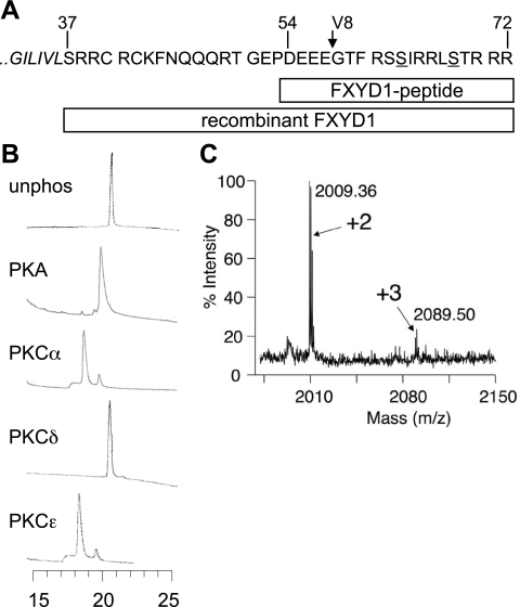
The fusion protein of glutathione S-transferase (GST) and the intracellular region of FXYD1 was expressed and purified by standard techniques, and the GST affinity tag was removed by cleavage with thrombin (GE Healthcare). As a result of the cloning, the recombinant FXYD1 COOH terminus used in this study has an additional NH2-terminal glycine residue, which is followed by a serine equivalent to serine 37 of full-length, processed canine FXYD1: the first residue of the intracellular region. Figure 1A shows the sequences of the FXYD1 peptide and recombinant FXYD1 used in this study.
Fig. 1.
In vitro phosphorylation of FXYD1. A: diagrammatic representation of the FXYD1 peptides used in this study. The end of the transmembrane domain (italicized) and intracellular region of canine FXYD1 are shown, with the known phosphorylation sites S63 and S68 underlined. The site of cleavage by V8 is marked with an arrow. FXYD1 peptide was residues 54-72. The entire intracellular region of FXYD1 (37-72) was produced recombinantly. B: HPLC analysis of in vitro phosphorylated FXYD1 peptide. Peptides were phosphorylated with the kinases indicated, then analyzed on a C18 reverse-phased column. Chromatograms are temporally aligned (retention time in minutes is shown at the bottom), and absorbance at 214 nm is shown plotted against time. Phosphorylation reduces the affinity of the peptide for the C18 matrix and causes earlier elution. PKA phosphorylates the peptide at one site, PKCα and ɛ at two sites, but PKCδ not at all. C: matrix-assisted laser desorption/ionization mass spectrometry (MALDI) analysis of in vitro phosphorylated recombinant FXYD1, digested with V8. Unphosphorylated material digested with V8 yields peaks at mass-to-charge ratio (m/z) 1849.07 (58-72, containing the phosphorylation sites) and 2625.19 (37-57, not shown). Phosphorylation by PKCα shifts the peak at 1849.07 to two peaks: m/z 2009.36 (+2: theoretical m/z of dual phosphorylated peptide 2009.01) and m/z 2089.50 (+3: theoretical m/z of triply phosphorylated peptide 2088.97). Note that MALDI relies on the selection of singly charged (in this case positive) ions, meaning quantitative comparison of peak intensities of dual and triply phosphorylated species is meaningless, because less triply phosphorylated peptide will form the MH+ ion.
In vitro phosphorylation.
All in vitro phosphorylation reactions were in a buffer consisting of 20 mM Tris, pH 7.4, 15 mM MgCl2, 1 mM dithiothreitol (DTT), and 1 mM ATP. For phosphorylation reactions involving PKC, a lipid-activating cocktail (which also contained calcium) was also included. cAMP was not required for PKA activity. Each reaction contained peptide substrate at a final concentration 10–50 μM and 20 units of kinase, where 1 unit is defined as the amount of kinase required to transfer 1 pmol phosphate from ATP to substrate per minute at 30°C. We confirmed the activity of all kinases against suitable positive control peptides (Upstate Biotechnology). All reactions were incubated at 37°C and allowed to proceed to completion unless indicated otherwise (90–120 min).
HPLC analysis of in vitro phosphorylated FXYD1.
In vitro phosphorylation reactions were halted by adjusting them to 0.1% trifluoroacetic acid (TFA) and analyzed by HPLC (Shimadzu) using a reversed-phase C18 column (Jones Chromatography), as described previously (22). Peptide elution was detected by monitoring the absorbance at 214 nm of the buffer exiting the column. The identity of peptide peaks was confirmed by collecting and drying them, followed by analysis using matrix-assisted laser desorption/ionization mass spectrometry (MALDI).
Isolation and culture of adult rat ventricular myocytes.
Male Wistar rats (250–300 g) were housed and handled in accordance with the Institute for Laboratory Animal Research Guide for Care and Use of Laboratory Animals. Animal studies were approved by the local ethics committees at King's College London and University of Dundee. Adult rat ventricular myocytes (ARVM) were isolated and cultured in serum-free medium 199 (Invitrogen, Paisley, UK) supplemented with taurine (5 mM), carnitine (2 mM), and creatine (2 mM) for 18–24 h on laminin-coated 6- and 12-well culture plates, as described in detail previously (27).
Drugs.
Drugs were applied to myocytes bathed in standard culture medium 18–24 h after plating. With the following exceptions, all drug stocks were 1,000× working concentration dissolved in PBS, stored at −20°C, and were not refrozen after thawing. Isoprenaline was dissolved in PBS immediately before use. Forskolin and PMA were dissolved in 95% ethanol in water and stored at −20°C. H89 and bis were dissolved in DMSO and stored at −20°C. Bis was protected from light. All drugs were applied in the dark at 37°C. All appropriate vehicle controls were carried out; however, for simplicity of presentation, these data are not shown.
Production of phosphospecific antibodies.
Antibodies phosphospecific for threonine 69 (T69)-phosphorylated FXYD1 (CP69), and dual S68- and T69-phosphorylated FXYD1 (CP689), were raised in sheep at Diagnostics Scotland (Edinburgh, UK). This procedure is described in detail elsewhere (19). Briefly, synthetic phosphopeptides representing residues 64 to 72 of rat FXYD1 were coupled separately to bovine serum albumin and keyhole limpet hemocyanin, mixed, then injected into sheep. For antibody CP69, the antigen was IRRLST*RRR, and for CP689, we used IRRLS*T*RRR, where asterisks denote the phosphorylated residues. Antisera were affinity purified on CH-Sepharose to which the relevant phosphopeptide had been coupled. Antibodies were used for immunoblotting in the presence of 10 μg/ml of the unphosphorylated peptide antigen to neutralize any antibodies that recognized the unphosphorylated form of FXYD1. Two additional blocking peptides, IRRLS*TRRR and IRRLST*RRR, were also used with antibody CP689, such that the remaining “unblocked” antibody was phosphospecific for FXYD1 dual phosphorylated at residues 68 and 69.
Immunoprecipitation.
For immunoprecipitation of FXYD1 phosphorylation states, cultured ARVM were lysed in 1% Triton X-100 in 20 mM HEPES, pH 7.4, supplemented with 1 mM EDTA, a protease inhibitor cocktail (Sigma), phosphatase inhibitor cocktail 1 (Sigma), and additional phosphatase inhibitors (2 mM sodium vanadate, 5 mM sodium fluoride, 2 mM sodium pyrophosphate, and 2 mM sodium β-glycerophosphate). Samples were gently agitated for 15 min at 4°C, and insoluble material was removed by centrifugation at 15,000 g for 5 min at 4°C. Immunoprecipitating antibodies were added to the supernatant, and following agitation for 2 h at 4°C, immune complexes were harvested for 2 h with immobilized protein A. Beads were washed three times with 1% Triton X-100 in 20 mM HEPES, pH 7.4, supplemented with protease and phosphatase inhibitors, and were then resuspended in SDS-PAGE sample buffer.
Coimmunoprecipitation.
For coimmunoprecipitation of FXYD1 and Na+-K+-ATPase α1-subunit, cultured ARVM were lysed in PBS supplemented with 6 mg/ml octaethylene glycol monododecyl ether (C12E8, Sigma), plus the protease and phosphatase inhibitors described above. Samples were agitated gently for 30 min at 4°C and diluted 1:1 with PBS (plus protease and phosphatase inhibitors), and insoluble material was removed by centrifugation (15,000 g, 5 min). Supernatants were agitated overnight at 4°C with anti-Na+-K+-ATPase α1-subunit monoclonal C464.6 (Santa Cruz Biotechnology), immune complexes were harvested with protein G Sepharose for 2 h at 4°C, and beads were washed five times with 0.5 mg/ml C12E8 in PBS supplemented with protease and phosphatase inhibitors. To avoid the confounding presence of mouse IgG in immunoprecipitated samples, successful immunoprecipitation of Na+-K+-ATPase α-subunit was confirmed using an antibody raised in chickens (Chemicon).
SDS-PAGE and immunoblotting.
Optimal resolution of FXYD1 was achieved using gels based on the Tris-tricine buffer system. Immunoblotting was carried out as described previously (25).
Whole cell patch clamping of cultured ARVM.
ARVM were perfused with 50 μM forskolin, 300 nM PMA, or 0.1% (vol/vol) ethanol (vehicle) for 25 min at 35°C. Whole cell patch-clamp recordings were established between 5 and 25 min after drug application, and Na+/K+ pump current was measured as the potassium-dependent current inhibited on switching from 5 mM to 0 mM potassium solution at 0 mV. Pipette filling solution was (in mM) 90 CsCH3O3S, 35 NaCH3O3, 8 CsCl, 15 NaCl, 1 MgCl2, 10 HEPES, 5 EGTA, 5 MgATP, and 5 creatine phosphate, pH 7.2. External solution was (in mM) 140 NaCl, 1 MgCl2, 2 NiCl2, 1 BaCl2, 5 or 0 KCl, 10 glucose, 10 HEPES, and 0.5 procaine, pH 7.4, at 35°C. Pipette resistance was 1–2 MΩ, and access resistance following establishment of a seal and membrane rupture was 3.3 MΩ.
RESULTS
Phosphorylation of FXYD1 peptide.
The phosphorylation of a FXYD1 peptide by PKA, PKCα, δ, and ɛ was investigated using the same peptide as in a previous study to identify PKA and PKC phosphorylation sites (32). Phosphorylation events were detected using HPLC as a shift in the elution time of the FXYD1 peptide from a reversed-phase C18 column. The more hydrophilic the peptide, the earlier it eluted and therefore the more phosphorylation events have occurred. Figure 1 indicates that PKA causes the elution time to shift from 21.5 min to 19 min, while PKCα and ɛ cause a shift to predominantly 18 min with a minor component eluting at 19 min. PKCδ does not phosphorylate the FXYD1 peptide, because the elution time is not shifted. Hence, PKA phosphorylates the FXYD1 peptide at a single site, and PKCα and ɛ phosphorylate two sites. Peptide peaks were collected, dried, and analyzed by MALDI to confirm that peptide masses were consistent with the phosphorylation stoichiometries determined by HPLC (data not shown). In addition, Edman sequencing of peptides phosphorylated in the presence of γ32P-ATP confirmed that PKA phosphorylated residue 15 of the peptide (equivalent to serine 68 in full-length FXYD1) and PKCα phosphorylated residues 10 and 15 (equivalent to serines 63 and 68). The phosphorylation pattern of this FXYD1 peptide was therefore identical to that previously reported (32).
Effect of prephosphorylation by PKA on subsequent phosphorylation by PKC.
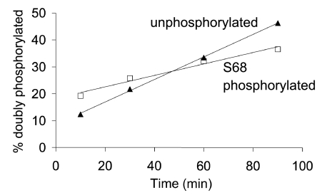
Researchers have previously noted that phosphorylation of FXYD1 peptides by PKC is limited if they are first phosphorylated by PKA (16); however, this remains an anecdotal observation that has not been rigorously and quantitatively investigated. We compared PKC phosphorylation of FXYD1 peptide with PKC phosphorylation of FXYD1 peptide that had been prephosphorylated by PKA, then HPLC purified. Figure 2 shows the result of an experiment using PKCα. The rate of conversion of substrate to the doubly phosphorylated species (assessed by HPLC) is plotted as the percentage of the total peptide that is doubly phosphorylated against time. Note that over the period shown in Fig. 2, accumulation of doubly phosphorylated peptide was linear with respect to time, probably indicating that in this example PKCα was operating at Vmax. Note also that the rate of accumulation of doubly phosphorylated species (the gradient of the linear regression lines shown in Fig. 2) is twice as fast if the starting material is unphosphorylated compared with if it is PKA phosphorylated. So, even though the prephosphorylated peptide is closer to the reaction end point (that is, it already has one phosphate incorporated), its conversion to that end point is slower than if no phosphate is present. A similar result was found for PKCɛ (not shown). In three independent experiments, that rate of conversion of unphosphorylated species to doubly phosphorylated was found to be 2.1 ± 0.1 times faster for PKCα and 1.6 ± 0.2 times faster for PKCɛ. Note, however, that both reactions reached completion if given sufficient time (not shown).
Fig. 2.
The effect of prephosphorylation of S68 on phosphorylation of the FXYD1 peptide by PKC. Unphosphorylated (▴) and PKA phosphorylated (□) FXYD1 peptide was phosphorylated with PKCα. Measurement of peak area of the HPLC-purified starting material ensured that substrate concentration was identical between the two reactions. Reaction progress was monitored by analyzing samples by HPLC: conversion of the starting material to a doubly phosphorylated species was measured by calculating the area of the doubly phosphorylated peak from each sample of each reaction. The percentage of peptide in each sample that had reached the doubly phosphorylated state is plotted as a function of the reaction time. The gradient of the linear regression lines are 0.21 (S68 phosphorylated starting material) and 0.42 (unphosphorylated starting material), indicating that conversion to the doubly phosphorylated state by PKCα proceeds twice as quickly for unphosphorylated starting material compared with S68 phosphorylated starting material.
Phosphorylation of recombinant FXYD1.
We expressed the intracellular region of canine FXYD1 as a GST fusion protein and removed the GST affinity tag by cleaving with thrombin after purification. This 37-residue recombinant fragment was phosphorylated in vitro using the same protocol as that for FXYD1 peptide.
We were unable to purify and phosphorylate sufficient recombinant FXYD1 for full phosphopeptide mapping using mass spectrometry. MALDI analysis of in vitro phosphorylated recombinant wild-type FXYD1 indicated the presence of singly and doubly phosphorylated peptides following phosphorylation by PKA, and doubly and triply phosphorylated peptides following PKCα or ɛ (Fig. 1C). Hence additional phosphorylation sites are present in recombinant FXYD1 compared with the FXYD1 peptide. While we were unable to definitively map these additional phosphorylation sites, we hypothesized that the additional site for PKC isoforms was T69.
Phosphospecific antibodies.
To investigate in vitro phosphorylation of FXYD1, and signaling pathways terminating in FXYD1 phosphorylation in cardiac myocytes, we raised antibodies to phosphorylated T69 (CP69), and dual phosphorylated S68 and T69 (CP689). The reactivity of these and existing antibodies in dot blots of in vitro phosphorylated recombinant wild-type and mutant FXYD1 is shown in Fig. 3. Antibody CP63 (25) is phosphospecific for phosphorylated serine 63. It reacts with wild-type, S68A, T69A, and S68T69A phosphorylated by α- and ɛ-PKC isoforms, but not with S63A. It also reacts with wild-type, S68A, T69A, and S68T69A phosphorylated by PKA, but barely at all with S63A. The additional PKA phosphorylation site in recombinant FXYD1 (compared with the FXYD1 peptide) is therefore probably S63, which has not previously been reported to be a PKA site. Figure 3 also indicates that antibody CP68 (24, 25) is specific for phosphorylated serine 68, although note that its binding is severely limited by mutation of the adjacent T69 to alanine. We have recently raised a new antibody to phosphorylated S68, which indicates that phosphorylation of S68 is indeed normal in T69A (not shown). CP68 shows no reactivity toward unphosphorylated FXYD1, and CP63 shows approximately 10-fold selectivity for phosphorylated over unphosphorylated FXYD1. Antibody CP69 reacts with PKCα and PKCɛ phosphorylated wild-type FXYD1 but not with mutant T69A or S68T69A. Antibody CP689 also reacts with PKCα and PKCɛ phosphorylated wild-type FXYD1 but not with mutant T69A, S68A, or S68T69A. Binding of antibodies CP69 and CP689 to in vitro phosphorylated recombinant wild-type FXYD1 was abolished by preincubation with the appropriate phosphorylated blocking peptide (not shown).
Fig. 3.
Characterization of FXYD1 phosphospecific antibodies. A: the reactivity of CP63, CP68, CP69, and CP689 to in vitro phosphorylated recombinant FXYD1 and its point mutants was investigated by dot blotting in vitro phosphorylation reactions that had proceeded to completion. Antibody CP69 was preincubated with 10 μg/ml unphosphorylated blocking peptide, and antibody CP689 was preincubated with unphosphorylated blocking peptide plus two singly phosphorylated blocking peptides (all 10 μg/ml), as described in materials and methods. All phosphospecific antibodies show the appropriate reactivity, although note that mutation T69A severely limits the ability of antibody CP68 to bind to FXYD1 phosphorylated at serine 68. Antibody CP689 shows very weak binding to mutant S68A phosphorylated by PKC (i.e., phosphorylated at T69 only); however, the majority of signal generated by this antibody is for dual phosphorylated S68 and T69 [compare with signal from wild type (WT) and S63A]. B: time course of phosphorylation of wild-type recombinant FXYD1 by PKA and PKC. Phosphorylation of S63 by PKA is very slow compared with S68 (phosphorylation of S68 is complete within 10 min, but phosphorylation of S63 requires 90 min). Phosphorylation of T69 by PKCɛ severely limits binding of antibody CP68 to phosphorylated S68.
Interestingly, when PKC is added to a reaction after phosphorylation of wild-type FXYD1 by PKA, binding of antibody CP68 is reduced, as binding of antibodies CP69 and CP689 is increased. Figure 3B shows the progress of an in vitro phosphorylation reaction in which PKA and PKC are applied sequentially to recombinant wild-type FXYD1. Samples were taken periodically and dot blotted with all four phosphospecific antibodies. When PKA is applied, S68 is phosphorylated quickly and S63 slowly. Application of PKC isoforms after PKA causes phosphorylation of T69, and this has a negative impact on binding of antibody CP68. If PKC is applied first, S63 and S68 are rapidly phosphorylated, and T69 phosphorylated more slowly; again, this reduces binding of CP68 (not shown). Hence, adjacent phosphorylation of T69 has a negative effect on the affinity of CP68 for its substrate. CP68 will underestimate phosphorylation of S68 in the presence of adjacent phosphorylated T69.
Signaling to FXYD1 phosphorylation in adult rat cardiac myocytes.
We investigated signaling to FXYD1 phosphorylation in ARVM isolated by enzymatic dispersion of the heart. Since this isolation procedure is stressful, ARVM were allowed to recover before signaling pathways were probed. We routinely find that freshly isolated myocytes are considerably less responsive to agonists than myocytes that have been cultured for 18–24 h (not shown). Therefore, ARVM were cultured on laminin-coated 12-well culture dishes before application of agonists.
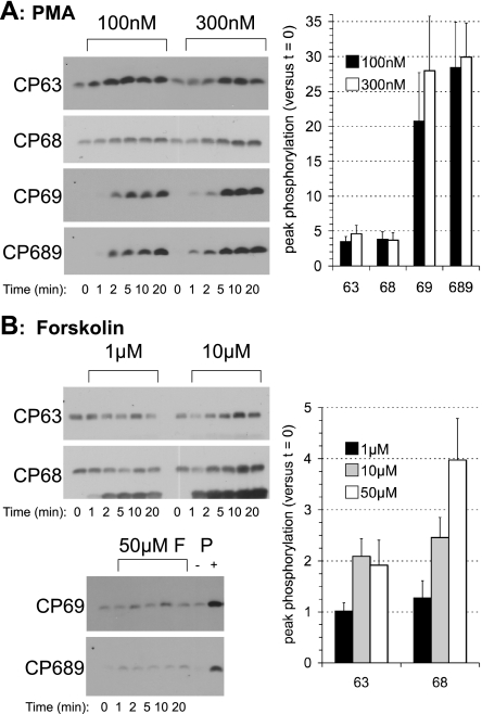
To reveal both basal and stimulated phosphorylation of the three candidate sites in FXYD1, we first investigated signaling by PKA and PKC to FXYD1 by maximally activating these kinases with forskolin and PMA, respectively (hence bypassing G protein-coupled receptors). Dose-response curves were constructed using 1, 10 and 50 μM forskolin and 100 and 300 nM PMA, and the development of a response was measured at several points in the first 20 min after drug application. For each phosphospecific FXYD1 antibody, the response in Fig. 4 is expressed as the increase in FXYD1 phosphorylation over signal strength at time zero (untreated ARVM). Figure 4 shows representative phosphospecific immunoblots for selected agonist concentrations and mean data for all agonist concentrations investigated. Figure 4A shows the response to PMA, and Fig. 4B shows the response to forskolin. The response to the highest dose of agonist was not greater than the next highest dose. Hence we used 50 μM forskolin and 300 nM PMA in all subsequent studies.
Fig. 4.
Phosphorylation of FXYD1 in cultured adult rat ventricular myocytes (ARVM) following treatment with PMA (A) and forskolin (B). PKC activation results in sustained phosphorylation of S63, S68, and T69 (A). Activation of PKA results in phosphorylation of S63 and S68, but not T69. Note that antibody CP68 cross-reacts with phospholamban phosphorylated at serine 16. A positive control [PMA-treated ARVM (P)] is shown for antibodies CP69 and CP689 in B. Representative immunoblots for 2 concentrations of agonist with each phosphospecific antibody and mean peak responses normalized to the signal at time zero (t = 0) are shown (n ≥ 6, means ± SE). F, forskolin.
Figure 4A shows that T69 phosphorylation and dual phosphorylation of S68 and T69 are observed following activation of PKC but not PKA. Note that while we observe phosphorylation of all three sites when ARVM are treated with PMA, phosphorylation of T69 is extremely low in untreated cells compared with S63 and S68. When normalizing to phosphorylation in untreated cells, relative changes in T69 phosphorylation are consequently much larger than relative changes in S63 and S68 phosphorylation. In Fig. 4B, antibody CP68 cross-reacts with serine 16 phosphorylated phospholamban (PLB, bottom band). It is noteworthy that phosphorylation of PLB is observed at concentrations of forskolin (1 μM) that induce no phosphorylation of FXYD1. Figure 4B also indicates that activation of PKA with forskolin induces phosphorylation of S63 that is of similar magnitude to the response to PMA, suggesting that S63 is a substrate for PKA in ARVM as well as in vitro. S68 phosphorylation elicited by PKA activation (detected by antibody CP68 in Fig. 4B) is greater than that seen when PKC is activated (Fig. 4A; however, antibody CP68 will underestimate the extent of S68 phosphorylation in these samples because of phosphorylation at adjacent T69). Hence Fig. 4 indicates that T69 of FXYD1 is a substrate for PKC in intact cells as well as in vitro, but that phosphorylation at T69 in untreated cells is substantially less than at the established sites S63 and S68.
Receptor-mediated FXYD1 phosphorylation.
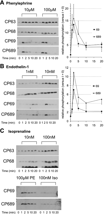
We next investigated phosphorylation of FXYD1 induced by PKA and PKC activation through cell surface receptors. Since the adrenergic signaling pathway is heavily implicated in FXYD1 phosphorylation, we used the β-adrenergic agonist isoprenaline (10 and 100 nM) to activate PKA, and the α-adrenergic agonist phenylephrine (10 and 100 μM) to activate PKC. The β-receptor antagonist atenolol (10 μM) was applied 2 min before phenylephrine to ensure that all responses were due to its effects on α-receptors. Additionally, we used endothelin-1 (1 and 10 nM) to activate PKC independent of adrenergic receptors. Figure 5 shows the responsiveness of ARVM to these drugs. Both PKC agonists (phenylephrine and endothelin-1) and the PKA agonist (isoprenaline) induce phosphorylation of S63 and S68. T69 is also phosphorylated following application of PKC agonists. Unlike S63 and S68, T69 phosphorylation is not sustained in the continued presence of these agonists; phosphorylation peaks 1–2 min after application and returns almost to basal levels after 10–20 min, even in the continued presence of drug. The transient phosphorylation of T69 was concentration dependent: in cells treated with 1 nM endothelin-1, we did not observe a peak in T69 phosphorylation (Fig. 5B), only a slowly developing increase. Dual phosphorylation of S68 and T69 follows an almost identical pattern to T69 phosphorylation, both in terms of the magnitude and the timescale of the response, suggesting that it is T69 rather than S68 phosphorylation that is the limiting factor in the accumulation of dual phosphorylated S68 and T69.
Fig. 5.
Receptor-mediated FXYD1 phosphorylation in cultured ARVM. α-Adrenoceptor activation with phenylephrine (A) causes sustained phosphorylation of S63 and S68, but transient phosphorylation of T69. Endothelin type A receptor activation with endothelin-1 (B) causes qualitatively very similar effects. β-Adrenoceptor activation with isoprenaline (C) causes sustained phosphorylation of S63 and S68 but not T69. A positive control for antibodies CP69 and CP689 [phenylephrine-treated ARVM (PE)] is shown alongside isoprenaline-treated cells (Iso; C). Representative immunoblots are shown for each phosphospecific antibody. For simplicity of presentation, only mean data for antibodies CP69 and CP689 at the highest agonist dose, normalized to the signal at time zero, are shown in A and B (n ≥ 6, means ± SE).
Basal phosphorylation of FXYD1.
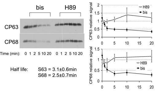
Researchers have previously noted that basal phosphorylation of FXYD1 in ARVM maintained in culture for 72 h is high: 16% at S63, and 46% at S68 (28). We have also previously observed relatively small increases in FXYD1 phosphorylation in response to agonists (25), possibly suggesting that FXYD1 phosphorylation is high in the resting state. In the present study, the maximum increase in S63 phosphorylation is approximately threefold, and at S68 fourfold. This is in contrast to the changes observed in PLB phosphorylation; in this and our previous study, PLB is not detected phosphorylated in the basal state. We therefore investigated the mechanisms underlying FXYD1 phosphorylation in the basal state, by applying PKA and PKC inhibitors and then measuring their acute effect on FXYD1 phosphorylation. Figure 6 indicates that while the PKA-specific inhibitor H89 (10 μM) is without effect on FXYD1 phosphorylation, the PKC inhibitor bis (1 μM) causes a rapid and sustained decrease in phosphorylation of both S63 and S68. These data were fit with single exponentials over the first 5 min of the bis treatment. We find the half-lives of S63- and S68-phosphorylated FXYD1 are 3.1 ± 0.6 min and 2.5 ± 0.7 min, respectively.
Fig. 6.
Effect of acute inhibition of PKA and PKC on basal phosphorylation of S63 and S68 in unstimulated cultured ARVM. While the PKA inhibitor H89 (10 μM) is without effect on basal FXYD1 phosphorylation, in the presence of the PKC inhibitor bisindolylmaleimide (bis, 1 μM), FXYD1 is rapidly dephosphorylated at S63 and S68. The first 5 min of bis treatment were fitted with a single exponential, giving half-lives of 3.1 ± 0.6 min and 2.5 ± 0.7 min for phosphorylated S63 and S68, respectively.
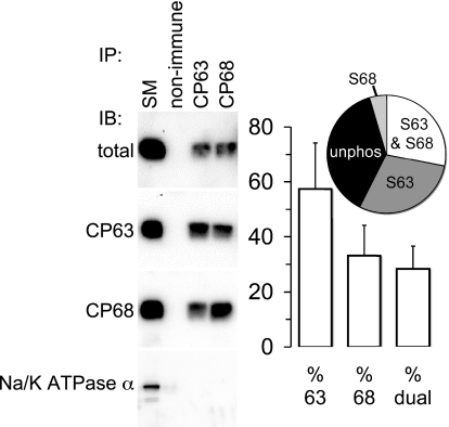
To assess the relative amount of FXYD1 phosphorylated in the basal state, we immunoprecipitated FXYD1 from unstimulated, cultured ARVM using the phosphospecific antibodies CP63 and CP68, then immunoblotted the immunoprecipitation reactions with our NH2 terminus-specific FXYD1 antibody raised in chickens (N1), which does not discriminate between phosphorylation states (11). Since both CP63 and CP68 have very low but detectable binding to unphosphorylated FXYD1, immunoprecipitation reactions were set up so that the efficiency of precipitation was <100% for each phosphorylation state. We reasoned that since the affinity of each antibody is so much greater for its particular phosphorylation state than for the unphosphorylated material, then as long as the antibody was not in excess it would only precipitate the appropriate phosphorylation state. The amount of antibody used in each immunoprecipitation reaction was determined empirically.
These immunoprecipitation reactions used 1% Triton X-100 to solubilize membrane proteins. In our hands this disrupts the FXYD1-Na+-K+-ATPase protein complex (Fig. 7), so we reasoned that it would also disrupt any FXYD1-FXYD1 complexes. Since it has been reported that FXYD1 forms multimers when expressed in heterologous cells (3), it is clearly key in the interpretation of these experiments that different FXYD1 phosphorylation states are not coimmunoprecipitated.
Fig. 7.
Basal phosphorylation of S63 and S68 in cultured ARVM. Triton X-100-solubilized FXYD1 was immunoprecipitated (IP) with nonimmune serum, and antibodies CP63 and CP68, and immunoblotted (IB) with an antibody that does not distinguish between phosphorylation states (total). Immunoprecipitated FXYD1 is expressed as percentage of that found in the starting material (SM). The FXYD1/Na+-K+-ATPase α1-subunit interaction is disrupted by the presence of 1% Triton X-100 in the immunoprecipitation buffer (Na+-K+-ATPase α-subunit is not detected copurifying with FXYD1). A full explanation of the calculations of percent phosphorylation is in the text. Mean data indicating percent basal phosphorylation at each residue from 5 independent experiments are shown (means ± SE). Inset: pie chart showing the distribution of FXYD1 between phosphorylated at S63 only, phosphorylated at S68 only, dual phosphorylated (S63 and S68), and unphosphorylated states in unstimulated ARVM.
Figure 7 shows the results of one immunoprecipitation experiment. In the representative immunoblots shown, 38% of S63-phosphorylated FXYD1 has been precipitated, which represents 16% of total FXYD1, so 100% of S63-phosphorylated FXYD1 is equivalent to 41% of total FXYD1: S63 is 41% phosphorylated in these cells. Likewise, 40% of S68-phosphorylated FXYD1 has been precipitated, which is 22% of the total, meaning that FXYD1 is 54% phosphorylated at S68 in these cells. These experiments also allow us to calculate the percentage of FXYD1 that is phosphorylated at both S63 and S68 in these cells. The 38% of S63-phosphorylated FXYD1 immunoprecipitated (lane CP63 in Fig. 7) represents 24% of the S68-phosphorylated FXYD1 in the starting material, so 100% of S63-phosphorylated FXYD1 is 63% of the S68-phosphorylated FXYD1. Since S68 is 54% of the total FXYD1 in these cells, dual phosphorylated FXYD1 is 54% of 63%; i.e., 34%. We can also calculate the percentage of dual phosphorylated FXYD1 the other way around. The 40% of S68-phosphorylated FXYD1 precipitated (lane CP68 in Fig. 7) represents 32% of the S63-phosphorylated FXYD1 in the starting material, so 100% of S68-phosphorylated FXYD1 is 80% of the S63-phosphorylated FXYD1. Since S63 is 41% phosphorylated in these cells, dual phosphorylated FXYD1 is 41% of 80%; i.e., 33%. In five independent experiments, we found that S63-, S68-, and dual (S63 and S68)-phosphorylated FXYD1 represent 57%, 33%, and 28% of total FXYD1, respectively, and hence we calculate that 38% of FXYD1 is phosphorylated at neither residue in the basal state.
Functional effects of FXYD1 phosphorylation on the Na+-K+-ATPase.
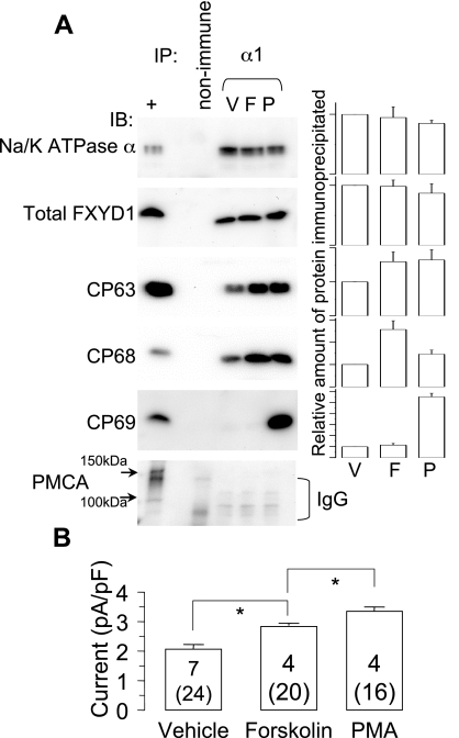
To assess the effect of phosphorylation of FXYD1 at T69 on Na+-K+-ATPase function, we compared the effects of PMA (300 nM), forskolin (50 μM), and PMA and forskolin applied together. Since we are yet to identify an agonist capable of eliciting T69 phosphorylation alone, the major difference between the effect of PMA and forskolin on FXYD1 is T69 phosphorylation (Fig. 4; the increase in S63 and S68 phosphorylation elicited by the drugs are very similar), and therefore the functional effect of T69 phosphorylation is the difference between the two. PMA elicits T69 phosphorylation when applied after forskolin (not shown). We immunoprecipitated Na+-K+-ATPase α1-subunit under conditions that favor copurification of FXYD1 and confirmed that, in forskolin-treated ARVM, more S63- and S68-phosphorylated FXYD1 is associated with Na+-K+-ATPase, and, in PMA-treated ARVM, more S63-, S68-, and T69- phosphorylated FXYD1 is found associated with Na+-K+-ATPase (Fig. 8A). In agreement with previous studies, we found no change in the total amount of FXYD1 copurifying with Na+-K+-ATPase following kinase activation (2, 3, 11, 25).
Fig. 8.
Functional effect of T69 phosphorylation on Na+-K+ pump currents. A: Na+-K+-ATPase α1-subunit was immunoprecipitated under conditions favoring copurification of FXYD1. Control immunoprecipitations used nonimmune mouse serum and untreated ARVM. +, Untreated ARVM. Representative immunoblots are shown, and the bar graphs alongside each immunoblot show the mean relative amounts of each protein immunoprecipitated, normalized to the amount purified from vehicle-treated cells (n = 5). Equal amounts of Na+-K+-ATPase α1 and copurifying total FXYD1 were immunoprecipitated from ARVM treated with vehicle [0.1% vol/vol ethanol (V)], 50 μM forskolin (F), or 300 nM PMA (P) for 10 min at 37°C. The specificity of coimmunoprecipitation was confirmed by blotting for plasma membrane Ca2+-ATPase (PMCA). We routinely observe bands at ∼140 kDa and ∼95 kDa when probing ARVM lysates for PMCA: neither form is immunoprecipitated in these reactions; however, the mouse IgG used in immunoprecipitated samples is seen. In cells treated with forskolin, copurifying FXYD1 was more phosphorylated at S63 and S68 compared with control. In cells treated with PMA, copurifying FXYD1 was more phosphorylated at S63, S68, and T69. B: cultured ARVM were treated with vehicle (0.1% vol/vol ethanol), forskolin (50 μM), PMA (300 nM). Recordings were made in the whole cell mode, with 50 mM Na+ in the pipette. Drugs were applied on the microscope stage at 35°C, and cells were left for a minimum of 5 min before establishing a seal and rupturing the membrane, to avoid disrupting signaling pathways by dialyzing the cell contents before a response developed. Recordings were made from multiple cells for up to 25 min after drugs were applied. Na+-K+ pump current was defined as the current sensitive to the removal of extracellular K. Data are means ± SE. Numbers are the number of cells (in parentheses) and number of animals (each animal gives one data point). *P < 0.05 (Student's t-test).
ARVM were voltage clamped in the ruptured patch mode, with pipette Na+ at 50 mM, such that the Vmax of the Na+-K+-ATPase was assessed. Na+-K+-ATPase current (Ipump) was defined as the current sensitive to the removal of extracellular K+, which we have previously shown is identical to the ouabain-sensitive current (25). Drugs were applied before sealing onto the myocyte and breaking into the cell, to avoid dialyzing cell contents and disrupting signaling pathways. Whole cell currents in the presence and absence of K+ were measured 2 min after membrane rupture, which was between 5 and 25 min after application of the drugs, a time at which FXYD1 phosphorylation is at steady state (Fig. 4). Note that this method of establishing a seal after applying a drug does not allow us to monitor the development of a response or to record Ipump before and after drug application; however, we considered it important to avoid cell dialysis at a time when signaling events were occurring.
Figure 8B indicates that PKA activation with forskolin and PKC activation with PMA causes a stimulation of Ipump. Hence the phosphorylation state of FXYD1 is intimately linked to Na+-K+-ATPase activity: FXYD1 phosphorylation is associated with an increase in Na+-K+-ATPase Vmax. T69 phosphorylation causes Na+-K+-ATPase activation additional to that elicited by phosphorylation at S63 and S68; PMA alone elicits a larger increase in Ipump (to 3.4 ± 0.2 pA/pF from 2.1 ± 0.2 pA/pF in vehicle-treated cells) than forskolin (2.9 ± 0.1 pA/pF). Parenthetically, when PMA was applied 10 min after forskolin, we also observed an increase in Ipump (3.3 ± 0.1 pA/pF) compared with forskolin alone (data not shown).
DISCUSSION
The present study has identified a new phosphorylation site in FXYD1, T69, which was not identified by other studies that mapped FXYD1 phosphorylation sites (32). T69 is phosphorylated by PKC isoforms found in cardiac myocytes—PKCα and/or ɛ, but probably not PKCδ—according to our in vitro phosphorylation experiments. The failure of previous studies to identify this phosphorylation site probably lies in the use of a FXYD1 peptide rather than the entire COOH terminus to map these sites. We ourselves also find that PKC isoforms are unable to phosphorylate T69 of a 19-amino acid peptide representing ∼50% of the FXYD1 COOH terminus; it is only when the entire intracellular region is offered as a substrate that T69 phosphorylation is observed. This FXYD1 peptide presumably lacks the necessary secondary structure to present T69 in an appropriate manner to PKC. The three-dimensional NMR structure of full-length FXYD1 in micelles (albeit not associated with Na+-K+-ATPase) indicates that T69 is fully exposed to the aqueous phase and will therefore be accessible to PKC isoforms (31).
Threonine 69 of FXYD1.
A notable property of receptor-mediated T69 phosphorylation in cardiac myocytes is its transient nature, particularly when compared with S63 and S68 phosphorylation, which persists in the continued presence of agonist. This is in contrast to the effect of direct PKC activation with PMA, which elicits a similar degree of T69 phosphorylation, but in a sustained manner. The time course of T69 phosphorylation in response to phenylephrine treatment is remarkably consistent: always peaking at 1–2 min, very substantially reduced at 5 min, and barely above basal at 10 min. Since the maximum achievable increase in T69 phosphorylation is approximately 30-fold (following treatment with 300 nM PMA), basal phosphorylation is at most ∼3%, meaning that 10 min after application of 100 μM phenylephrine, at most 15% of FXYD1 is phosphorylated at T69. This failure to maintain the response in the continued presence of agonist must indicate that receptor activation is associated with either a rapid loss of driving force to the kinase phosphorylating T69 (but not S63 and S68), and/or the activation of a pathway to specifically dephosphorylate T69 (but not S63 and S68) that is absent when PKC is directly activated with PMA. In addition, T69 phosphorylation appears to be very much lower than S63 and S68 in unstimulated ARVM: 3% vs. 57% and 33%, respectively. Not only does this suggest that the mechanisms leading to T69 phosphorylation and dephosphorylation are different, it is also tempting to speculate that the different control of T69 phosphorylation implies a different functional role for this residue.
Sustained T69 phosphorylation causes a significant additional increase in Na+/K+ pump Vmax compared with the functional effect of phosphorylating S63 and S68 alone. It has been suggested that T69 phosphorylation in a kidney cell line may be required for transport of FXYD1 from the endoplasmic reticulum to the cell surface (15). FXYD1 predominantly resides in the sarcolemma and t-tubules in cardiac myocytes (25, 33), and the transient nature of agonist-induced T69 phosphorylation would probably be insufficient to induce trafficking from an intracellular store: sustained PMA-induced PKC activation for >5 min is required to induce FXYD1 trafficking in kidney cells (15).
Of more relevance to this study is the functional effect of threonine 17 phosphorylation of PLB on the activity of the sarcoplasmic reticulum Ca2+-ATPase SERCA2a. There is sequence similarity in the phosphorylation sites between PLB and FXYD1: RSAIRRAS16T17 for PLB and RSS63IRRLS68T69 for FXYD1. Threonine 17 of PLB is phosphorylated by CaM kinase II, which does not phosphorylate FXYD1 (21). Phosphorylation of serine 16 of PLB precedes that at threonine 17 (26), much as we have seen in our study. Although preferential phosphorylation of T17 has been reported in response to rapid pacing (12), heart rate rarely, if ever, is substantially elevated in the absence of β-adrenoceptor receptor occupation and PKA activation. Hence, it is not clear whether in a physiological setting threonine 17 of PLB is ever phosphorylated in the absence of serine 16 phosphorylation (17). The functional effect on SERCA2a of PLB phosphorylation at threonine 17 is indistinguishable from the effect of phosphorylation at serine 16. Given that receptor-mediated, PKC-induced T69 phosphorylation is not sustained, it is debatable whether the increase in Na+/K+ pump current that T69 phosphorylation elicits over that induced by S63 and S68 phosphorylation is physiologically important for the myocyte. So, much like threonine 17 of PLB, the physiological relevance of T69 phosphorylation of FXYD1 remains unclear for the moment.
Physiological role of FXYD1 phosphorylation.
In general, we find that higher concentrations of PKA agonists are required to cause phosphorylation of FXYD1 than PLB (Fig. 4B; 1 μM forskolin induces phosphorylation of PLB but not FXYD1). This is possibly a reflection of the opposing physiological roles of these phosphorylation events: PLB phosphorylation is associated with positive inotropy and lusitropy, through an increase in SERCA activity. FXYD1 phosphorylation leading to Na+-K+-ATPase activation is negatively inotropic because it increases the driving force for Ca2+ extrusion by NCX. Recent observations in FXYD1-knockout animals have led researchers to propose that the physiological role of FXYD1 is to temper the rise in intracellular Na+ during sympathetic stimulation (9). We would go further, because our biochemical evidence suggests that Na+-K+-ATPase activation by FXYD1 may only be necessary to oppose sodium overload at the top of the PKA agonist dose-response curve. Whatever the physiological consequences of this phenomenon, it suggests the biochemical events leading to FXYD1 phosphorylation by PKA are different from those that occur when PKA phosphorylates PLB, because they occur over a different range of agonist concentrations.
The changes in Na+/K+ pump current associated with FXYD1 phosphorylation observed in this study are similar to our previous electrophysiological studies (22, 25), and in line with that reported for PKC-mediated FXYD1 phosphorylation in mouse ventricular myocytes (13): 35–60% increases in current following FXYD1 phosphorylation. The dynamic range of FXYD1-mediated Na+-K+-ATPase regulation in ventricular myocytes therefore appears relatively small, particularly when considering the additional Na+ influx that will occur during adrenergic stimulation, through enhanced NCX activity, (up to) 2–3× Na+ entry through voltage-gated channels, and enhanced Na+-dependent substrate uptake. Once again, our data are consistent with a model in which FXYD1 exerts a tonic inhibition on the Na+-K+-ATPase, which is relieved upon FXYD1 phosphorylation. Unlike PLB regulation of SERCA, we observe increases in Na+-K+-ATPase Vmax when FXYD1 is phosphorylated, whether by PKA or PKC.
This study did not set out to establish that FXYD1 is solely responsible for kinase-mediated regulation of Na+-K+-ATPase in cardiac myocytes, and we cannot exclude the possibility that in ARVM treated with forskolin or PMA, phosphorylation of the Na+-K+-ATPase α-subunit may influence catalytic activity. However, the evidence that PKA may directly phosphorylate the α-subunit is weak at best (25, 29), and in other cell types, PKC-mediated phosphorylation principally regulates α-subunit subcellular distribution rather than catalytic activity (4, 10). Acute kinase regulation of Na+-K+-ATPase is absent in FXYD1-knockout animals (8, 13); hence in this study we ascribe the kinase-mediated changes in Na+-K+-ATPase activity observed (Fig. 8) to changes in phosphorylation of the associated FXYD1, with the proviso that these changes in ATPase activity are correlated with (not conclusively caused by) FXYD1 phosphorylation.
The relatively high basal phosphorylation of FXYD1 (at S63 and S68) means that the system can respond in either direction. Acute hormonal regulation of Na+-K+-ATPase can be both stimulation through FXYD1 phosphorylation (as in this study), and inhibition through dephosphorylation. Again, this differs from the PLB-SERCA relationship, because PLB is not phosphorylated in the basal state. Physiologically, the reason for the substantial basal phosphorylation of FXYD1 is presumably to have a system that can respond in either direction; however, we are yet to identify an intervention that causes dephosphorylation of FXYD1. Basally, we find phosphorylated FXYD1 throughout myocyte membranes, but more concentrated in t-tubules (data not shown), suggesting a role in the regulation of excitation-contraction coupling.
PKC and FXYD1.
PMA-induced activation of PKC causes sustained translocation of PKC isoforms to the membrane, whereas receptor-mediated PKC activation is usually characterized by transient translocation (5). With this in mind, the pattern of T69 phosphorylation of FXYD1 much more closely mirrors the membrane localization of PKC isoforms during receptor-mediated PKC activation. Indeed, given the pattern of PKC activation in cardiac myocytes, it is perhaps more surprising to find sustained phosphorylation of S63 and S68 following endothelin-1 or phenylephrine application than it is to find that T69 is phosphorylated transiently, especially given that S63 and S68 may clearly be very avidly dephosphorylated in the absence of resting PKC “tone” (acute application of bisindolylmaleimide; Fig. 6).
The very substantially different control of phosphorylation of the adjacent PKC phosphorylation sites S68 and T69 is another remarkable property of FXYD1. This is evidently an area that requires further investigation, but our data suggest that phosphorylation of S63 and S68 is achieved by a PKC isoform that is relatively active in unstimulated cells, whose receptor-mediated activation is sustained, and which is likely different from the isoform that phosphorylates T69. It is also possible that a different phosphatase is responsible for T69 dephosphorylation. We estimate that 2 min after application of 100 μM phenylephrine, phosphorylated T69 is dephosphorylated with a half-life is ∼4 min, which is similar to that observed for S63 and S68 in unstimulated cells (Fig. 6). Whether the avid dephosphorylation of T69 following receptor-mediated PKC activation reflects the activity of a phosphatase that is active regardless of the state of the myocyte, or one that is stimulated as a result of receptor activation, remains to be seen.
PKA and FXYD1.
Other investigators do not observe phosphorylation of S63 by PKA (8); however, we find that recombinant FXYD1 may be phosphorylated by PKA in vitro, and PKA activation in ARVM results in phosphorylation at S63. The reason for this discrepancy is unclear; it may represent a species difference, because we do not observe isoprenaline-induced phosphorylation of S63 in mouse ventricular myocytes (data not shown). S68 is undoubtedly the primary PKA site in FXYD1; Fig. 3B indicates that, in vitro, PKA-induced S63 phosphorylation develops very slowly compared with S68 phosphorylation. In ARVM, we observe significant day-to-day variation in the ability of isoprenaline to induce S63 phosphorylation, while S68 phosphorylation remains very consistent. When considering the FXYD1 peptide, we found that phosphorylation of S63 by PKC isoforms is slowed if the peptide is prephosphorylated at S68 by PKA (Fig. 2). The interfering effect of PKA activation on S63 phosphorylation in ARVM means that we are unable to extend this observation to intact cells.
In summary, we have identified a new phosphorylation site for PKC in FXYD1: threonine 69. Phosphorylation of this site is different from the previously described serines 63 and 68 in that it is very low in unstimulated cells, and not maintained in the presence of PKC agonists. T69 phosphorylation is associated with stimulation of Na+-K+-ATPase over that achieved by S63 and S68 phosphorylation.
GRANTS
This work was supported by grants from the British Heart Foundation (to M. J. Shattock and W. Fuller), the Medical Research Council (to M. J. Shattock and L. M. McLatchie), a Tenovus Scotland Research Grant (to W. Fuller), and a Research Councils UK Academic Fellowship (to W. Fuller).
Acknowledgments
The technical assistance of Semka Dashnyam and Dola Akanmu is gratefully acknowledged for help with cell isolation.
REFERENCES
- 1.Bers DM, Barry WH, Despa S. Intracellular Na+ regulation in cardiac myocytes. Cardiovasc Res 57: 897–912, 2003. [DOI] [PubMed] [Google Scholar]
- 2.Bossuyt J, Ai X, Moorman JR, Pogwizd SM, Bers DM. Expression and phosphorylation of the Na-pump regulatory subunit phospholemman in heart failure. Circ Res 97: 558–565, 2005. [DOI] [PubMed] [Google Scholar]
- 3.Bossuyt J, Despa S, Martin JL, Bers DM. Phospholemman phosphorylation alters its fluorescence resonance energy transfer with the Na/K-ATPase pump. J Biol Chem 281: 32765–32773, 2006. [DOI] [PubMed] [Google Scholar]
- 4.Chibalin AV, Pedemonte CH, Katz AI, Feraille E, Berggren PO, Bertorello AM. Phosphorylation of the catalyic α-subunit constitutes a triggering signal for Na+,K+-ATPase endocytosis. J Biol Chem 273: 8814–8819, 1998. [DOI] [PubMed] [Google Scholar]
- 5.Clerk A, Bogoyevitch MA, Anderson MB, Sugden PH. Differential activation of protein kinase C isoforms by endothelin-1 and phenylephrine and subsequent stimulation of p42 and p44 mitogen-activated protein kinases in ventricular myocytes cultured from neonatal rat hearts. J Biol Chem 269: 32848–32857, 1994. [PubMed] [Google Scholar]
- 6.Crambert G, Fuzesi M, Garty H, Karlish S, Geering K. Phospholemman (FXYD1) associates with Na,K-ATPase and regulates its transport properties. Proc Natl Acad Sci USA 99: 11476–11481, 2002. [DOI] [PMC free article] [PubMed] [Google Scholar]
- 7.Crambert G, Geering K. FXYD proteins: new tissue-specific regulators of the ubiquitous Na,K-ATPase. Sci STKE 2003: RE1, 2003. [DOI] [PubMed] [Google Scholar]
- 8.Despa S, Bossuyt J, Han F, Ginsburg KS, Jia LG, Kutchai H, Tucker AL, Bers DM. Phospholemman-phosphorylation mediates the beta-adrenergic effects on Na/K pump function in cardiac myocytes. Circ Res 97: 252–259, 2005. [DOI] [PubMed] [Google Scholar]
- 9.Despa S, Tucker AL, Bers DM. Phospholemman-mediated activation of Na/K-ATPase limits [Na]i and inotropic state during beta-adrenergic stimulation in mouse ventricular myocytes. Circulation 117: 1849–1855, 2008. [DOI] [PMC free article] [PubMed] [Google Scholar]
- 10.Feschenko MS, Sweadner KJ. Phosphorylation of Na,K-ATPase by protein kinase C at Ser18 occurs in intact cells but does not result in direct inhibition of ATP hydrolysis. J Biol Chem 272: 17726–17733, 1997. [DOI] [PubMed] [Google Scholar]
- 11.Fuller W, Eaton P, Bell JR, Shattock MJ. Ischemia-induced phosphorylation of phospholemman directly activates rat cardiac Na/K-ATPase. FASEB J 18: 197–199, 2004. [DOI] [PubMed] [Google Scholar]
- 12.Hagemann D, Kuschel M, Kuramochi T, Zhu W, Cheng H, Xiao RP. Frequency-encoding Thr17 phospholamban phosphorylation is independent of Ser16 phosphorylation in cardiac myocytes. J Biol Chem 275: 22532–22536, 2000. [DOI] [PubMed] [Google Scholar]
- 13.Han F, Bossuyt J, Despa S, Tucker AL, Bers DM. Phospholemman phosphorylation mediates the protein kinase C-dependent effects on Na+/K+ pump function in cardiac myocytes. Circ Res 99: 1376–1383, 2006. [DOI] [PubMed] [Google Scholar]
- 14.Kelly CE, Ram ML, Francis SA, Houle TD, Cala SE. Identification of a cytoskeleton-bound form of phospholemman with unique C-terminal immunoreactivity. J Membr Biol 202: 127–135, 2004. [DOI] [PubMed] [Google Scholar]
- 15.Lansbery KL, Burcea LC, Mendenhall ML, Mercer RW. Cytoplasmic targeting signals mediate delivery of phospholemman to the plasma membrane. Am J Physiol Cell Physiol 290: C1275–C1286, 2006. [DOI] [PubMed] [Google Scholar]
- 16.Lu KP, Kemp BE, Means AR. Identification of substrate specificity determinants for the cell cycle-regulated NIMA protein kinase. J Biol Chem 269: 6603–6607, 1994. [PubMed] [Google Scholar]
- 17.Mattiazzi A, Mundina-Weilenmann C, Guoxiang C, Vittone L, Kranias E. Role of phospholamban phosphorylation on Thr17 in cardiac physiological and pathological conditions. Cardiovasc Res 68: 366–375, 2005. [DOI] [PubMed] [Google Scholar]
- 18.Molitoris BA, Kinne R. Ischemia induces surface membrane dysfunction. Mechanism of altered Na+-dependent glucose transport. J Clin Invest 80: 647–654, 1987. [DOI] [PMC free article] [PubMed] [Google Scholar]
- 19.Morton S, Davis RJ, McLaren A, Cohen P. A reinvestigation of the multisite phosphorylation of the transcription factor c-Jun. EMBO J 22: 3876–3886, 2003. [DOI] [PMC free article] [PubMed] [Google Scholar]
- 20.Mounsey JP, Lu KP, Patel MK, Chen ZH, Horne LT, John JE, 3rd Means AR, Jones LR, Moorman JR. Modulation of Xenopus oocyte-expressed phospholemman-induced ion currents by co-expression of protein kinases. Biochim Biophys Acta 1451: 305–318, 1999. [DOI] [PubMed] [Google Scholar]
- 21.Palmer CJ, Scott BT, Jones LR. Purification and complete sequence determination of the major plasma membrane substrate for cAMP-dependent protein kinase and protein kinase C in myocardium. J Biol Chem 266: 11126–11130, 1991. [PubMed] [Google Scholar]
- 22.Pavlovic D, Fuller W, Shattock MJ. The intracellular region of FXYD1 is sufficient to regulate cardiac Na/K ATPase. FASEB J 21: 1539–1546, 2007. [DOI] [PubMed] [Google Scholar]
- 23.Presti CF, Jones LR, Lindemann JP. Isoproterenol-induced phosphorylation of a 15-kilodalton sarcolemmal protein in intact myocardium. J Biol Chem 260: 3860–3867, 1985. [PubMed] [Google Scholar]
- 24.Rembold CM, Ripley ML, Meeks MK, Geddis LM, Kutchai HC, Marassi FM, Cheung JY, Moorman JR. Serine 68 phospholemman phosphorylation during forskolin-induced swine carotid artery relaxation. J Vasc Res 42: 483–491, 2005. [DOI] [PMC free article] [PubMed] [Google Scholar]
- 25.Silverman BZ, Fuller W, Eaton P, Deng J, Moorman JR, Cheung JY, James AF, Shattock MJ. Serine 68 phosphorylation of phospholemman: acute isoform-specific activation of cardiac Na/K ATPase. Cardiovasc Res 65: 93–103, 2005. [DOI] [PubMed] [Google Scholar]
- 26.Simmerman HK, Jones LR. Phospholamban: protein structure, mechanism of action, and role in cardiac function. Physiol Rev 78: 921–947, 1998. [DOI] [PubMed] [Google Scholar]
- 27.Snabaitis AK, Muntendorf A, Wieland T, Avkiran M. Regulation of the extracellular signal-regulated kinase pathway in adult myocardium: differential roles of G(q/11), Gi and G(12/13) proteins in signalling by alpha1-adrenergic, endothelin-1 and thrombin-sensitive protease-activated receptors. Cell Signal 17: 655–664, 2005. [DOI] [PubMed] [Google Scholar]
- 28.Song J, Zhang XQ, Ahlers BA, Carl LL, Wang J, Rothblum LI, Stahl RC, Mounsey JP, Tucker AL, Moorman JR, Cheung JY. Serine 68 of phospholemman is critical in modulation of contractility, [Ca2+]i transients, and Na+/Ca2+ exchange in adult rat cardiac myocytes. Am J Physiol Heart Circ Physiol 288: H2342–H2354, 2005. [DOI] [PubMed] [Google Scholar]
- 29.Sweadner KJ, Feschenko MS. Predicted location and limited accessibility of protein kinase A phosphorylation site on Na-K-ATPase. Am J Physiol Cell Physiol 280: C1017–C1026, 2001. [DOI] [PubMed] [Google Scholar]
- 30.Sweadner KJ, Rael E. The FXYD gene family of small ion transport regulators or channels: cDNA sequence, protein signature sequence, and expression. Genomics 68: 41–56, 2000. [DOI] [PubMed] [Google Scholar]
- 31.Teriete P, Franzin CM, Choi J, Marassi FM. Structure of the Na,K-ATPase regulatory protein FXYD1 in micelles. Biochemistry 46: 6774–6783, 2007. [DOI] [PMC free article] [PubMed] [Google Scholar]
- 32.Walaas SI, Czernik AJ, Olstad OK, Sletten K, Walaas O. Protein kinase C and cyclic AMP-dependent protein kinase phosphorylate phospholemman, an insulin and adrenaline-regulated membrane phosphoprotein, at specific sites in the carboxy terminal domain. Biochem J 304: 635–640, 1994. [DOI] [PMC free article] [PubMed] [Google Scholar]
- 33.Zhang XQ, Qureshi A, Song J, Carl LL, Tian Q, Stahl RC, Carey DJ, Rothblum LI, Cheung JY. Phospholemman modulates Na+/Ca2+ exchange in adult rat cardiac myocytes. Am J Physiol Heart Circ Physiol 284: H225–H233, 2003. [DOI] [PubMed] [Google Scholar]


