Abstract
Although organelle movement in higher plants is predominantly actin-based, potential roles for the 17 predicted Arabidopsis myosins in motility are only just emerging. It is shown here that two Arabidopsis myosins from class XI, XIE, and XIK, are involved in Golgi, peroxisome, and mitochondrial movement. Expression of dominant negative forms of the myosin lacking the actin binding domain at the amino terminus perturb organelle motility, but do not completely inhibit movement. Latrunculin B, an actin destabilizing drug, inhibits organelle movement to a greater extent compared to the effects of AtXIE-T/XIK-T expression. Amino terminal YFP fusions to XIE-T and XIK-T are dispersed throughout the cytosol and do not completely decorate the organelles whose motility they affect. XIE-T and XIK-T do not affect the global actin architecture, but their movement and location is actin-dependent. The potential role of these truncated myosins as genetically encoded inhibitors of organelle movement is discussed.
Keywords: Golgi, mitochondria, motility, myosin, peroxisome
Introduction
Plant organelle movement can be extremely dynamic, and is dependent upon the actin cytoskeleton (Boevink et al., 1998; Nebenführ et al., 1999; Jedd and Chua, 2002; Mano et al., 2002; Mathur et al., 2002; Van Gestel et al., 2002) and most probably driven by one or more myosin motors. In silico studies have identified 17 Arabidopsis myosins (Reddy and Day, 2001) which fall into two classes; class VIII consists of four members, class XI comprises 13. The vast majority of studies implicating myosins in plant organelle movement have primarily been derived from immunocytochemistry (Liebe and Quader, 1994; Miller et al., 1995; Yokota et al., 1995; Radford and White, 1998; Reichelt et al., 1999) and the use of chemical inhibitors (Nebenführ et al., 1999; Jedd and Chua, 2002; Mano et al., 2002). In addition, studies have utilized biochemical isolation and in vitro motility assays (Yokota and Shimmen, 1994; Yokota et al., 1999; Yokota, 2000; Tominaga et al., 2003; Hachikubo et al., 2007), and expression of truncated myosins (Li and Nebenführ, 2007; Reisen and Hanson, 2007; Avisar et al., 2008; Golomb et al., 2008; Peremyslov et al., 2008).
The basic myosin structure contains an ATPase-dependent actin-binding domain at the amino terminus followed by a regulatory ‘neck’ domain containing IQ motifs, which may bind light chains and calcium/calmodulin, and a carboxy tail domain. Homology studies indicate that the mammalian class V myosins closely resemble those in plant class XI (Li and Nebenführ, 2007) and the class V tail domain has been implicated in cargo binding (Rodriguez and Cheney, 2002). Truncation studies, where expression of the tail domain alone saturates and acts as a dominant negative form of the myosin, have shown inhibition of cargo motility. For instance, the class V myosin Myo5c tail domain affects transferrin trafficking in HeLa cells (Rodriguez and Cheney, 2002) and the class I myosin Myo1e tail domain inhibits endocytosis in the same cells (Krendel et al., 2007). Similar effects on organelle movement were not reported for the transient expression of Arabidopsis myosin tail truncations in recent studies by Li and Nebenführ (2007) and Reisen and Hanson (2007).
A systematic screen of the Arabidopsis myosins carried out by generating N terminal fusions between a fluorescent reporter and the C terminal tail domains of a large number of Arabidopsis myosins is presented here. The aim was to determine which myosin, if any, is involved in Golgi movement. Only two of the myosin fusions cloned to date appeared to affect Golgi and also mitochondrial and peroxisome movement. Both of these belong to Class XI, termed XIE and XIK. Other studies on XIK have recently shown that independent Arabidopsis T-DNA mutants are defective in tip growth (Ojangu et al., 2007; Peremyslov et al., 2008). Also, transient expression studies in Nicotiana benthamiana reported that RNAi or overexpression of untagged truncated tail domains of the NbXIK homologue inhibits peroxisome, mitochondrial, and Golgi movement (Avisar et al., 2008), and similar effects were observed in an A. thaliana XIK T-DNA insertion mutant, and Arabidopsis overexpressing the AtXIK tail domain (Peremyslov et al., 2008). Similar effects of an N terminal YFP fusion to AtXIK tail truncation (AtXIK-T) on organelle movement through heterologous expression in Nicotiana tabacum are reported here, thus indicating conservation of XIK function between Arabidopsis and tobacco. In addition, XIK tail location is demonstrated, evidence is provided that tail truncation movement is actin dependent, and it is shown that AtXIE tail domain (AtXIE-T) also has a drastic effect on organelle movement. Comparisons between AtXIK-T, AtXIE-T, and Latrunculin B effects on organelle movement are quantified, and it is shown that transient expression of these YFP myosin tail fusions do not disrupt another energy-dependent, cytoskeletal-independent process, thus indicating limited effects on cell viability. Both of the latter points provide a quantifiable platform for use of these tail fusions as genetically encoded tools in perturbing organelle movement both in stable and transient assays.
Materials and methods
Generation of XIE-T and XIK-T tail fusions
Myosins AtXIE-T and AtXIK-T were amplified by RT-PCR (using the Superscript III one step RT-PCR Platinum Taq HiFi kit, Invitrogen) from total RNA extracted (using the Nucleospin RNA II kit, Macherey-Nagel) from Arabidopsis floral (buds, whole flowers) tissue or cell suspension cultures, respectively. Samples were directly cloned into pDONOR 207 and subsequently into binary vectors 35S-eYFP-CassetteA-nos:pCAMBIA 1300 (Sparkes et al., 2005) and pMDC32 (Curtis and Grossniklaus, 2003) using standard gateway cloning technology. Primers used were; XIE Forward 5′-GGGGACAAGTTTGTACAAAAAAGCAGGCTTCCCGCCAATGCAGCTCAAAATGGCTTCAAG-3′, XIE reverse 5′-GGGGACCACTTTGTACAAGAAAGCTGGGTCTTAGTCAGAACATGGCAATAG-3′, XIK forward 5′-GGGGACAAGTTTGTACAAAAAAGCAGGCTTCCCGCCAATGACACTTAAGATGGCCGCACG-3′, XIK reverse 5′-GGGGACCACTTTGTACAAGAAAGCTGGGTCTTACGATGTACTGCCTTCTTTAC-3′. XIE-T and XIK-T clones matched the predicted sequence, however, XIE-T resulted in three amino acid substitutions (R885G, N1048D, L1524P), one within a predicted coiled coil domain (N1048D).
Expression and imaging
Agrobacterium tumefaciens GV3101 mp90 was transformed with binary vectors 35S-eYFP-XIE-T-nos::pCAMBIA 1300 and 35S-eYFP-XIK-T-nos::pCAMBIA 1300 using the Hofgens freeze–thaw procedure (Hofgen and Willmitzer, 1988). Nicotiana tabacum leaf epidermal cells were infiltrated with agrobacteria containing relevant binary vectors according to Sparkes et al. (2006) using the following optical densities; 0.1 (eYFP)-XIK-T and (eYFP)-XIE-T, ST-CFP, CFP-SKL, GFP-HDEL 0.04, 0.1 β ATPase-GFP at OD600.
Leaf pieces were excised and expression monitored by laser scanning confocal microscopy using a Zeiss LSM META 510 confocal microscope. Where indicated 5 mm2 leaf samples were treated with 25 μm Latrunculin B for 30 min. Dual labelling was visualized using line switching and the 458 nm and 514 nm to excite CFP and eYFP, respectively, with bandpass filters 470–500 nm and 530–600 nm for CFP and eYFP, respectively. Subsequent image manipulation was carried out using Adobe Photoshop (Adobe Systems Inc.). For movement analysis, cells were first imaged to check for co-expression of organelle marker and XIE-T/XIK-T, and subsequently ‘fast scanning’ (peroxisomes 7.58 fs−1, Golgi 5.29 fs−1) was carried out by only capturing data to measure organelle movement, choosing a small region of interest (ROI), and scanning at 256×256 pixel digital resolution. All the movies pertaining to a particular type of organelle were captured using the same settings and ROI image capture size to enable direct comparisons of organelle movement between movies generated from the same fluorescent marker pair.
A region of interest was photobleached, 20–40 μm2 for ST-CFP with 20 iterations of 100% 405 nm laser power, and fluorescence recovery was monitored over time. Fluorescence units were converted into a percentage scale where pre-bleaching represents 100%, and post-bleach and recovery are a percentage of the pre-bleach value.
Motility measurements
Peroxisome movement was captured over 26.4 s with 200 images per movie (7.58 fs−1), and Golgi bodies were imaged over 18.9 s with 100 images per movie (5.29 fs−1). The data were analysed using Volocity 3.0 software (Improvision, Coventry, UK) and measurements generated for each organelle track identified were assessed. To determine whether differences in each pair of observed velocity/displacement rate distributions were statistically significant, the Kolmogorov–Smirnov (KS) test, implemented via a modified FORTRAN routine (Press et al., 1992) was used.
Results
Isolation of XIE and XIK tail domains
The predicted amino acid sequences for both XIE (At1g54560) and XIK (At5g20490) were analysed and the protein domains encoding for the myosin head and calmodulin IQ domains were identified. The tail domains immediately after the last predicted IQ domain for XIE and XIK were cloned and N terminal fusions to eYFP were generated. The tail domains were predicted to contain several distinct regions such as a coiled coil and a dilute domain (http://pfam.sanger.ac.uk/, http://groups.csail.mit.edu/cb/paircoil2/, and http://www.coiled-coil.org/arabidopsis/).
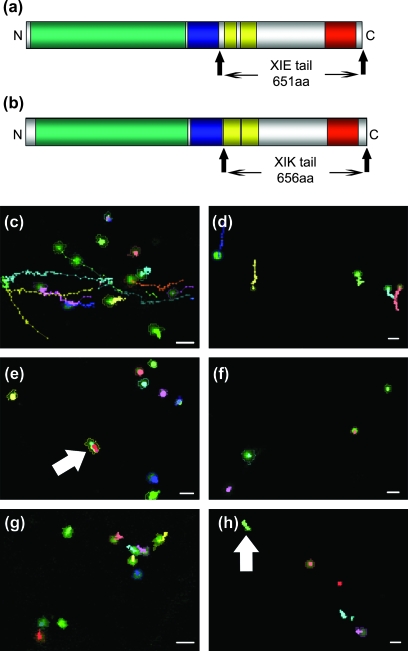
XIE and XIK tail domains (XIE-T and XIK-T) comprising the carboxy terminal 651 aa and 656 aa, respectively, were generated (Fig. 1a, b). Since they lack the amino terminal myosin motor (actin binding and ATPase domains) and regulatory IQ domains, they should, in theory, bind to and label cargo without attaching to microfilaments.
Fig. 1.
Expression of eYFP-XIE-T or eYFP-XIK-T in tobacco leaf epidermal cells perturbs Golgi and peroxisome movement. A schematic to scale representation of AtXIE-T (a) and AtXIK-T (b) is shown and the tail domain is highlighted. Where the predicted myosin motor actin binding domain (green), IQ motif region (blue), coiled coil domains (yellow), and dilute region (red) are depicted. Movies of Golgi bodies (c, e, g) or peroxisomes (d, f, h) were generated and analysed using ‘Volocity’ tracking software. Tracks (lines, where each connecting point represents one time point in the movie sequence) from representative movies of each organelle (outlined objects) were generated and are shown in each panel. Cells co-expressing eYFP-XIE-T (e, f) or eYFP-XIK-T (g, h) and control cells only expressing organelle markers (c, d) are presented. The myosin tail fusion location is not shown. Expression of eYFP-XIK-T or eYFP-XIE-T severely perturbs Golgi and peroxisome movement over the time-course of the movie. Note, some organelles still display an obvious directional movement, albeit reduced, in the presence of eYFP-XIK-T or eYFP-XIE-T tail domain fusions (arrows). Scale bar 2 μm.
XIE-T and XIK-T fusions affect Golgi, peroxisome, and mitochondrial movement
Fluorescent fusions of XIE-T and XIK-T were transiently co-expressed in tobacco epidermal cells with fluorescent markers for Golgi (ST-CFP; Brandizzi et al., 2002) or peroxisomes (CFP-SKL; Sparkes et al., 2005). Initial observations indicated that cells co-expressing eYFP-XIE-T or eYFP-XIK-T with markers for either Golgi or peroxisomes resulted in slower Golgi and peroxisome movement when compared with cells only expressing markers for these organelles. In order to quantify the effects on organelle movement, at least ten independent movie sequences of organelle movement were captured of cells co-expressing the myosin tails and an organelle marker. Movies were captured using a high scanning rate (peroxisomes, 7.58 fs−1; Golgi, 5.29 fs−1) over a short period of time in order to track fast-moving objects. Movies were then analysed using ‘Volocity’ tracking software in order to generate tracks for each individual organelle (Fig. 1). The peroxisome (Fig. 1d, f, h) and Golgi body (Fig. 1c, e, g) tracks show that expression of eYFP-XIE-T (Fig. 1e, f) or eYFP-XIK-T (Fig. 1g, h) affects movement of both organelles (Fig. 1; see Supplementary movie 1 at JXB online).
Statistical analysis of movie sequences revealed figures for track velocity, displacement rate, and meandering index, which relay information regarding organelle velocity and directionality of movement (saltatory versus unidirectional). Track velocity is the length of the entire track over time. Displacement rate is a measure of the shortest distance between the beginning and end of a track over time, and therefore does not cover the entire length of the track. Organelles displaying high levels of saltatory motion will have a track velocity, but a low displacement rate compared with organelles moving away from their point of origin. Therefore, the meandering index, defined as displacement rate divided by track velocity, is given as an indication of how straight a track is with a value of one relating to a straight unidirectional track. Therefore, directional movement will have a higher meandering index than organelle tracks displaying saltatory movements over a short distance.
Both peroxisomes and Golgi bodies display a range of movements in terms of variable velocities, displacement rates, and, therefore, meandering indexes. Merely recording averages is insufficient as, in some cases, the standard deviation can be close to the average. Therefore, these measurements are displayed as cumulative distribution plots which provide a better descriptive tool to assess and compare the movements of organelles under varying treatments.
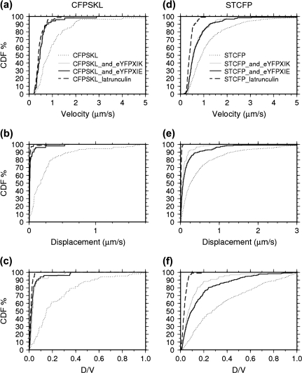
Cumulative distribution plots were generated for each experimental condition; ST-CFP control, co-expression of ST-CFP with eYFP-XIE-T or eYFP-XIK-T, and similarly for the peroxisome marker CFP-SKL (Fig. 2). CDF plots for velocity (Fig. 2a, d), displacement rate (Fig. 2b, e) and meandering index (Fig. 2c, f) are shown.
Fig. 2.
Statistical analysis of the effects of eYFP-XIE-T and eYFP-XIK-T on Golgi and peroxisome movement. Cumulative distribution function (CDF) plots of tracked peroxisomes (CFP-SKL, a, b, c) and Golgi bodies (ST-CFP, d, e, f) from at least 10 independent movies under various experimental conditions were generated. Organelle track velocity (a, d), displacement rates (shortest distance between the beginning and end of a track over time) (b, e), and meandering index (displacement rate divided by the track velocity) (c, f) are shown. Tracks from untreated control samples only expressing markers for Golgi bodies (n=543, dotted line) or peroxisomes (n=108, dotted line) are shown. Golgi body tracks from samples treated with Latrunculin B (n=106, dashed line) or co-expressing eYFP-XIE-T (n=243, black line) or eYFP-XIK-T (n=125, grey line) are slower and have lower rates than control samples, with Latrunculin B treatment having the lowest rates. Similar results are also apparent for peroxisome movement [peroxisome control (n=108), peroxisome sample treated with Latrunculin B (n=71), peroxisome sample co-expressing eYFP-XIE-T (n=52), or eYFP-XIK-T (n = 79)].
It is clearly evident that expression of eYFP-XIK-T and eYFP-XIE-T (Fig. 2a, d, black and grey line plots) significantly perturb organelle movement (both velocity and displacement rates) compared with cells expressing the organelle marker alone (Fig. 2a, d, dotted line plot; Table 1, P > 0.9); Golgi body mean track velocity is c. 2-fold lower, and there is a c. 3-fold and 6-fold reduction in mean displacement rate for eYFP-XIE-T and eYFP-XIK-T tails, respectively, peroxisome average track velocity is c. 2-fold lower, and there is a c. 9-fold and 10-fold reduction in mean displacement rate for eYFP-XIE-T and eYFP-XIK-T, respectively (see Supplementary Table 1 at JXB online). In order to determine whether this effect is as severe as inhibiting organelle movement with the actin depolymerizing agent Latrunculin B, samples expressing the organelle markers were treated with the drug, and movies were captured in the same way as for previous samples. The results indicate that Latrunculin B (Fig. 2d, black dashed line) has a more severe effect on Golgi movement (c. 3-fold reduction in mean velocity and c. 38-fold reduction in mean displacement rate) than eYFP-XIE/XIK-T expression alone (c. 2-fold lower, and c. 3-fold and 6-fold reduction in mean displacement rate for eYFP-XIE-T and eYFP-XIK-T tails, respectively; see Supplementary Table 1 at JXB online). However, eYFP-XIE/XIK-T's effects are not significantly different to Latrunculin B with regard to peroxisome velocity (Fig. 2a); c. 2-fold reduction in mean track velocity for all treatment conditions. They are however, significantly different in terms of displacement rate (Fig. 2b, c. 24-fold reduction in mean displacement rate compared to 9–10-fold reduction with eYFP-XIE/XIK-T expression; see Supplementary Table 1 at JXB online), thus indicating a directional movement of peroxisomes in cells expressing eYFP-XIE/XIK-T compared with those treated with Latrunculin B. Directional rather than oscillatory movement is corroborated by the CDF plots of the meandering index, where values approaching 1 relate to a straight track. As can be seen from the plots (Fig. 2f), the meandering index for the vast majority of the Golgi bodies tracked are higher than those monitored after Latrunculin B treatment, and Golgi bodies in cells co-expressing eYFP-XIE-T/XIK-T fusions have lower meandering index than in control cells which are only expressing the fluorescent organelle marker. There is also a difference in meandering index values for tracked peroxisomes under different experimental conditions, however the differences are not as obvious from the CDF plot for Latrunculin B and eYFP-XIE-T/XIK-T expression. However, there is c. 9-, 7-, and 6-fold reduction in peroxisome mean meandering index values after Latrunculin B treatment, or in cells co-expressing eYFP-XIE-T or eYFP-XIK-T, respectively (see Supplementary Table 1 at JXB online). The significant differences are limited by the time-course over which the movies were taken. The intrinsic nature of data acquisition, manipulation, and tracking prohibited analysis of movies over a longer time frame. However, visual observation of movement over a longer time frame indicated that both peroxisomes and Golgi in myosin tail-expressing cells do still display directional movement, albeit at a much slower rate to control cells (see arrows in Fig. 1e, h for very short directional tracks, and see global positioning of Golgi bodies in Fig. 7a–g).
Table 1.
K–S test outcomes for data represented in Fig. 2
| (a) Peroxisome velocity (V) in the absence (control) or presence of Latrunculin B (Lat B) or expression of eYFP-XIK-T or eYFP-XIE-T. | ||||
| Control | Lat B | eYFP-XIK-T | eYFP-XIE-T | |
| V | V | V | V | |
| Control V | 0 | 1 | 1 | 1 |
| Lat B V | 1 | 0 | 0.73208 | 0.92122 |
| eYFP-XIK-T V | 1 | 0.73208 | 0 | 0.43946 |
| eYFP-XIE-T V |
1 |
0.92122 |
0.43946 |
0 |
| (b) Peroxisome displacement rates (D) in the absence (control) or presence of Latrunculin B (Lat B) or expression of eYFP-XIK-T or eYFP-XIE-T. | ||||
| Control | Lat B | eYFP-XIK-T | eYFP-XIE-T | |
| D |
D |
D |
D |
|
| Control D | 0 | 1 | 1 | 1 |
| Lat B D | 1 | 0 | 1 | 0.97799 |
| eYFP-XIK-T D | 1 | 1 | 0 | 0.99639 |
| eYFP-XIE-T D |
1 |
0.97799 |
0.99639 |
0 |
| (c) Golgi body velocity (V) in the absence (control) or presence of Latrunculin B (Lat B) or expression of eYFP-XIK-T or eYFP-XIE-T. | ||||
| Control | Lat B | eYFP-XIK-T | eYFP-XIE-T | |
| V |
V |
V |
V |
|
| Control V | 0 | 1 | 1 | 1 |
| Lat B V | 1 | 0 | 1 | 1 |
| eYFP-XIK-T V | 1 | 1 | 0 | 0.65211 |
| eYFP-XIE-T V |
1 |
1 |
0.65211 |
0 |
| (d) Golgi body displacement rates (D) in the absence (control) or presence of Latrunculin B (Lat B) or expression of eYFP-XIK-T or eYFP-XIE-T. | ||||
| Control | Lat B | eYFP-XIK-T | eYFP-XIE-T | |
| D |
D |
D |
D |
|
| Control D | 0 | 1 | 1 | 1 |
| Lat B D | 1 | 0 | 1 | 1 |
| eYFP-XIK-T D | 1 | 1 | 0 | 0.98614 |
| eYFP-XIE-T D | 1 | 1 | 0.98614 | 0 |
In order to determine whether two datasets are significantly different at the 90% confidence level (corresponding to a P value of 0.9 or greater), the Kolmogorov–Smirnov (K–S) test was used. This test compares two continuous distributions and the probabilities of these distributions being different to one another. The P values from the K–S test comparing the datasets represented in Figs 2 and 5 are shown in Tables 1 and 2, respectively.
Fig. 7.
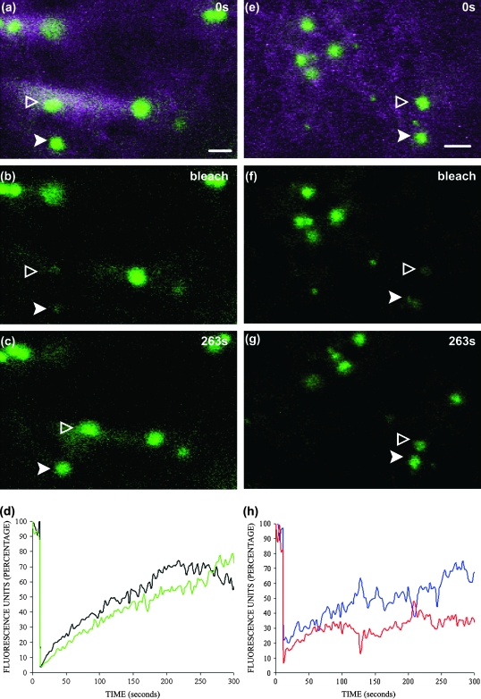
FRAP of ST-CFP in cells co-expressing eYFP-XIE-T or eYFP-XIK-T. ST-CFP within Golgi bodies (arrowheads) were bleached and recovery monitored over time in tobacco epidermal cells co-expressing either eYFP-XIE-T (a, b, c, d) or eYFP-XIK-T(e, f, g, h). Images shown were taken pre-bleach (a, e), immediately after bleaching (b, f) and 263 s after bleaching (c, g). Golgi bodies are still motile and so recovery of fluorescence is not at a steady rate due to movement of the organelle within the focal plane (graphs d and h). Curves are representative from at least five independent FRAP experiments.
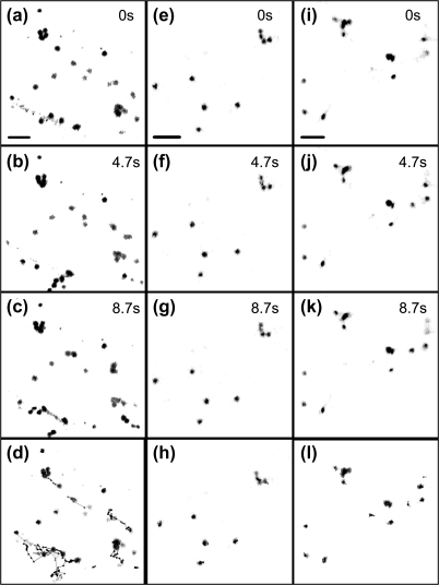
Expression of untagged myosin tails had a similar effect on Golgi body movement as the tagged tails, indicating that the eYFP tag did not affect the function of the myosin tail domains (Fig. 3). Quantitative analysis of Golgi body movement in the presence of untagged XIK-T/XIE-T was not undertaken due to the inherent difficulties in confirming untagged construct expression in cells.
Fig. 3.
Expression of untagged XIE-T or XIK-T in tobacco epidermal cells perturbs Golgi body movement. Images taken from movies of cells expressing the Golgi body marker ST-CFP (a–c) or with untagged XIE-T (e–g) or untagged XIK-T (i–k) are shown. Note, global differences in Golgi positioning (black spots) over time in control cells (a–c) compared with Golgi location in cells expressing untagged XIE-T or XIK-T. Images generated during this time frame were subjected to ‘Volocity’ track analysis. The tracks generated for each organelle under control (d), XIE-T (h) or XIK-T (l) expression are shown, clearly indicating Golgi movement in control versus myosin tail expression are different. Time is shown in seconds on each panel; scale bar 5 μm.
The effects of eYFP-XIE-T/XIK-T on mitochondrial dynamics were also tested. It is clear that they have a similar effect in perturbing movement; however, this effect was not quantified owing to difficulties in accurately determining dynamics of such a dense population of organelles (see Supplementary movie 1 at JXB online).
XIE-T and XIK-T fusions do not completely decorate peroxisomes or Golgi
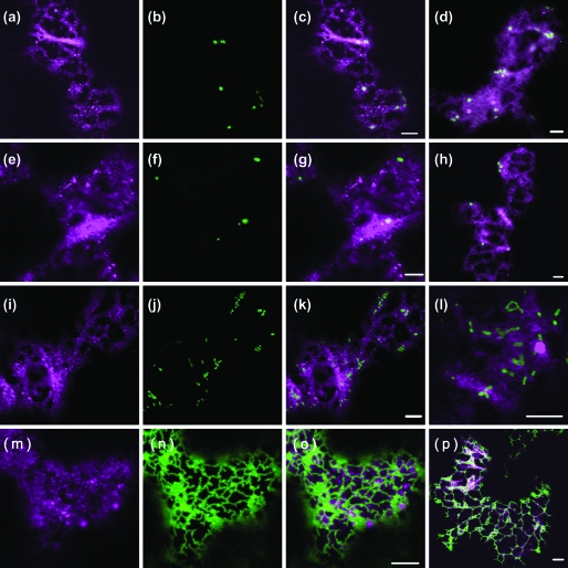
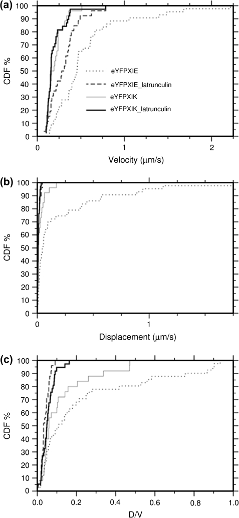
The dramatic effect of XIE-T/XIK-T expression on peroxisome, Golgi, and mitochondrial movement could lead to the hypothesis that the myosin tails are binding to these organelles, and are potentially acting in a dominant negative manner over the native full-length proteins. However, observations of cells co-expressing these myosin tails and fluorescent fusion markers for Golgi, peroxisomes, and mitochondria indicate that eYFP-XIE-T and eYFP-XIK-T do not completely decorate the periphery of these organelles, but instead appear dispersed throughout the cytosol (Fig. 4). The same observation was made when eYFP-XIE-T/XIK-T were co-expressed with fluorescent organelle markers for mitochondria (β ATPase signal sequence-GFP; Logan and Leaver, 2000) and the ER (GFP-HDEL; Batoko et al., 2000; Fig. 4). Tobacco leaf epidermal cells contain large vacuoles, thus restricting the cytosol and its contents to the cortex and, as eYFP-XIE-T/XIK-T is ubiquitous throughout the cytosol, it is difficult to discern whether the tails are associated with or are merely in close association with cargo. Both eYFP-XIE-T and eYFP-XIK-T locate to large and small puncta, and are present throughout the cytosol. eYFP-XIE-T, however, also appears to locate to more small highly motile puncta than eYFP-XIK-T. The large number and high speed of these small puncta makes it impossible to track the individual structures over time (see Supplementary movies 2 and 3 at JXB online). Therefore, in order to determine the rate of movement of eYFP-XIE-T and eYFP-XIK-T, the larger slower puncta were tracked (Fig. 5). eYFP-XIE-T (Fig. 5, grey dashed line) display significantly higher velocity (c. 3-fold increase in mean track velocity) and displacement rates (c. 8-fold increase in mean displacement rates: see Supplementary Table 1 at JXB online) than eYFP-XIK-T (Fig. 5, grey line; Table 2, P > 0.9).
Fig. 4.
Co-expression of eYFP-XIE-T or eYFP-XIK-T with various fluorescent organelle markers in tobacco epidermal cells. eYFP-XIE-T (magenta a, e, i, m) was co-expressed with fluorescent markers for Golgi bodies (ST-CFP, b), peroxisomes (CFP-SKL, f), mitochondria (β ATPase signal peptide-GFP, j), and the ER (GFP-HDEL, n). The merged images indicate that eYFP-XIE-T is closely associated, but does not solely decorate the periphery of Golgi bodies (c), peroxisomes (g), mitochondria (k), or the ER (o). Merged images of cells co-expressing eYFP-XIK-T (magenta) with fluorescent markers (green) for Golgi bodies (d), peroxisomes (h), mitochondria (l), and ER (p) also show a close association of eYFP-XIK-T with these organelles. Both eYFP-XIE-T and eYFP-XIK-T are present in large and small puncta. eYFP-XIE-T is in more numerous small puncta than eYFP-XIK-T, whereas eYFP-XIK-T is more diffuse throughout the cytosol than eYFP-XIE-T (compare c with d). Scale bar 5 μm.
Fig. 5.
Statistical analysis of eYFP-XIE-T and eYFP-XIK-T movement. CDF plots were generated of eYFP-XIE-T and eYFP-XIK-T large puncta tracks generated using ‘Volocity’ software. Track velocity (a), displacement rates (shortest distance between the beginning and end of a track over time, b), and meandering index (displacement rate divided by the track velocity, c) are shown. Tracks were generated from samples treated with Latrunculin B and from untreated cells. eYFP-XIK-T Latrunculin B treatment (n=39) black line, untreated cells (n=27) grey line, eYFP-XIE-T Latrunculin B treatment (n=27) black dashed line, and untreated cells (n=44) grey dotted line.
Table 2.
K-S test outcomes for data represented in Fig. 5
| (a) eYFP-XIE-T/K velocity (V) in the presence or absence of Latrunculin B (Lat B). | ||||
| eYFP-XIK-T | eYFP-XIK-T Lat B | EYFP-XIE-T | eYFP-XIE-T Lat B | |
| V |
V |
V |
V |
|
| eYFP-XIK-T V | 0 | 0.37458 | 1 | 0.96922 |
| eYFP-XIK-T Lat B V | 0.37458 | 0 | 1 | 0.9914 |
| eYFP-XIE-T V | 1 | 1 | 0 | 0.9973 |
| eYFP-XIE-T L, B V |
0.96922 |
0.9914 |
0.9973 |
0 |
| (b) eYFP-XIE-T/K displacement rates (D) in the presence or absence of Latrunculin B (Lat B). | ||||
| eYFP-XIK-T | eYFP-XIK-T Lat B | eYFP-XIE-T | eYFP-XIE-T Lat B | |
| D |
D |
D |
D |
|
| eYFP-XIK-T D | 0 | 0.8562 | 0.99597 | 0.74186 |
| eYFP-XIK-T Lat B D | 0.8562 | 0 | 1 | 0.29129 |
| eYFP-XIE-T D | 0.99597 | 1 | 0 | 1 |
| eYFP-XIE-T Lat B D | 0.74186 | 0.29129 | 1 | 0 |
In order to determine whether two datasets are significantly different at the 90% confidence level (corresponding to a P value of 0.9 or greater), the Kolmogorov–Smirnov (K–S) test was used. This test compares two continuous distributions and the probabilities of these distributions being different to one another. The P values from the K–S test comparing the datasets represented in Figs 2 and 5 are shown in Tables 1 and 2, respectively.
XIE-T/XIK-T and the actin cytoskeleton
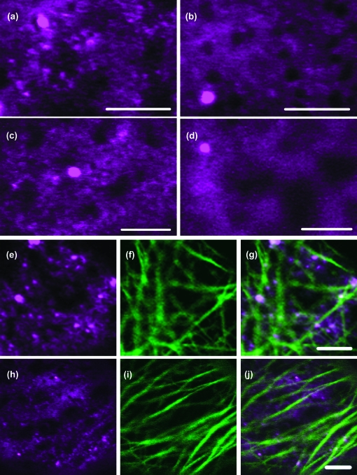
Arabidopsis XIE and XIK are predicted to encode for myosins and the effect of expressing a dominant negative tail region confirms they have a role in organelle motility. In order to determine whether their movement is dependent upon the actin cytoskeleton, movement analysis in the presence of Latrunculin B was carried out and compared with the rates described above (Fig. 5). Analysis indicates that Latrunculin B perturbs movement resulting in puncta exhibiting more saltatory than directional motion (i.e they have a lower meandering index when compared to samples not treated with Latrunculin B; c. 2-fold reduction in mean meandering index of eYFP-XIK-T after Latrunculin B treatment, and c. 6-fold reduction for eYFP-XIE-T; see Supplementary Table 1 at JXB online). In addition, the distribution of both eYFP-XIE-T and eYFP-XIK-T altered upon treatment with Latrunculin B (Fig. 6a–d). The numerous small puncta were reduced in number along with the appearance of a diffuse cytoplasmic pool of fluorescence (Fig. 6b, d) when compared with untreated cells (Fig. 6a, c). The large puncta did not appear to be affected, leading us to suspect that they might be aggregates caused by overexpression, although many exhibited directional movement (Fig. 5).
Fig. 6.
eYFP-XIE-T and eYFP-XIK-T association with the actin cytoskeleton. eYFP-XIE-T (a, b) and eYFP-XIK-T(c, d) were expressed in tobacco epidermal cells. Images were taken of cells treated with Latrunculin B (b, d) and control untreated cells (a, c). After treatment there are fewer small puncta, but the large puncta still remain. eYFP-XIE-T (magenta, e–g) and eYFP-XIK-T (magenta, h–j) were expressed with an actin marker, GFP-FABD2 (green, f, i). The global architecture of the actin appears unchanged by myosin tail expression, and the myosin tails puncta are frequently closely associated with the actin filaments. Scale bar 5 μm.
Since eYFP-XIE-T/XIK-T do not perfectly decorate peroxisomes, mitochondria, or Golgi, an alternative interpretation as to how they could affect organelle movement is through perturbing the actin cytoskeleton itself. To test this, eYFP-XIE-T or eYFP-XIK-T were co-expressed with an actin marker, GFP-FABD2 (Ketelaar et al., 2004; Sheahan et al., 2004) (Fig. 6e–j). Based on the location of GFP-FABD2, expression of the myosin tails did not appear to affect the global architecture of the actin cytoskeleton, although in many instances the myosin tail puncta appeared in close proximity to the actin microfilaments.
XIE-T and XIK-T fusions do not affect the trafficking of a fluorescent Golgi marker
Whilst XIE-T and XIK-T perturb the movement of Golgi, peroxisomes, and mitochondria, it was necessary to assess whether they had a detrimental effect on the cell viability, thus affecting other active processes such as trafficking of the Golgi marker (ST-CFP) from the ER. Previous studies have shown that the trafficking of the Golgi marker, ST-GFP, from the ER is energy-dependent but probably independent of the cytoskeleton. This was demonstrated by FRAP analysis through monitoring the recovery of ST-GFP into photobleached Golgi bodies over time in the presence and absence of Latrunculin B (Brandizzi et al., 2002). In these studies the half-time recovery of ST-GFP in Golgi bodies could only be determined in static Golgi after treatment with Latrunculin B. Since XIE-T and XIK-T do not completely inhibit Golgi body movement, the same inherent difficulties in obtaining a half-time of recovery also applied. Therefore, it was determined whether ST-CFP could recover over time after photobleaching in a similar manner to motile Golgi bodies (daSilva et al., 2004). Several independent bleaching studies of ST-CFP in cells co-expressing eYFP-XIE-T or eYFP-XIK-T showed that ST-CFP could recover after photobleaching (Fig. 7). Representative recovery curves of these experiments are shown (Fig. 7d, h) where recovery is plotted as a percentage of the pre-bleach value. Recovery rates varied, but did not appear to conform to an exponential increase over time due to the motility of the Golgi bodies around the FRAP measurement area.
Discussion
The main conclusions from this study are that expression of two Arabidopsis myosin XIE and XIK tail domains; (i) perturbs both Golgi, peroxisome, and mitochondrial movement in tobacco epidermal cells, (ii) that this effect is not as severe as that observed after depolymerization of the actin cytoskeleton with Latrunculin B treatment, (iii) XIE/XIK-T fusions do not co-locate to these organelles and display significantly different dynamics to one another, which are actin-dependent, and (iv) XIE-T/XIK-T fluorescent fusion protein constructs are motile when expressed in leaf cells.
The majority of myosins are molecular motors required for actin-dependent processes. These processes are generally thought of as active movement of cargo; however, there are examples where they appear to be required for anchoring or maintaining organelle/cargo integrity (Allan et al., 2002; Tyska et al., 2005).
The myosin family in higher plants appears to show functional redundancy; T-DNA insertional mutants of 11 of the 13 class XI myosins did not display an altered gross morphology compared to control plants (Peremyslov et al., 2008), expression of fluorescent fusions of several different truncated myosin isoforms labelled peroxisomes in tobacco epidermal cells (Li and Nebenführ, 2007) and three different isoforms were immunodetected on Golgi bodies and mitochondria in tobacco pollen tubes (Romagnoli et al., 2007). However, without carrying out a detailed study on the potential truncated remnants expressed in T-DNA lines, and assessing the potential functional role(s) of these peptides in vivo, or comparing temporal–spatial expression patterns under various physiological conditions, it remains to be seen how much true functional redundancy exists within the higher plant myosin gene family.
Immunocytochemical and overexpression studies of fluorescent fusions have reported the subcellular locations of ATM1, an Arabidopsis class VIII myosin, to plasmodesmata (Reichelt et al., 1999; Golomb et al., 2008), developing cell plate (Van Damme et al., 2004) and was implicated in endocytosis (Golomb et al., 2008), truncations of Mya1 (Li and Nebenführ, 2007) and Mya2 to peroxisomes (Hashimoto et al., 2005; Li and Nebenführ, 2007; Reisen and Hanson, 2007), Mya1 to Golgi (Li and Nebenführ, 2007), and various class XI truncations to unknown motile structures (Li and Nebenführ, 2007; Reisen and Hanson, 2007). Studies in other higher plant systems such as tobacco have isolated putative myosins and reported myosin-based dynamics (Tominaga et al., 2003).
XIE-T and XIK-T perturb peroxisome, Golgi body and mitochondrial movement
It is clear that XIE-T and XIK-T fusions severely perturb peroxisome, Golgi body, and mitochondrial movement, although these effects are not as total as those observed after actin depolymerization with Latrunculin B. Whilst mitochondrial dynamics were not quantified, it is obvious that both eYFP-XIE-T and eYFP-XIK-T have a more marked effect on peroxisome displacement rates compared with Golgi bodies (3–6-fold compared with 9–10-fold), but both tail fusions had c. 2-fold reduction in average Golgi and peroxisome velocities.
The effect of the myosin tails does not extend to trafficking of the Golgi membrane marker ST-CFP from the ER to Golgi bodies, and therefore corroborates the findings of Brandizzi et al. (2002); membrane protein transport between the endoplasmic reticulum and the Golgi is cytoskeleton independent. This also confirms that the effects of XIE-T and XIK-T expression is a primary and not a secondary effect due to loss of cell viability, which is further supported by the viable Arabidopsis T-DNA insertional XIK mutants (Peremyslov et al., 2008).
Both tail fusions appear to have no effect on the global architecture of the actin cytoskeleton, and do not completely decorate peroxisomes, Golgi, ER, or mitochondria, but are diffuse within the cytosol and located to highly motile small and slower larger puncta of unknown composition. Formation or maintenance of the small puncta, however, appears to be actin-dependent based on the reduced number after actin depolymerization on treatment with Latrunculin B.
In previous expression studies of tagged truncated XIK tail domains there were no reports of perturbation of organelle movement (Li and Nebenführ, 2007; Reisen and Hanson, 2007). Li and Nebenführ's (2007) main focus was determining the cargo binding regions in the tails of several class XI myosin. Reisen and Hanson (2007) expressed a shorter truncated AtXIK fusion in Arabidopsis leaf epidermal cells which primarily located to punctate structures and to a lesser extent the cytosol compared to the observations reported here in N. tabacum epidermal cells. The punctate structures displayed an average velocity of c. 1.1±0.25 μm s−1 compared with the 0.2±0.098 μm s−1 described here. Discrepancies in rates could relate to expression of the tail fusion in tobacco epidermal cells compared with Arabidopsis epidermal cells, levels of expression, and the physiological response to different transformation procedures (leaf infiltration versus biolistic bombardment). However, it is interesting to note that Reisen and Hanson did not report on the possible effects of their XIK tail fusion on organelle dynamics.
Recently, it was reported that transient expression of untagged and RNAi Nicotiana benthamiana XIK tail domain, and truncations of this region, effect organelle (peroxisome, Golgi, and mitochondria) movement in N. benthamiana leaf epidermal cells (Avisar et al., 2008). A similar effect was demonstrated in Arabidopsis with T-DNA insertional mutants in XIK, and lines overexpressing an untagged A. thaliana XIK tail domain (Peremyslov et al., 2008). This study used a similar experimental approach, but eYFP fusions of A. thaliana XIK and XIE tail domain were transiently expressed in N. tabacum leaf epidermal cells permitting location data to be collected and at the same time allowing the dynamics and actin dependency to be studied. The organelles were also tracked using ‘Volocity’ software, but at an image rate capture that was 10-fold faster than in previous myosin papers.
Expression of the untagged AtXIK tail in Arabidopsis elongated epidermal leaf cells near the central vein resulted in c. 3–5-fold reduction in mean Golgi and peroxisome velocity (Peremyslov et al., 2008), whereas it was found here that eYFP-AtXIK-T fusion resulted in c. 2-fold reduction in these rates in N. tabacum leaf epidermal ‘pavement’ cells. In N. benthamiana, the untagged NbXIK tail resulted in c. 10- or 15-fold reduction in mean velocity of Golgi stacks or peroxisomes, respectively (Avisar et al., 2008). The differences in mean velocity rates could reflect an impairment of XIK tail function by the eYFP fusion; heterologous versus homologous expression as Avisar et al. (2008) noted that AtMya2 (XI-2) had a similar effect on Golgi and peroxisome dynamics in Arabidopsis, but NbXI-2 had a more detrimental effect on peroxisome movement than Golgi bodies in N. benthamiana. Peremyslov et al. (2008) also noted that AtXIK T-DNA insertional mutants displayed c. 2-fold reduction in Golgi and peroxisome mean velocity in roots compared with 3–5-fold reduction in leaves indicating cell-type-specific differences. Lastly, differences in image capture rates and the subsequent tracking analysis can result in different calculations of organelle velocity and displacement.
Therefore, the study presented here corroborates recently documented effects of XIK tail domain on organelle movement, but also provides novel information regarding AtXIK tail location, movement, and actin-dependent distribution and dynamics. Similar effects are evident for the AtXIE tail domain, an effect not reported to date. Taken together, these results show, as in mammalian class V myosins, that the tail domain of certain plant myosins (XIK, XIE, Mya2) can be used as a dominant negative tool to perturb some aspects of subcellular movement (Avisar et al., 2008; Peremyslov et al., 2008).
Based on these observations, the overriding question is how do XIE and XIK tail domains inhibit peroxisome, Golgi, and mitochondrial movement, whilst themselves still exhibiting vectorial movement that appears to be actin dependent?
Sequence prediction indicates that XIE-T and XIK-T contain coiled coil and dilute domains (pfam Accession no. PF01843). Such domains are implicated in protein–protein interactions which could result in dimerization of the protein. Two class XI myosins from Chara and tobacco have been shown by rotary shadowing to form dimers (Yamamoto et al., 1995; Tominaga et al., 2003), with the tobacco myosin dimer possessing high velocity movement along actin (Tominaga et al., 2003).
Based on the structural predictions and effects of XIE-T/XIK-T on peroxisome/Golgi/mitochondrial movement, one could hypothesize that overexpression of XIE/XIK tail domains may induce dimers and heterodimers with full-length native XIE/XIK in the cell. Thus, the highly motile small puncta could be heterodimers containing tail and full-length XIE/XIK trapped in the actin-bound state, and the larger less motile puncta are a mixture of tail/tail or full-lenth XIE/XIK homodimers, and heterodimers of tail and full-lenth XIE/K. In this model, the movement of the myosin constructs would be due to actin turnover or treadmilling which results in ‘dragging’ of the heterodimer through the cytosol. Upon depolymerization of the actin cytoskeleton the small highly motile actin-bound heterodimers would dissociate and effectively disappear. MyoV studies indicates that the tail domain can affect the head domain activity (Krementsov et al., 2004; Li et al., 2004; Wang et al., 2004), moreover, based on structural studies it was proposed that the myoV recycles back to its ‘starting’ position to pick up new cargo by simply hanging on to the actin and the treadmilling of actin recycles and drags it back to pick up more cargo (Liu et al., 2006). Formation of the large eYFP-XIE-T/XIK-T puncta, which it is assumed are induced by overexpression, is independent on actin as they would contain a mixture of non-actin-bound tail homodimers and heterodimers. We do not believe that these structures are simple protein aggregates as, on photobleaching, they show a rapid cycling of protein (data not shown). This model is consistent with the location and motility of eYFP-XIE-T/XIK-T. However, since the coiled coil domain in the tail region of AtXIK is dispensable for its effect on organelle movement (Avisar et al., 2008), dimerization/association of the tail domains would have to be through another domain/interacting partner.
Alternatively, it would not be unreasonable to presume that the effect on Golgi, peroxisome and mitochondria motility could be a result of the tail domains titrating accessory factors required for the recruitment of XIE/K to the cargo membrane or to other proteins associated with the cargo membrane. A similar situation has been described in mammalian cells where it has been shown that mice mutants defective in Rab27a (ashen) or myosin Va (dilute) display altered coat colour due to reduced accumulation of melansomes at the periphery of skin melanocytes (Wu et al., 2001; Seabra and Coudrier, 2004). Rab27a along with melanophilin forms part of a complex required for melanosome attachment to myosin Va (Wu et al., 2002a, b). Other examples of Rab GTPase action on myosins have been described (Seabra and Coudrier, 2004). Therefore, in the XIE/K tail overexpression studies presented here, the tail domains could, in effect, be interacting with and titrating out factors required for the interaction between XIE/K with peroxisomes, Golgi, and mitochondria. Cargo movement is not completely inhibited due to lower numbers of native full-lenth XIE/K able to interact with these organelles
Since the basic biochemistry, interacting partners, and kinetics of Arabidopsis XIE/K are unknown, further studies are required to test this hypothesis.
XIE-T and XIK-T as genetically encoded inhibitors of organelle movement
A major caveat in studying actin-myosin dynamics is that none of the current techniques allows for a non-invasive regulated in vivo approach to studying the effects of inhibiting organelle movement on various aspects of plant physiology; for example, pathogen response, development, gravitropism. Whilst chemical inhibition can be a powerful tool, side-effects have been identified, which may not be related to inhibition of the target molecule per se. The efficacy of BDM, the commonest inhibitor of myosin in plant studies, is often questioned (McCurdy, 1999). The development of two genetically encoded inhibitors which affect Golgi, peroxisome and mitochondria movement several days after expression has been presented here. Whilst the exact mode of action of these myosin tail fusions is yet to be elucidated the advantage of being able to control activation genetically in a temporal and spatial manner will be invaluable in the further study of the role of organelle motility in cell, tissue and plant development and response under various physiological (biotic and abiotic) conditions.
Supplementary data
Supplementary data in the form of a table and three movies can be found at JXB online.
Supplementary Table 1. Mean values and standard deviations for velocity, displacement rate, and meandering index for tracked Golgi bodies and peroxisomes under various treatment conditions (where Lat B is Latrunculin B treatment), and XIK-T/XIK-E large puncta in N. tabacum epidermal cells.
Supplementary Movie 1. Organelle dynamics are affected by eYFP-XIE-T and eYFP-XIK-T expression in N. tabacum leaf epidermal cells. Fluorescent markers targeted to Golgi (A–C), peroxisomes (D–F) and mitochondria (G–I) were coexpressed in N. tabacum leaf epidermal cells with eYFP-XIE-T (B, E, H) or eYFP-XIK-T (C, F, I). Movies were generated over the same time frame for each type of organelle, thus demonstrating that eYFP-XIE-T and eYFP-XIK-T perturb organelle movement when compared to control movies (A,D,G).
Supplementary Movie 2. eYFP-XIE-T dynamics in N. tabacum leaf epidermal cells. eYFP-XIE-T labels both cytosol and puncta (large slow puncta and numerous highly motile smaller punctate structures) when expressed in N. tabacum leaf epidermal cells.
Supplementary Movie 3. eYFP-XIK-T dynamics in N. tabacum leaf epidermal cells. eYFP-XIK-T labels large puncta, cytosol, and occasional small punctate structures when expressed in N. tabacum leaf epidermal cells.
Supplementary Material
Acknowledgments
We would like to thank John Runions for help with image analysis, Janet Evins for general laboratory running and maintenance of plant material, and John Runions, Katja Graumann, and Jenny Schoberer for FRAP advice. We would also like to thank D Logan and DW McCurdy for the β-ATPase-GFP and GFP-FABD2 constructs, respectively, and Isabel Bermudez for critical reading of the manuscript. The BBSRC, Oxford Brookes University, and the Leverhulme Trust are acknowledged for funding various aspects of this research.
References
- Allan VJ, Thompson HM, McNiven MA. Motoring around the Golgi. Nature Cell Biology. 2002;4:E236–E242. doi: 10.1038/ncb1002-e236. [DOI] [PubMed] [Google Scholar]
- Avisar D, Prokhnevsky AI, Makarova KS, Koonin EV, Dolja VV. Myosin XI-K is required for rapid trafficking of Golgi stacks, peroxisomes and mitochondria in leaf cells of Nicotiana benthamiana. Plant Physiology. 2008;146:1098–1108. doi: 10.1104/pp.107.113647. [DOI] [PMC free article] [PubMed] [Google Scholar]
- Batoko H, Zheng HQ, Hawes C, Moore I. A Rab1 GTPase is required for transport between the endoplasmic reticulum and Golgi apparatus and for normal Golgi movement in plants. The Plant Cell. 2000;12:2201–2218. doi: 10.1105/tpc.12.11.2201. [DOI] [PMC free article] [PubMed] [Google Scholar]
- Boevink P, Oparka K, Santa-Cruz S, Martin B, Betteridge A, Hawes C. Stacks on tracks:the plant Golgi apparatus traffics on an actin/ER network. The Plant Journal. 1998;15:441–447. doi: 10.1046/j.1365-313x.1998.00208.x. [DOI] [PubMed] [Google Scholar]
- Brandizzi F, Snapp EL, Roberts AG, Lippincott-Schwartz J, Hawes C. Membrane protein transport between the endoplasmic reticulum and the Golgi in tobacco leaves is energy-dependent but cytoskeleton-independent: evidence from selective photobleaching. The Plant Cell. 2002;14:1293–1309. doi: 10.1105/tpc.001586. [DOI] [PMC free article] [PubMed] [Google Scholar]
- Curtis M, Grossniklaus U. A Gateway cloning vector set for high-throughput functional analysis of genes in planta. Plant Physiology. 2003;133:462–469. doi: 10.1104/pp.103.027979. [DOI] [PMC free article] [PubMed] [Google Scholar]
- daSilva LLP, Snapp EL, Denecke J, Lippincott-Schwartz J, Hawes C, Brandizzi F. Endoplasmic reticulum export sites and Golgi bodies behave as single mobile secretory units in plant cells. The Plant Cell. 2004;16:1753–1771. doi: 10.1105/tpc.022673. [DOI] [PMC free article] [PubMed] [Google Scholar]
- Golomb L, Abu-Abied M, Belausov E, Sadot E. Different subcellular localizations and functions of Arabidopsis myosin VIII. BMC Plant Biology. 2008;8 doi: 10.1186/1471-2229-8-3. DOI:10.1186/1471-2229-8-3. [DOI] [PMC free article] [PubMed] [Google Scholar]
- Hachikubo Y, Ito K, Schiefelbein J, Manstein DJ, Yamamoto K. Enzymatic activity and motility of recombinant Arabidopsis myosin XI, MYA1. Plant and Cell Physiology. 2007;48:886–891. doi: 10.1093/pcp/pcm054. [DOI] [PubMed] [Google Scholar]
- Hashimoto K, Igarashi H, Mano S, Nishimura M, Shimmen T, Yokota E. Peroxisomal localization of a myosin XI isoform in Arabidopsis thaliana. Plant and Cell Physiology. 2005;46:782–789. doi: 10.1093/pcp/pci085. [DOI] [PubMed] [Google Scholar]
- Hofgen R, Willmitzer L. Storage of competent cells for Agrobacterium transformation. Nucleic Acids Research. 1988;16:9877. doi: 10.1093/nar/16.20.9877. [DOI] [PMC free article] [PubMed] [Google Scholar]
- Jedd G, Chua NH. Visualization of peroxisomes in living plant cells reveals acto-myosin-dependent cytoplasmic streaming and peroxisome budding. Plant and Cell Physiology. 2002;43:384–392. doi: 10.1093/pcp/pcf045. [DOI] [PubMed] [Google Scholar]
- Ketelaar T, Alwood EG, Anthony R, Voigt B, Menzel D, Hussey PJ. The actin-interacting protein AIP1 is essential for actin organization and plant development. Current Biology. 2004;14:145–149. doi: 10.1016/j.cub.2004.01.004. [DOI] [PubMed] [Google Scholar]
- Krementsov DN, Krementsova EB, Trybus KM. Myosin V: regulation by calcium, calmodulin, and the tail domain. Journal of Cell Biology. 2004;164:877–886. doi: 10.1083/jcb.200310065. [DOI] [PMC free article] [PubMed] [Google Scholar]
- Krendel M, Osterweil EK, Mooseker MS. Myosin 1E interacts with synaptojanin-1 and dynamin and is involved in endocytosis. FEBS Letters. 2007;581:644–650. doi: 10.1016/j.febslet.2007.01.021. [DOI] [PMC free article] [PubMed] [Google Scholar]
- Li J, Nebenführ A. Organelle targeting of myosin XI is mediated by two globular tail domains with separate cargo binding sites. Journal of Biological Chemistry. 2007;282:20593–20602. doi: 10.1074/jbc.M700645200. [DOI] [PubMed] [Google Scholar]
- Li XD, Mabuchi K, Ikebe R, Ikebe M. Ca2+-induced activation of ATPase activity of myosin Va is accompanied with a large conformational change. Biochemical and Biophysical Research Communications. 2004;315:538–545. doi: 10.1016/j.bbrc.2004.01.084. [DOI] [PubMed] [Google Scholar]
- Liebe S, Quader H. Myosin in onion (Allium cepa) bulb scale epidermal cells: involvement in dynamics of organelles and endoplasmic-reticulum. Physiologia Plantarum. 1994;90:114–124. [Google Scholar]
- Liu J, Taylor DW, Krementsova EB, Trybus KM, Taylor KA. Three-dimensional structure of the myosin V inhibited state by cryoelectron tomography. Nature. 2006;442:208–211. doi: 10.1038/nature04719. [DOI] [PubMed] [Google Scholar]
- Logan DC, Leaver CJ. Mitochondria-targeted GFP highlights the heterogeneity of mitochondria shape, size, and movement within living plant cells. Journal of Experimental Botany. 2000;51:865–871. [PubMed] [Google Scholar]
- Mano S, Nakamori C, Hayashi M, Kato A, Kondo M, Nishimura M. Distribution and characterization of peroxisomes in arabidopsis by visualization with GFP: dynamic morphology and actin- dependent movement. Plant and Cell Physiology. 2002;43:331–341. doi: 10.1093/pcp/pcf037. [DOI] [PubMed] [Google Scholar]
- Mathur J, Mathur N, Hulskamp M. Simultaneous visualization of peroxisomes and cytoskeletal elements reveals actin and not microtubule-based peroxisome motility in plants. Plant Physiology. 2002;128:1031–1045. doi: 10.1104/pp.011018. [DOI] [PMC free article] [PubMed] [Google Scholar]
- McCurdy DW. Is 2,3-butanedione monoxime an effective inhibitor of myosin-based activities in plant cells? Protoplasma. 1999;209:120–125. doi: 10.1007/BF01415707. [DOI] [PubMed] [Google Scholar]
- Miller DD, Scordilis SP, Hepler PK. Identification and localization of 3 classes of myosins in pollen tubes of Lilium longiflorum and Nicotiana alata. Journal of Cell Science. 1995;108:2549–2563. doi: 10.1242/jcs.108.7.2549. [DOI] [PubMed] [Google Scholar]
- Nebenführ A, Gallagher LA, Dunahay TG, Frohlick JA, Mazurkiewicz AM, Meehl JB, Staehelin LA. Stop-and-go movements of plant Golgi stacks are mediated by the acto-myosin system. Plant Physiology. 1999;121:1127–1141. doi: 10.1104/pp.121.4.1127. [DOI] [PMC free article] [PubMed] [Google Scholar]
- Ojangu EL, Jarve K, Paves H, Truve E. Arabidopsis thaliana myosin XIK-T is involved in root hair as well as trichome morphogenesis on stems and leaves. Protoplasma. 2007;230:193–202. doi: 10.1007/s00709-006-0233-8. [DOI] [PubMed] [Google Scholar]
- Peremyslov VV, Prokhnevsky AI, Avisar D, Dolja VV. Two class XI myosins function in organelle trafficking and root hair development in Arabidopsis thaliana. Plant Physiology. 2008;146:1109–1116. doi: 10.1104/pp.107.113654. [DOI] [PMC free article] [PubMed] [Google Scholar]
- Press WH, Flannery BP, Teukolsky SA, Vetterling WT. Numerical recipes. Cambridge University Press; 1992. [Google Scholar]
- Radford JE, White RG. Localization of a myosin-like protein to plasmodesmata. The Plant Journal. 1998;14:743–750. doi: 10.1046/j.1365-313x.1998.00162.x. [DOI] [PubMed] [Google Scholar]
- Reddy ASN, Day IS. Analysis of the myosins encoded in the recently completed Arabidopsis thaliana genome sequence. Genome Biology. 2001;2:1–18. doi: 10.1186/gb-2001-2-7-research0024. [DOI] [PMC free article] [PubMed] [Google Scholar]
- Reichelt S, Knight AE, Hodge TP, Baluska F, Samaj J, Volkmann D, Kendrick-Jones J. Characterization of the unconventional myosin VIII in plant cells and its localization at the post-cytokinetic cell wall. The Plant Journal. 1999;19:555–567. doi: 10.1046/j.1365-313x.1999.00553.x. [DOI] [PubMed] [Google Scholar]
- Reisen D, Hanson MR. Association of six YFP-myosin XI-tail fusions with mobile plant cell organelles. BMC Plant Biology. 2007;7 doi: 10.1186/1471-2229-7-6. doi:10.1186/1471-2229-7-6. [DOI] [PMC free article] [PubMed] [Google Scholar]
- Rodriguez OC, Cheney RE. Human myosin-Vc is a novel class V myosin expressed in epithelial cells. Journal of Cell Science. 2002;115:991–1004. doi: 10.1242/jcs.115.5.991. [DOI] [PubMed] [Google Scholar]
- Romagnoli S, Cai G, Faleri C, Yokota E, Shimmen T, Cresti M. Microtubule- and actin filament-dependent motors are distributed on pollen tube mitochondria and contribute differently to their movement. Plant and Cell Physiology. 2007;48:345–361. doi: 10.1093/pcp/pcm001. [DOI] [PubMed] [Google Scholar]
- Seabra MC, Coudrier E. Rab GTPases and myosin motors in organelle motility. Traffic. 2004;5:393–399. doi: 10.1111/j.1398-9219.2004.00190.x. [DOI] [PubMed] [Google Scholar]
- Sheahan MB, Staiger CJ, McCurdy DW. A green fluorescent protein fusion to actin-binding domain 2 of Arabidopsis Fimbrin highlights new features of a dynamic actin cytoskeleton in live plants cells. Plant Physiology. 2004;136:3968–3978. doi: 10.1104/pp.104.049411. [DOI] [PMC free article] [PubMed] [Google Scholar]
- Sparkes IA, Hawes C, Baker A. AtPEX2 and AtPEX10 are targeted to peroxisomes independently of known endoplasmic reticulum trafficking routes. Plant Physiology. 2005;139:690–700. doi: 10.1104/pp.105.065094. [DOI] [PMC free article] [PubMed] [Google Scholar]
- Sparkes I, Runions J, Kearns A, Hawes C. Rapid, transient expression of fluorescent fusion proteins in tobacco plants and generation of stably transformed plants. Nature Protocols. 2006;1:2019–2025. doi: 10.1038/nprot.2006.286. [DOI] [PubMed] [Google Scholar]
- Tominaga M, Kojima H, Yokota E, Orii H, Nakamori R, Katayama E, Anson M, Shimmen T, Oiwa K. Higher plant myosin XI moves processively on actin with 35 nm steps at high velocity. EMBO Journal. 2003;22:1263–1272. doi: 10.1093/emboj/cdg130. [DOI] [PMC free article] [PubMed] [Google Scholar]
- Tyska MJ, Mackey AT, Huang JD, Copeland NG, Jenkins NA, Mooseker M. Myosin-1a is critical for normal brush border structure and composition. Molecular Biology of the Cell. 2005;16:2443–2457. doi: 10.1091/mbc.E04-12-1116. [DOI] [PMC free article] [PubMed] [Google Scholar]
- Van Damme D, Bouget FY, Poucke KV, Inzé D, Geelen D. Molecular dissection of plant cytokinesis and phragmoplast structure: a survey of GFP-tagged proteins. The Plant Journal. 2004;40:386–398. doi: 10.1111/j.1365-313X.2004.02222.x. [DOI] [PubMed] [Google Scholar]
- Van Gestel K, Kohler RH, Verbelen JP. Plant mitochondria move on F-actin, but their positioning in the cortical cytoplasm depends on both F-actin and microtubules. Journal of Experimental Botany. 2002;53:659–667. doi: 10.1093/jexbot/53.369.659. [DOI] [PubMed] [Google Scholar]
- Wang F, Thirumurugan K, Stafford WF, Hammer JA, Knight PJ, Sellers JR. Regulated conformation of myosin V. Journal of Biological Chemistry. 2004;279:2333–2336. doi: 10.1074/jbc.C300488200. [DOI] [PubMed] [Google Scholar]
- Wu X, Rao K, Bowers MB, Copeland NG, Jenkins NA, Hammer JA. Rab27a enables myosin Va-dependent melanosome capture by recruiting the myosin to the organelle. Journal of Cell Science. 2001;114:1091–1100. doi: 10.1242/jcs.114.6.1091. [DOI] [PubMed] [Google Scholar]
- Wu XF, Wang F, Rao K, Sellers JR, Hammer JA. Rab27a is an essential component of melanosome receptor for myosin Va. Molecular Biology of the Cell. 2002b;13:1735–1749. doi: 10.1091/mbc.01-12-0595. [DOI] [PMC free article] [PubMed] [Google Scholar]
- Wu XFS, Rao K, Zhang H, Wang F, Sellers JR, Matesic LE, Copeland NG, Jenkins NA, Hammer JA. Identification of an organelle receptor for myosin-Va. Nature Cell Biology. 2002a;4:271–278. doi: 10.1038/ncb760. [DOI] [PubMed] [Google Scholar]
- Yamamoto K, Kikuyama M, Sutohyamamoto N, Kamitsubo E, Katayama E. Myosin from alga Chara: unique structure revealed by electron-microscopy. Journal of Molecular Biology. 1995;254:109–112. doi: 10.1006/jmbi.1995.0603. [DOI] [PubMed] [Google Scholar]
- Yokota E. Identification and characterization of higher plant myosins responsible for cytoplasmic streaming. Journal of Plant Research. 2000;113:511–519. [Google Scholar]
- Yokota E, McDonald AR, Liu B, Shimmen T, Palevitz BA. Localization of a 170 kda myosin heavy-chain in plant cells. Protoplasma. 1995;185:178–187. [Google Scholar]
- Yokota E, Shimmen T. Isolation and characterization of plant myosin from pollen tubes of lily. Protoplasma. 1994;177:153–162. [Google Scholar]
- Yokota E, Yukawa C, Muto S, Sonobe S, Shimmen T. Biochemical and immunocytochemical characterization of two types of myosins in cultured tobacco bright yellow-2 cells. Plant Physiology. 1999;121:525–534. doi: 10.1104/pp.121.2.525. [DOI] [PMC free article] [PubMed] [Google Scholar]
Associated Data
This section collects any data citations, data availability statements, or supplementary materials included in this article.


