Abstract
CXCL5 is a proangiogenic CXC-type chemokine that is an inflammatory mediator and a powerful attractant for granulocytic immune cells. Unlike many other chemokines, CXCL5 is secreted by both immune (neutrophil, monocyte, and macrophage) and nonimmune (epithelial, endothelial, and fibroblastic) cell types. The current study was intended to determine which of these cell types express CXCL5 in normal and malignant human prostatic tissues, whether expression levels correlated with malignancy and whether CXCL5 stimulated biologic effects consistent with a benign or malignant prostate epithelial phenotype. The results of these studies show that CXCL5 protein expression levels are concordant with prostate tumor progression, are highly associated with inflammatory infiltrate, and are frequently detected in the lumens of both benign and malignant prostate glands. Exogenous administration of CXCL5 stimulates cellular proliferation and gene transcription in both nontransformed and transformed prostate epithelial cells and induces highly aggressive prostate cancer cells to invade through synthetic basement membrane in vitro. These findings suggest that the inflammatory mediator, CXCL5, may play multiple roles in the etiology of both benign and malignant proliferative diseases in the prostate.
Introduction
The American Cancer Society estimates that there will be nearly 219,000 newly diagnosed cases of prostate cancer in the United States in 2007 and that an estimated 27,000 patients, or more than 10% of those diagnosed with prostate cancer, will die of this disease. It is also estimated that prostate cancer alone will account for almost 30% of cancer incidents in American men and almost 10% of cancer deaths. Although deaths due to prostate cancer have declined over the past decades, it is still the most commonly diagnosed cancer and accounts for the second highest death rate from cancer among American men [1]. Clearly, a better understanding of the biology of prostate cancer initiation and progression that would facilitate the development of strategies for the successful prevention or therapeutic intervention of this common malignancy is needed.
Although successful interventional therapeutic strategies for prostate cancer have hinged on hormonal ablation therapies, recent studies have shown that growth factors other than steroid hormones play key roles in the development and progression of prostate cancer. Among these molecules are a large class of immunomodulator proteins termed chemokines. Chemokines are soluble mediators involved in angiogenesis, cellular growth control, cellular motility, wound healing, and inflammatory responses [2]. Although extensively studied as part of the immune system, chemokines have recently been investigated as possible mediators of mammalian tumor development and progression. Our laboratory has shown that the CXC-type chemokine, CXCL12, is secreted at subnanomolar quantities by aging prostate stroma and stimulates both a proliferative and transcriptional response from both nontransformed and transformed prostate epithelial cells [3,4]. Several other laboratories have shown that stromally secreted CXCL12 stimulates prostate cancer cells to become motile and invade through both synthetic basement membrane and endothelial cell barriers in vitro, and promotes prostate tumor growth and metastasis in vivo [5–8].
More recently, another CXC-type chemokine, CXCL5, has been the focus of studies examining the role(s) of chemokines in tumorigenesis. Like other chemokines that recognize and bind the G-protein-coupled receptor CXCR2, CXCL5 is proangiogenic and is a powerful attractant for granulocytic immune cells. Unlike many other chemokines, however, CXCL5 is secreted by several cell types, including epithelial cells, endothelial cells, fibroblasts, neutrophils, monocytes, and macrophages [2,9]. Studies examining the expression of CXCL5 in human tumors and tumor cells in vitro have reported that CXCL5 transcripts are significantly upregulated in sporadic endometrioid endometrial adenocarcinomas compared to normal endometrium [10]. CXCL5 transcript and secreted protein have also been observed as significantly upregulated in cells derived from metastatic compared to primary head and neck squamous cell carcinomas, and RNA interference of the CXCL5 transcript reduced the ability of head and neck squamous carcinoma cells to migrate and invade through the Matrigel in vitro [11,12]. Studies by Park et al. [13] have shown that CXCL5 protein overexpression was associated with late-stage gastric cancer and high N stage, suggesting a role for CXCL5 in the progression of gastric cancer, specifically in lymph node metastasis. The current study was intended to investigate whether CXCL5 was expressed concordantly with prostate tumor progression, and whether CXCL5 could stimulate phenotypic responses in prostate epithelial cells consistent with malignant progression.
Methods
Construction of Tissue Microarray and Immunohistochemistry
A tissue microarray (TMA) was constructed from 99 prostate tissues including benign prostate, localized prostate cancer, and advanced hormone refractory metastatic prostate cancer. The radical prostatectomy series is part of the University of Michigan Prostate Cancer Specialized Program of Research Excellence (SPORE) Tissue Core. Metastatic prostate cancer cases were a part of the Rapid Autopsy series at the University of Michigan and supported by SPORE. All patients provided written informed consent, and this study was approved by the Institutional Review Board at the University of Michigan Medical School.
Immunostaining was performed on the TMA using standard avidin-biotin complex techniques and a mouse monoclonal antibody against CXCL5. The slide was pretreated by microwaving in Tris buffer, pH 9.0, for antigen retrieval. The TMA was then incubated overnight with primary CXCL5 antibody (Human CXCL5/ENA-78 MAb clone 33160; R&D Systems, Minneapolis, MN) at a dilution of 1:25. CXCL5 expression was scored in a blinded fashion as negative (score = 1), weak (score = 2), moderate (score = 3), or strong (score = 4) based on the intensity of staining and the percentage of cells exhibiting that staining intensity. Product scores (intensity x percentage values) were calculated for all tissue cores, and the median product scores were determined for specific tissue types (benign glands, malignant glands, and metastases). These values were used in subsequent statistical analyses.
Cell Cultures
N15C6 and BPH-1 cells are nontransformed prostate epithelial cells; they grow continuously in culture but do not form colonies in soft agar or tumors in immunocompromised mice [14–16]. Both cell lines were maintained in 5% HIE media (Ham's F12; Mediatech Inc., Herndon, VA) with 5% FBS (Life Technologies, Inc.), 5 µg/ml insulin, 10 ng/ml epidermal growth factor, 1 µg/ml hydrocortisone (Sigma Chemical Co., St. Louis, MO), or in defined serum-free (SF) media supplemented to 5 mM ethanolamine (Sigma Aldrich), 10 mM HEPES (Sigma Aldrich), 5 µg/µl transferrin (Sigma Aldrich), 10 µM 3,3′,5-triiodo-l-thyronine (Sigma Aldrich), 50 µM sodium selenite (Sigma Aldrich), 0.1% BSA (JRH Biosciences, Lenexa, KS), 0.05 mg/ml gentamycin (Invitrogen, Carlsbad, CA), and 0.5 µg/ml fungizone (Cambrex Bioscience, Walkersville, MD). The androgen-sensitive LNCaP and 22Rv1 and the androgen-insensitive PC3-transformed prostate epithelial cell lines were maintained in 10% RPMI media or SF RPMI (0.1% BSA) and in antibiotics as described previously [17–19].
ELISA Assays
Semiconfluent cells were grown in SF media over a 48-hour period. The resulting conditioned media was collected, serially concentrated using centrifugal filters (Centriplus; Millipore, Billerica, MA) with a 3-kDa molecular weight cutoff, and assessed using the R&D Systems DuoSet kit for Human CXCL5/ENA-78. All reactions were performed in duplicate, and the resulting values were averaged [3].
Cellular Growth Assays
Cellular proliferation, after plating cells at a density of 1000 cells/well, was assessed in triplicate in multiwell plates by counting cells after 24 and 96 hours of incubation as described previously [14]. To assess the effects of exogenous CXCL5 on cellular proliferation, recombinant human CXCL5 (254-X; R&D Systems) was added at the desired concentration in SF media (alone for control) to each well. The cells were refed at 24 hours of growth period and counted at 24 and 96 hours of growth period. Averages and standard deviations of the cell number were calculated for each time point under each media condition to permit statistical analysis. For the antibody blockade experiments, 10,000 cells/well were plated in triplicate in 24-well dishes and preincubated with mouse anti-human CXCR2 (AHR1532X; Invitrogen) or mouse anti-human CXCR4 (555971; BD Pharmingen, San Diego, CA) at 1 µg/ml for 1 hour before adding CXCL5. Cells were maintained in media containing appropriate antibodies for the entirety of the experiment and were counted at 24 and 96 hours of growth.
For the measurement of apoptosis, nucleosomal DNA was assayed using Cell Death Detection ELISA kit (Roche Diagnostics, Indianapolis, IN). Cells were seeded into six-well plates and grown overnight; and triplicate wells were changed to SF media alone or SF media supplemented with 10 pM CXCL5 for N15C6 cells or 100 pM CXCL5 for LNCaP cells. Cells were trypsinized, collected, washed, and counted at 0, 24, 48, and 72 hours, respectively. Twenty thousand cells were extracted with 200 µl of incubation buffer for 30 minutes at room temperature and then centrifuged, after which the supernatant was subjected to ELISA according to the manufacturer's protocols (Roche Diagnostics). The fraction of cells exhibiting apoptosis was calculated as the difference in absorbance measured at 405 nm and at the reference wavelength of 490 nm after adjusting for background absorbance at both wavelengths. Averages and standard deviations were calculated for each time point under each condition to permit statistical analysis.
Invasion Assays
The ability to invade through the synthetic basement membrane was measured by plating 15,000 cells/well onto Matrigel-coated membranes in duplicate using 24-well BD Matrigel Invasion Chambers (Becton-Dickinson, Franklin Lakes, NJ). Cells were plated in complete media and left untreated or pretreated either with 1 µg/ml anti-CXCR2 (#CMC203; Cell Sciences, Canton, MA) or with 1 µg/ml anti-CXCR4 (555971; BD Pharmingen) for 2 hours and were then exposed to complete media only or to 20 nM CXCL5 as a chemoattractant added to the companion plate. After 24 hours of incubation, the upper surface of the membranes was scrubbed to remove noninvasive cells, and the cells that had invaded to the lower surface of the Matrigel-coated membranes or to the bottom of the companion plate were fixed, stained, and counted. The number of invaded cells was calculated as the average number of cells present on the lower surface of the Matrigel-coated membrane and on the companion plate counted over four low-power (magnification, x 10) microscopic fields per membrane over both wells per experimental condition. Fold invasion was calculated as the number of invaded cells counted under experimental conditions divided by the number of invaded cells counted under control conditions, with relative invasion under control conditions equivalent to one-fold.
Western Blot and Protein Analyses
Cells were lysed, proteins were resolved by electrophoresis, and electroblot analysis was carried out as described previously [3,4]. Proteins were detected using antibodies against CXCR2 (#CMC203; Cell Sciences), phospho-extracellular signal-regulated kinase (ERK) 1/2 (#9101; Cell Signaling, Danvers, MA), total ERK 1/2 (#9102; Cell Signaling), phospho-p65 (#100-401-264; Rockland Immunochemicals), total p65 (#100-4165; Rockland Immunochemicals), signal transducer and activator of transcription-3 (STAT3) (#9139; Cell Signaling), phospho-STAT3 (#9138; Cell Signaling), β-actin (#sc-1615; Santa Cruz Biotechnology, Santa Cruz, CA), or tubulin (#DM1A; Millipore) in conjunction with an enhanced chemiluminescent (ECL) detection system (Millipore). Secondary antibodies included goat anti-rabbit (#7074; Cell Signaling) and goat antimouse (#sc-2005; Santa Cruz Biotechnology), and both were used at a concentration of 1:5000. Immunoblots shown are representative of triplicate experiments. Densitometric quantitation of immunoblot films was accomplished by scanning the original films and converting the .tiff files to grayscale. Images were inverted, and mean band intensities were measured using ImageJ (www.nih.gov). The mean intensity of adjacent background was also measured for each band and was subtracted from band intensity.
Quantitative Real-Time Polymerase Chain Reaction
All quantitative real-time assays were conducted as previously described using a real-time polymerase chain reaction (PCR) system (7900 HT; Applied Biosystems, Foster City, CA) and reagents [6]. Cells were grown to 70% confluence in 60-mm dishes before RNA purification using the Trizol reagent (Invitrogen Life Technologies). For all experiments, 1 µg of RNA was reverse-transcribed by using Superscript III reverse transcriptase (Invitrogen). The resulting cDNA was diluted to 1:100. Real-time PCR was performed by using Assays-on-Demand (Applied Biosystems) according to the manufacturer's instructions. Reactions were performed in triplicate, including no-template controls and an endogenous control probe, RPLP0 (ribosomal protein, large, P0), to assess template concentration. Cycle numbers to threshold were calculated by subtracting averaged control from averaged experimental values at each time point, and Fold Gene Expression was calculated by raising these values exponential to the base 2. Fold transcript values obtained at time 0 are set and normalized to one-fold to permit comparison with subsequent time points. Carboxyfluorescein (FAM)-conjugated, gene-specific assays were Hs00152928_m1 for the early growth response 1 (EGR1) gene and Hs99999902_m1 for the control, RPLP0.
Statistical Analysis
Densitometric data from multiple experiments were averaged and standard deviations were calculated for graphical depiction and statistical analysis. Similarities and differences between median-product score distributions between the different histologic subtypes represented on the tissue microarray data were evaluated using Kruskal-Wallis and Wilcoxon rank sum tests. All other data were assessed by t test or by analysis of variance, with P < .05 considered statistically significant.
Results
CXCL5 Protein Expression Levels Are Significantly Elevated in Primary and Metastatic Prostate Tumors
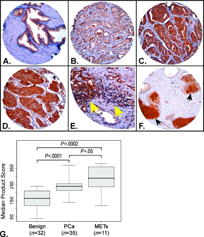
CXCL5 protein expression in human prostate tissues was evaluated by immunohistochemical analysis of a tissue microarray comprising 360 cores obtained from 99 tissue samples, of which 226 cores representing 83 separate tissue samples could be evaluated. These included benign glands (n = 32 tissues, 76 cores), primary prostate tumors (PCa; n = 35 tissues, 84 cores), metastatic prostate tumors (METs, n = 11 tissues, 29 cores), and miscellaneous other histologic subtypes [benign prostatic hyperplasia, n = 7 tissues, 15 cores; prostatic intraepithelial neoplasia (PIN), n = 5 tissues, 8 cores; prostatic atrophy, n = 9 tissues, 12 cores; prostatic inflammatory atrophy, n = 2 tissues, 3 cores]. The CXCL5 protein exhibited diffuse cytoplasmic cellular localization by immunostaining, which was largely confined to epithelial cell types in both benign and malignant glands. As shown in Figure 1, CXCL5 immunostaining was significantly higher in PCa compared to benign glands (P < .0001) and was significantly higher in METs compared to PCa (P = .05) or benign glands (P = .0002). CXCL5 immunostaining was also significantly higher in PIN than benign glands (P = .0008), although only five PIN lesions that could be evaluated were present on the TMA (not shown). Taken together, these data show that CXCL5 protein expression levels increased concordantly with prostate tumor progression. Further evaluation of CXCL5 protein expression levels among PCa revealed a trend toward higher expression levels in tumors exhibiting combined Gleason scores of 7 or greater (n = 25) compared to those with combined Gleason scores of 6 or less (n = 8) (P = .055) (Figure 1, B and C); some of these cancer areas were associated with stromal inflammation (Figure 1E).
Figure 1.
CXCL5 protein expression is concordant with prostate cancer progression. Shown are representative panels from a hematoxylin and eosin-stained, high-density tissue microarray probed with antibody against CXCL5, as follows: (A) Benign glands demonstrating weak staining. (B) PCa (Gleason sum 3 + 3) demonstrating weak staining. (C) PCa (Gleason sum 4 + 4) demonstrating moderate to strong staining. (D) Hormone refractory METs demonstrating strong staining. (E) PCa demonstrating moderate to strong staining associated with stromal inflammatory component (yellow arrows point to areas of inflammation). (F) Benign glands demonstrating strongly staining luminal secretions (black arrows). Original magnifications, x100. Panel E has been enlarged further, x4, to illustrate the area of inflammatory infiltrate concomitant with CXCL5 protein expression. (G) Boxplot depicting median product score distributions of protein expression levels for benign glands, malignant glands from PCa, and malignant areas from METs and P values associated with the statistical evaluation of these distributions.
CXCL5 immunostaining was associated with stromal inflammation in 50% (6 of 12) of cores containing atrophic glands and 100% (3 of 3) of cores containing prostatic inflammatory atrophy lesions. CXCL5 protein immunostaining was also detected in the luminal space of glands histologically characterized as benign (6 of 75 cores, 8.0%), hyperplastic (4 of 5 cores, 27%), PIN (2 of 8 cores, 25%), or malignant glands (6 of 84 cores, 7%), consistent with active secretion of CXCL5 into the lumen (Figure 1F). It should be noted that cores from two different cases containing hyperplastic glands demonstrated both luminal and inflammation-associated CXCL5 staining affecting the same glands.
CXCL5 Induces a Proliferative Response in Prostate Cancer Cells In Vitro
Because the studies on TMA demonstrated that CXCL5 protein expression levels increased concordantly with prostate tumor progression, we wished to investigate whether CXCL5 promoted a phenotypic response consistent with prostate tumor development and/or progression in vitro. To accomplish this, we first determined whether nontransformed N15C6 and BPH-1, or transformed LNCaP and PC3, prostate epithelial cells expressed CXCR2, the receptor for CXCL5, or endogenously secreted CXCL5. Western blot analysis of protein lysates prepared from nontransformed BPH-1 and N15C6 cells, and transformed LNCaP and PC3 cells, immunoblotted and probed with an antibody against CXCR2 revealed a band corresponding to a molecular weight of 40 kDa, consistent with that of CXCR2. Although the quality of the antibody precluded precise quantitation of the amount of CXCR2 protein, the immunoblot data shows that the relative amount of CXCR2 protein expressed was variable between cell lines (Figure 2A). ELISA of media conditioned by the same four cell lines showed that LNCaP secreted very low levels of CXCL5, on the order of less than 10 pg/ml into the media. N15C6 cells secreted 10-fold higher levels of CXCL5 into the media than LNCaP, and both BPH-1 and PC3 cells secreted approximately 100-fold higher levels of CXCL5 into the media than LNCaP (Figure 2B). Taken together, these results showed that the levels of CXCR2 and CXCL5 protein expressed by the four prostate epithelial cell lines examined was variable and did not appear to be directly related to whether the cells were phenotypically transformed or nontransformed.
Figure 2.
Nontransformed and transformed prostate epithelial cells express the CXCL5 receptor and endogenously secrete CXCL5. (A) Immunoblot analysis of protein lysates prepared from transformed PC3 and LNCaP, and nontransformed N15C6 and BPH-1 prostate epithelial cells probed with antibodies specific for the CXCL5 receptor, CXCR2, and loading control, β-actin. Primary antibody concentrations used were 1:1000 for CXCR2 and 1:5000 for β-actin. (B) Protein levels (pg/ml) of CXCL5 present in media conditioned by transformed LNCaP and PC3 or nontransformed N15C6 or BPH-1 cells prostate epithelial cells were determined by ELISA. The graph shows the pg/ml CXCL5 detected plotted on a logarithmic scale (y axis).
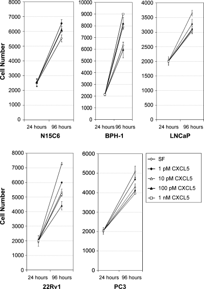
Because the cell lines tested for endogenous CXCL5 secretion were immortalized and highly proliferative, and expressed the appropriate receptor for CXCL5, we tested whether exogenously administered CXCL5 could stimulate a proliferative response in these cells. For these experiments, nontransformed (N15C6 and BPH-1), androgen-responsive transformed (22Rv1 and LNCaP) or androgen-insensitive transformed (PC3) prostate epithelial cells were seeded in triplicate, then grown for 3 days in SF media or the same media supplemented with increasing doses of CXCL5. These studies showed that CXCL5 initiates a moderate, but reproducible and statistically significant (P < .05), proliferative response in N15C6, BPH-1, LNCaP, and 22Rv1, but not PC3, prostate cancer cells in vitro (Figure 3). Both N15C6 and BPH-1 cells proliferated to significantly higher levels in response to almost all concentrations of CXCL5 tested compared to growth in SF media alone (Figure 3). LNCaP and 22Rv1 cells responded similarly to each other and proliferated significantly in response to only very low levels (1 or 10 pM) of CXCL5. Higher levels of CXCL5, e.g., 100 pM and 1 nM, were actually growth-suppressive in these cells (Figure 3).
Figure 3.
Prostate epithelial cells proliferate in response to CXCL5. Nontransformed N15C6 or BPH-1 cells, androgen-sensitive transformed LNCaP or 22Rv1, or androgen-insensitive transformed PC3 cells, were plated at 1000 cells/well in triplicate in SF media, harvested, and counted 24 or 96 hours later. The mean and standard deviation (error bars) of cell numbers obtained at each time point for growth in SF media is indicated by white diamonds, in SF media supplemented to 1 pM CXCL5 by black diamonds, to 10 pM CXCL5 by white triangles, to 100 pM CXCL5 by black triangles, and to 1 nM CXCL5 by white squares. The experiments shown are representative of replicate proliferation assays.
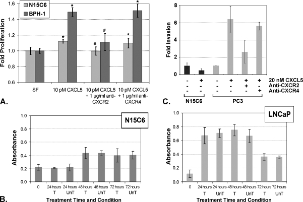
The specificity of the CXCL5-mediated proliferative response in the nontransformed N15C6 and BPH-1 cells was next tested through antibody blockade experiments. For these studies, the cells were pretreated with an antibody against the CXCL5-specific receptor, CXCR2, or that of an unrelated chemokine receptor, CXCR4. The results of these experiments showed that pretreatment with the antibody against the CXCL5-specific receptor, CXCR2, significantly (P < .001) ablated the ability of CXCL5 to stimulate proliferation in both cell lines. However, pretreatment with an antibody directed against the unrelated chemokine receptor, CXCR4, did not ablate the CXCL5-mediated proliferative response in either cell line (Figure 4A). Additional experiments showed that the fraction of cells exhibiting apoptosis was similar in untreated cells and in those treated with CXCL5 for both nontransformed N15C6 and transformed LNCaP cells (Figure 4B). Taken together, these studies showed that the observed CXCL5-mediated proliferative response was dependent on interactions with the G-protein-coupled receptor, CXCR2, which specifically recognizes this chemokine. Moreover, these studies showed that CXCL5 promoted a pro-proliferative, rather than an antiapoptotic, response in prostate epithelial cells.
Figure 4.
CXCL5-stimulated proliferative and invasive responses. (A) N15C6 (light gray bars) or BPH-1 (dark gray bars) nontransformed prostate epithelial cells proliferated to significantly higher levels when grown for 72 hours in SF media supplemented with 10 pM CXCL5 than those grown in SF alone (*P < .001). Preincubation of the cells for 1 hour with 1 µg/ml antibody against CXCR2, the receptor for CXCL5, followed by supplementation with CXCL5 and maintenance of growth in CXCL5 + anti-CXCR2-containing media significantly ablated the proliferative response (#P < .001). In contrast, cellular growth after preincubation with an antibody against an unrelated chemokine receptor, CXCR4, followed by supplementation with CXCL5 and maintenance of growth in CXCL5 + anti-CXCR4-containing media was similar to that observed for non-pretreated cells grown in CXCL5-supplemented media and was significantly higher than that in SF alone (*P < .001). All data are shown normalized to growth in unsupplemented SF, which was set at one-fold. (B) N15C6 (LEFT) or LNCaP (RIGHT) cells were grown in SF media (untreated, UnT) or SF media supplemented with 10 pM CXCL5 for N15C6 or 100 pM CXCL5 for LNCaP (treated, T) for the times indicated. The cells were then harvested and assessed for nucleosomal DNA fragmentation. The fraction of cells exhibiting apoptosis plotted on the y axis was calculated as the difference in absorbance measured at 405 nm and at the reference wavelength of 490 nm after adjusting for background absorbance at both wavelengths. No significant differences in the fraction of cells exhibiting apoptosis were observed between treated and untreated cells at any time point, demonstrating that CXCL5 does not promote antiapoptotic responses in these cells. (C) Fifteen thousand each of N15C6 (black bars) or PC3 (gray bars) cells were plated onto Matrigel-coated membranes and were exposed to complete media or complete media supplemented with 20 nM CXCL5 for 24 hours. After 24 hours, the cells that migrated and invaded through the Matrigel were stained and counted. N15C6 cells did not demonstrate an invasive response to treatment with CXCL5. However, approximately six-fold more PC3 cells migrated through the synthetic basement membrane, Matrigel, in response to 20 nM CXCL5 compared to vehicle (control, set at one-fold) (*P < .05). PC3 cell invasion through the Matrigel in response to CXCL5 was significantly inhibited by pretreatment with 1 µg/ml blocking antibody (anti-CXCR2) (#P < .05) but not by pretreatment with nonspecific antibody (anti-CXCR4) (*P < .05).
CXCL5 Stimulates Prostate Cancer Cell Migration and Invasion In Vitro
Several studies have shown that the CXC-type chemokine, CXCL12, stimulates the migration and invasion of prostate cancer cells in vitro and in vivo [5–8] and that migration in vitro was observed using a concentration of CXCL12 in the 10 to 20 nM range. Therefore, we examined whether CXCL5 could also stimulate acquisition of an invasive phenotype by prostate cancer cells. Using a modified Boyden chamber assay, 15,000 nontransformed N15C6 or transformed PC3 cells were plated onto Matrigel-coated membranes (upper wells) and were exposed to complete media or complete media supplemented with 20 nM CXCL5 for 24 hours. After 24 hours, the cells that migrated and invaded through the Matrigel were stained and counted. As seen in Figure 4C, significantly more PC3 cells migrated through the Matrigel in response to CXCL5 than to the vehicle (*P < .05). In contrast, N15C6 cells did not migrate through the Matrigel in response to CXCL5. To test the specificity of the CXCL5-mediated invasive response, PC3 cells were pretreated for 2 hours with 1 µg/ml blocking antibody (anti-CXCR2) or nonspecific antibody (anti-CXCR4) in the bottom wells for 12 hours, then treated with vehicle or 20 nM CXCL5. These experiments showed that significantly more PC3 cells migrated through the Matrigel in response to CXCL5 than to the vehicle (*P < .05) and that this activity was blocked by an antibody against the receptor to CXCL5, CXCR2, but not against the receptor to CXCL12, CXCR4 (Figure 4C). Thus, transformed PC3 prostate epithelial cells demonstrated a robust and specific migratory/invasive response to CXCL5 in vitro. In contrast, non-transformed N15C6 cells did not migrate or invade through the Matrigel in response to CXCL5.
CXCL5 Activates Both Mitogen-Activated Protein Kinase and Phosphoinositide 3-Kinase Signaling in Prostate Epithelial Cells
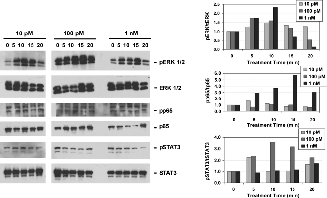
We and others have shown that another CXC-type chemokine, CXCL12, interacts with its primary receptor, CXCR4, to activate downstream signaling events involving the mitogen-activated protein kinase (MAPK) and/or phosphoinositide 3-kinase (PI3K), and/or Janus kinases/signal transducers and activators of transcription (JAK/STAT) pathways in prostate epithelial cells [3,4]. To begin to test whether CXCL5 activated similar pathways associated with prostate epithelial cellular proliferation or invasion, N15C6 or LNCaP cells were treated with increasing doses of CXCL5 and assessed for activation of ERK 1/2 (MAPK pathway), the p65 subunit of nuclear factor-kappa B (NF-κB; PI3K pathway), or STAT3 (JAK/STAT pathway). As seen in Figure 5, nontransformed N15C6 cells rapidly and transiently phosphorylated ERK 1/2 and STAT3 when treated with either subnanomolar (10 or 100 pM) or nanomolar (1 nM) levels of CXCL5, whereas NF-κB subunit activation was evident only after treatment with 1 nM CXCL5. Transformed LNCaP cells rapidly and transiently phosphorylated both ERK 1/2 and the p65 subunit of NF-κB on treatment with subnanomolar (10 or 100 pM) levels of CXCL5 (Figure 6). However, activation of either ERK 1/2 or NF-κB was not evident in LNCaP cells treated with nanomolar levels of CXCL5 (Figure 6), and activation of STAT3 was not observed in LNCaP cells treated with either subnanomolar or nanomolar levels of CXCL5 (not shown).
Figure 5.
CXCL5 activates MAPK signaling in nontransformed N15C6 prostate epithelial cells. Nontransformed N15C6 cells rapidly and transiently phosphorylated ERK 1/2 and STAT3 when treated with either subnanomolar (10 or 100 pM) or nanomolar (1 nM) levels of CXCL5, whereas NF-κB subunit activation was evident only after treatment with 1 nM CXCL5. Primary antibody concentrations used were 1:500 for phospho-ERK, 1:500 for phospho-65 (NF-κB), 1:1000 for phospho-STAT3, 1:1000 for total ERK, 1:1000 for total p65, and 1:2000 for total STAT3. A total of 20 µg of protein lysate was electrophoresed per well. Immunoblots are shown on the left, and corresponding densitometric evaluations of the same blots are shown on the right. Phosphorylation relative to total protein quantitated from the immunoblot is shown in the densitometric plots as phospho/total protein.
Figure 6.
CXCL5 activates both MAPK and PI3K signaling in transformed LNCaP prostate epithelial cells. Transformed LNCaP cells rapidly and transiently phosphorylated both ERK 1/2 and the p65 subunit of NF-κB on treatment with subnanomolar (10 or 100 pM) levels of CXCL5. Immunoblots are shown in the top panel, and corresponding densitometric evaluations of the same blots are shown in the bottom panel. Phosphorylation relative to total protein quantitated from the immunoblot is shown in the densitometric plots as phospho/total protein. A total of 100 µg of protein lysate was electrophoresed per well. Primary antibody concentrations used were as described for Figure 5.
CXCL5 Stimulates EGR1 Gene Transcription
We have recently shown that another CXC-type chemokine, CXCL12, stimulates a complex and robust transcriptional response in both nontransformed N15C6 and transformed LNCaP prostate epithelial cells [5]. In particular, CXCL12 activated transcription of EGR1, which encodes a C2H2-type zinc-finger protein induced by mitogenic stimulation and shown to stimulate tumor cell growth, play a role in tumor progression, and stimulate angiogenesis and improved survival of tumor cells [4,20]. Therefore, we investigated whether CXCL5 stimulated a transcriptional response in nontransformed or transformed prostate epithelial cells. As shown in Figure 7, nontransformed N15C6 cells treated with subnanomolar levels (1–100 pM) CXCL5 rapidly accumulated four- to eight-fold more EGR1 transcript than vehicle-treated cells. Similarly treated transformed LNCaP cells accumulated two- to three-fold more EGR1 transcript than vehicle-treated cells, but only at the lowest CXCL5 concentration (10 pM) tested.
Figure 7.
CXCL5 stimulates a transcriptional response in both nontransformed and transformed prostate epithelial cells. Quantitative real-time PCR of RNA purified from N15C6 cells (left) or LNCaP cells (right) treated with subnanomolar CXCL5 as shown demonstrates rapid and robust transcription of the EGR1 gene significantly higher than levels obtained at time 0 (set at one-fold) (*P < .05). Data shown are averaged from three or more separate experiments per time point per concentration of CXCL5 examined.
Discussion
The studies reported here are the first to demonstrate that CXCL5 protein expression increases concordantly with prostate tumor progression. Moreover, CXCL5 protein expression was observed as equivalently more abundant in PIN lesions (P < .0008) and in primary tumors (P < .0001) compared to benign glands, suggesting that CXCL5 protein upregulation is an early event during prostate tumorigenesis. These findings parallel those reported for the CXCL5 receptor, CXCR2, which was also characterized by higher protein expression levels in malignant prostate glands and high-grade PIN lesions compared to benign glands [21]. The observation that CXCL5 expression was slightly elevated in metastatic tumors compared to primary tumors (P = .05) also implies that expression of this protein remains high, and may increase, during prostate cancer metastasis. Studies by Park et al. [13] have shown that CXCL5 protein overexpression was associated with late-stage gastric cancer and high N stage, suggesting a role for CXCL5 in the progression of gastric cancer, specifically in lymph node metastasis. Taken together, these data indicate that CXCL5 expression increases concordantly with tumor progression in prostate and other cancers.
CXCL5 is a member of a proangiogenic subgroup of the CXC-type chemokine family of small, secreted proteins [2,9]. Luminal staining suggestive of active secretion of CXCL5 protein was observed in approximately 10% to 25% of tissue cores examined containing benign, hyperplastic, PIN, or malignant glands. Other studies have shown that concentrations of CXCL5 in peritoneal fluid are markedly elevated in women with severe endometriosis, a disease characterized by ectopic proliferation of highly vascularized endometrial tissue [10,22]. Miyazaki et al. [11] recently reported that cells derived from a gastric cancer lymph node metastasis, but not from a primary tumor from the same patient, actively secreted CXCL5. Data reported in this study show that both nontransformed and transformed prostate epithelial cells secrete varying levels of CXCL5 and express CXCR2, the CXCL5 receptor. Taken together, these studies all associate CXCL5 secretion and CXCR2 expression with benign or malignant proliferative diseases. Indeed, the results of dose-response experiments conducted as part of the studies reported here demonstrated that nontransformed N15C6 and BPH-1 cells, as well as transformed androgen-sensitive 22Rv1 and LNCaP cells, although not transformed androgen-insensitive PC3 cells, responded proliferatively to exogenous CXCL5. Further testing of two of these cell lines, N15C6 and LNCaP, for percentage of cells exhibiting apoptosis showed that CXCL5 does not promote an antiapoptotic response, suggesting that the observed increase in cell number consequent to exposure to CXCL5 is largely a pro-proliferative response.
Of these cell lines examined in this study, only PC3 cells have been examined by others for a proliferative response to CXCL5 in vitro or in vivo. Moore et al. [23] reported that treatment of PC3 cells with 100 ng/ml (approximately 12.5 nM) did not elicit a proliferative response. These data correlate well with our study, which further demonstrated that PC3 cells did not demonstrate a proliferative response in vitro to either sub- or suprananomolar levels of exogenously administered CXCL5. The study by Moore et al. [23] also reported a positive correlation between the in vivo production of CXCL5 and the exponential growth of PC3 xenografts in vivo, although this effect on growth may be due to the angiogenic induction of endothelial cell, rather than PC3 cell, proliferation.
It is unclear why some transformed prostate epithelial cells, e.g., LNCaP and 22Rv1, proliferate in response to CXCL5, but others, e.g., PC3, do not. It is tempting to speculate that the presence or absence of the androgen receptor in these cells may modulate whether they can respond proliferatively to CXCL5. However, both N15C6 and BPH-1 cells also responded proliferatively to CXCL5, yet BPH-1 cells do not express the androgen receptor, and N15C6 cells express only very low levels of this protein [16, and unpublished observations]. Therefore, it is possible that both androgen-dependent and -independent mechanisms can be involved in the observed CXCL5-mediated proliferative response.
Although the proliferative response of N15C6, BPH-1, LNCaP, and 22Rv1 prostate epithelial cells was highly reproducible, it was also relatively modest and, for the most part, confined to subnanomolar levels of CXCL5. It was clearly specific to CXCL5/CXCR2 interactions, as both N15C6 and BPH-1 cells failed to proliferate in response to CXCL5 after pretreatment with antibody against the CXCL5 receptor, but not antibody against an unrelated chemokine receptor, CXCR4. The results of these experiments are consistent with those from other studies conducted in other tumor types, which have shown that CXCL5 promotes the proliferation of human head and neck squamous carcinoma cells in vitro and in vivo [12], that heterotrophic renal cell carcinomas fail to proliferate in CXCR2-null mice [24], and that tumors in the transgenic adenocarcinoma of the mouse prostate model (TRAMP)/CXCR2-null mice were significantly smaller than those in TRAMP/CXCR2 +/+ mice and had reduced angiogenesis [25]. Taken together, these results suggest that CXCL5 stimulates a proliferative response in both nontransformed and some transformed prostate epithelial cells, thus, may promote abnormal cellular proliferation and facilitate the development of both benign and malignant proliferative disease of the prostate. The observation of abundant and elevated CXCL5 expression in metastatic compared to primary tumors indicated that CXCL5 may promote expression of the invasive phenotype required for tumor metastasis. Indeed, CXCL5 is a chemotaxic agent, and is a key modulator of immune cell trafficking, especially for granulocytic immune cells. Experiments reported here show that CXCL5 exerts a strong and specific chemotactic effect on highly malignant, transformed PC3 prostate cancer cells, which was substantial enough to draw the prostate cancer cells through a Matrigel synthetic basement membrane barrier. In contrast, nontransformed N15C6 cells failed to migrate through the Matrigel in response to the same concentration of CXCL5. Previous studies by Reiland et al. [26] showed that PC3 cells migrated through the reconstituted basement membrane in response to 36 nM interleukin-8, another proangiogenic CXC-type chemokine, and that this response was CXCR2-dependent. Another study reported by Miyazaki et al. [12] showed that RNA interference of the CXCL5 transcript reduced the ability of head and neck squamous carcinoma cells to migrate and invade through the Matrigel in vitro. Together, these studies suggest that CXCL5 may promote metastasis in multiple tumor types in a CXCR2-dependent manner.
Our laboratory recently reported that subnanomolar levels of another chemokine, CXCL12, stimulated intracellular signaling through both MAPK (specifically, MEK/ERK) and PI3K (specifically, NF-κB) pathways associated with cellular proliferation [4,5]. The current study shows that CXCL5 also signals through these pathways, as well as through the JAK/STAT pathway. In contrast to CXCL12, however, CXCL5 stimulates ERK phosphorylation in both nontransformed N15C6 and transformed LNCaP cells, as well as STAT3 activation in N15C6 cells. Both MAPK-dependent and -independent mechanisms have been shown to activate the transcription factor, ELK-1, which in turn can activate the promoters of multiple genes containing serum response elements (e.g., EGR1) or ternary complex factor binding sites [27–31]. Data reported here show that CXCL5 stimulates the transcription of EGR1 by both nontransformed N15C6 and transformed LNCaP cells. The observation of similarities, as well as differences, between intracellular signaling events elicited by CXC-type chemokines raises the possibility that these chemokines may use both common and unique cellular mechanisms to induce similar phenotypic responses, e.g., proliferation, migration/invasion, and gene transcription, in prostate cancer cells. If so, the responses of benign or malignant prostate epithelial cells to endogenously produced (CXCL5) or exogenously produced (CXCL12) chemokines may be the result of a complex interplay between costimulated and differentially stimulated signaling pathways.
In addition to the expression and biologic activities of CXCL5 in connection with tumor development, CXCL5 expression by epithelial cells has been frequently observed in conjunction with inflammatory responses. For example, CXCL5 protein expression by the intestinal mucosa has been observed in association with inflammatory conditions such as Crohn disease, ulcerative colitis, and acute appendicitis, and by the exocrine tissue of the pancreas associated with chronic pancreatitis [9]. Our studies reported here show that the CXCL5 protein expression levels are concurrent with tumor progression and are coincident with the presence of granulocytic inflammatory cells. Inflammatory infiltrate in the prostate coincident with benign prostatic enlargement and prostate cancer has been frequently observed, provoking intense interest in the potential role of inflammatory mediators in the etiology of both diseases [32–37]. The studies reported here for the inflammatory mediator, CXCL5, are consistent with these observations. In addition, these and other studies show that CXCL5 promotes the proliferation and metastasis of both nontransformed and transformed cells in vitro and in vivo. Taken together, these data provide rationale for exploring potential functional links between inflammatory mediators and the development of benign hypertrophic or malignant neoplastic proliferative disease in the prostate.
Footnotes
This work was supported by the National Institutes of Health awards from the George M. O'Brien Center for Urologic Research at the University of Michigan 1 P50 DK065313 (J.A.M.), the University of Michigan Comprehensive Cancer Center support grant 5 P30 CA46592 (J.A.M., A.M.C.), the University of Michigan Prostate Cancer Specialized Program of Research Excellence (SPORE) grant 5 P50 CA068568 (A.M.C.), W81XWH-07-1-0385 from the Department of Defense's Congressionally Directed Medical Research Program (J.A.M.), and funds awarded by Domino's Pizza, L.L.C. (J.A.M.)
References
- 1.Jemal A, Siegel R, Ward E, Murray T, Xu J, Thun MJ. Cancer statistics, 2007. CA Cancer J Clin. 2007;57:43–66. doi: 10.3322/canjclin.57.1.43. [DOI] [PubMed] [Google Scholar]
- 2.Koch AE. Chemokines and their receptors in rheumatoid arthritis: future targets? Arthritis Rheum. 2005;52:710–721. doi: 10.1002/art.20932. [DOI] [PubMed] [Google Scholar]
- 3.Begley L, Monteleon C, Shah RB, Macdonald JW, Macoska JA. CXCL12 overexpression and secretion by aging fibroblasts enhance human prostate epithelial proliferation in vitro. Aging Cell. 2005;4(6):291–298. doi: 10.1111/j.1474-9726.2005.00173.x. [DOI] [PubMed] [Google Scholar]
- 4.Begley LA, MacDonald JW, Day ML, Macoska JA. CXCL12 activates a robust transcriptional response in human prostate epithelial cells. J Biol Chem. 2007;282(37):26767–26774. doi: 10.1074/jbc.M700440200. [DOI] [PubMed] [Google Scholar]
- 5.Singh S, Singh UP, Grizzle WE, Lillard JW., Jr CXCL12-CXCR4 interactions modulate prostate cancer cell migration, metalloproteinase expression and invasion. Lab Invest. 2004;84(12):1666–1676. doi: 10.1038/labinvest.3700181. [PMID: 15467730] [DOI] [PubMed] [Google Scholar]
- 6.Mochizuki H, Matsubara A, Teishima J, Mutaguchi K, Yasumoto H, Dahiya R, Usui T, Kamiya K. Interaction of ligand-receptor system between stromal-cell-derived factor-1 and CXC chemokine receptor 4 in human prostate cancer: a possible predictor of metastasis. Biochem Biophys Res Commun. 2004;320(3):656–663. doi: 10.1016/j.bbrc.2004.06.013. [PMID: 15240098] [DOI] [PubMed] [Google Scholar]
- 7.Taichman RS, Cooper C, Keller ET, Pienta KJ, Taichman NS, McCauley LK. Use of the stromal cell-derived factor-1/CXCR4 pathway in prostate cancer metastasis to bone. Cancer Res. 2002;62(6):1832–1837. [PMID: 11912162] [PubMed] [Google Scholar]
- 8.Darash-Yahana M, Pikarsky E, Abramovitch R, Zeira E, Pal B, Karplus R, Beider K, Avniel S, Kasem S, Galun E, et al. Role of high expression levels of CXCR4 in tumor growth, vascularization, and metastasis. FASEB J. 2004;18(11):1240–1242. doi: 10.1096/fj.03-0935fje. [PMID: 15180966] [DOI] [PubMed] [Google Scholar]
- 9.Walz A, Schmutz P, Mueller C, Schnyder-Candrian S. Regulation and function of the CXC chemokine ENA-78 in monocytes and its role in disease. J Leukoc Biol. 1997;62(5):604–611. doi: 10.1002/jlb.62.5.604. [DOI] [PubMed] [Google Scholar]
- 10.Wong YF, Cheung TH, Lo KW, Yim SF, Siu NS, Chan SC, Ho TW, Wong KW, Yu MY, Wang VW, et al. Identification of molecular markers and signaling pathway in endometrial cancer in Hong Kong Chinese women by genome-wide gene expression profiling. Oncogene. 2007;26:1971–1982. doi: 10.1038/sj.onc.1209986. [DOI] [PubMed] [Google Scholar]
- 11.Miyazaki H, Patel V, Wang H, Ensley JF, Gutkind JS, Yeudall WA. Growth factor-sensitive molecular targets identified in primary and metastatic head and neck squamous cell carcinoma using microarray analysis. Oral Oncol. 2006;42(3):240–256. doi: 10.1016/j.oraloncology.2005.07.006. [DOI] [PubMed] [Google Scholar]
- 12.Miyazaki H, Patel V, Wang H, Edmunds RK, Gutkind JS, Yeudall WA. Down-regulation of CXCL5 inhibits squamous carcinogenesis. Cancer Res. 2006;66(8):4279–4284. doi: 10.1158/0008-5472.CAN-05-4398. [DOI] [PubMed] [Google Scholar]
- 13.Park JY, Park KH, Bang S, Kim MH, Lee JE, Gang J, Koh SS, Song SY. CXCL5 overexpression is associated with late stage gastric cancer. J Cancer Res Clin Oncol. 2007;133(11):835–840. doi: 10.1007/s00432-007-0225-x. [DOI] [PubMed] [Google Scholar]
- 14.Begley L, Keeney D, Beheshti B, Squire JA, Kant R, Chaib H, MacDonald JW, Rhim J, Macoska JA. Concordant copy number and transcriptional activity of genes mapping to derivative chromosomes 8 during cellular immortalization in vitro. Genes Chromosomes Cancer. 2006;45(2):136–146. doi: 10.1002/gcc.20274. [DOI] [PubMed] [Google Scholar]
- 15.Macoska JA, Paris P, Collins C, Andaya A, Beheshti B, Chaib H, Kant R, Begley L, MacDonald JW, Squire JA. Evolution of 8p loss in transformed human prostate epithelial cells. Cancer Genet Cytogenet. 2004;154(1):36–43. doi: 10.1016/j.cancergencyto.2004.02.013. [DOI] [PubMed] [Google Scholar]
- 16.Hayward SW, Dahiya R, Cunha GR, Bartek J, Deshpande N, Narayan P. Establishment and characterization of an immortalized but nontransformed human prostate epithelial cell line: BPH-1. In Vitro Cell Dev Biol Anim. 1995;31(1):14–24. doi: 10.1007/BF02631333. [DOI] [PubMed] [Google Scholar]
- 17.Horoszewicz JS, Leong SS, Kawinski E, Karr JP, Rosenthal H, Chu TM, Mirand EA, Murphy GP. LNCaP model of human prostatic carcinoma. Cancer Res. 1983;43(4):1809–1818. [PubMed] [Google Scholar]
- 18.Sramkoski RM, Pretlow TG, II, Giaconia JM, Pretlow TP, Schwartz S, Sy MS, Marengo SR, Rhim JS, Zhang D, Jacobberger JW. A new human prostate carcinoma cell line, 22Rv1. In Vitro Cell Dev Biol Anim. 1999;35(7):403–409. doi: 10.1007/s11626-999-0115-4. [DOI] [PubMed] [Google Scholar]
- 19.Kaighn ME, Narayan KS, Ohnuki Y, Lechner JF, Jones LW. Establishment and characterization of a human prostatic carcinoma cell line (PC-3) Invest Urol. 1979;17(1):16–23. [PubMed] [Google Scholar]
- 20.Adamson ED, Mercola D. Egr1 transcription factor: multiple roles in prostate tumor cell growth and survival. Tumour Biol. 2002;23:93–102. doi: 10.1159/000059711. [DOI] [PubMed] [Google Scholar]
- 21.Murphy C, McGurk M, Pettigrew J, Santinelli A, Mazzucchelli R, Johnston PG, Montironi R, Waugh DJ. Nonapical and cytoplasmic expression of interleukin-8, CXCR1, and CXCR2 correlates with cell proliferation and microvessel density in prostate cancer. Clin Cancer Res. 2005;11(11):4117–4127. doi: 10.1158/1078-0432.CCR-04-1518. [DOI] [PubMed] [Google Scholar]
- 22.Suzumori N, Katano K, Suzumori K. Peritoneal fluid concentrations of epithelial neutrophil-activating peptide-78 correlate with the severity of endometriosis. Fertil Steril. 2004;81:305–308. doi: 10.1016/j.fertnstert.2003.08.011. [DOI] [PubMed] [Google Scholar]
- 23.Moore BB, Arenberg DA, Stoy K, Morgan T, Addison CL, Morris SB, Glass M, Wilke C, Xue YY, Sitterding S, et al. Distinct CXC chemokines mediate tumorigenicity of prostate cancer cells. Am J Pathol. 1999;154(5):1503–1512. doi: 10.1016/S0002-9440(10)65404-1. [DOI] [PMC free article] [PubMed] [Google Scholar]
- 24.Mestas J, Burdick MD, Reckamp K, Pantuck A, Figlin RA, Strieter RM. The role of CXCR2/CXCR2 ligand biological axis in renal cell carcinoma. J Immunol. 2005;175(8):5351–5357. doi: 10.4049/jimmunol.175.8.5351. [DOI] [PubMed] [Google Scholar]
- 25.Shen H, Schuster R, Lu B, Waltz SE, Lentsch AB. Critical and opposing roles of the chemokine receptors CXCR2 and CXCR3 in prostate tumor growth. Prostate. 2006;66(16):1721–1728. doi: 10.1002/pros.20476. [DOI] [PubMed] [Google Scholar]
- 26.Reiland J, Furcht LT, McCarthy JB. CXC-chemokines stimulate invasion and chemotaxis in prostate carcinoma cells through the CXCR2 receptor. Prostate. 1999;41(2):78–88. doi: 10.1002/(sici)1097-0045(19991001)41:2<78::aid-pros2>3.0.co;2-p. [DOI] [PubMed] [Google Scholar]
- 27.Chung KC, Gomes I, Wang D, Lau LF, Rosner MR. Raf and fibroblast growth factor phosphorylate Elk1 and activate the serum response element of the immediate early gene pip92 by mitogen-activated protein kinase-independent as well as -dependent signaling pathways. Mol Cell Biol. 1998;18(4):2272–2281. doi: 10.1128/mcb.18.4.2272. [DOI] [PMC free article] [PubMed] [Google Scholar]
- 28.Mohney RP, Das M, Bivona TG, Hanes R, Adams AG, Philips MR, O'Bryan JP. Intersectin activates Ras but stimulates transcription through an independent pathway involving JNK. J Biol Chem. 2003;278(47):47038–47045. doi: 10.1074/jbc.M303895200. [DOI] [PubMed] [Google Scholar]
- 29.Torii S, Yamamoto T, Tsuchiya Y, Nishida E. ERK MAP kinase in G cell cycle progression and cancer. Cancer Sci. 2006;97(8):697–702. doi: 10.1111/j.1349-7006.2006.00244.x. [Review] [DOI] [PMC free article] [PubMed] [Google Scholar]
- 30.Buchwalter G, Gross C, Wasylyk B. Ets ternary complex transcription factors. Gene. 2004;324:1–14. doi: 10.1016/j.gene.2003.09.028. [DOI] [PubMed] [Google Scholar]
- 31.Sun Q, Chen G, Streb JW, Long X, Yang Y, Stoeckert CJ, Miano JM. Defining the mammalian CArGome. Genome Res. 2006;16(2):197–207. doi: 10.1101/gr.4108706. [DOI] [PMC free article] [PubMed] [Google Scholar]
- 32.Nickel JC, Downey J, Young I, Boag S. Asymptomatic inflammation and/or infection in benign prostatic enlargement. BJU Int. 1999;84:976–981. doi: 10.1046/j.1464-410x.1999.00352.x. [DOI] [PubMed] [Google Scholar]
- 33.Gerstenbluth RE, Seftel AD, MacLennan GT, Rao RN, Corty EW, Ferguson K, Resnick MI. Distribution of chronic prostatitis in radical prostatectomy specimens with up-regulation of bcl-2 in areas of inflammation. J Urol. 2002;167:2267–2270. [PubMed] [Google Scholar]
- 34.Roehrborn CG, Kaplan SA, Noble WD, Lucia MS, Slawin KM, McVary KT, Kusek JW, Nyberg LM. The impact of acute or chronic inflammation in baseline biopsy on the risk of clinical progression of BPE: results from the MTOPS study. J Urol. 2005;173(4 Suppl):346. [American Urological Association Meeting 2005; Abstract #1277] [Google Scholar]
- 35.De Marzo AM, Marchi VL, Epstein JI, Nelson WG. Proliferative inflammatory atrophy of the prostate: implications for prostatic carcinogenesis. Am J Pathol. 1999;155:1985–1992. doi: 10.1016/S0002-9440(10)65517-4. [DOI] [PMC free article] [PubMed] [Google Scholar]
- 36.Putzi MJ, De Marzo AM. Morphologic transitions between proliferative inflammatory atrophy and high-grade prostatic intraepithelial neoplasia. Urology. 2000;56:828–832. doi: 10.1016/s0090-4295(00)00776-7. [DOI] [PubMed] [Google Scholar]
- 37.Palapattu GS, Sutcliffe S, Bastian PJ, Platz EA, De Marzo AM, Isaacs WB, Nelson WG. Prostate carcinogenesis and inflammation: emerging insights. Carcinogenesis. 2004;26(7):1170–1181. doi: 10.1093/carcin/bgh317. [DOI] [PubMed] [Google Scholar]


