Abstract
KEULE is required for cytokinesis in Arabidopsis thaliana. We have positionally cloned the KEULE gene and shown that it encodes a Sec1 protein. KEULE is expressed throughout the plant, yet appears enriched in dividing tissues. Cytokinesis-defective mutant sectors were observed in all somatic tissues upon transformation of wild-type plants with a KEULE–green fluorescent protein gene fusion, suggesting that KEULE is required not only during embryogenesis, but at all stages of the plant's life cycle. KEULE is characteristic of a Sec1 protein in that it appears to exist in two forms: soluble or peripherally associated with membranes. More importantly, KEULE binds the cytokinesis-specific syntaxin KNOLLE. Sec1 proteins are key regulators of vesicle trafficking, capable of integrating a large number of intra- and/or intercellular signals. As a cytokinesis-related Sec1 protein, KEULE appears to represent a novel link between cell cycle progression and the membrane fusion apparatus.
Keywords: cytokinesis, Sec1, cell plate, syntaxin, Arabidopsis
Introduction
Cytokinesis is the partitioning of the cytoplasm after nuclear division. Spatial and temporal regulation are key issues underlying this process. The new cleavage plane needs to be oriented with respect to the mother cell and, to avoid losing or bisecting the nucleus, cytokinesis can only be initiated once the nuclear cycle is complete. Although actin and tubulin dynamics during cytokinesis have been well characterized in plant, yeast, and animal cells, the membrane dynamics of cytokinesis represent a new frontier in our understanding of cell division. It is clear that the de novo addition of membranes is required for cytokinesis in plant, yeast, and animal cells. The origin and dynamics of these new membranes are best understood in plants. Careful electron microscopy analyses of synchronized tobacco cells (complemented by in vivo imaging; Nebenführ et al. 2000) have shown how Golgi-derived vesicles fuse to build delicate, tubular membrane networks at the equator of a dividing cell (Samuels et al. 1995). The membrane network undergoes several rearrangements, such as the appearance of membrane coats and clathrin-coated pits, which have been detailed in a five-staged model of plant cytokinesis (Samuels et al. 1995). At the molecular genetic level, the vesicle trafficking syntaxin gene, KNOLLE, has been shown to be specifically required for cytokinesis in somatic plant cells (Lukowitz et al. 1996; Lauber et al. 1997). It has recently been shown that syntaxins are also implicated in cytokinesis in somatic animal cells (Jantsch-Plunger and Glotzer 1999). However, little is known about the regulation of these syntaxins.
Although sharing some common features, cytokinesis in plant and animal cells differs in several ways. Plant cells build complex cell walls in the brief period of time between late anaphase and telophase. This requires an efficient and rapid mobilization of resources and is achieved by targeting Golgi-derived vesicles carrying cell wall components to the equator of a dividing cell. The nascent cell wall, assembled within the membrane network formed by vesicle fusion, undergoes a complex process of maturation during which callose is removed and cellulose and pectins are added (for review see Assaad et al. 1997). This maturation process appears perturbed in mutants with defective cell walls, such as cyt1 (Nickle and Meinke 1998) and korrigan (Zuo et al. 2000).
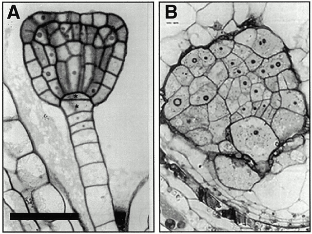
Given the complexity of plant cytokinesis, surprisingly few genes have been identified by mutation. Genes required for cytokinesis in somatic plant cells fall into two classes. Although genes in the first class are required for the proper orientation of the plane of division, genes in the second class are required for the execution of cytokinesis. fas or tonneau and tangled (for review see Smith 1999) mutants define the first class of cytokinesis genes. The cyd mutants of pea (Liu et al. 1995) and Arabidopsis (Yang et al. 1999b) and the KNOLLE (Lukowitz et al. 1996) and KEULE (Assaad et al. 1996) genes of Arabidopsis fall into the second class. keule and knolle mutants die as seedlings in which rudiments of all body parts can be recognized and are characterized by the incidence of grossly enlarged, irregularly shaped cells (Assaad et al. 1996; Lukowitz et al. 1996). Light and electron microscopy showed that dividing cells in cyd, knolle, and keule mutants are often multinucleate, with gapped or incomplete cross walls, which defines these mutants as cytokinesis defective (Liu et al. 1995; Assaad et al. 1996; Lukowitz et al. 1996). The multinucleate cells are invariably enlarged (Assaad et al. 1996) and account for the rough surface and bloated appearance of these cytokinesis mutants. The keule phenotype (recapitulated in Fig. 1) highlights the importance of cytokinesis in cellular differentiation and morphogenesis in plants (Assaad et al. 1996).
Figure 1.
Cytokinesis defects in keule embryos A and B depict histological embryo sections (A) wild-type (triangular) (B) keule (delayed in their development). Note the large, irregularly shaped multinucleate cells in keule mutants. Bar: (A) 50 μm; (B) 20 μm.
In this study, we describe the cloning of the KEULE gene and present evidence that it encodes a Sec1 homologue. There are two conserved homologues of KEULE in the Arabidopsis genome. Sec1 proteins (note that this designates the whole Sec1 superfamily and not only yeast Sec1 orthologues implicated in exocytosis) are key regulators of vesicle trafficking, a complex process regulated at the levels of vesicle formation, transport, tethering/docking, and fusion. Sec1 proteins regulate the last two steps, namely tethering/docking and membrane fusion, by interacting with syntaxins (Halachmi and Lev 1996) and several other proteins (Butz et al. 1998; Peterson et al. 1999). Given KEULE's gene identity, we investigate its potential role in processes such as cellular elongation and root hair development, which are, like cytokinesis, mediated by vesicle trafficking. We show that KEULE behaves like a Sec1 protein with respect to the nature of its apparent membrane association. In addition, KEULE binds the cytokinesis-specific syntaxin KNOLLE (Lukowitz et al. 1996; Lauber et al. 1997) in in vitro binding assays. KEULE and KNOLLE are not only some of the few genes of Arabidopsis with a primary defect in the execution of cytokinesis, but also some of the only vesicle trafficking genes of Arabidopsis for which mutant phenotypes have been described.
Materials and Methods
Genetic Techniques and Phenotypic Analysis
The majority of the keule alleles have been described previously (Assaad et al. 1996); MM125 was generated in an x-ray screen. A cytokinesis-defective line nonallelic to keule and designated “keule-like” (obtained from the same screen as keule) was used in controls. Samples for scanning electron micrography and clearing preps were prepared as described (Assaad et al. 1996). To visualize root hairs, seedlings were briefly dipped in 0.05% methylene blue, rinsed in water, and imaged. All images shown were processed with the Photoshop® and/or Illustrator® software (Adobe).
Fine Mapping of the KEULE Gene
For mapping, the R227 allele of keule in the Landsberg background was crossed to wild-type Niederzenz. The restriction fragment length polymorphisms (RFLPs) m322 and m219 were shown to flank KEULE on either side and we therefore developed CAPS markers for these loci (Lukowitz et al. 1996). DNA from 1,000 F2 plants was prepared from single rosette leaves for preselecting recombinants with these CAPS markers according to a cetyltrimethylammonium bromide (CTAB)-based miniprep protocol we developed for this purpose. Briefly, a single rosette leaf was ground in 0.2 ml of 2× CTAB buffer (2% [wt/vol] CTAB, 1.4 M NaCl, 100 mM Tris HCl, pH 8.0, 20 mM EDTA) and the mixture incubated at 65°C for at least 20 min. The polysaccharides were extracted with chloroform/isoamylalcohol and the nucleic acids contained in the aqueous supernatant precipitated with 2.5 vol of ethanol.
Molecular Cloning
All standard molecular techniques were performed according to Sambrook et al. 1989. For Southern analysis, genomic DNA was prepared on cesium chloride gradients as described previously (Leutwiler et al. 1984); DNA was transferred onto H-bond N1 membranes (Amersham Pharmacia Biotech) by alkaline transfer and probes were labeled with a megaprime labeling kit (Amersham Pharmacia Biotech). Hybridizations were carried out in 10-ml volume in hybridization tubes (Southern blots) or boxes (library screens). For library screens, SSC was replaced by SSPE. X-ray film (Eastman Kodak Co.) was used for detection. Yeast artificial chromosome (YAC) and bacterial artificial chromosome (BAC) methods have been described previously (Choi et al. 1995; Lukowitz et al. 1996). Screens of cDNA libraries of young inflorescences (Weigel et al. 1992) were carried out in duplicate and probed with gel-purified BAC fragments (JetSorb kit). cDNAs were sequenced with the pSK+ forward and reverse primers.
DNA from the mutant alleles of keule was prepared on pools of 10–30 mutant seedlings using the CTAB miniprep protocol with the modification that two CTAB/chloroform extractions were carried out. We sequenced independently amplified PCR products from different DNA preparations for each allele. DNA for wild-type or mutant allele sequencing was PCR amplified using the following primer pairs: CTTCATCGGCTTCGTCTCG and CAAGTGACTTAGCAGCTCGG for the 5′ end of the coding sequences (60°C annealing); GCGTGCTTGAATGTGATGG and CTCTACGTGGAGAGAGAGCTTG for the central sequences (58°C annealing); and CGACCTTATCAGAGAGCAAGG and TGAACTGTGCAGGATCGTCC (60°C annealing) for the 3′ end of the gene. In all instances we used 36 cycles for amplification from genomic DNA and 20 cycles for amplification from plasmid or BAC DNA. The PCR products were purified over QIAquick PCR purification columns (QIAGEN). Sequencing reactions were carried out with an ABI prism semiautomated sequencer using Big Dye™ (PE Biosystems).
Sequence Analysis
Sequence analysis was carried out with Sequence Navigator. We predicted coding sequences from DNA sequence with the help of the Baylor College of Medicine Fgenep and the Netstart program [available at http://www.arabidopsis.org]. Alignments and phylogenetic analysis were carried out with the ClustalW algorithm available on the internet (Baylor College of Medicine). Pairwise sequence similarity was assessed with the Lipman-Pearson algorithm (Megalign, DNA Star).
Analysis of Transcription
For reverse transcription (RT)-PCR analysis, RNA from 200 pooled mutant seedlings or 30 wild-type seedlings was prepared with Trizol (GIBCO BRL) as described by the manufacturer. RT-PCR was carried out with a SUPERSCRIPT™ II, RNaseH− reverse transcriptase kit (Stratagene) on 2 μg RNA according to the manufacturer's directions using the gene-specific primers described above.
Complementation of Yeast Sec1 and Sly1 Mutants
The mutant lines rsy785 sec1-1 and rsy sly1 were obtained from R. Scheckman (University of California at Berkeley, Berkeley, CA) and D. Gallwitz (University of Göttingen, Göttingen, Germany). The full length KEULE coding sequences (amplified from cDNA with the following primer pair: ATAATAAAGCGGCCGCATGTCGTACTCTGACTCC and GGGGTACCTCATATTTGGAGATCGTC, three cycles at 55°C annealing followed by 18 cycles at 65°C annealing, PCR product digested with KpnI and Not1) were introduced in between the KpnI and NotI sites of the pSAL4 vector, under transcriptional control of the copper-inducible CUP1 promoter (Mascorro-Gallardo et al. 1996). The yeast strains were transformed (lithium acetate method) and plated on minimal SD medium without uracil in order to select for the recombinant pSAL4 URA bearing plasmid (Sherman et al. 1986). Yeast strains were incubated at 25°C (permissive temperature) and 37°C (restrictive temperature) in the presence of 0, 20, or 100 μM copper sulfate. 100 μM copper sulfate induced sufficient KEULE expression for detection on Western blots, but did not confer on either temperature-sensitive mutant strain the ability to grow at the restrictive temperature.
Constructs and Plant Transformation
P35S:GFP-KEULE NH2- and COOH-terminal green fluorescent protein (GFP) fusions were constructed by inserting the KEULE coding sequence (amplified from cDNA using the following primer pairs: CGGGATCCGATGTCGTACTCTGACTCC and CGCGGATCCTCATATTTGGAGATCGTC [NH2-terminal fusion] or CGGGATCCTCGGTCTTCGTCTCGTCG and CGGGATCCTTGGAGATCGTCTAAAG [COOH-terminal fusion], annealing at 50°C, 3 cycles, followed by 18 cycles at 65°C [NH2-terminal] or 70°C [COOH-terminal]; PCR products digested with BamH1) in the BamHI sites of the binary vectors pEGAD (NH2-terminal fusion; Cutler et al. 2000) or pEZ-NL (COOH-terminal fusion; from D. Ehrhardt [available at http://deepgreen.stanford.edu/html/vectors.html]). Plants were transformed by infiltration as described previously (Cutler et al. 2000). Transgenics were imaged for GFP fluorescence by confocal microscopy as described previously (Cutler et al. 2000).
Mutant Rescue
A rescue construct (see Fig. 2 C) was designed by replacing the P35S promoter and GFP sequences in the P35S:GFP-KEULE NH2-terminal fusion construct in pEGAD (above) with a genomic stretch spanning the KEULE 5′ upstream sequences, 5′ UTR, the first eight exons, as well as the first seven introns. (Insert amplified from Columbia genomic DNA with primer pair: CAGGCCTGCTTAAACTCCCATTCTCAACCC and CGTACTGCTGGGAATTCC; 60°C annealing; PCR product and vector digested with EcoRI and StuI.) For mutant rescue, we used the x-ray allele MM125, which harbors a 156-bp deletion (see Fig. 2 E). Heterozygous plants were transformed and their progeny were analyzed by PCR for segregants homozygous for the MM125 locus rescued by the construct. Mutant rescue was observed with the rescue construct, but not with the GFP-KEULE fusion construct described above.
Figure 2.
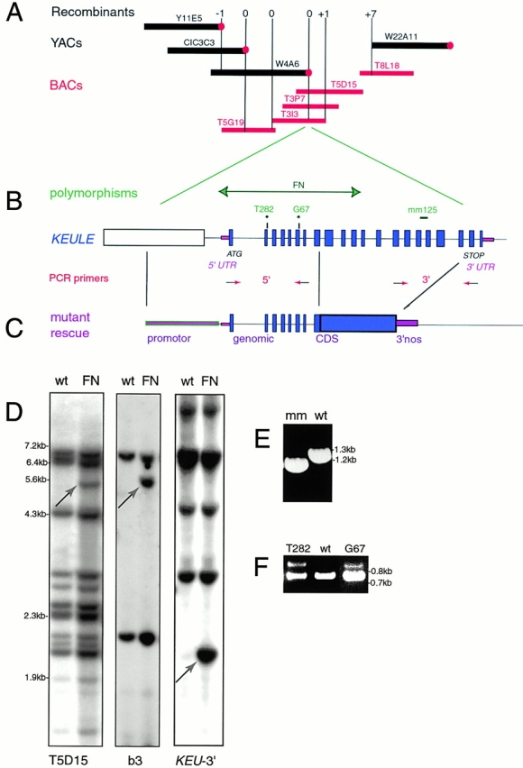
Cloning and molecular characterization of KEULE. (A) Selected portion of the chromosome walk spanning the KEULE region. The BACs (red bars) span the gap in the YAC contig (black bars), which was otherwise only covered by the large (600–800 kb) CIC YACS CIC12A9, CIC12H10, and CIC9G11. The vertical, purple lines represent RFLP markers used for mapping and correspond to polymorphic YAC or BAC ends (filled circles represent left ends of YACs). The numbers above the bars represent the number of recombinants between the marker and keule. The −1 to +1 recombination interval corresponds to 130 kb. Y, YUP; EW, Eric Ward YACs. All BACs are from the Texas A&M University library. Not to scale. (B) Structure of the KEULE locus. The KEULE gene spans 5 kb and includes 21 exons (filled, blue boxes) and 20 introns. The upstream gene (b3 cDNA, below) is represented as an open rectangle. The intergenic region is very small (only 352 bp). Mutations in four alleles are indicated. The two breakpoints in the fast-neutron allele lie in the intergenic region, and in the middle of the KEULE gene. The EMS-induced T282 and G67 mutations lie at intron/exon junctions. MM125 is a small x-ray–induced 156-bp deletion which spans 72 bp of coding and 72 bp of intron sequences. Arrows with red arrowheads represent primers used for amplifying the 5′ and 3′ ends of the coding region. (C) A construct sufficient for mutant rescue consists of genomic sequences fused to cDNA sequences as shown. (D) Fast-neutron–induced sequence polymorphisms at the KEULE locus. Southern blots of wild-type Lansdberg DNA (wt) or of DNA heterozygous for fast-neutron (FN)-induced keule mutations were probed with the BAC T5D15 (left) with the adjacent cDNA (b3, middle) or with a PCR product corresponding to the 3′ end of the KEULE gene (right). Dra1 polymorphisms (green-headed arrows) are shown. The 3′ end of the KEULE gene detects a Dra1 polymorphism distinct from the one detected by b3. (E) PCR analysis of genomic DNA of seedlings homozygous for the MM125 x-ray–induced allele of KEULE detects a deletion at the 3′ end of the coding sequences (3′ PCR primers used). (F) RT-PCR analysis of heterozygous mutant seedlings from the EMS-induced alleles T282 and G67 reveals a small increase in exon length (98 and 85 bp expected, respectively) at the 5′ end of the coding sequences.
Antibody Generation
A fusion containing the full length KEULE gene tagged NH2-terminally to six His residues in the BamHI site of pET15b (Novagen) was constructed using the same primers as for the P35S:GFP-KEULE construct. High levels of overexpression of the full length KEULE protein (50 mg/ml) were obtained in Escherichia coli (BL21/DE3). The insoluble protein was purified by isolation of the inclusion bodies and nickel affinity chromatography under denaturing conditions (TB055 manual; Novagen), followed by gel filtration chromatography. Chickens were inoculated and boosted twice with denatured and in vitro–renatured protein, then boosted four additional times with acrylamide gel slices containing the KEULE protein (courtesy of Dr. M. Erhard, Department of Veterinary Sciences, Ludwig Maximillians University, Munich, Germany). IgG was purified from egg yolks by diluting 10-fold in TBS, pH 7.3, centrifugation (15 min, 5,000 rpm, 4°C), addition to the supernatant of 0.4 ml dextran sulfate per egg yolk, followed (after 10 min) by 1 ml 1 M CaCl2 per gram of egg yolk, centrifugation, dialysis of the supernatant against TBS, pH 7.3, selective precipitation of IgG by addition of 26 g ammonium sulfate per 100-ml preparation, centrifugation, resuspension of the IgG pellet in TBS and extensive dialysis against TBS, and PBS. We affinity purified the antibodies over a column (AminoLink; Pierce Chemical Company) to which purified inclusion bodies of full length KEULE had been coupled (according to Pierce Chemical Company's AminoLink instructions, but in the presence of 6 M guanidine hydrochloride).
Peptide antibodies were generated in rabbits against peptides encompassing amino acids 470–490 of KEULE (peptide CSLLGSAVDAKKNTPGGFTL) and amino acids 500–527 of the KEULE homologue AtSec1a (peptide CMNQSSHKEESEARTGSVRK). The peptides (synthesized by the Protein and Nucleic Acid facility) were conjugated to malemide-activated KLH by virtue of an NH2-terminal cysteine residue for injection (CoCalico). After four to five boosts, the antiserum was affinity purified over columns in which the peptides were conjugated to short linkers via their free SH groups (SulfoLink; Pierce Chemical Company).
Freund's adjuvant was used for all injections. Initial inoculations were carried out with 200–400 μg protein/peptide, and boosts with ∼100 μg protein/peptide. The specific antibodies were eluted off the column in two successive rounds using the high salt gentle elute buffer from Pierce Chemical Company followed by 6 M guanidine HCl. All antibodies were tested on Western blots for specificity and sensitivity.
Cofillin and protein disulfide isomerase rabbit antibodies used as load controls are commercially available from Rose Biotechnology (RB10101 and RB10109). The KNOLLE antibody has been described elsewhere (Lauber et al. 1997).
For immunoprecipitation using the chicken antibody, we coupled the dialyzed guanidine–eluted fraction to AminoLink beads (Pierce Chemical Company) and incubated these with root tip extracts prepared in HKE buffer as described below for the syntaxin binding assays.
Western Analysis
Standard methods were used for Western analysis (Sambrook et al. 1989). 12.5% SDS-PAGE gels were blotted onto PVF Immobilon-P membranes (Millipore) by tank transfer. Primary antibodies were as described above. Secondary antibodies were HRP-conjugated goat anti–rabbit (Bio-Rad Laboratories) or rabbit anti–chicken (Pierce Chemical Company). The Supersignal West Pico chemiluminescent substrate (Pierce Chemical Company) was used for detection with x-ray film. Exposure times varied from 1 min to overnight, as the KEULE protein is not abundant. Protein extracts for gel electrophoresis were prepared by homogenizing fresh tissue in an equal volume (wt/vol) of 2× sample buffer (final protein concentration ∼10 mg/ml). For root samples, plants were grown in liquid culture (MS salts, 2% sucrose, B5 vitamins). Roots were disected to separate the tips (dividing) from the lengths (elongating).
Cell Fractionation and Membrane Association
Fractionation experiments were carried out in HKE buffer (50 mM Hepes-KOH, pH 7.5, 10 mM potassium acetate, 1 mM EDTA, 0.4 M sucrose, 1 mM DTT, 0.1 mM PMSF, proteinase inhibitor cocktail for plants [P-9599; Sigma-Aldrich]) according to Lauber et al. 1997. For solubilization, the rinsed P100K pellets were resuspended in HKE buffer by sonication; membrane clumps were removed by centrifugation at 10,000 g and a single sample was divided into four tubes. These tubes were incubated with 3 M sodium chloride, 0.1 M sodium carbonate, pH 10.9 or 11.5, 2% SDS or water as a negative control. After 1–3 h on ice with occasional vortexing, the fractions were again pelletted for 1 h at 100,000 g. In some cases, proteins in the supernatant were concentrated by deoxycholate/TCA precipitation. In brief, deoxycholate was added to the samples at a final concentration of 100 μg/ml. After 30 min on ice, a standard TCA precipitation was carried out.
Syntaxin Binding
We have fused a truncated version of the KNOLLE protein, lacking the COOH-terminal membrane anchor, to the glutathione S-transferase (GST) protein and to the 11–amino acid T7 tag; both tags fused NH2-terminally. For GST-KNOLLE we amplified the insert from KNOLLE cDNA with primer pairs CGGGATCCAATCGTTTATGAGTTAC and GGACCCGGGGCTGTTTCTCTGATGACTC (annealing 61°C), and introduced it in between the BamHI and SmaI sites of pGex3X (Promega). T7-KNOLLE was constructed in pET17b (Novagen) using the following primer pair: GGGGATCCGATGAACGACTTGATGACG and CCGCTCGAGCTAGCTGTTTCTCTGATGACTC (65 °C annealing, amplification from cDNA, PCR product and vector digested with BamHI and XhoI). The fusion proteins were overexpressed in the bacterial strain BL21/DE3 (∼10 and 50 mg/ml were achieved for GST-KNOLLE and T7-KNOLLE, respectively). In vitro binding assays were carried out essentially as described by the manufacturer (manual TB125; Novagen), with the modification that the plant extracts were prepared in HKE buffer (see Cell Fractionation above). In brief, 2–10 ml bacterial cultures containing ∼100 μg of fusion protein was incubated with 50 μl T7 antibody–agarose beads (Novagen). 1 ml plant extract (prepared from 0.5 g tissue, protein concentration ∼10 mg/ml) was homogenized in HKE buffer (see above), debris pelleted by centrifugation at 1,000 g, and the supernatant was supplemented with 1% Triton X-100 to solubilize membranes. The plant extracts were incubated with the loaded beads in batch for 1 h at room temperature. The beads were washed extensively in the same buffer and boiled for 5 min in 100 μl of Laemmli sample buffer to elute the bound proteins. 10-μl aliquots of the samples were loaded onto 12.5% SDS-PAGE gels, transferred to nylon membranes, and subjected to Western analysis.
Results
Positional Cloning of KEULE
A map-based approach was taken to clone the KEULE gene, which had previously been rough-mapped to the left arm of chromosome I (Assaad et al. 1996). We established a PCR-based alternative to RFLP mapping and screened a segregating population of 1,000 plants for recombinants in the vicinity of KEULE. By chromosome walking, we generated a YAC contig comprising >24 clones and spanning >1,500 kb or 2 cM, as well as a BAC contig to bridge the gap in the relevant interval of the YAC contig (Fig. 2 A). As the recombination frequency around the KEULE gene was 10 times lower than the genomic average, we were not able to narrow down the interval containing the gene further than 130 kb, in spite of the large mapping population (Fig. 2 A). The approaches taken to identify KEULE in 130 kb of unsequenced, overlapping BAC clones were cDNA library screening combined with a search for allele-specific polymorphisms. We identified 10 genes, including a Sec1 homologue, by cDNA library screens (Assaad, F., and Y. Huet, unpublished observations). Similarly, using whole BACs to probe a fast-neutron–induced allele of KEULE, we detected two visible polymorphisms which mapped in the vicinity of the Sec1 homologue (Fig. 2 D).
To confirm the coidentity of the Sec1 gene and KEULE, we looked for sequence polymorphisms between the wild-type and mutant alleles at this locus and carried out complementation analysis. The fast-neutron–induced allele of keule has a complex rearrangement (probably an inversion) with a breakpoint in the middle of the Sec1 homologue and a second breakpoint upstream of the gene (Fig. 2 D). An x-ray–induced keule allele harbors a 156-bp deletion in the 3′ region of the gene (Fig. 2 E), encompassing 78 bp of coding sequences and a small intron. We found that two EMS-induced alleles had (G to A) point mutations which inactivated splice sites at different introns in the 5′ region of the coding sequence. That unspliced introns in fact remain in transcripts from these alleles was confirmed by RT-PCR (Fig. 2 F). A construct consisting of the KEULE genomic stretch, spanning a region from 1-kb upstream to the first third of the gene, fused to the cDNA and 3′nos elements (Fig. 2 D) complements keule mutants. Thus, KEULE encodes a Sec1 homologue.
Molecular Characterization of the KEULE Gene
We compared the sequences of six KEULE cDNAs with 7 kb genomic DNA encompassing the gene and the upstream region. Sequence analysis with web-based tools (Netstart, which predicts start sites for transcription) suggested that two cDNAs are full length and that the KEULE gene spans a genomic interval of 5 kb, including 20 introns (Fig. 2 D). Upstream of KEULE, the intergenic region is only 352 nucleotides long. The predicted transcription unit is 2.4 kb in length and encodes a protein of 667 amino acids. This predicted size was confirmed by Northern analysis and RT-PCR (Fig. 2 F and not shown), as well as Western analysis (see Fig. 5 A).
Figure 5.
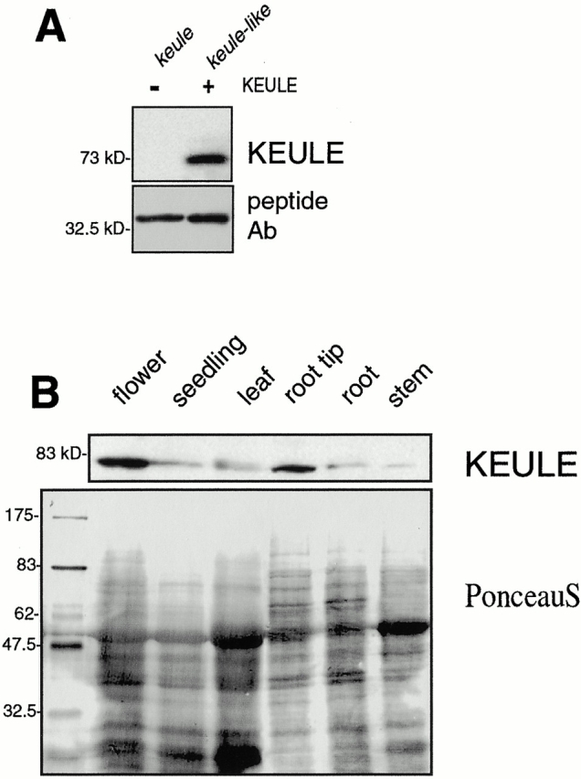
KEULE antibody and expression of the KEULE protein visualized by Western analysis. (A) The peptide antibody specifically recognizes a 73-kD band in keule-like mutants (described in Materials and Methods) but no band of the size of KEULE in keule mutants. Bottom panel shows the dominant contaminating band revealed by this peptide antibody. Both lanes are loaded with 50 mutant seedlings (left, fast-neutron–induced keule allele; right, cytokinesis-defective line G235) homogenized in 10 μl of sample buffer. (B) KEULE is expressed throughout the plant (as seen after longer exposures of the blot; expression in root shown in Fig. 7 B and in seedling in Fig. 8 C) and appears enriched in dividing tissues of Arabidopsis, namely the root tips and inflorescence meristems, designated “flower” in the figure. The membrane was stained with Ponceau S to monitor loading.
Northern analysis of mRNA from flower buds, leaves, root lengths, and root tips suggests that KEULE is expressed in all plant tissues tested, be they mostly dividing (flower buds, root tips), elongating (roots), or quiescent (leaves) (not shown). Thus, in contrast to KNOLLE, which is expressed only in mitotically active but not quiescent cells, KEULE is expressed throughout the plant.
The KEULE gene is 28–30% identical (49–50% similar) to worm (unc18), fly (Rop), and mammalian (munc18/n-Sec1) Sec1 homologues (Rop shown in Fig. 3). The homology is spread over the entire coding region. The Sec1 “signature,” as defined by Halachmi and Lev 1996, consists of 23 highly conserved residues (boxed in Fig. 3). KEULE contains 21 of these 23 residues (Fig. 3). One of the nonidentical residues has been replaced by a conserved residue. The KEULE protein appears hydrophilic, as expected for a Sec1. The nature of the fast-neutron– and x-ray–induced mutations described above and the observation that all 19 alleles of keule have very comparable phenotypes are a strong indication that we have identified null alleles of keule.
Figure 3.
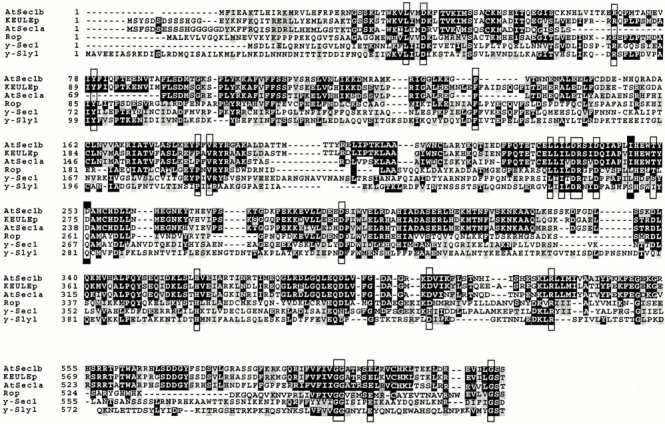
Sequence alignment of the KEULE protein. KEULE has two closely related homologues in Arabidopsis, AtSec1a and AtSec1b. These three proteins are clearly much closer to each other than to their homologues in other organisms. The Sec1 signature was defined before the publication of plant Sec1s as consisting of 23 highly conserved residues, boxed in this figure. Note that KEULE contains 21 of these 23 residues. The two residues that do not fit in this signature are identical in all three Arabidopsis Sec1s and in one instance, they are replaced by a conserved amino acid (I in lieu of L, KEULE amino acid 269). Rop, Drosophila nSec1 homologue; Sly1, yeast Sec1 homologue involved in ER-Golgi transport; accession numbers are to be found in Halachmi and Lev 1996. Plant Sec1 homologue sequence data are available from GenBank/EMBL/DDBJ under accession nos.: KEULE, AF331066; AtSec1a, AF335539; AtSec1b, CAB40953. Note that we do not show the entire proteins but only the domains with higher degrees of conservation. Black boxes highlight identical residues and gray boxes shade conserved residues.
BLAST searches with the KEULE coding sequence revealed two conserved homologues in the Arabidopsis genome: KEULE is 61% identical to AtSec1a and 65% identical to AtSec1b at the protein level (Fig. 3). Phylogenetic analysis shows that these three Sec1s cluster in a group of their own and have no true orthologue, even though their closest homologues are Sec1s required for exocytosis in fly, worm, and mammalian cells (Fig. 3; Sanderfoot et al. 2000). As the closest homologues of KEULE in yeast are Sly1p (22% identical; Dascher et al. 1991) and Sec1p (20% identical; Carr et al. 1999) (Fig. 3), we expressed KEULE in these mutant backgrounds under transcriptional control of an inducible promoter (see Materials and Methods). Although expression of the KEULE protein was detected by Western analysis in these yeast strains, KEULE failed to complement the yeast Sec1 and Sly1 mutants (see Materials and Methods).
keule-like Cytokinesis-defective Sectors in GFP-KEULE Transgenics
GFP fusions (NH2- and COOH-terminal) of the KEULE full length sequence under the transcriptional control of the CaMV 35S promoter (a strong, fairly constitutive promoter) were introduced into wild-type Arabidopsis plants. The gene fusions failed to rescue the mutant phenotype (see Materials and Methods) and we therefore do not present the GFP fluorescence here as this was weakly and/or erroneously expressed and has unfortunately not enabled us to localize the KEULE protein. Interestingly, cytokinesis-defective mutant sectors (such as depicted in Fig. 4 D) appeared on a small fraction (∼6–7%, or 40 affected individuals in a population of ∼600 plants) of the wild-type transformants (with the NH2-terminal fusion). These were seen on all somatic organs of the plants including petals and sepals (Fig. 4D and Fig. F) as well as the apical meristem (Fig. 4 E) but not on the carpels, siliques, or stamens (Fig. 4 D). Cellular analysis of these sectors revealed the presence of large, bloated cells (Fig. 4 F, confocal analysis not shown) characteristic of keule mutants (Fig. 4A and Fig. B). Sectored plants exhibited striking clusters or rosettes of siliques (Fig. 4 G) which may in part reflect the differential effect of the transgene on somatic versus reproductive organs. Sectored flowers similar to those shown in Fig. 4 D were also observed with the COOH-terminal GFP fusion (data not shown).
Figure 4.
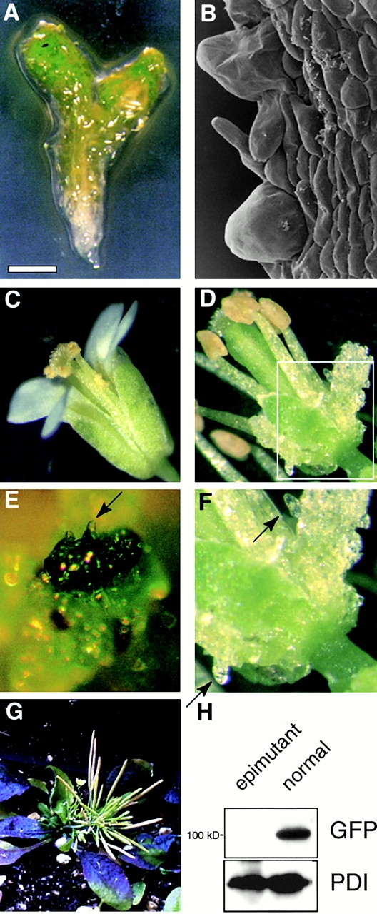
KEULE is required for cytokinesis in somatic cells throughout the plant life cycle. (A) keule seedling, with its characteristic bloated surface. (B) Scanning electron micrograph of a keule seedling (hypocotyl), showing the bloated cells at the surface layer. (C) Wild-type flower. D–G are epimutations at the keule locus due most likely to cosuppression in transformants harboring a P35S::GFP-KEULE gene fusion. (D) Sectors are seen on petals and sepals, but not on the carpels or stamens. (E) Sector taking over the entire apical meristem. (F) Bloated, keule-like surface cells (arrows) in somatic sector boxed in D. (G) Lines with large numbers of mutant sectors (such as E) matured as a cluster or large rosette of fertile silliques. (H) Using an anti-GFP antibody, we fail to detect the GFP-KEULE fusion protein in cytokinesis-defective sectored plants (epimutant), but do see it in nonsectored siblings (normal). Anti-protein disulfide isomerase (PDI) was used as a loading control. Bar: (A) 300 μm; (B) 45 μm; (E) 200 μm; (F) 170 μm.
Monitoring GFP fluorescence as well as protein levels by Western analysis showed that the endogenous KEULE protein and the GFP fusion protein were downregulated in cytokinesis-defective sectors, but not in corresponding tissues from normal siblings (Fig. 4 H shows the fusion protein; endogenous KEULE and GFP fluorescence not shown). These observations, as well as the incidence of somatic sectors rather than a uniform phenotype affecting the whole plant, are consistent with the interpretation that the keule-like defects are due to cosuppression (a phenomenon whereby the expression of both a transgene and its endogenous homologues is suppressed, see Assaad et al. 1993 and references therein) rather than overexpression or dominant negative effects. The sectors indicate that KEULE, or a closely related homologue, is required not only during embryogenesis but for cytokinesis in somatic cells throughout the plant life cycle.
Generation of KEULE-specific Antibodies and KEULE Expression
As KEULE is a member of a conserved gene family in Arabidopsis, generating specific antibodies has only been possible by using peptides as antigens. We generated and affinity purified a rabbit antibody against a synthetic peptide specific to KEULE (see Materials and Methods). This antibody recognizes a 73-kD band which is specific to KEULE, as evidenced by the observation that it is present in cytokinesis-defective mutants nonallelic to keule (designated keule-like in Fig. 5 A), but never in null keule mutants (Fig. 5 A), even after overexposure of the blots. This KEULE-specific band is of the predicted size based on the coding sequence. However, there are two contaminating lower molecular bands revealed by this peptide antibody (Fig. 5 A, lower panel shows the dominant one). These have precluded the use of the antibody in immunoprecipitation or immunolocalization experiments, but have not hindered Western analysis. As judged by comparing extracts from keule mutants, transgenic plants carrying epitope-tagged KEULE, and wild-type plants, KEULE can migrate anomalously at ∼100 kD or higher in SDS-PAGE gels (an example is given in Fig. 6 A). This is especially pronounced when reductant levels are <100 mM DTT and may be due to intramolecular (or possibly even intermolecular) disulfide bridges.
Figure 6.
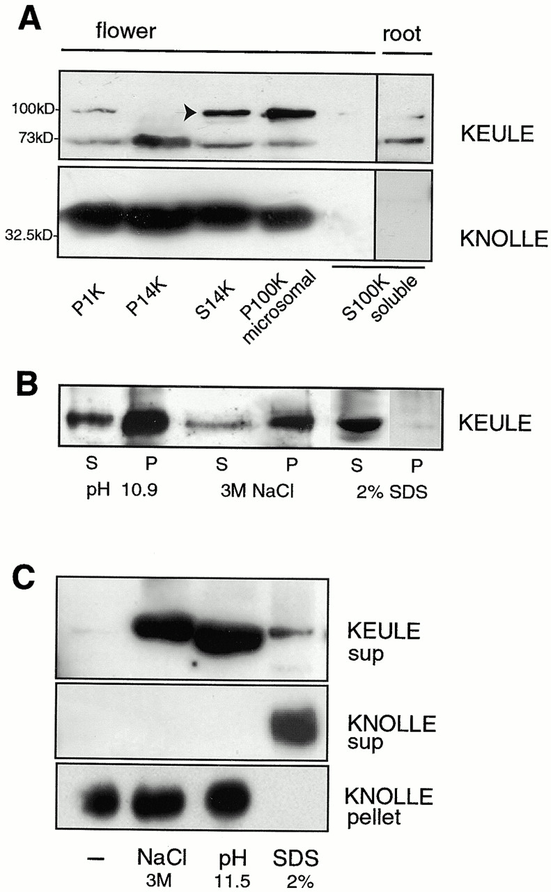
KEULE is peripherally associated with membranes. (A) KEULE appears to be membrane associated: KEULE is present in both the heavy membrane (P14K) and microsomal (P100) fractions. Longer exposure of the blot shows that KEULE is found in the soluble fraction as well. KEULE is shown to be in the soluble fraction of roots (right). As a control, the syntaxin KNOLLE is shown to be present in the membrane but not the soluble (S100K) fractions (bottom). P, pellet; S, supernatant. Note: not equiloaded, the membrane fractions are fivefold more concentrated than the soluble fractions. The arrow points to the KEULE-specific band which migrates anomalously at 100 kD in this sample (run with <100 mM DTT). It is not clear why this band is absent in the P14K fraction. (B and C) Membrane association is peripheral. KEULE can be released from the microsomes (P100K) if these are incubated with high salt (3 M NaCl), if the pH is increased (0.1 M NaCO3, pH 10.9–11.5), or with 2% SDS. (B) Solubilzation is complete with 2% SDS but only partial with high salt and high pH. (C) KEULE and KNOLLE differ with respect to the nature of their membrane association. In contrast to KEULE, KNOLLE was released from the microsomal fraction by SDS but not by high salt or high pH. (Although it appears that less KEULE protein is released from membranes by SDS than by high salt or high pH, this is most likely an artefact due to incomplete deoxycholate/TCA precipitation in the presence of SDS; see Materials and Methods for sample preparation. As in B, the majority of the KEULE protein remains in the pellets; not shown as grossly overexposed.) sup, supernatant.
Western analysis shows that KEULE is expressed in all tissues (Fig. 5 B). In addition, KEULE appears enriched in dividing tissues (root tips and inflorescence meristems or “flowers”; Fig. 5 B), consistent with its cytokinesis-defective phenotype. Although both Northern and Western analyses suggest that KEULE is expressed throughout the plant, the apparent enrichment observed in dividing tissues is observed at the protein level but not at the level of transcription.
KEULE Appears To Be Peripherally Associated with Membranes
KEULE is present in both the membrane and cytosolic fractions, as are other Sec1 proteins (Halachmi and Lev 1996). In cell fractionation experiments, the heavy membranes, including the Golgi and mitochondria, pellet at 14,000 g and the microsomes, including vesicles, pellet at 100,000 g. KEULE is present in both the heavy and light membrane fractions (Fig. 6 A) and is also found in the soluble fraction (the supernatant after a 100,000 g spin; Fig. 6 A). The KNOLLE antibody was used to control for the purity of the fractions. As shown previously (Lauber et al. 1997), KNOLLE is exclusively in the membrane fractions (Fig. 6 A, bottom). KEULE has been found in both the soluble and membrane fractions of all the tissues tested, including seedlings, roots, stems, leaves, and flowers.
As expected for Sec1s, which are membrane associated by virtue of interactions with membrane proteins, KEULE can be released from the microsomal fraction by treatments, such as high salt (3 M Nacl) and high pH (pH 10.9 or 11.5), that disrupt protein–protein interactions without solubilizing the membranes (Fig. 6B and Fig. C). By contrast, the membrane-anchored syntaxin KNOLLE is only released into the soluble fraction by treatments which disrupt membranes (2% SDS; Fig. 6 C). As shown in Fig. 6 B, KEULE is fully solubilized by 2% SDS but only partially solubilized by 3 M NaCl and high pH, the majority of the protein remaining in the pellet or membrane fraction. This is consistent with KEULE having a stable, high affinity association with its membrane receptor, as suggested for its homologues r-VPS45 and AtVPS45 (Bock et al. 1997; Bassham and Raikhel 1998).
Testing for a KEULE–KNOLLE Interaction
To test whether KEULE behaves like other SecI proteins, we examined its ability to bind syntaxins. Candidates for interacting partners are KNOLLE, a cytokinesis-specific syntaxin (Lukowitz et al. 1996; Lauber et al. 1997), as well as its closely related homologues, of which there are eight in the Arabidopsis genome (Sanderfoot et al. 2000). The KNOLLE gene was modified by the addition of a sequence corresponding to the 11–amino acid T7 tag (Novagen). This small epitope has a high affinity for the T7 monoclonal antibody, commercially available in a bead-bound form (α-T7 agarose beads). Recombinant protein produced in E. coli was bound to α-T7 agarose beads, which were incubated with plant extracts from root, flower, and leaf tissues. Bead-bound proteins were subjected to Western analysis. To control for nonspecific binding of proteins from the E.coli and plant extracts to the α-T7 agarose beads, we used a GST-KNOLLE protein fusion in lieu of the T7-KNOLLE fusion. This negative control leaves the active site of the T7 antibody available for non-specific interactions and was therefore chosen over a vector/tag only control. As seen in the Coomassie-stained samples (Fig. 7 A), T7-KNOLLE but not GST-KNOLLE binds the α-T7 agarose beads. The peptide antibody specific to KEULE detects KEULE in the T7-KNOLLE pull down lanes but not in the negative control (Fig. 7 B, bottom). This shows that native KEULE from plant extracts is capable of binding T7-KNOLLE–loaded beads in these assays. Formally, this binding could be indirect, mediated by other proteins from the bacterial or plant extracts. However, as Sec1 proteins are known to bind syntaxins, we are most likely witnessing a direct interaction.
Figure 7.
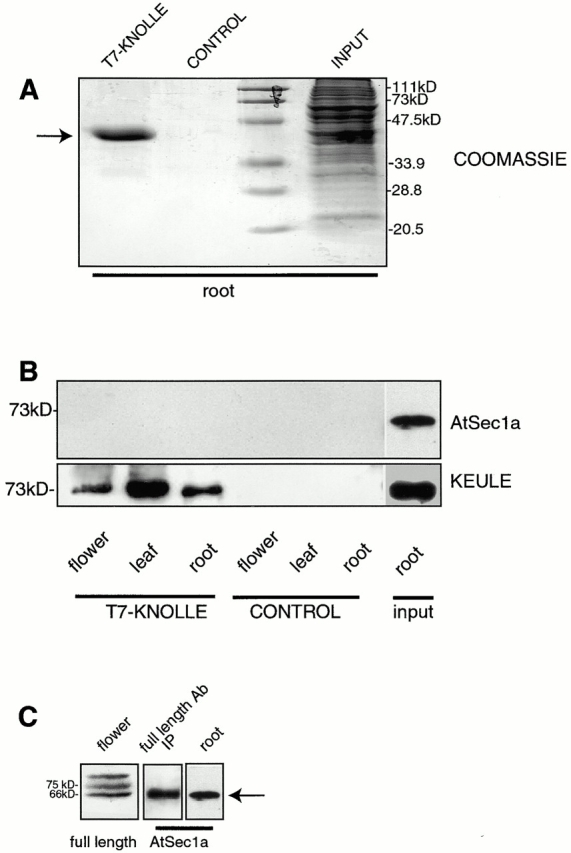
KEULE and KNOLLE interact. T7-KNOLLE was bacterially overexpressed and bound to α-T7 agarose beads. In the control lanes, bacterially expressed GST-KNOLLE was used in lieu of T7-KNOLLE. The loaded beads were incubated with protein extracts from flowers, leaves, and root lengths. 10% of the bead-bound proteins (T7-KNOLLE, CONTROL) or 1% of the plant extracts (INPUT) are loaded. (A) Coomassie-stained gel of beads incubated with root extract, or of root extract. Arrow points to T7-KNOLLE. GST-KNOLLE fails to bind the α-T7 agarose beads. (B) Westerns were probed with a peptide antibody against the highly conserved KEULE homologue AtSec1a (top) and with the KEULE peptide antibody (bottom). Longer exposures of the upper panel reveal the AtSec1a band in all six lanes, with no differential behavior between the experiment and negative control. The input lanes are loaded with the root extracts used for this experiment. (C) Specificity of the AtSec1a antibody. The AtSec1a peptide antibody recognizes a band at the expected size, 66 kD (arrow), in plant extracts (root). To confirm that this indeed corresponds to AtSec1a, we show that the antibody cross-reacts with a 66-kD protein immunoprecipitated (IP) by an antibody raised against the full length KEULE protein (middle). As expected based on sequence analysis (see Fig. 3), the full length antibody reveals three bands in plant extracts; the lower one presumambly corresponds to AtSec1a (left).
To test for the specificity of this interaction, we investigated the ability of the KEULE homologue AtSec1a to bind KNOLLE in these assays. To this end, we generated a peptide antibody specific to AtSec1a. A band of the expected size at ∼66 kD is observed with this antibody (antibody specificity discussed in Fig. 7 C). Interestingly, the closely related KEULE homologue AtSec1a does not bind the T7-KNOLLE affinity column in the same series of assays (Fig. 7 B, top). This suggests that the KEULE–KNOLLE interaction is highly specific.
Is KEULE Cytokinesis Specific?
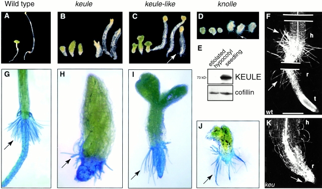
Given KEULE's gene identity, we considered the possibility that, in addition to its role in cytokinesis, KEULE might regulate other vesicle trafficking events in the plant cell. We investigated two processes that, like cytokinesis, require an extensive remobilization of resources mediated via membrane trafficking: cell elongation and root hair growth. A striking example of rapid cell elongation in plants occurs in etiolated seedlings: hypocotyls of seedlings germinated in the dark are roughly fivefold longer than light-germinated controls (Fig. 8 A). To test whether keule and other cytokinesis-defective mutants were capable of elongation, we germinated these in the dark. keule and keule-like mutants are capable of elongation (Fig. 8B and Fig. C), although not to the same extent as the wild-type (Fig. 8 A; compare up to roughly threefold elongation in the mutants with roughly fivefold elongation in the wild-type). Hypocotyl length is determined largely by cell expansion, but also by cell division, and the effect of keule and keule-like mutants on the extent of elongation is likely to be a (direct or indirect) consequence of the cytokinesis defect. In the case of knolle seedlings, which also appear capable of elongation (Fig. 8 C), this experiment is complicated by the severe germination defect of knolle mutants. We also tested whether KEULE is enriched in rapidly elongating cells as it is in dividing cells. We detected only low levels of KEULE expression in etiolated hypocotyls (Fig. 8 E). Similarly, KEULE is weakly expressed in the stems (mostly quiescent or elongating tissue, Fig. 5 B) and is present at higher levels in the dividing (root tip) than in the elongating moiety (root length) of the root (Fig. 5 B; see Materials and Methods for sample preparation). We conclude that KEULE is not absolutely required for cell elongation.
Figure 8.
Cell elongation and root hair growth in keule mutants. (A–D) Mutant seedlings were germinated in the light (left) or dark (right). keule (B), keule-like (C), and knolle (D) mutants are capable of elongation, but not to the same extent as wild-type (A). (E) KEULE is only weakly expressed in etiolated hypocotyls. Anticofillin antibody was used as a load control. (F–K) Root hairs are absent (K) or stunted and radially swollen (H) in keule mutants, but of normal length in other cytokinesis-defective mutants. Root hairs are stained with methylene blue. Each keule allele exhibits a range of phenotypes, though the range may differ in a given allele. In contrast to keule mutants, keule-like (I) and knolle (J) mutants grow long root hairs. (F and K) Clearing preparation of wild-type (F) and keule (K) seedling. Note that the basal portion of keule seedlings have root-like characteristics. Arrows point to root hairs. h, hypocotyl; r, root; t, root tip. KEULE alleles: MM125 in B and H; G67 in K. Bars: (F) 270 μm; (K) 200 μm.
We also investigated the ability of keule mutant seedlings to grow root hairs (Fig. 8, F–K). In this respect, keule mutants have a striking (and hitherto unpublished) phenotype, namely stunted and radially swollen (Fig. 8 H) or absent root hairs (Fig. 8 K). This has been seen in all 19 alleles of KEULE. By contrast, other cytokinesis-defective mutants such as keule-like and knolle seedlings grow long root hairs (Fig. 8I and Fig. J). Thus, KEULE is required not only for cytokinesis but also for root hair development, which is known to arise via tip growth.
Discussion
We present numerous lines of evidence that KEULE is a Sec1 gene. These include fine mapping, allele-specific polymorphisms, complementation analysis, the presence of cytokinesis-defective sectors in wild-type transformants, as well as the absence of the predicted gene product in null mutants. This inferred gene identity readily accounts for the observed defects in vesicle fusion (Waizenegger et al. 2000) and cytokinesis (Assaad et al. 1996) in keule mutants.
To determine whether the sequence similarity between KEULE and Sec1 genes translates into functional homology, we tested KEULE's properties in vivo and in vitro. KEULE is characteristic of a Sec1 protein in all respects. It exists in two forms, soluble and membrane associated. It is peripherally associated with membranes, being released from membranes by treatments which disrupt protein–protein interactions. Finally, KEULE acts as a syntaxin-binding protein in in vitro binding assays.
Sec1 proteins are thought to regulate syntaxins both positively and negatively, although the latter is controversial and may vary for different Sec1 proteins (see below). The observation that vesicles accumulate but do not fuse at the equator of dividing cells in keule and knolle embryos (Waizenegger et al. 2000) indicates that, like the syntaxin KNOLLE, KEULE is required for vesicle fusion and plays a positive role in this process. Whether or not it also plays a negative role in regulating vesicle fusion remains to be determined.
KEULE and KNOLLE Interact
To date, there are four lines of evidence that KEULE and KNOLLE interact. First, the genes mutate to the same phenotype (Assaad et al. 1996; Lukowitz et al. 1996). Second, the gene identities are in and of themselves suggestive of an interaction as Sec1s, and syntaxins are known to form tight complexes. Third, in this study we have been able to demonstrate that the proteins bind each other in in vitro binding assays. Finally, double mutant analysis has provided evidence that the two genes interact in vivo (Waizenegger et al. 2000). Although cytokinesis is impaired but not blocked in keule and knolle mutants, which survive till the seedling stage, it is completely abolished in knolle keule double mutants which die as large, single celled multinucleate embryos (Waizenegger et al. 2000). The synthetic lethality (or greater than additive enhancement) of the knolle keule double mutant phenotype provides genetic evidence that KEULE and KNOLLE are involved in the same process (Waizenegger et al. 2000). Although synthetic lethality implies that two gene products act concertedly, it does not necessarily point to a direct interaction.
A direct regulation of KNOLLE by KEULE would account for the synthetic lethality of the knolle keule double mutant phenotype. However, the double mutant phenotype is also an indication that the two genes have additional functions independently of each other. Indeed, if KEULE's sole function were to regulate KNOLLE, one would not expect the knolle keule double mutants to be any different from knolle null mutants. In other terms, one would expect epistasis rather than synthetic lethality. As mentioned below, we expect KEULE to interact not only with KNOLLE but with other syntaxins as well. As a Sec1 protein, KEULE is also likely to have a variety of interaction partners other than syntaxins (see below). Similarly, syntaxins have a large number of interacting partners other than Sec1 proteins, such as v-SNARES, SNAP25, and NSF (see below). Thus, the double mutant phenotype is consistent with KEULE and KNOLLE having a direct interaction in addition to several interactions with other partners.
KEULE Is a Member of a Gene Family
BLAST searches of the Arabidopsis genome with the KEULE sequence revealed the presence of two highly conserved homologues. A certain degree of functional redundancy between KEULE and its Arabidopsis homologues would best explain the attenuated nature of the keule mutant phenotype, in particular when this is contrasted to the extreme phenotype of the knolle keule double mutant. This redundancy could only be partial, as the KEULE gene mutates to seedling lethality. Furthermore, KEULE and its highly conserved homologue, AtSec1a, behave differentially with respect to their ability to bind KNOLLE. It is thus interesting to note that although KEULE and its homologue AtSec1a are closely related, their functions appear to have diverged.
Although KEULE's closest homologues are required for exocytosis, phylogenetic analysis places KEULE and its homologues in a class of their own. Consistently, KEULE failed to rescue the yeast Sec1 and Sly1 mutants. Whereas there are only single counterparts to the Sec1 genes SlyI, VPS45, and VPS33 in plants (Sanderfoot et al. 2000), there are three members in the KEULE family. Similarly, KNOLLE belongs to the largest subfamily of syntaxins in Arabidopsis, with eight closely related homologues (Sanderfoot et al. 2000), but only weak homology to yeast and animal syntaxins (Lukowitz et al. 1996). It thus appears that Arabidopsis has several novel vesicle trafficking genes that may fulfill diverse but as yet largely undetermined functions (Sanderfoot et al. 2000).
KEULE Is Required for Cytokinesis throughout the Plant Life Cycle
As KEULE mutates to seedling lethality (Assaad et al. 1996), it has been difficult to ascertain its role in the plants' life cycle past the seedling stage. To this effect, the cytokinesis-defective somatic sectors observed on wild-type transformants are highly informative. Such sectors were observed on all the somatic tissues, but not on the reproductive organs, namely the siliques, carpels, and stamens. These results suggest that KEULE (or a closely related homologue) is required not only during embryogenesis, but for cytokinesis in somatic cells throughout the plant life cycle. Formally, the absence of keule-like sectors in the reproductive organs cannot be taken to imply that KEULE is not required in these organs, as it could be due to the heterologous promotor used to drive KEULE expression in transgenics. However, it may be relevant that several plant genes such as stud, tetraspore, and sidecar pollen specifically affect cytokinesis during pollen development (for review see Heese et al. 1998) and that neither KEULE nor KNOLLE appears to be required during gametophytic development (Assaad et al. 1996; Lauber et al. 1997). Consistent with a role for KEULE after embryogenesis, we have been unable to regenerate keule mutant seedlings in tissue culture and have observed cytokinesis defects in the apical meristem of mutant seedlings (Assaad et al. 1996). In addition, KEULE transcripts are expressed in all tissues, including the reproductive organs, and the KEULE gene product is expressed throughout the plant, predominantly in adult meristematic tissues such as the root tip and flower buds.
Is KEULE Cytokinesis Specific?
Is KEULE cytokinesis specific, or is cytokinesis simply the most sensitive or visible process perturbed in the mutant? We had in previous analyses thus far failed to detect any role for KEULE other than in the execution of cytokinesis. Electron microscopy did not reveal any defects in the morphology of membrane systems such as the plastids, mitochondria, peroxisomes, ER, and Golgi in keule embryos (Assaad et al. 1996). The presence of a cuticle on KEULE embryos suggests that secretion is not affected (Assaad et al. 1996). Surface expansion appears unaffected, as keule cells grow to large sizes. Finally, as mutation at the KEULE locus has no gametophytic effect (Assaad et al. 1996), pollen tip growth is unlikely to be affected. In this study, we have found that the KEULE protein appears to be enriched in dividing tissues and to interact with the cytokinesis-specific syntaxin KNOLLE. These data support a primary function for KEULE in the execution of cytokinesis.
We have furthered our phenotypic analysis of keule mutants by monitoring cell elongation and root hair development in mutant seedlings. These are events which, like cytokinesis, are mediated by vesicle trafficking. Cell elongation does not appear to be grossly affected in seedling sections and only low levels of KEULE protein were detected in elongating tissues such as stems, root lengths, and etiolated hypocotyls. Finally, keule seedlings were capable of elongation when germinated in the dark. We conclude that KEULE is not absolutely required for cell elongation.
By contrast, KEULE is required for root hair growth: root hairs are absent or stunted and radially swollen in keule mutants. This phenotype is not observed in knolle and keule-like mutants, which develop long root hairs, and is therefore unlikely to be a secondary consequence of a primary defect in cytokinesis. As the basal region of keule seedlings have root-like characteristics (Assaad et al. 1996), it is equally unlikely that this is due to developmental anomalies resulting from the cytokinesis defect. Furthermore, anomalies in cellular differentiation in keule mutants have been observed in discontinuous patches, but never in complete files of cells (Assaad et al. 1996). Root hairs grow rapidly by a process of tip growth. KEULE's potential role in this and related processes merits further attention.
A survey of the Arabidopsis genome revealed 6 Sec1 genes and 24 syntaxins (Sanderfoot et al. 2000). There is thus only one Sec1 gene for four syntaxins and it is to be expected that KEULE might interact not only with KNOLLE, but with additional syntaxins as well. A working model to be tested in future experiments is that, whereas some syntaxins such as KNOLLE may be specific in their functions, Sec1 proteins such as KEULE may specifically regulate a well-defined number of cellular processes by interacting with multiple syntaxins. Phenotypic analyses of keule and knolle mutants support this hypothesis.
The Role of Sec1 Proteins in Regulating Vesicle Fusion
Although the crystal structure of the nSec1–syntaxin 1a complex has been elucidated, the biological role of this complex as well as its relevance to other sec1–syntaxin interactions is unclear. Vesicle targeting and fusion require a specific “lock and key” interaction between syntaxins (t-SNAREs) on target membranes and v-SNAREs on vesicle membranes. SNARE interactions, however, are promiscuous rather than selective (Yang et al. 1999a) and the specificity of membrane fusion is ensured by key regulators such as Sec1 and Rab proteins. Based on studies of neuronal cells and molecules, it is thought that nSec1 binds syntaxins in a closed form and opens or activates these, thereby playing a positive role in vesicle fusion (Misura et al. 2000 and references therein). In addition, nSec1 is thought to negatively regulate vesicle fusion by sterically inhibiting core complex (syntaxin/v-SNARE/adapter four-helix bundle) formation. By contrast, the yeast Sec1 is thought to bind not the closed syntaxin but rather the open form bundled with other SNAREs in core complexes (Carr et al. 1999).
The crystal structure of the neuronal Sec1–syntaxin complex shows that the large Sec1 arcs around the smaller syntaxin, which fits snuggly into a central cavity (Misura et al. 2000). A large surface of the bound Sec1 protein would thereby be available for additional interactions. In fact, Sec1 proteins have been shown to interact in vitro and/or in vivo with a variety of proteins other than syntaxins, such as Rab effectors and MINTs. The Rab effector Vac1p is a long adapter protein that binds Rab, phosphatidylinositol, and the Sec1 protein VPS45 in yeast vacuolar targeting (Peterson et al. 1999). The Sec1-interacting MINT proteins form tripartite complexes which couple membrane traffic to adhesion molecules, membrane receptors, and channels at the neural synapse (Butz et al. 1998). In these examples, Sec1 proteins constitute key links between the membrane fusion machinery and Rab and phosphatidylinositol signaling, or between exocytosis and the functional asymmetry and development of neural synapses. The emerging picture is that Sec1 proteins are the receptors or sensors of various forms of inter- or intracellular signaling, and that they may regulate vesicle fusion, via their interaction with syntaxins, in response to these signals.
We have characterized a regulator of vesicle trafficking, namely a Sec1 gene, with a role in cytokinesis. Although Sec1 proteins and syntaxins have been shown to interact in a variety of organisms and cell types, the cell cycle context of this interaction in the case of KEULE and KNOLLE is novel. The cell cycle regulation of KEULE's association with membranes, of its interaction with KNOLLE, and of its intracellular localization will be of great interest.
The multinucleate phenotype characteristic of keule mutants indicates that new nuclear cycles are initiated even if cytokinesis has not been completed. By contrast, cell wall stubs are only observed in multinucleate cells and never in cells with a single nucleus (Assaad et al. 1996). Thus the onset of cytokinesis is, as predicted, coordinated with the completion of anaphase. As the execution of cytokinesis in plants is accomplished by vesicle trafficking, there must be a link between cell cycle progression and membrane trafficking. The molecular identity of this link remains to be determined. By analogy to the potential role of Sec1 proteins in yeast and animal cells, an intriguing possibility is that KEULE may integrate cell cycle signals and transduce them to the cytokinetic vesicle fusion machinery by virtue of an interaction with the syntaxin KNOLLE.
Acknowledgments
We are especially grateful to Heddy Bendjaballah for help with establishing the BAC contig and to Regine Kahmann for her support. Dr. Mikhael Erhard helped us generate chicken antibodies. The scanning electron micrograph was taken by Prof. Gehard Wanner and the knolle seedling image by Wolfgang Lukowitz. Gerti Glaesser histoligically characterized the cytokinesis-defective keule-like line. Ramon Torres Ruiz and Jeff Dangl provided the fast-neutron–induced allele of keule. Many thanks to a large number of colleagues, especially Chris Koch, Max Busch, Reinhardt Kunze, Dianne Bassham, Tony Sanderfoot, Richard Scheller, Martin Steegmaier, and all the members of the Sommerville lab for stimulating discussions and technical tips. Thanks to Chris Sommerville, Dave Ehrhardt, Maren Heese, Wolfgang Lukowitz, Jason Bock, Dario Bonetta, Sean Cutler, Dominique Bergman, Natasha Raikhel, and an anonymous reviewer for useful suggestions and/or critical evaluation of the manuscript. The Arabidopsis Ohio stock center provided YAC and BAC filters and clones, as well as cDNA libraries.
F. Assaad was supported by a long term EMBO fellowship followed by an Hochshulsonderprogramm III stipend from the University of Munich. This research was funded by a Leibniz award from the Deutsche Forschungsgemeinschaft and a European Union Biotechnology Program Framework IV grant to G. Jürgens, by a grant from the US Department of Energy (DE-FG02-00ER20133) to Chris Sommerville, and by Deutsche Forschungsgemeinschaft grant AS110/2-1 to F. Assaad.
Footnotes
Abbreviations used in this paper: BAC, bacterial artificial chromosome; CTAB, cetyltrimethylammonium bromide; GFP, green fluorescent protein; GST, glutathione S-transferase; RFLP, restriction fragment length polymorphism; RT, reverse transcription; YAC, yeast artificial chromosome.
References
- Assaad F.F., Tucker K.L., Signer E.R. Epigenetic repeat-induced gene silencing (RIGS) in Arabidopsis . Plant Mol. Biol. 1993;22:1067–1085. doi: 10.1007/BF00028978. [DOI] [PubMed] [Google Scholar]
- Assaad F., Mayer U., Wanner G., Jürgens G. The keule gene is involved in cytokinesis in Arabidopsis . Mol. Gen. Genet. 1996;253:267–277. doi: 10.1007/pl00008594. [DOI] [PubMed] [Google Scholar]
- Assaad F.F., Lukowitz W., Mayer U., Jürgens G. Cytokinesis in somatic plant cells. Plant Physiol. Biochem. 1997;35:175–182. [Google Scholar]
- Bassham D.C., Raikhel N.V. An Arabidopsis VPS45 homolog implicated in protein transport to the vacuole. Plant Physiol. 1998;117:407–415. doi: 10.1104/pp.117.2.407. [DOI] [PMC free article] [PubMed] [Google Scholar]
- Bock J.B., Klumperman J., Davanger S., Scheller R. Syntaxin 6 functions in trans-Golgi network vesicle trafficking. Mol. Biol. Cell. 1997;8:1261–1271. doi: 10.1091/mbc.8.7.1261. [DOI] [PMC free article] [PubMed] [Google Scholar]
- Butz S., Okamoto M., Südhof T. A tripartite protein complex with the potential to couple synaptic vesicle exocytosis to cell adhesion in brain. Cell. 1998;94:773–782. doi: 10.1016/s0092-8674(00)81736-5. [DOI] [PubMed] [Google Scholar]
- Carr C.M., Grote E., Munson M., Hughson F.M., Novick P.J. Sec1p binds to SNARE complexes and concentrates at sites of secretion. J. Cell Biol. 1999;146:333–344. doi: 10.1083/jcb.146.2.333. [DOI] [PMC free article] [PubMed] [Google Scholar]
- Choi S., Creelman R.A., Mullet J.E., Wing R.A. Construction and characterization of a bacterial artificial chromosome library of Arabidopsis thaliana . Plant Mol. Biol. Reporter. 1995;13:124–128. [Google Scholar]
- Cutler S.R., Ehrhardt D.W., Griffitts J.S., Sommerville C.R. Random GFP::cDNA fusions enable visualization of subcellular structures in cells of Arabidopsis at high frequency. Proc. Natl. Acad. Sci. USA. 2000;97:3718–3723. doi: 10.1073/pnas.97.7.3718. [DOI] [PMC free article] [PubMed] [Google Scholar]
- Dascher C., Ossig R., Gallwitz D., Schmitt H.D. Identification and structure of four yeast genes (SLY) that are able to suppress the functional loss of YPT1, a member of the Ras superfamily. Mol. Cell Biol. 1991;11:872–885. doi: 10.1128/mcb.11.2.872. [DOI] [PMC free article] [PubMed] [Google Scholar]
- Halachmi N., Lev Z. The Sec1 familya novel family of proteins involved in synaptic transmission and general secretion. J. Neurochem. 1996;66:889–897. doi: 10.1046/j.1471-4159.1996.66030889.x. [DOI] [PubMed] [Google Scholar]
- Heese M., Mayer U., Jürgens G. Cytokinesis in flowering plantscellular processes and developmental integration. Curr. Opin. Plant Biol. 1998;1:486–491. doi: 10.1016/s1369-5266(98)80040-x. [DOI] [PubMed] [Google Scholar]
- Jantsch-Plunger V., Glotzer M. Depletion of syntaxins in the early Caenorhabditis elegans embryo reveals a pole for membrane fusion events in cytokinesis. Curr. Biol. 1999;9:738–745. doi: 10.1016/s0960-9822(99)80333-9. [DOI] [PubMed] [Google Scholar]
- Lauber M.H., Waizenegger I., Steinmann T., Schwarz H., Mayer U., Hwang I., Lukowitz W., Jürgens G. The Arabidopsis KNOLLE Protein is a cytokinesis-specific syntaxin. J. Cell Biol. 1997;139:1485–1493. doi: 10.1083/jcb.139.6.1485. [DOI] [PMC free article] [PubMed] [Google Scholar]
- Leutwiler L.S., Hough-Evans B.R., Meyerowitz E.M. The DNA of Arabidopsis thaliana . Mol. Gen. Genet. 1984;194:15–23. [Google Scholar]
- Liu C., Johnson S., Wang T.L. Cyd, a mutant of pea that alters embryo morphology is defective in cytokinesis. Dev. Genet. 1995;16:321–331. [Google Scholar]
- Lukowitz W., Mayer U., Jürgens G. Cytokinesis in the Arabidopsis embryo involves the syntaxin-related KNOLLE gene product. Cell. 1996;84:61–71. doi: 10.1016/s0092-8674(00)80993-9. [DOI] [PubMed] [Google Scholar]
- Mascorro-Gallardo J.O., Covarrubias A.A., Gaxiola R. Construction of a CUP1 promoter-based vector to modulate gene expression in Saccharomyces cerevisiae . Gene. 1996;119:169–171. doi: 10.1016/0378-1119(96)00059-5. [DOI] [PubMed] [Google Scholar]
- Misura K.M.S., Scheller R.H., Weis W. Three-dimensional structure of the neuronal-Sec1-syntaxin 1a complex. Nature. 2000;404:355–362. doi: 10.1038/35006120. [DOI] [PubMed] [Google Scholar]
- Nebenführ A., Frohlick J.A., Staehelin L.A. Redistribution of Golgi stacks and other organelles during mitosis and cytokinesis in plant cells. Plant Physiol. 2000;124:135–151. doi: 10.1104/pp.124.1.135. [DOI] [PMC free article] [PubMed] [Google Scholar]
- Nickle T., Meinke D. A cytokinesis-defective mutant of Arabidopsis (cyt1) characterized by embryonic lethality, incomplete walls, and excessive callose accumulation. Plant J. 1998;15:321–332. doi: 10.1046/j.1365-313x.1998.00212.x. [DOI] [PubMed] [Google Scholar]
- Peterson M.R., Burd C.G., Emr S.D. Vac1p coordinates Rab and phosphatidylinositol 3-kinase signaling in Vps45p-dependent vesicle docking/fusion at the endosome. Curr. Biol. 1999;9:159–162. doi: 10.1016/s0960-9822(99)80071-2. [DOI] [PubMed] [Google Scholar]
- Sambrook J., Fritsch E.F., Maniatis T. Molecular CloningA Laboratory Manual. Cold Spring Harbor Press; Cold Spring Harbor, NY: 1989. [Google Scholar]
- Samuels A.L., Giddings T.H., Staehelin L.A. Cytokinesis in tobacco BY-2 and root tip cellsa new model of cell plate formation in higher plants. J. Cell Biol. 1995;130:1345–1357. doi: 10.1083/jcb.130.6.1345. [DOI] [PMC free article] [PubMed] [Google Scholar]
- Sanderfoot A.A., Assaad F.F., Raikhel N.V. The Arabidopsis thaliana genomean abundance of SNAREs. Plant Physiol. 2000;124:1558–1569. doi: 10.1104/pp.124.4.1558. [DOI] [PMC free article] [PubMed] [Google Scholar]
- Sherman F., Fink G.R., Hicks J.B. Laboratory Course Manual for Methods in Yeast Genetics. Cold Spring Harbor Press; Cold Spring Harbor, NY: 1986. [Google Scholar]
- Smith L.G. Divide and conquer, cytokinesis in plant cells. Curr. Opin. Plant Biol. 1999;2:447–453. doi: 10.1016/s1369-5266(99)00022-9. [DOI] [PubMed] [Google Scholar]
- Waizenegger I., Lukowitz W., Assaad F., Schwarz H., Jürgens G., Mayer U. The Arabidopsis KNOLLE and KEULE genes interact to promote vesicle fusion during cytokinesis. Curr. Biol. 2000;10:1371–1374. doi: 10.1016/s0960-9822(00)00775-2. [DOI] [PubMed] [Google Scholar]
- Weigel D., Alvarez J., Smyth D.R., Yanofsky M.F., Meyerowitz E.M. LEAFY controls meristem identity in Arabidopsis . Cell. 1992;69:843–859. doi: 10.1016/0092-8674(92)90295-n. [DOI] [PubMed] [Google Scholar]
- Yang B., Gonzalez L., Prekeris R., Steegmaier M., Advani R.J., Scheller R.H. SNARE interactions are not selectiveimplications for membrane fusion specificity J. Biol. Chem 274 1999. 5649 5653a [DOI] [PubMed] [Google Scholar]
- Yang M., Nadeau J.A., Zhao L.M., Sack F.D. Characterization of a cytokinesis defective (cyd1) mutant of Arabidopsis J. Exp. Bot 50 1999. 1437 1446b [DOI] [PubMed] [Google Scholar]
- Zuo J., Niu Q.W., Nishizawa N., Wu Y., Kost B., Chua N.H. Korrigan, an Arabidopsis endo-1,4-beta-glucanase localizes to the cell plate by polarized targeting and is essential for cytokinesis. Plant Cell. 2000;12:1137–1152. doi: 10.1105/tpc.12.7.1137. [DOI] [PMC free article] [PubMed] [Google Scholar]


