Abstract
Only mature B lymphocytes can enter the lymphoid follicles of spleen and lymph nodes and thus efficiently participate in the immune response. Mature, long-lived B lymphocytes derive from short-lived precursors generated in the bone marrow. We show that selection into the mature pool is an active process and takes place in the spleen. Two populations of splenic B cells were identified as precursors for mature B cells. Transitional B cells of type 1 (T1) are recent immigrants from the bone marrow. They develop into the transitional B cells of type 2 (T2), which are cycling and found exclusively in the primary follicles of the spleen. Mature B cells can be generated from T1 or T2 B cells.
Keywords: B cell development, transitional B cells, spleen, CD45, Bruton's tyrosine kinase
Mice with genetic deletions of elements participating in the B cell receptor signaling cascade display developmental arrest at the T1 or T2 stage. The analysis of these defects showed that the development of T2 and mature B cells from T1 precursors requires defined qualitative and quantitative signals derived from the B cell receptor and that the induction of longevity and maturation requires different signals.
In the adult mouse, B cells are generated in the bone marrow. Their development requires the successful rearrangement of the Ig H and L chain gene loci and the surface expression of the B cell antigen receptor (BCR)1 1.
The BCR is composed of the membrane-bound Ig molecule and the Ig-α/Ig-β heterodimer. The Ig-α/Ig-β heterodimer is indispensable for the surface expression and signaling function of the BCR 2. In the early phases of development in the bone marrow, B cells only express IgM. Immature B cells have a low density of IgM (IgMdull) and are still resident in the parenchyma of the bone marrow. They develop into IgMbright transitional B cells 3 and move toward and through the bone marrow sinusoids before migrating to the periphery 4. Few of the transitional B cells enter the mature, long-lived B cell compartment. Out of the 2 × 10⁷ IgM+ B cells that develop daily in the bone marrow of the mouse, 10% reach the spleen and only 1–3% enter the mature B cell pool 5 6. Mature B cells coexpress IgM and IgD 7. IgM+IgD+ B cells in the bone marrow are thought to be B cells that have completed their maturation process in the periphery and return to the bone marrow as mature recirculating B cells. In the normal mouse, only mature B cells are long lived, can recirculate with the lymph and the blood, and are able to enter the lymphoid follicles of spleen and lymph nodes. These abilities are very important to mount an efficient immune response. Only B cells that can enter the follicle have access to antigen deposited on the follicular dendritic cells and are able to initiate the germinal center reaction and then rapidly produce antigen-specific, high-affinity Abs.
The rules that regulate the size, diversity, and quality of the long-lived B cell pool are therefore important but largely unknown. It is, however, likely that signals from the BCR control these differentiation events 8 9 10 11. Indeed, mice with natural or targeted deletions or mutations of genes encoding elements involved in BCR signaling often show abnormal development of B cells and are therefore useful tools to study the molecular requirements for lymphocyte differentiation.
The deletion of the cytoplasmic part of Ig-α in mb1 Δc/Δc mice 12 still permits expression of the IgM-BCR on the cell surface, and B cell development in the bone marrow is compromised but not completely blocked. However, the mature B cell pool is absent in the periphery.
CD45 is a tyrosine phosphatase expressed in alternatively spliced forms on the surface of B and T cells and cells of the myeloid lineage. In mice deficient for CD45 13, thymocyte development is blocked at the CD4, CD8 double-positive stage. The early stages of B cell differentiation in the bone marrow are normal, but B cell development is blocked in the spleen 14. CD45 positively regulates BCR signaling. In B cells deficient for CD45, BCR cross-linking fails to elicit calcium influxes from the extracellular space and induce B cell proliferation 14. These defects can be explained at least partially by the positive regulatory role that CD45 exerts on the Src family kinase Lyn. Lyn plays a fundamental role in BCR signaling 15.
Bruton's tyrosine kinase (Btk) is a 77-kD nonreceptor tyrosine kinase that is specifically expressed in myeloid cells and B lymphocytes 16. Btk plays a complex role in BCR signaling. It is rapidly activated by Src family kinases after BCR cross-linking and interacts with a number of ligands participating in the BCR signaling cascade. Btk has an NH2-terminal plekstrin homology (PH) domain that interacts with the PI-3 kinase product phosphatidylinositol 3 4 5 triphosphate (PI[3,4,5]P3) and with other signaling molecules, like protein kinase C (PKC) 17 and G proteins 18. The interaction with PI(3,4,5)P3 regulates the recruitment of Btk to the cell membrane and thus the intensity and duration of extracellular calcium fluxes 19. The localization of Btk to the cell membrane might also be influenced by the interaction with PKC. PKC activity, in turn, is modulated by Btk. The association with G protein results in an increase of the catalytic activity of Btk 20. The PH domain is essential for the function of Btk. CBA/N mice, which represent the prototype for the murine X-linked immunodeficiency (xid), have a mutation (Arg28→Cys) in the PH domain 21. B cell development is blocked at an immature stage in the spleens of CBA/N mice 22; the same defect is observed in mice with a complete deletion of the Btk domain 23.
We have analyzed the late stages of B cell development in normal mice and in mice with mutations of Ig-α, CD45, and Btk, elements that all participate in the transduction of signals from the BCR. We confirm that specific signals derived from the BCR are indispensable for the survival of B cells that have just left the bone marrow and demonstrate that the quality of these signals regulates their further differentiation into mature, long-lived B cells.
Materials and Methods
Mouse Strains.
CD45−/− 13 and RAG-2−/− 24 mice have been described before 13 21. Mutant mice and mice of standard strains (C57BL/6, CBA/J, and CBA/N) were bred and maintained in our animal facilities, with the exception of the mb1 Δc/Δc–deficient 12 and the mb1 Δc/Δc-CD45−/− double-mutant mice, which were bred at the Basel Institute of Immunology (Basel, Switzerland). Adult mice were 6–8 wk old.
Flow Cytometry.
Single-cell suspensions, prepared from different organs, or peripheral blood samples were depleted of erythrocytes by lysis with Gey's solution. For three-color fluorescence surface staining, 106 cells per sample were incubated with varying combinations of FITC-, PE-, Cy5-, and biotin-labeled Abs. Streptavidin–RED670 (GIBCO BRL) was used as second-step reagent. Apoptotic cells were detected using merocyanine 540 (Sigma Chemical Co.) at 1 μg/ml. Data was collected on a FACScan™ or FACStarPLUS™ flow cytometer (Becton Dickinson) and analyzed using CELLQuest™ software (Becton Dickinson). The following mAbs were used: anti-IgM (clone 2911), anti-IgD (clone 11.26c), anti-CD21 (clones 7G6 and 7E9), anti-CD23 (clone B3B4), anti-B220 (clone RA3-6B2), and anti-HSA (heat-stable antigen; clone M1/69). They were prepared and labeled in our laboratory or purchased from PharMingen. PE-labeled goat F(Ab)2 anti–IgM was purchased from Caltag Labs., and Cy5-labeled goat anti–IgM was from Jackson ImmunoResearch Labs., Inc.
Bromodeoxyuridine Labeling and Cell Cycle Analysis.
The thymidine analogue bromodeoxyuridine (BrdU; 1 mg/ml) was freshly prepared every day and administered in the mouse drinking water. Normal water was given after a 3–5-d labeling period. BrdU incorporated into the DNA during the application period was detected by flow cytometry, using a protocol that allowed both examination of BrdU incorporation and surface phenotype 6. At each point of measurement, two mice per experimental group were analyzed. Anti-BrdU Abs were purchased from Becton Dickinson and BrdU from Calbiochem Corp.
For cell cycle analysis, splenic and bone marrow cells were first stained with FITC- and CY5-labeled Abs directed against surface markers. Cells were then fixed with 70% ethanol. Propidium iodide (10 μg/ml) was added after a 30-min treatment with RNase.
Transfer Experiments.
Splenic cells from a pool of 8 and, in a second experiment, 20 1-wk-old C57BL/6 mice were depleted of erythrocytes and dead cells by density gradient centrifugation (Ficoll Paque; Pharmacia Biotech). The percentage of transitional type 1 (T1) B cells in the preparation was measured by flow cytometry. Cells were injected into the tail veins of adult RAG-2−/− mice in 200 μl PBS. Each mouse received 2 × 10⁶ B cells.
Sorted transitional type 2 (T2) cells (106) were injected into the tail veins of adult RAG-2−/− mice. The spleens of recipient mice were analyzed by flow cytometry after 24 and 48 h. Three independent experiments were performed.
Histology.
Cryostatic sections (6 μm) of spleens were fixed with cold acetone and then stained with fluorescent Abs. Slides were analyzed with a Leica Confocal Laser Scanning Microscope (model TCS 4D). For FITC, the excitation wavelength was 488 nm, and the emitted fluorescence was collected with a BP 520 filter. TRITC was excited at 568 nm and fluorescence was collected with an LP 590 filter. TRITC-labeled IgM was from Jackson ImmunoResearch Labs., Inc. and anti-MAdCAM was from PharMingen.
Results
Transitional B Cells of Normal Mice Can Be Separated into Two Subsets, T1 and T2, Based on Expression of IgD and CD21.
We have recently described an intermediate stage in the development of B cells in the bone marrow, the transitional B cell stage 3, and we have shown, as later studies have confirmed 25, that transitional B cells are the target of negative selection. Transitional B cells express high amounts of IgM (IgMbright) and low amounts of IgD (IgDdull). Based on the surface expression of IgM and IgD, transitional B cells (indicated as T in Fig. 1 A) can be distinguished from IgMdullIgD− immature B cells and from IgMdullIgDbright mature B cells (Fig. 1 A, indicated by M; prototypic mature B cells are lymph node B cells). Transitional B cells are found not only in the bone marrow but also in the blood and spleen (Fig. 1 A, top panels). In the bone marrow, 15–20% of all B lymphocytes have the phenotype of transitional B cells, whereas in the blood they are 15–20% and in the spleen 10–15% of all B cells. In the lymph node, transitional B cells are not found.
Figure 1.
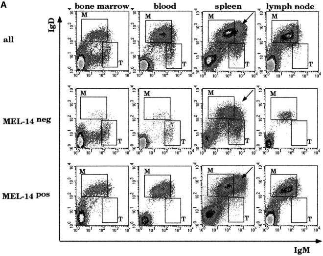
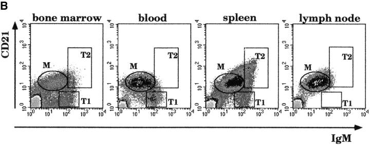
Transitional B cells of normal mice. Three-color flow cytometric analysis of cells isolated from bone marrow, blood, spleens, and lymph nodes of adult C57BL/6 mice. 100,000 events were collected. (A) Log density plots showing IgD and IgM expression. Top, all cells; center, MEL-14− cells; bottom, MEL-14+ cells. M, mature B cells; T, transitional B cells. Arrows, cells that are indicated as T2 in panel B. (B) Cells were stained with Abs to IgM and CD21. M, mature B cells; T1, CD21− and T2, CD21bright transi-tional B cells. (C) Splenocytes were stained with Abs to IgM, IgD, and CD23. The plots show the IgD and IgM staining of cells separated on the basis of the expression of CD23. CD23− B cells include IgMbright IgD− T1, and MZ B cells and all IgM− cells in the spleen (T cells and macrophages). Only B cells are positive for CD23. They are mostly T2 and mature (M) B cells. (D) Splenocytes were stained with Abs to IgD, CD21, and IgM and separated into IgD− or IgD+ cells. IgD− T1 and MZ B cells can be distinguished on the basis of CD21 expression. T2 and mature B cells are both in the gate of the IgD+ B cells but are either bright (T2) or dull (M) for IgM.
Lymphocyte entry in the lymph nodes is dependent on l-selectin, an adhesion molecule that facilitates migration through the high endothelial venules 26. We used the anti–l-selectin Ab MEL-14 to separate bone marrow, blood, spleen, and lymph node cells in MEL-14− and MEL-14+ cells (Fig. 1 A, center and bottom panels, respectively). Transitional B cells were exclusively MEL-14− (Fig. 1 A, center panels, gate T). Mature B cells were instead almost all positive for MEL-14. Only a minor fraction (<10%) of the mature B cells in the spleen and lymph node was negative for MEL-14. Our analysis suggests that transitional B cells leave the bone marrow with the blood. The absence of l-selectin on transitional B cells is consistent with their inability to enter the lymph node and their preferential migration to the spleen. A second population of IgMbright B cells is found exclusively in the spleen and expresses, in contrast to transitional B cells, both IgD and l-selectin (Fig. 1 A, arrows). We have used CD21 as a marker to further analyze this population. CD21, or complement receptor type 2 (CR-2), binds to C3 complement components 27 and is expressed in a developmentally regulated way. It is not present on immature B cells but is expressed on mature B cells. By staining with Abs to IgM and CD21, we could divide the population of IgMbright B cells into two populations, which we have called transitional 1 (T1) and transitional 2 (T2). T1 B cells lack CD21, whereas T2 B cells express CD21 in higher amounts (CD21bright) than mature B cells (Fig. 1 B). T2 B cells are found exclusively in the spleen but not in the bone marrow, blood, or lymph node (Fig. 1 B).
Relationship of T2 B Cells to Previously Described B Cell Populations.
Recently, CD21brightIgMbright B cells in the spleen have been identified as marginal zone (MZ) B cells 28. The MZ is mainly populated by B cells and macrophages and surrounds the lymphoid follicle outside the marginal sinus. MZ B cells do not express the differentiation markers IgD and CD23, which are also absent on immature and transitional B cells in the bone marrow 3. Splenic CD23− B cells are IgMbright and IgD− (Fig. 1 C, left), whereas CD23+ B cells coexpress IgM and IgD (Fig. 1 C, right). A more precise discrimination of the different B cell populations in the spleen is based on comparing the expression of CD21 and IgM on IgD− or IgD+ B cells (Fig. 1 D). IgMbrightIgD− B cells include T1 B cells, which are CD21−, and MZ B cells, which are CD21bright (Fig. 1 D, left). IgM+IgD+ B cells include T2 and mature B cells (Fig. 1 D, right). Therefore, we can identify two populations of IgMbrightCD21bright B cells in the spleen: (i) the MZ B cells (3–5% of the splenic B cells), which lack IgD and CD23 and (ii) T2 B cells (15–20%), which express both IgD and CD23. T1 B cells represent 5–10% of splenic B cells, with mature B cells corresponding to the remainder.
T2 cells thus represent a distinct population of IgMbright IgDbrightCD21brightCD23+ B cells. This phenotype distinguishes them from T1, MZ, and mature B cells. Moreover, they are found exclusively in the spleen and not in the lymph node or bone marrow. We found T2 B cells in the spleens of adult mice of different genetic backgrounds (C57BL/6, BALB/c, CBA/J, or NMRI), independent of intentional immunization or housing conditions (conventional, specific pathogen free, and germ free; results not shown).
T1 B Cells Are the Precursors of T2 and Mature B Cells.
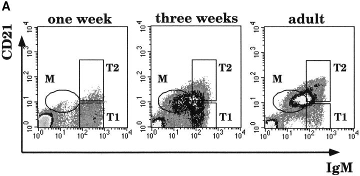
To investigate the developmental relationship between T1, T2, and mature B cells, we studied their distribution among splenocytes of mice at various ages. In the adult mouse, T1, T2, and mature B cells were present in the expected proportions (Fig. 2 A, right). In the spleens of 1-wk-old mice, all B cells were T1 B cells (Fig. 2 A, left). T2 and mature B cells started to appear at 2 wk of age (not shown) and were clearly detectable in the spleen of 3-wk-old mice (Fig. 2 A, center). At this time, the T1 population was still larger than in the adult mice (40%), and the fractions of T2 and mature B cells were reduced. MZ B cells are not present in the spleens of newborn mice; they are first detectable in the spleen at 4 wk of age 28.
Figure 2.
T1 B cells are the precursors of T2 and mature B cells. (A) Splenocytes of 1- (left) and 3- (center) wk-old and adult (right) C57BL/6 mice were stained with Abs to CD21 and IgM and analyzed by flow cytometry. (B) Splenic cells from recipient RAG-2−/− mice were analyzed at the indicated times after transfer of 2 × 10⁶ splenic B cells from a pool of 1-wk-old mice. Cells were stained with Abs to IgM, IgD, and CD21. Top panels, IgM vs. IgD staining. Bottom panels, the CD21 vs. IgM profile of IgD+IgM+ donor B cells. 200,000 events were collected. Data is shown as dot plots to highlight the few transferred cells that home to the spleen. In the dot plots corresponding to the control (adult) spleen, only 5% of the collected events are shown.
To study the developmental potential of neonatal T1 B cells, we transferred splenocytes from 1-wk-old mice into adult RAG-2−/− mice, which lack B and T cells 24. Donor splenocytes were injected into the mouse tail veins, and the spleens of recipient mice were analyzed 5, 24, and 48 h after transfer. The expression patterns of IgD and IgM showed that 5 h after transfer, all donor B cells still had the T1 phenotype (Fig. 2 B, top). IgD+ and IgMdull cells were found 24 h after transfer (Fig. 2 B, top). Mature B cells and T2 B cells were clearly detectable after 48 h (Fig. 2 B, top, labeled M and T2, respectively). This was confirmed by the analysis of CD21 expression on B cells (B cells were defined as being positive for IgM and/or IgD). In Fig. 2 B, bottom, the expression patterns of CD21 and IgM are represented. 48 h after transfer, T2 B cells were 52% of all B cells, whereas mature B cells were 36% (Fig. 2 B, bottom). Both T2 and mature B cells expressed CD23 (not shown). In a second experiment, the fate of transferred cells was followed for 7 d after injection. At this time, mature and T2 B cells were still detectable, but ∼50% of the transferred cells had downregulated IgD and CD23 and could be considered phenotypically MZ B cells (not shown). In both experiments, the transferred cells constituted ∼1% of all splenocytes of recipient mice.
These experiments demonstrate that in the adult spleen, neonatal T1 B cells develop to T2 and mature B cells in 2 d and to MZ B cells in 7 d. Therefore, neonatal T1 B cells do not have an intrinsic defect that prevents their further maturation 29 but most likely, the microenvironment of the neonatal spleen does not support the late phases of B cell development, from T1 to T2 and to mature B cells.
T2 B Cells Develop into Mature B Cells.
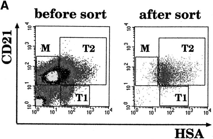
What is the developmental relationship between T2 and mature B cells? T2 and mature B cells could independently develop from the T1 population, or T2 B cells could represent an intermediate stage of development, preceding the mature stage. To address this question, we isolated T2 B cells from the spleen of a normal adult mouse, transferred them into RAG-2−/− mice, and analyzed their developmental potential. To separate T2 from T1 and mature B cells, we did not use the IgM and IgD antigen receptors as markers, because the staining procedure could have initiated BCR-mediated signals and, therefore, influenced B cell survival and differentiation. T1, T2, and mature B cells were instead identified on the basis of their expression of HSA and CD21. The expression of HSA is developmentally regulated in B cells. It is high in early phases of development in the bone marrow, and it is downregulated in mature B cells. Recent bone marrow immigrant B cells express higher amounts of HSA than mature B cells 5. By staining with Abs to HSA and CD21, we could distinguish T1, T2, and mature B cells. T1 B cells are HSAbright and lack CD21. T2 B cells are also HSAbright but express CD21, and mature B cells are HSAdull and CD21+ (Fig. 3 A, before sort). In further experiments, we confirmed the accuracy of the discrimination by using IgM and IgD as additional markers (not shown). The purity of sorted T2 B cells (Fig. 3 A, after sort) was also controlled by staining an aliquot with Abs to IgM and IgD. Sorted T2 B cells expressed high amounts of both IgM and IgD and, therefore, corresponded to bona fide T2 B cells.
Figure 3.
T2 B cells develop into mature B cells in the spleen. (A) Spleen cells of adult mice were stained with Abs to HSA and CD21, and T1, T2, and mature (M) B cells were identified (before sort). CD21+HSAbright T2 B cells were sorted (after sort) and transferred into adult RAG-2−/− recipient mice. (B) The spleens of recipient RAG-2−/− mice were analyzed 24 h after transfer and compared with the spleen of an adult control mouse. T1, T2, and mature B cells were identified on the basis of the expression of HSA, B220, IgM, and IgD. The plots show the IgD and IgM staining of cells that were positive for B220 and either bright or dull for HSA.
106 sorted HSAbrightCD21+ T2 B cells were injected into the tail veins of RAG-2−/− mice. Recipient mice were killed 24 h later, and splenocytes were stained with Abs to B220, HSA, IgD, and IgM. As control, a normal mouse was analyzed; the staining pattern of T1 and T2 B cells is shown in Fig. 3 B (control). Both populations were undetectable in the mice that had received the T2 transplant. At least 95% of the B cells isolated from host RAG-2−/− spleens were B220+HSAdull mature B cells (Fig. 3 B, 24 h). Three independent experiments were performed, and recipient mice were also analyzed 48 h after transplantation, always with comparable results (not shown).
Our experiments demonstrate that T2 B cells develop into mature B cells in the spleen. This step of differentiation is associated to the downregulation of IgM and HSA. Our data does not exclude the possibility that at least a fraction of the T1 B cells can also directly develop into mature B cells.
T2 B Cells Are in the Primary Follicle.
To study the localization of T1, T2, and mature B cells in the spleens of normal mice, we stained sections with TRITC-coupled Abs to IgM and with FITC-coupled Abs to IgD (Fig. 4 A). IgMbrightIgD− MZ and T1 B cells appear in red, T2 B cells, which coexpress high amounts of IgM and IgD, appear in yellow, and mature B cells, which are bright for IgD and have downregulated IgM, are green (Fig. 4). T1 B cells are in the outer periarteriolar lymphoid sheet (PALS) close to the primary follicle. T2 and mature B cells are together inside the follicle. Mature B cells can also be seen in the outer PALS and in the red pulp. MZ B cells, as expected, surround the follicle. Fig. 4 B shows an enlargement of the bordering area between outer PALS and follicle, where single T1 (red; indicated by the single-headed arrow) and T2 cells (yellow; indicated by the double-headed arrow) can be seen, together with mature B cells (bright green).
Figure 4.

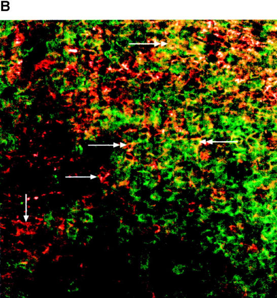
T2 B cells are in the primary follicle of normal mice. (A) Sections of normal spleen were fixed and stained with TRITC-labeled goat anti–mouse IgM and FITC-labeled anti-mouse IgD. Green and red fluorescence were measured separately by confocal laser microscopy, and the pictures obtained were then overlaid (magnification 100). M, mature B cells. (B) The sector indicated by the square in A was scanned with a 250-fold amplification to better visualize single cells. T1 B cells (red) are indicated by the single-headed arrow and T2 (yellow) by the double-headed arrow. Mature B cells are green.
B Cell Development Is Arrested at the T2 B Cell Stage in the Spleens of CD45−/− and CBA/N Mice.
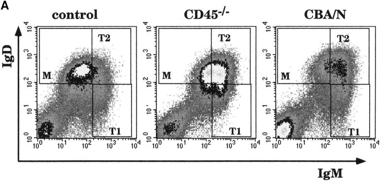
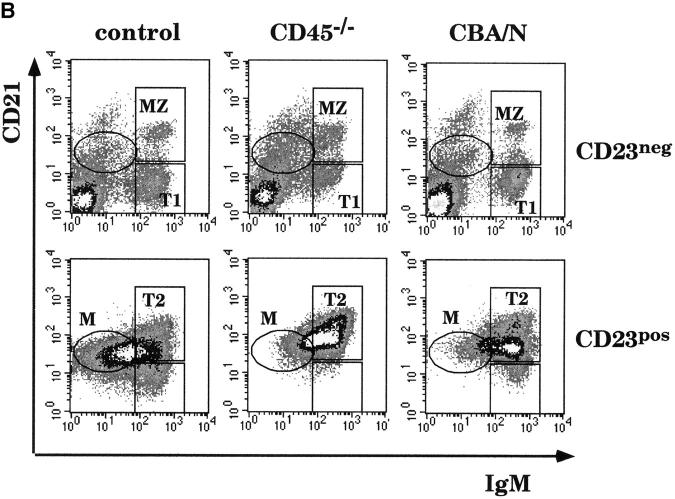
Development of mature B cells is compromised in mice deficient for CD45 and in mice mutant for Btk (CBA/N). We stained splenocytes of normal control, CD45−/−, and CBA/N mice with Abs to IgM, IgD, CD21, and CD23. Most of the splenic B cells of CD45−/− and CBA/N mice are phenotypically identical to the T2 B cells of normal mice. They are IgMbright IgDbright (Fig. 5 A, gate T2) and CD23+CD21bright (Fig. 5 B, bottom panels). T2 B cells, however, are present in a higher percentage: in CD45−/− mice, they represent 49% and in CBA/N mice 44% of all B cells, as compared with 15–20% in normal mice. The fraction of T1 B cells is only slightly reduced in size in CD45−/− mice and is normal in CBA/N mice.
Figure 5.
B cell development is blocked at the T2 stage in CD45−/− and CBA/N mice, but the number and the phenotype of MZ B cells is normal. Flow cytometric analysis of splenic B cells from normal C57BL/6 (control) and CD45−/− and CBA/N mice. (A) Cells were stained with Abs to IgM and IgD. T1, T2, and mature (M) B cells are boxed. (B) Cells were stained with Abs to CD23, CD21, and IgM and separated into CD23− and CD23+ cells. T1, T2, MZ, and mature B cells were identified as in Fig. 1 C. The fraction of T2 B cells represented 23% of all B cells in the normal mouse spleen, 55% in the CD45−/− spleen, and 50% in the CBA/N spleen.
Surprisingly, MZ B cells are present in the expected proportions and have a normal phenotype in CD45 and CBA/N mutant mice. We calculated the percentages of T1, T2, and MZ B cells in normal and mutant mice based on the stainings shown in Fig. 5 B. In normal mice, T1 B cells (IgMbrightCD23−CD21−) represent 7% and MZ B cells (IgMbrightCD23−CD21+) 3% of all B cells. The fraction of T1 B cells is slightly reduced in the CD45−/− spleen (4%); the fraction of MZ B cells is normal (3%). In the CBA/N mouse, both T1 and MZ B cells are slightly increased in percentage (13 and 5%, respectively). Prototypic mature B cells are not found in these mice: the cells in gate M (mature) in Fig. 5 express more IgM and CD21 than normal mature B cells.
The genetic background does not influence the distribution of T1, T2, and mature cells in the spleen, because the relative size and phenotype of the three populations is identical in normal C57BL/6 and CBA/J mice, which are genetically matched controls for CD45−/− and CBA/N mice, respectively (not shown). Therefore, in all of the following experiments, we used C57BL/6 mice as controls.
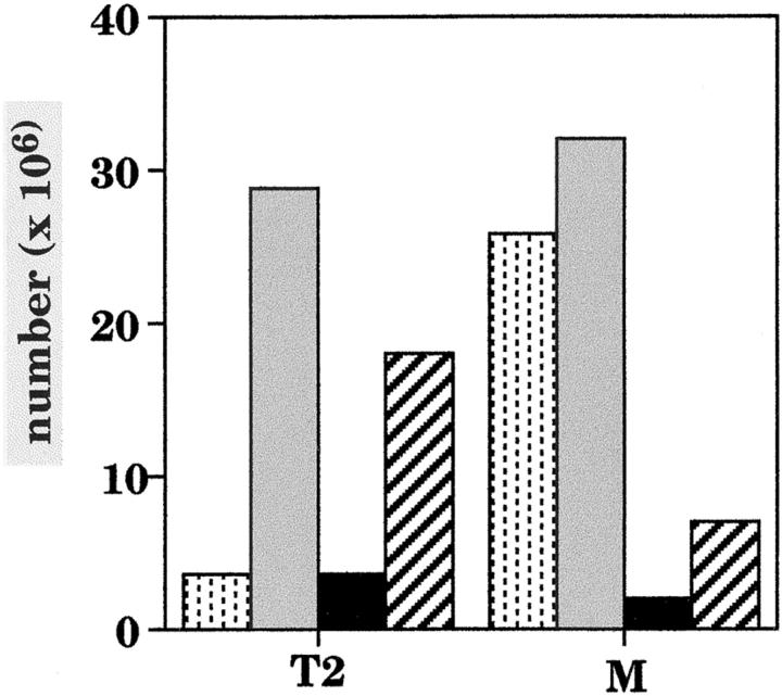
Although the frequency of T2 B cells is similarly increased in CD45−/− and CBA/N mice, their absolute number is dramatically different in the two mutant mice. In the spleens of CD45−/− mice, the absolute number of T2 B cells is six to eight times larger than in control mice, whereas in CBA/N mice, the size of the T2 population is normal (Fig. 6). In contrast, the total number of the phenotypically aberrant mature B cells is reduced in the CBA/N mice but normal in CD45−/− mice. These data demonstrate that the defect in B cell development that leads to an arrest at the T2 stage is not identical in the two mutant mice. To support this conclusion, we backcrossed CD45−/− to CBA/N mice. In double-mutant mice, the number of T2 B cells is five times higher and the number of mature B cells is four times lower than in normal mice (Fig. 6). These results prove that CD45 plays a role in determining the number of cells in the T2 B cell pool, whereas Btk either facilitates the entry of T2 B cells into the mature B cell pool or prolongs the survival of mature B cells.
Figure 6.
Absolute number of T2 and mature (M) splenic B cells in normal, CD45−/−, and CBA/N and in CD45−/−CBA/N double-mutant mice. Cell numbers were calculated using the gates shown in Fig. 5. Stippled bar, normal; gray bar, CD45−/−; black bar, CBA/N; hatched bar, double mutant.
T2 B Cells Proliferate in the Spleen; Proliferation Is Deregulated in CD45−/ − Mice.
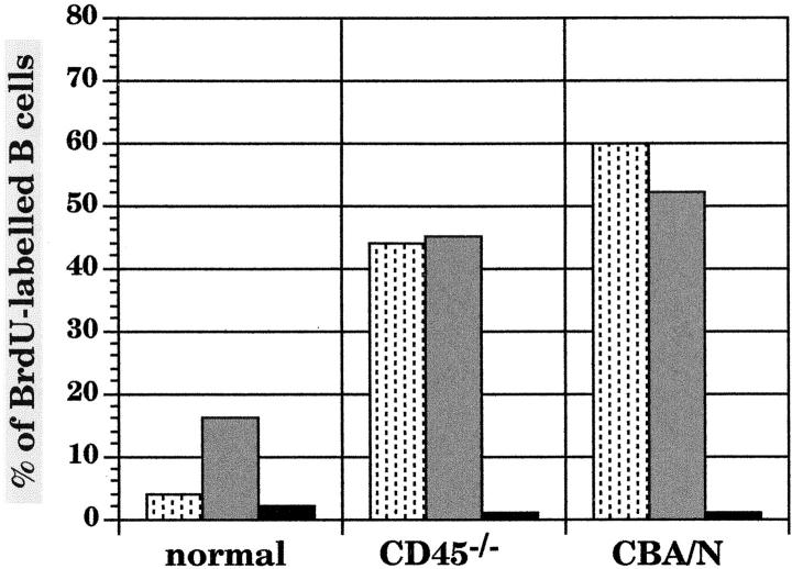
In normal mice, mature B cells are long lived, and recent splenic immigrant B cells from the bone marrow are short lived, with a life span of 3 d after their arrival in the spleen. Subsequently, they are either incorporated into the long-lived pool or eliminated 5 30. We determined the frequency and phenotypic distribution of short- and long-lived B cells in CD45 and Btk mutant mice using the BrdU labeling technique. Mice were analyzed 24, 72, and 120 h after the onset of the treatment. 24 h after the beginning of the BrdU administration, the fraction of labeled cells was 4% in the spleens of normal mice (Fig. 7) and rose to a maximal 16% after 3 d of continuous treatment. In CD45−/− and CBA/N mice, about half of the splenic B cells incorporated BrdU in 24 h. The percentage of labeled cells remained constant thereafter. Our results confirm the life span analysis of splenic B cells of normal mice but indicate that in mutant mice, labeled B cells have a life span of only 1 d.
Figure 7.
Kinetics of BrdU labeling of B lymphocytes of normal and mutant mice. Mice received BrdU (1 mg/ml) in their drinking water for the indicated times. Splenic B cell populations were identified on the basis of three different surface staining protocols: IgM vs. IgD, IgM vs. CD21, and IgD vs. CD22. The BrdU content of the cells was determined by flow cytometry. The experiments were repeated three times, and comparable results were obtained. The columns represent the percentages of BrdU-labeled B cells in normal and mutant mice analyzed 24 (stippled bars) and 72 h (gray bars) after the beginning of the BrdU administration. The percentage of B cells still labeled 18 d after a 3-d treatment with BrdU (18-d chase; black bars) is also shown. The absolute numbers of labeled B cells in the normal spleen were 2 × 10⁶ after 24 h, 5.6 × 10⁶ after 3 d, and 0.9 × 10⁶ after the 18-d chase. In the CD45−/− mouse, 35 × 106 B cells were labeled after 24 h and 37.8 × 10⁶ after 3 d. 0.8 × 10⁶ B cells were still labeled after the 18-d chase. The CBA/N mouse had 2.4 × 10⁶ labeled B cells on day 1 and 3.2 × 10⁶ on day 3. Only 0.1 × 10⁶ cells were still labeled after the chase.
Splenic B cells that are labeled after a short BrdU pulse represent the balance of (a) cells derived from cycling bone marrow precursors, (b) labeled cells that died during the treatment period, and (c) cells that entered the cell cycle in the spleen. This last factor is not considered significant in the normal mouse. However, because B cell numbers and labeling rates were normal in the bone marrow of CD45−/− mice (not shown), the large number of B cells labeled with BrdU can only be explained by proliferation in the spleen. Therefore, we studied cell cycle progression of T1, T2, and mature B cells in spleen and bone marrow. The different populations were identified by surface staining with anti-IgM and anti-CD21 Abs, and DNA content was measured with propidium iodide. As expected, in spleens and bone marrow of normal and mutant mice, 93–97% of the mature and T1 B cells are in the G0–G1 phase of the cell cycle (Table ). Surprisingly, 15% of the T2 B cells of normal mice are in the G2–M phase of the cell cycle. In CD45−/− mice, 33% and in CBA/N mice, 21% of the T2 B cells are in the G2–M phase. In this type of analysis, MZ B cells are included in the T2 B cell population. Although they represent a minor fraction, we have also measured cell cycle progression in IgMbrightIgDbright (proper T2 B cells), IgMbrightIgD− (T1 and MZ B cells), and IgM+IgDbright cells (mature B cells). The results confirmed the notion that T1, MZ, and mature B cells are noncycling cells, whereas the T2 B cells are a population of cycling cells (Table , bottom). Therefore, entrance into the cell cycle appears to be a normal event at the T2 B cell stage, but proliferation is limited. In contrast, proliferation is deregulated in the absence of CD45.
Table 1.
Cell Cycle Analysis of T1, T2, and Mature B Cells
| Mature | T1 | T2 and MZ | ||
|---|---|---|---|---|
| Control | spleen | 3 | 3 | 15 |
| bone marrow | 5 | 5 | — | |
| CD45 | spleen | 3 | 2 | 33 |
| bone marrow | 5 | 3 | — | |
| CBA/N | spleen | 4 | 3 | 21 |
| bone marrow | 7 | 5 | — | |
| Mature | T1 and MZ | T2 | ||
| Control spleen | 3 | 1 | 17 | |
| CD45spleen | 4 | 1 | 31 | |
| CBA/N spleen | 4 | 4 | 31 | |
Top: spleen and bone marrow cells were stained with Abs to IgM and CD21. After ethanol fixation, propidium iodide was used to label DNA. For cell cycle analysis, cells were gated in T1, T2, and M, as indicated in Fig. 1 B, and the DNA content was measured in these subpopulations. In bone marrow, T2 B cells are undetectable. Percentages of B cells in the G2–M phase of the cell cycle are given.
Bottom: splenic cells were stained with Abs to IgM and IgD before DNA labeling. T1, MZ, T2, and mature B cells were identified as described in the text.
Entrance into the Mature Pool Is Impaired in CBA/N Mice.
The recruitment rate into the long-lived pool was measured by pulse–chase experiments. The fraction of BrdU-labeled B cells was measured 18 d after a 3-d labeling pulse with BrdU. As few as 14% of the B cells that had incorporated BrdU over a 3-d period entered the long-lived pool in the spleens of normal mice, compared with an even lower 2% in CD45−/− and 2% in CBA/N mice (Fig. 7). Therefore, the number of splenic B cells labeled after 3 d of continuous BrdU administration corresponds to the number of short-lived B cells, whereas long-lived B cells are mostly unlabeled. The absolute numbers of these cell populations in the spleen are given in Table . We also calculated the number of labeled B cells that were still detectable 18 d after the BrdU pulse and that, as we have seen, correspond to the small percentage of short-lived B cells that were recruited into the long-lived pool. In CBA/N mice, the number of short-lived splenic B cells was identical to the number in control mice, but in CD45−/− mice, it was sevenfold higher. The total number of long-lived cells is normal in CD45 mutant mice but reduced sixfold in CBA/N mice. Accordingly, the number of B cells that entered the long-lived pool was significantly reduced in CBA/N mice but normal in CD45−/− mice (Table ).
Table 2.
Absolute and Relative Numbers of Short- and Long-lived B Cells in the Spleens of Normal and Mutant Mice
| Short-lived | Long-lived | Short→long-lived | |
|---|---|---|---|
| Control | 5.6 (100%) | 29.4 (100%) | 0.9 (100%) |
| CD45−/− | 37.8 (670%) | 46.2 (157%) | 0.8 (89%) |
| CBA/N | 5.2 (92%) | 4.8 (16%) | 0.1 (11%) |
The absolute number (× 106) of labeled (short-lived) and unlabeled (long-lived) B cells in the spleens of control, CD45−/−, and CBA/N mice was calculated after 3 d of continuous treatment with BrdU. The number of labeled B cells detected 18 d after the 3-d BrdU treatment period corresponds to the number of B cells that became long lived. Numbers of labeled and unlabeled B cells in mutant mice are given in parentheses as percentages of the numbers found in control mice  .
.
The short life span of a major part of B cells in the mutant mice and the low fraction of cells that enter the long-lived pool implies that the death rate is very high among these cells. We measured the fraction of apoptotic cells in normal and mutant mice with merocyanine 540, a pigment that binds to the membranes of cells in the early phases of the apoptotic process 31 32. Whereas only 10% of the T2 B cells bound merocyanine in the normal spleen, 30% of them were apoptotic in CD45−/− mice and 25% were apoptotic in CBA/N mice (data not shown).
In conclusion, BrdU labeling experiments and cell cycle analysis confirm that the defects in CD45−/− and CBA/N mice are not identical. In CD45−/− mice, B cells extensively proliferate at the T2 stage in the spleen, but the number of long-lived B cells is normal. In CBA/N mice, the apparent increase of T2 B cells is due instead to the reduction of the long-lived mature B cells. In addition, in both mutant mice, long-lived B cells had an abnormal phenotype, showing that the ability to survive can be dissociated from the phenotype in mutant B cells.
The BCR Is Indispensable for the Development of T2 and Mature B Cells.
It has been suggested that B cells are selected into the mature B cell pool by antigen 8 9 10 11. Selection, therefore, would depend on the engagement of the BCR. To analyze the role of the BCR in the development of T1, T2, and mature B cells, we analyzed the late stages of B cell development in mb1 Δc/Δc mice 12. These mice have a complete deletion of the cytoplasmic tail of the Ig-α component of the BCR, which causes a severe block in the development of peripheral B cells 12. In the spleens of these mice, the number of T1 B cells is 20% of normal, but T2, mature, and MZ B cells are undetectable (Fig. 8, left). This finding confirms that the signaling function of the BCR is essential for the development of T2, mature, and MZ B cells from T1 B cells.
Figure 8.
Development of T2 and mature B cells is blocked in mice lacking the cytoplasmic tail of Ig-α. Spleen cells of mb1 Δc/Δc (left), CD45−/− (center), and mb1 Δc/Δc-CD45−/− double-mutant (right) mice were stained with Abs to CD21 and IgM. Flow cytometric profiles are shown. Dead cells, which fail to exclude the dye propidium iodide, were excluded from the analysis. 100,000 events were collected.
To prove that development of T2 B cells requires signaling through the BCR, we backcrossed CD45−/− to mb1/Δc/Δc mice. In double-deficient mice, the enlarged T2 population present in the spleens of CD45−/− mice does not develop (Fig. 8; compare CD45−/− and double mutant). This indicates that a BCR-mediated signal is required for the development and proliferation of T2 B cells in the spleens of CD45−/− mice.
Primary Follicles of Mutant Mice Show a Normal Architecture.
To study splenic architecture in the mutant mice, we stained spleen sections with Abs against MAdCAM-1, a mucosal vascular addressin that is expressed on the cells lining the marginal sinus in the spleen 33. B cells were labeled with anti-IgM Abs (Fig. 9). In the spleen of the control mouse, the primary follicle (F) is surrounded by a thin layer of MAdCAM-1–positive endothelial cells (green; indicated by arrows). MZ B cells (MZ) express high amounts of IgM and encircle the follicle (F) outside of the marginal sinus. Follicles of CD45−/− mice have a larger size but a normal distribution of MZ and follicular B cells. MZ and follicular B cells also have a normal distribution in the spleens of CBA/N mice 34. CBA/N mice, however, have very small lymphoid follicles. The normal distribution of B cells in the spleens of mutant mice shows that their T2 B cells can enter the follicle and that the MZ has a normal proportion, in accordance with the normal fraction of these cells in the spleen.
Figure 9.
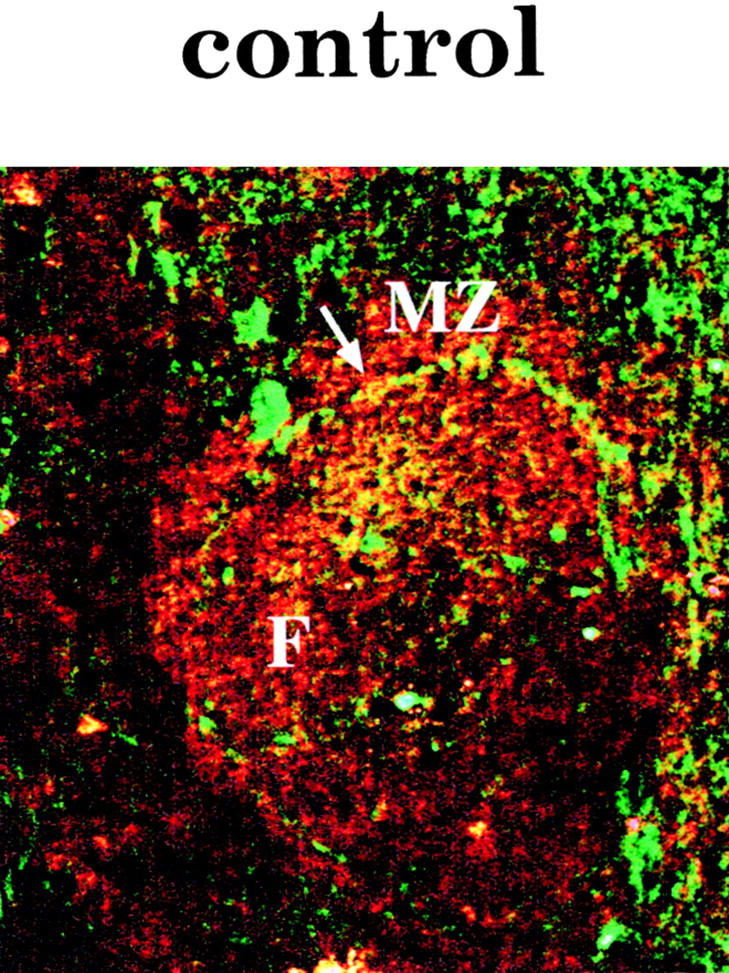

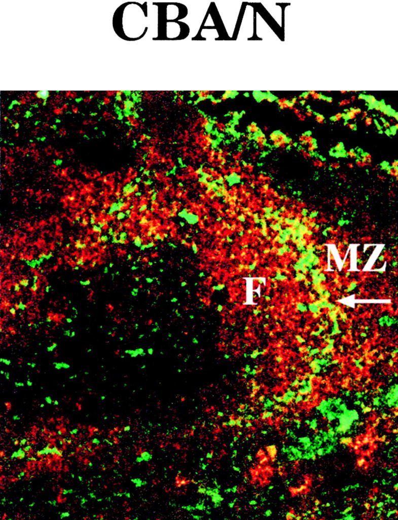
Primary follicles of mutant mice show normal architecture. Cryostatic sections of spleens from normal and mutant mice were stained with TRITC-labeled goat anti–mouse IgM Abs. Anti–MAdCAM-1 Abs (from rats) were counterstained with FITC-labeled goat anti–rat Abs. Green and red fluorescence was measured separately by confocal laser microscopy, and the pictures obtained were then overlaid (magnification 100). The arrow points to the thin layer of MAdCAM-1–positive endothelial cells of the marginal sinus. F, follicle.
Discussion
The differentiation pathway from stem cell to mature B lymphocyte can be divided into several stages, characterized by differentiation processes, proliferation phases, and control steps. The progression of B cells along this pathway is probably best understood assuming that it is governed by the principle of conditional survival. In each developmental stage, specific genetic programs are completed in discrete environments. To progress from one stage to the next, the cells have to meet specific requirements set by the new developmental stage and the new environment. In the early phases of differentiation in the bone marrow, the ability to productively rearrange the H and L chain genes is an essential requirement 35. The signaling function of the BCR plays an equally important role 36. It has recently been shown that the presence of the BCR is also indispensable for the survival of mature B cells in the periphery 37. This study suggests that the mere presence of the signaling complex organized around the IgM and IgD molecules generates a tonic signal necessary for the survival of mature B cells. It could not, however, address the question of whether this minimal constitutive signal is also sufficient to induce the differentiation of B cells recently generated in the bone marrow into mature B cells.
Studies of B lymphocyte population dynamics have shown that entry into the pool of the mature long-lived B cells is a highly selective event. Only 1–3% of the B lymphocytes leaving the bone marrow every day enter the pool; the remainder of the newly generated B cells are eliminated 5 11 38. The requirements that B cells have to meet at the time of selection into the long-lived pool and the site where selection takes place are largely unknown. We show here that selection into the mature pool is an active process and takes place in the spleen. Two populations of splenic B cells were identified as precursors for mature B cells, the T1 and the T2 B cells. Their development into mature B cells requires defined qualitative and quantitative signals derived from the BCR. We finally show that the induction of longevity and maturation requires different signaling pathways.
A Functional BCR Is Indispensable for the Progression from T1 to T2 and Mature B Cell Stage in the Spleen.
In the spleen, we have identified four different B cell populations by flow cytometry: T1, T2, mature, and MZ B cells. Their phenotypes are summarized in Table . T1 B cells originate from the bone marrow and can be detected in the marrow, blood, and spleen but not in the lymph nodes (Fig. 1 A). Entry into the lymph nodes depends on the expression of the homing receptor l-selectin (CD62L) 26. In both mice treated with anti–l-selectin Ab (MEL-14 Ab) 39 and mice deficient for l-selectin 40, the number of B and T cells is drastically reduced in the lymph nodes but is increased in the spleen. Our observations are in accordance with these reports: T1 B cells lack the expression of l-selectin (Fig. 1 A) and home to the spleen, not to the lymph nodes. They enter the spleen via the terminal arterioles of the red pulp and marginal sinus 41. B cells that do not express CD21 and l-selectin have recently been described as nonrecirculating B cells in the blood. This subset of B cells only homed to the spleen and was excluded from the lymphatic recirculation pathway 42. These cells are probably T1 B cells in transit from the bone marrow to the spleen.
Table 3.
Phenotypic Description and Tissue Distribution of Immature, T1, T2, Mature, and MZ B Cells
| IMM | T1 | T2 | M | MZ | |
|---|---|---|---|---|---|
| IgM | + | +++ | +++ | + | +++ |
| IgD | − | − | +++ | +++ | − |
| CD21 | − | − | +++ | ++ | +++ |
| CD23 | − | − | ++ | ++ | − |
| Location | BM | BM, B, S | S | BM, B, S, LN | S |
Expression of the indicated markers is given as negative (−), weakly positive (+), positive (++), and strongly positive (+++). The location where the various cell types are found is also indicated: bone marrow (BM), blood (B), spleen (S), and lymph nodes (LN). IMM, immature.
In normal mice, T2 B cells are only found in the spleen. Phenotypically, they appear to be an intermediate stage of differentiation between the T1 and the mature B cell stage. Like T1 B cells, they still express high amounts of the early hematopoietic marker HSA 5 and the recently described marker of immature B cells 43, recognized by the mAb 493 (our unpublished observations). Like mature B cells, they express IgD, CD23, and l-selectin. T2 B cells appear in an activated state in vivo: they are large, they express B7-2 (not shown), and a significant fraction of them is in the G2–M phase of the cell cycle (Table ).
MZ B cells are also found in the spleen, in a specialized area that surrounds the white pulp outside the marginal sinus 44, and are phenotypically different from T1 and T2 B cells (Fig. 1c and Fig. d). MZ B cells are nonrecirculating, long-lived B cells 45.
To establish the ontogenetic order of appearance of T1, T2, and mature B cells, we injected T1 B cells intravenously into RAG-2−/− mice. T1 B cells developed into T2 and mature B cells 48 h after the injection into adult RAG-2−/− mice (Fig. 2 B). This experiment demonstrates that T1 B cells are the precursors of T2 and mature B cells but does not establish the developmental relationship between T2 and mature B cells. T2 and mature B cells could represent two independent, end-stage populations generated from the T1 pool. The relatively immature phenotype of T2 B cells and their exclusive presence in the spleen, the only organ where T1 are also found, however, suggest another possible scenario: T2 B cells may represent a developmental checkpoint between T1 and mature B cells. To study the fate of T2 B cells, we transferred them into RAG-2−/− mice. T2 B cells developed into mature B cells in 24 h (Fig. 3). Our data shows that T2 B cells represent an intermediate stage of development that feeds into the mature pool. However, we cannot and will not exclude the possibility that at least a fraction of the T1 B cells can directly develop into mature B cells.
T1 and T2 B cells are found in different microenvironments in the spleen. T1 B cells are located in the red pulp and the outer PALS, whereas T2 and mature B cells are located in the follicle (Fig. 4). The development of T2 and mature B cells from T1 precursors and their specific localization in different microenvironments in the spleen might be influenced by the signaling capacity of the BCR. Indeed, it was recently demonstrated that B cells deficient for Syk, a tyrosine kinase that plays a fundamental role in BCR signaling, migrate from the bone marrow to the spleen but fail to enter the primary follicle and remain in the red pulp and the PALS 46. In addition, it has been shown that encounter with antigen influences the migration of B cells from the outer PALS to the follicle 47. It is therefore likely that T1 B cells can enter the primary follicle only if they received a signal in the outer PALS and became T2 B cells. Because entrance into the primary follicles of the spleen is impaired in B cells of mice deficient for the chemokine BLR-1 48, it is possible that B cells acquire the ability to respond to BLR-1 at the T2 stage.
To analyze the role of the signaling capacity of the BCR in this phase of development, we have studied mice with genetic defects of signaling elements involved in the BCR pathway. In mice lacking the cytoplasmic tail of Ig-α (mb1 Δc/Δc mice), the signaling function of the BCR is severely impaired. The number of B cells in the bone marrow is reduced to 20% of normal 12. We show here that B cells in the spleen are arrested at the T1 stage of development (Fig. 8). These findings demonstrate that the final steps of B cell development in the spleen, from T1 to T2, mature, and MZ B cells require a functional BCR. The fact that the early stages of B cell development are impaired in the bone marrow of mb1 Δc/Δc mice, whereas the late stages are completely abolished in the spleen, may reflect a more strict requirement for a perfectly functional BCR in the final phases of differentiation.
In mice deficient for the tyrosine phosphatase CD45, B cells with a proper mature phenotype are missing, whereas the T2 population is six- to eightfold larger than in normal mice (Table ). T2 B cells proliferate in the spleens of CD45−/− mice. At least 30% of them are in the G2–M phase of the cell cycle. Surprisingly, also in normal and CBA/N mice, 15–20% of the T2 B cells are in the G2–M phase (Table ). In contrast, both T1 and mature B cells are in the G0–G1 phase. This result suggests that the BCR-mediated activation event responsible for the progression from the T1 to T2 stage results in the proliferation of a large fraction of the T2 cells. Proliferation is deregulated in the absence of CD45. When CD45−/− mice were backcrossed to mb1 Δc/Δc mice, the development of T2 B cells was completely blocked (Fig. 8). Therefore, a BCR-mediated signal is necessary for the development of T1 to T2 B cells in the spleen and induces a proliferative response at this stage.
Given the large number of short-lived B cells in CD45−/− mice, a low recruitment rate into the long-lived pool (2% as compared with 16% in normal mice) still ensures an almost normal influx into the long-lived pool (Table ). The low recruitment rate may reflect a limited supply of resources necessary to support the survival of B cells 49.
The CD45 tyrosine phosphatase positively regulates and amplifies BCR-derived signals. The tyrosine phosphatase SHP-1 has the opposite function and reduces and terminates signals generated by the BCR 50. Development of mature B cells was rescued when CD45−/− mice were back-crossed to motheaten mice, which lack SHP-1 activity 51. Also, the deletion of CD22 52, which recruits SHP-1 to the BCR and negatively regulates B cell signaling, rescues the development of mature B cells in CD45−/− mice (Wardemann, H., F. Loder, M.C. Lamers, and R. Carsetti, manuscript in preparation). Signal strength is therefore an essential factor in determining the fate of B cells in the late phases of development from T1 to mature B cells. A strong signal is necessary for the development of mature B cells, but a weak signal is sufficient for the development of T2 and MZ B cells.
More complex is the interpretation of the CBA/N defect. Also in this case, T2 and MZ B cells develop but long-lived mature B cells are absent. The effect of Btk on B cell survival can only partially explain the defect. The overexpression of bcl-2 53, which belongs to a family of proteins that inhibits apoptosis and prolongs B cell life span, leads to the increase of T2 B cells but does not rescue the development of mature B cells. It is, therefore, likely that Btk plays a dual role in B cells: it controls their life span and also regulates their final differentiation to mature B cells. Accordingly, several downstream effectors and partners of Btk have been identified, including members of the bcl-2 family but also elements of the mitogen-activated protein (MAP) kinase pathway 20.
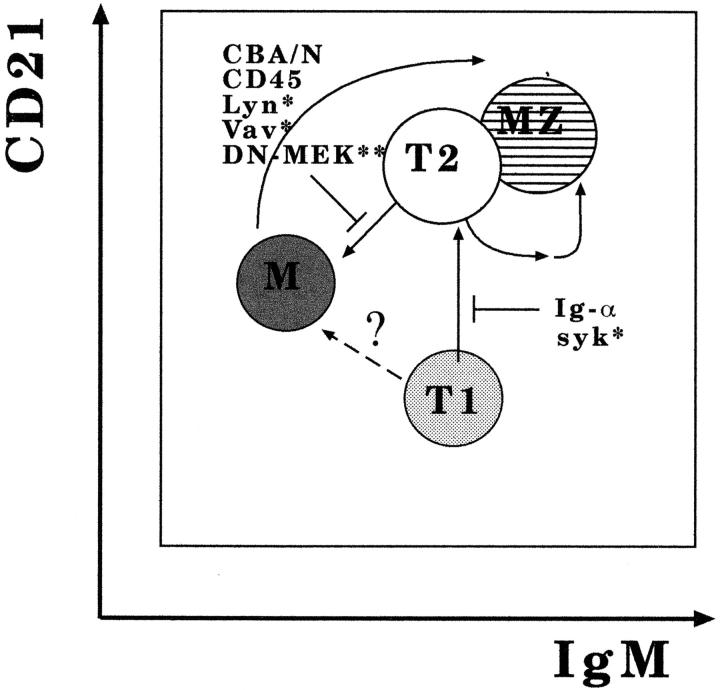
Little is known about the nature of events downstream of the BCR necessary for B cell maturation. We have evidence that the MAP kinase pathway plays an important role in these processes. For instance, T2 B cells become mature B cells in vitro upon transient and strong activation of the MAP kinase pathway, whereas mice that are transgenic for a dominant-negative form of MAP kinase/extracellular regulated kinase (MEK) show a developmental arrest at the T2 stage (Carsetti, R., personal observation). The high expression of CD21 on T2 B cells could play an important role in this process. CD21 forms a complex with the CD19 coreceptor. Cross-linking of the BCR with the CD21–CD19 complex strongly activates the MAP kinase pathway 54 and may result in the generation of mature from T2 B cells. T2 B cells that do not find their ligands may remain in the spleen as T2 B cells. A summary of our view of the developmental pathways of B cells in the spleen is given in Fig. 10.
Figure 10.
Proposed developmental pathways of B cells in the spleen. The B cell subpopulations described in this paper are depicted as they would appear in a flow cytometric profile after staining with anti-IgM and anti-CD21 Abs. T1 B cells recently immigrated from the bone marrow develop into T2 B cells if they receive a BCR-mediated signal. This signal is insufficient in Ig-α– and Syk-deficient mice. Progression of T2 B cells into the mature long-lived pool (M) is blocked in mice with qualitative and quantitative defects in BCR signaling function, as in Btk-, Vav-, Lyn-, and CD45−/− mice, and in transgenic mice with a dominant-negative form of MEK (DN-MEK), which is only expressed in B cells. MZ B cells are thought to derive from mature B cells but are also found in mice with a developmental arrest at the T2 stage, leaving open the possibility that they can also be derived from T2 B cells. It is presently unclear whether T1 B cells can directly differentiate into mature cells in the spleen. Solid arrows, major pathways; broken arrows, possible pathways; ⊥, pathway blocked; *inferred from the literature; **our unpublished data.
Implications.
Our data demonstrate that two factors regulate the development of T1 into T2 and mature B cells: the signaling function of the BCR and the microenvironment of the adult spleen. In a simplified model, strength and duration of BCR-mediated signals determine the outcome of antigen receptor–induced B cell activation: death or survival, proliferation, or differentiation. Three factors regulate signal strength: the antigen, the antigen receptor, and the signal transduction machinery. We have shown that mutations affecting the components of the BCR signal transduction cascade severely influence the late stages of B cell development in the spleen. In normal mice, the choice between proliferation and differentiation might depend mostly on the structure of the ligand, which regulates the extent of cross-linking, and on the affinity of the BCR for this ligand, which also affects the intensity of the signal. Experiments with transgenic mice have clearly shown that B cells that have a high affinity for self-antigens, and therefore receive a strong signal upon binding, are deleted. Positive selection in the mature pool may happen at a much lower signaling threshold, at a level where the quality and composition of the BCR signal transduction apparatus is of fundamental importance.
This model predicts that B cells expressing certain V region specificities would be selected only into the T2 or MZ pools and never into the mature pool and vice versa. Indeed, previous findings are compatible with this view. In mice, where the rearranged T15 VH region gene was introduced in its natural location, 5′ of the Ig intron enhancer, all B cells expressed the transgenic VH region in their Abs. Splenic B cells had the phenotype of T2 B cells, and the mature population was absent 55. In a transgenic mouse model, it has recently been shown that B cells expressing a multireactive IgM transgene derived from fetal liver B cells are selected into the MZ 56 but do not become mature B cells.
What is the ligand that drives the B cells during the last stages of differentiation? We do not have an answer to this important question. In germ-free mice, the number of mature B cells is strongly reduced, whereas the number of T2 B cells is normal (data not shown). T cells are not necessary for the development of T2 and mature B cells, because both populations are present in mice unable to generate T cells (data not shown). Large, activated B cells have been previously described in the spleens of normal mice and were called naturally activated B cells. They are probably generated by the low-affinity interaction with autoantigens. Naturally activated B cells are thought play an important role in the construction of the B cell repertoire and secrete the natural antibodies found in the sera of mice and humans 57 58. Natural Abs play a role in the defence against common pathogens and may represent a linkage between innate and adaptive immunity 59. T2 B cells might be identical to this population. Alternatively, T2 B cells could have a function in the first-line, rapid, and thymus-independent defense against infectious agents. Recruitment of T2 B cells in this type of immune response might be facilitated by the preactivated state of this cell type by the high density of the BCR and complement receptors. In agreement with this possibility, encapsulated bacteria, which induce a rapid thymus-independent immune response in normal individuals, cause a life-threatening disease in splenectomized and hyposplenic patients. This acute form of sepsis, known as OPSI (overwhelming post-splenectomy infection), frequently results in the death of the patient 60.
The identification of two new stages of B cell development in the spleen allows a better definition of the phenotype of mice with defects of the BCR signaling pathway. Indeed, a survey of the published data suggests that B cell development may be blocked at the T1 stage in Syk-deficient mice 46 but at the T2 stage in both Lyn 61 62 and Vav mutant 63 64 mice (Fig. 10). The role of the spleen in the development of T2 and mature B cells can be studied thanks to the availability of mice lacking spleens or having severely altered splenic architecture.
Finally, preliminary experiments have shown that T2 B cells are also present in the human spleen and, under certain pathophysiological conditions, also outside of the spleen. Our study should help provide a better understanding of the rules and signals that regulate B cell survival, differentiation, and activation in health and disease in mice and in humans.
Acknowledgments
We thank Dr. Horst Mossmann and Mrs. Uta Stauffer for their invaluable help with the maintenance and breeding of animals and Drs. Roberta Pelanda and Thomas Boehm for critically reading the manuscript.
This work was funded in part by a Deutsche Forschungsgemeinschaft grant (Leibniz Prize to Dr. Michael Reth). B. Mutschler and R. Carsetti are supported by a grant from the Deutsche Krebshilfe (Mildred Scheel Stiftung, Germany).
Present addresses are as follows: F. Loder, Howard Florey Institute, University of Melbourne, Melbourne 3052, Australia; R.J. Ray and C.J. Paige, Ontario Cancer Institute, Toronto, Ontario M5G 2C1, Canada.
Footnotes
1used in this paper: BCR, B cell antigen receptor; BrdU, bromodeoxyuridine; Btk, Bruton's tyrosine kinase; HSA, heat-stable antigen; MAP, mitogen-activated protein; MZ, marginal zone; PALS, periarteriolar lymphoid sheath; PH, plekstrin homology; PKC, protein kinase C; T1, transitional type 1; T2, transitional type 2
References
- Rolink A., Melchers F. B-cell development in the mouse. Immunol. Lett. 1996;54:157–161. doi: 10.1016/s0165-2478(96)02666-1. [DOI] [PubMed] [Google Scholar]
- Reth M. The B-cell antigen receptor complex and co-receptors. Immunol. Today. 1995;16:310–313. doi: 10.1016/0167-5699(95)80141-3. [DOI] [PubMed] [Google Scholar]
- Carsetti R., Kohler G., Lamers M.C. Transitional B cells are the target of negative selection in the B cell compartment. J. Exp. Med. 1995;181:2129–2140. doi: 10.1084/jem.181.6.2129. [DOI] [PMC free article] [PubMed] [Google Scholar]
- Jacobsen K., Osmond D.G. Microenvironmental organization and stromal cell associations of B lymphocyte precursor cells in mouse bone marrow. Eur. J. Immunol. 1990;20:2395–2404. doi: 10.1002/eji.1830201106. [DOI] [PubMed] [Google Scholar]
- Allman D.M., Ferguson S.E., Lentz V.M., Cancro M.P. Peripheral B cell maturation. II. Heat-stable antigen(hi) splenic B cells are an immature developmental intermediate in the production of long-lived marrow-derived B cells. J. Immunol. 1993;151:4431–4444. [PubMed] [Google Scholar]
- Melchers F., Rolink A., Grawunder U., Winkler T.H., Karasuyama H., Ghia P., Andersson J. Positive and negative selection events during B lymphopoiesis. Curr. Opin. Immunol. 1995;7:214–227. doi: 10.1016/0952-7915(95)80006-9. [DOI] [PubMed] [Google Scholar]
- Yuan D., Witte P.L. Transcriptional regulation of mu and delta gene expression in bone marrow pre-B and B lymphocytes. J. Immunol. 1988;140:2808–2814. [PubMed] [Google Scholar]
- Gu H., Tarlinton D., Muller W., Rajewsky K., Forster I. Most peripheral B cells in mice are ligand selected. J. Exp. Med. 1991;173:1357–1371. doi: 10.1084/jem.173.6.1357. [DOI] [PMC free article] [PubMed] [Google Scholar]
- Rajewsky K. Clonal selection and learning in the antibody system. Nature. 1996;381:751–758. doi: 10.1038/381751a0. [DOI] [PubMed] [Google Scholar]
- Viale A.C., Coutinho A., Heyman R.A., Freitas A.A. V region dependent selection of persistent resting peripheral B cells in normal mice. Int. Immunol. 1993;5:599–605. doi: 10.1093/intimm/5.6.599. [DOI] [PubMed] [Google Scholar]
- Coutinho A. Lymphocyte survival and V-region repertoire selection. Immunol. Today. 1993;14:38–40. doi: 10.1016/0167-5699(93)90323-D. [DOI] [PubMed] [Google Scholar]
- Torres R.M., Flaswinkel H., Reth M., Rajewsky K. Aberrant B cell development and immune response in mice with a compromised BCR complex. Science. 1996;272:1804–1808. doi: 10.1126/science.272.5269.1804. [DOI] [PubMed] [Google Scholar]
- Kishihara K., Penninger J., Wallace V.A., Kundig T.M., Kawai K., Wakeham A., Timms E., Pfeffer K., Ohashi P.S., Thomas M.L. Normal B lymphocyte development but impaired T cell maturation in CD45-exon 6 protein tyrosine phosphatase-deficient mice. Cell. 1993;74:143–156. doi: 10.1016/0092-8674(93)90302-7. [DOI] [PubMed] [Google Scholar]
- Benatar T., Carsetti R., Furlonger C., Kamalia N., Mak T., Paige C.J. Immunoglobulin-mediated signal transduction in B cells from CD45-deficient mice. J. Exp. Med. 1996;183:329–334. doi: 10.1084/jem.183.1.329. [DOI] [PMC free article] [PubMed] [Google Scholar]
- Pao L.I., Cambier J.C. Syk, but not Lyn, recruitment to B cell antigen receptor and activation following stimulation of CD45− B cells. J. Immunol. 1997;158:2663–2669. [PubMed] [Google Scholar]
- Rawlings D.J., Witte O.N. The Btk subfamily of cytoplasmic tyrosine kinasesstructure, regulation and function. Semin. Immunol. 1995;7:237–246. doi: 10.1006/smim.1995.0028. [DOI] [PubMed] [Google Scholar]
- Yao L., Kawakami Y., Kawakami T. The pleckstrin homology domain of Bruton tyrosine kinase interacts with protein kinase C. Proc. Natl. Acad. Sci. USA. 1994;91:9175–9179. doi: 10.1073/pnas.91.19.9175. [DOI] [PMC free article] [PubMed] [Google Scholar]
- Tsukada S., Simon M.I., Witte O.N., Katz A. Binding of beta gamma subunits of heterotrimeric G proteins to the PH domain of Bruton tyrosine kinase. Proc. Natl. Acad. Sci. USA. 1994;91:11256–11260. doi: 10.1073/pnas.91.23.11256. [DOI] [PMC free article] [PubMed] [Google Scholar]
- Bolland S., Pearse R.N., Kurosaki T., Ravetch J.V. SHIP modulates immune receptor responses by regulating membrane association of Btk. Immunity. 1998;8:509–516. doi: 10.1016/s1074-7613(00)80555-5. [DOI] [PubMed] [Google Scholar]
- Satterthwaite A.B., Li Z., Witte O.N. Btk function in B cell development and response. Semin. Immunol. 1998;10:309–316. doi: 10.1006/smim.1998.0123. [DOI] [PubMed] [Google Scholar]
- Thomas J.D., Sideras P., Smith C.I., Vorechovsky I., Chapman V., Paul W.E. Colocalization of X-linked agammaglobulinemia and X-linked immunodeficiency genes. Science. 1993;261:355–358. doi: 10.1126/science.8332900. [DOI] [PubMed] [Google Scholar]
- Scher I., Titus J.A., Finkelman F.D. The ontogeny and distribution of B cells in normal and mutant immune-defective CBA/N micetwo-parameter analysis of surface IgM and IgD. J. Immunol. 1983;130:619–625. [PubMed] [Google Scholar]
- Khan W.N., Alt F.W., Gerstein R.M., Malynn B.A., Larsson I., Rathbun G., Davidson L., Muller S., Kantor A.B., Herzenberg L.A. Defective B cell development and function in Btk-deficient mice. Immunity. 1995;3:283–299. doi: 10.1016/1074-7613(95)90114-0. [DOI] [PubMed] [Google Scholar]
- Shinkai Y., Rathbun G., Lam K.-P., Oltz E.M., Stewart V., Mendelsohn M., Charron J., Datta M., Young F., Stall A.M. RAG-2 deficient mice lack mature lymphocytes owing to inability to initiate V(D)J rearrangement. Cell. 1992;68:855–867. doi: 10.1016/0092-8674(92)90029-c. [DOI] [PubMed] [Google Scholar]
- Melamed D., Benschop R.J., Cambier J.C., Nemazee D. Developmental regulation of B lymphocyte immune tolerance compartmentalizes clonal selection from receptor selection. Cell. 1998;92:173–182. doi: 10.1016/s0092-8674(00)80912-5. [DOI] [PubMed] [Google Scholar]
- Tedder T.F., Steeber D.A., Chen A., Engel P. The selectinsvascular adhesion molecules. FASEB J. 1995;9:866–873. [PubMed] [Google Scholar]
- Fearon D.T., Carter R.H. The CD19/CR2/TAPA-1 complex of B lymphocyteslinking natural to acquired immunity. Annu. Rev. Immunol. 1995;13:127–149. doi: 10.1146/annurev.iy.13.040195.001015. [DOI] [PubMed] [Google Scholar]
- Oliver A.M., Martin F., Gartland G.L., Carter R.H., Kearney J.F. Marginal zone B cells exhibit unique activation, proliferative and immunoglobulin secretory responses. Eur. J. Immunol. 1997;27:2366–2374. doi: 10.1002/eji.1830270935. [DOI] [PubMed] [Google Scholar]
- Monroe J.G. Tolerance sensitivity of immature-stage B cellscan developmentally regulated B cell antigen receptor (BCR) signal transduction play a role? J. Immunol. 1996;156:2657–2660. [PubMed] [Google Scholar]
- Forster I., Rajewsky K. The bulk of the peripheral B-cell pool in mice is stable and not rapidly renewed from the bone marrow. Proc. Natl. Acad. Sci. USA. 1990;87:4781–4784. doi: 10.1073/pnas.87.12.4781. [DOI] [PMC free article] [PubMed] [Google Scholar]
- McEnvoy L., Schlegel R.A., Williamson P., Del Buono B.J. Merocyanine 540 as a flow cytometric probe of membrane lipid organization in leukocytes. J. Leukoc. Biol. 1988;44:337–344. doi: 10.1002/jlb.44.5.337. [DOI] [PubMed] [Google Scholar]
- Mower D.A.J., Peckham D.W., Illera V.A., Fishbaugh J.K., Stunz L.L., Ashman R.F. Decreased membrane phospholipid packing and decreased cell size precede DNA cleavage in mature mouse B cell apoptosis. J. Immunol. 1994;152:4832–4842. [PubMed] [Google Scholar]
- Kraal G., Schornagel K., Streeter P.R., Holzmann B., Butcher E.C. Expression of the mucosal vascular addressin, MAdCAM-1, on sinus-lining cells in the spleen. Amer. J. Pathol. 1995;144:763–771. [PMC free article] [PubMed] [Google Scholar]
- Liu Y.J., Lortan J.E., Oldfield S., MacLennan I.C. CBA/N mice have marginal zone B cells with normal surface immunoglobulin phenotype. Adv. Exp. Med. Biol. 1988;237:105–111. doi: 10.1007/978-1-4684-5535-9_15. [DOI] [PubMed] [Google Scholar]
- Chen J., Alt F.W. Gene rearrangement and B-cell development. Curr. Opin. Immunol. 1993;5:194–200. doi: 10.1016/0952-7915(93)90004-c. [DOI] [PubMed] [Google Scholar]
- Turner M., Mee P.J., Costello P.S., Williams O., Price A.A., Duddy L.P., Furlong M.T., Geahlen R.L., Tybulewicz V.L. Perinatal lethality and blocked B-cell development in mice lacking the tyrosine kinase Syk. Nature. 1995;378:298–302. doi: 10.1038/378298a0. [DOI] [PubMed] [Google Scholar]
- Lam K.P., Kuhn R., Rajewsky K. In vivo ablation of surface immunoglobulin on mature B cells by inducible gene targeting results in rapid cell death. Cell. 1997;90:1073–1083. doi: 10.1016/s0092-8674(00)80373-6. [DOI] [PubMed] [Google Scholar]
- Rolink A., Haasner D., Melchers F., Andersson J. The surrogate light chain in mouse B-cell development. Int. Rev. Immunol. 1996;13:341–356. doi: 10.3109/08830189609061757. [DOI] [PubMed] [Google Scholar]
- Lepault F., Gagnerault M.C., Faveeuw C., Boitard C. Recirculation, phenotype and functions of lymphocytes in mice treated with monoclonal antibody MEL-14. Eur. J. Immunol. 1994;24:3106–3112. doi: 10.1002/eji.1830241229. [DOI] [PubMed] [Google Scholar]
- Arbones M.L., Ord D.C., Ley K., Ratech H., Maynard-Curry C., Otten G., Capon D.J., Tedder T.F. Lymphocyte homing and leukocyte rolling and migration are impaired in l-selectin-deficient mice. Immunity. 1994;1:247–260. doi: 10.1016/1074-7613(94)90076-0. [DOI] [PubMed] [Google Scholar]
- MacLennan J. B-cell development of peripheral B cells. Curr. Opin. Immunol. 1998;10:220–225. doi: 10.1016/s0952-7915(98)80252-5. [DOI] [PubMed] [Google Scholar]
- Young A.J., Marston W.L., Dessing M., Dudler L., Hein W.R. Distinct recirculating and non-recirculating B-lymphocyte pools in the peripheral blood are defined by coordinated expression of CD21 and l-selectin. Blood. 1997;90:4865–4875. [PubMed] [Google Scholar]
- Rolink A.G., Andersson J., Melchers F. Characterization of immature B cells by a novel monoclonal antibody, by turnover and by mitogen reactivity. Eur. J. Immunol. 1998;28:3738–3748. doi: 10.1002/(SICI)1521-4141(199811)28:11<3738::AID-IMMU3738>3.0.CO;2-Q. [DOI] [PubMed] [Google Scholar]
- Kumararatne D.S., MacLennan I.C. Cells of the marginal zone of the spleen are lymphocytes derived from recirculating precursors. Eur. J. Immunol. 1981;11:865–869. doi: 10.1002/eji.1830111104. [DOI] [PubMed] [Google Scholar]
- MacLennan I.C., Liu Y.J., Oldfield S., Zhang J., Lane P.J. The evolution of B-cell clones. Curr. Top. Microbiol. Immunol. 1990;159:37–63. doi: 10.1007/978-3-642-75244-5_3. [DOI] [PubMed] [Google Scholar]
- Turner M., Gulbranson-Judge A., Quinn M.E., Walters A.E., MacLennan I.C., Tybulewicz V.L. Syk tyrosine kinase is required for the positive selection of immature B cells into the recirculating B cell pool. J. Exp. Med. 1997;186:2013–2021. doi: 10.1084/jem.186.12.2013. [DOI] [PMC free article] [PubMed] [Google Scholar]
- Fulcher D.A., Basten A. B-cell activation versus tolerance—the central role of immunoglobulin receptor engagement and T-cell help. Int. Rev. Immunol. 1997;15:33–52. doi: 10.3109/08830189709068170. [DOI] [PubMed] [Google Scholar]
- Forster R., Mattis A.E., Kremmer E., Wolf E., Brem G., Lipp M. A putative chemokine receptor, BLR1, directs B cell migration to defined lymphoid organs and specific anatomic compartments of the spleen. Cell. 1996;87:1037–1047. doi: 10.1016/s0092-8674(00)81798-5. [DOI] [PubMed] [Google Scholar]
- Freitas A.A., Rocha B.B. Lymphocyte lifespanshomeostasis, selection and competition. Immunol. Today. 1993;14:25–29. doi: 10.1016/0167-5699(93)90320-K. [DOI] [PubMed] [Google Scholar]
- Ulyanova T., Blasioli J., Thomas M.L. Regulation of cell signaling by the protein tyrosine phosphatases, CD45 and SHP-1. Immunol. Res. 1997;16:101–113. doi: 10.1007/BF02786326. [DOI] [PubMed] [Google Scholar]
- Pani G., Siminovitch K.A., Paige C.J. The motheaten mutation rescues B cell signaling and development in CD45-deficient mice. J. Exp. Med. 1997;186:581–588. doi: 10.1084/jem.186.4.581. [DOI] [PMC free article] [PubMed] [Google Scholar]
- Nitschke L., Carsetti R., Ocker B., Kohler G., Lamers M.C. CD22 is a negative regulator of B-cell receptor signalling. Curr. Biol. 1997;7:133–143. doi: 10.1016/s0960-9822(06)00057-1. [DOI] [PubMed] [Google Scholar]
- Woodland R.T., Schmidt M.R., Korsmeyer S.J., Gravel K.A. Regulation of B cell survival in xid mice by the proto-oncogene bcl-2. J. Immunol. 1996;156:2143–2154. [PubMed] [Google Scholar]
- Dempsey P.W., Fearon D.T. Complementinstructing the acquired immune system through the CD21/CD19 complex. Res. Immunol. 1996;147:71–75. doi: 10.1016/0923-2494(96)87176-8. [DOI] [PubMed] [Google Scholar]
- Taki S., Meiering M., Rajewsky K. Targeted insertion of a variable region gene into the immunoglobulin heavy chain locus. Science. 1993;262:1268–1271. doi: 10.1126/science.8235657. [DOI] [PubMed] [Google Scholar]
- Chen X., Martin F., Forbush K.A., Perlmutter R.M., Kearney J.F. Evidence for selection of a population of multi-reactive B cells into the splenic marginal zone. Int. Immunol. 1997;9:27–41. doi: 10.1093/intimm/9.1.27. [DOI] [PubMed] [Google Scholar]
- Coutinho A., Kazatchkine M.D., Avrameas S. Natural autoantibodies. Curr. Opin. Immunol. 1995;7:812–818. doi: 10.1016/0952-7915(95)80053-0. [DOI] [PubMed] [Google Scholar]
- Lacroix-Desmazes S., Mouthon L., Coutinho A., Kazatchkine M.D. Analysis of the natural human IgG antibody repertoirelife-long stability of reactivities towards self antigens contrasts with age-dependent diversification of reactivities against bacterial antigens. Eur. J. Immunol. 1995;25:2598–2604. doi: 10.1002/eji.1830250929. [DOI] [PubMed] [Google Scholar]
- Carroll M.C., Prodeus A.P. Linkages of innate and adaptive immunity. Curr. Opin. Immunol. 1998;10:36–40. doi: 10.1016/s0952-7915(98)80028-9. [DOI] [PubMed] [Google Scholar]
- Reid M.M. Splenectomy, sepsis, immunization and guidelines. Lancet. 1994;344:970–971. doi: 10.1016/s0140-6736(94)91635-7. [DOI] [PubMed] [Google Scholar]
- Wang J., Koizumi T., Watanabe T. Altered antigen receptor signaling and impaired Fas-mediated apoptosis of B cells in Lyn-deficient mice. J. Exp. Med. 1996;184:831–838. doi: 10.1084/jem.184.3.831. [DOI] [PMC free article] [PubMed] [Google Scholar]
- Chan V.W., Meng F., Soriano P., DeFranco A.L., Lowell C.A. Characterization of the B-lymphocyte populations in Lyn-deficient mice and the role of Lyn in signal initiation and downregulation. Immunity. 1997;7:69–81. doi: 10.1016/s1074-7613(00)80511-7. [DOI] [PubMed] [Google Scholar]
- Tarakhovsky A., Turner M., Schaal S., Mee P.J., Duddy L.P., Rajewsky K., Tybulewicz V.L. Defective antigen receptor-mediated proliferation of B and T cells in the absence of Vav. Nature. 1995;374:467–470. doi: 10.1038/374467a0. [DOI] [PubMed] [Google Scholar]
- Zhang R., Alt F.W., Davidson L., Orkin S.H., Swat W. Defective signalling through the T- and B-cell antigen receptors in lymphoid cells lacking the vav proto-oncogene. Nature. 1995;374:470–473. doi: 10.1038/374470a0. [DOI] [PubMed] [Google Scholar]


