Abstract
Progress through mitosis is controlled by the sequential destruction of key regulators including the mitotic cyclins and securin, an inhibitor of anaphase whose destruction is required for sister chromatid separation. Here we have used live cell imaging to determine the exact time when human securin is degraded in mitosis. We show that the timing of securin destruction is set by the spindle checkpoint; securin destruction begins at metaphase once the checkpoint is satisfied. Furthermore, reimposing the checkpoint rapidly inactivates securin destruction. Thus, securin and cyclin B1 destruction have very similar properties. Moreover, we find that both cyclin B1 and securin have to be degraded before sister chromatids can separate. A mutant form of securin that lacks its destruction box (D-box) is still degraded in mitosis, but now this is in anaphase. This destruction requires a KEN box in the NH2 terminus of securin and may indicate the time in mitosis when ubiquitination switches from APCCdc20 to APCCdh1. Lastly, a D-box mutant of securin that cannot be degraded in metaphase inhibits sister chromatid separation, generating a cut phenotype where one cell can inherit both copies of the genome. Thus, defects in securin destruction alter chromosome segregation and may be relevant to the development of aneuploidy in cancer.
Keywords: separase; proteolysis; mitosis; chromosome; ubiquitin
Introduction
The essential role of protein degradation in mitosis was first inferred from the discovery of the mitotic cyclins that are degraded in each mitosis (Evans et al., 1983). Subsequently, other key regulators have been identified whose destruction is also required for progress through mitosis. Most prominent amongst these is securin, an inhibitor of sister chromatid separation (Holloway et al., 1993; Cohen-Fix et al., 1996; Funabiki et al., 1996b). Securin binds and inactivates separase, a protease that cleaves the Scc1 cohesin subunit responsible for sister chromatid cohesion (Cohen-Fix et al., 1996; Ciosk et al., 1998; Uhlmann et al., 1999; Zou et al., 1999; for review see Nasmyth et al., 2000; Uhlmann et al., 2000; Waizenegger et al., 2000). Other proteins, such as Ase1 in budding yeast, are required to stabilize the spindle and are degraded late in mitosis (Juang et al., 1997). Thus, one key to understanding mitosis is to determine how the right protein is degraded at the right time. For example, it is clearly essential that securin be degraded before Ase1 so that sister chromatids will separate before the spindle disassembles.
Some of the important players that mediate mitosis-specific proteolysis have been identified. These include the multisubunit anaphase promoting complex/cyclosome (APC/C)* that acts as an ubiquitin ligase and the Cdc20/fizzy and Cdh1/fizzy-related proteins that are required by the APC/C to recognize its substrates (for reviews see Peters, 1998; Morgan, 1999; Zachariae and Nasmyth, 1999; Vodermaier, 2001). APC/CCdc20 and APC/CCdh1 appear to have different substrate specificities (Visintin et al., 1997). In vitro, APC/CCdc20 recognizes proteins that contain a destruction box (D-box), a loosely conserved nine amino acid motif with the consensus RxxLxxxxN, whereas APC/CCdh1 is able to recognize proteins with either a D-box or a KEN box (Pfleger and Kirschner, 2000). Indeed, there are data to indicate that Cdc20 and Cdh1 bind directly to proteins with these motifs (Burton and Solomon, 2001; Hilioti et al., 2001; Pfleger et al., 2001; Schwab et al., 2001; for review see Vodermaier, 2001). Proteolysis directed against cyclin B1 by APC/CCdc20 is inhibited by the spindle checkpoint, and this underlies the difference in the timing of cyclin A2 and cyclin B1 destruction in mammalian cells (den Elzen and Pines, 2001; Geley et al., 2001). In somatic cells, Cdc20 is replaced later in mitosis by Cdh1 (Schwab et al., 1997; Sigrist and Lehner, 1997; Visintin et al., 1997; Fang et al., 1998; Kramer et al., 1998), but the exact time at which this occurs in mammalian cells has not been established. Current thinking based on evidence from budding yeast is that Cdh1 has to be dephosphorylated before it can bind to the APC/C and that this can only happen once the mitotic cyclin/cyclin-dependent kinases (CDKs) have been inactivated (Visintin et al., 1998; Zachariae et al., 1998; Jaspersen et al., 1999; for reviews see Morgan, 1999 and Zachariae and Nasmyth, 1999; Kramer et al., 2000).
Much of our understanding of when and how mitotic regulators are degraded in mitosis has come from studies using budding and fission yeast and from early embryonic systems such as Xenopus and Drosophila. In budding yeast, securin (Pds1p) is important but not essential for the proper timing of sister chromatid separation (Cohen-Fix et al., 1996; Yamamoto et al., 1996; Ciosk et al., 1998; Shirayama et al., 1999). Pds1p also has an important role to play in the response to DNA damage (Cohen-Fix and Koshland, 1999; Gardner et al., 1999; Sanchez et al., 1999; Tinker-Kulberg and Morgan, 1999; Wang et al., 2001). The stability of Pds1p is regulated by the Mec1p-dependent DNA damage response pathway, and a nondegradable Pds1p will arrest yeast cells in mitosis (Clarke et al., 1999; Gardner et al., 1999; Sanchez et al., 1999; Wang et al., 2001). In contrast, fission yeast with a nondegradable securin proceed with cytokinesis even though they are unable to separate their sister chromatids, resulting in a cut (chromosomes untimely torn) phenotype (Funabiki et al., 1996b). Hence fission yeast securin is called cut2. Both Pds1p and cut2 bind and inhibit separase to prevent sister chromatid separation, but both are also required for the proper functioning of the separase (Funabiki et al., 1996a; Uhlmann et al., 1999; Jensen et al., 2001). For example, fission yeast cut2 is required to load separase (cut1) onto the mitotic spindle. In Drosophila, securin is the product of the pimples gene (Stratmann and Lehner, 1996; Leismann et al., 2000; Jager et al., 2001). A nondegradable pimples protein also causes a cut phenotype but only at high levels; at low levels, nondegradable pimples will rescue a pimples mutant (Leismann et al., 2000).
Human securin (hsecurin) was identified as the product of the pituitary tumor transforming gene and shown to bind to human separase (Zou et al., 1999). Human securin, like Pds1p, cut2, and pimples, is required not only to inhibit the separase but also to generate the active form of the separase in an as yet unexplained manner (Jallepalli et al., 2001). Thus, a cell line without hsecurin does not prematurely separate its sister chromatids. Rather, it exhibits high chromosome loss and abnormal anaphases because of a reduction in the level of active separase (Jallepalli et al., 2001). Mice with a homozygous deletion for securin have also been reported, and these have developed into apparently normal 4-wk-old animals. However, in culture the embryonic fibroblasts from these animals spend more time in G2 phase and in mitosis (Mei et al., 2001).
Human securin can be ubiquitinated in vitro by both APC/CCdc20 and APC/CCdh1, and both a D-box and a KEN box have to be mutated to generate a nondegradable protein (Zur and Brandeis, 2001). Nondegradable hsecurin also causes a cut phenotype in which some chromatin is trapped in the cleavage furrow in a minority of cells, although the majority of the sister chromatids separate (Zur and Brandeis, 2001). However, it is still unknown when securin degradation is initiated in mitosis and how this relates to the destruction of other mitotic regulators, such as cyclin A2 and cyclin B1, and to the spindle checkpoint. Thus, we have analyzed securin degradation in living cells and find that its destruction resembles that of cyclin B1, being initiated at the beginning of metaphase well before sister chromatid separation. Furthermore, at least at high levels, cyclin B1–CDK1 can block anaphase, indicating that both securin and cyclin B1 must be degraded to allow sister chromatid separation. We also show that a securin with a mutant D-box but an intact KEN box is degraded later in mitosis, possibly indicating the time at which ubiquitination switches from mediation by APC/CCdc20 to APC/CCdh1. Lastly, we find that a nondegradable securin can cause all the sister chromatids to be inherited by one cell.
Results
Securin–fluorescent proteins are valid markers for endogenous securin
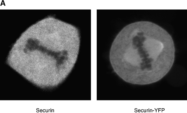
We established an in vivo assay for proteolysis using green fluorescent protein (GFP) fusion proteins (Clute and Pines, 1999; den Elzen and Pines, 2001). Because the level of GFP fluorescence was directly related to the amount of GFP fusion protein, we were able to follow proteolysis in real time by the decrease in GFP fluorescence. With this assay, we showed that cyclin A2 began to be degraded at, or just after, nuclear envelope breakdown, whereas cyclin B1 destruction began later, when the spindle checkpoint had been satisfied (i.e., when all the chromosomes were attached to both poles of the mitotic spindle) (Clute and Pines, 1999; den Elzen and Pines, 2001). To determine when securin destruction began in relation to cyclins A and B1, we tagged securin at the COOH terminus with cyan fluorescent protein (CFP), GFP, or yellow fluorescent protein (YFP). We were able to validate the securin–fluorescent proteins (FPs) as markers for endogenous securin by the following criteria. First, securin and the securin–FP chimaeras had the same localization pattern in interphase (unpublished data) and mitosis (Fig. 1 A). In interphase, the proteins were primarily nuclear, and in mitosis both securin and securin–YFP were localized throughout the cell and on the spindle but not on the chromosomes. Second, securin and the securin–FPs both bound to human separase (Fig. 1 B). Third, nondegradable versions of securin and securin–YFP gave identical phenotypes in mitosis, preventing chromosome separation (see below). Lastly, in fission yeast a securin (cut2)–GFP fusion protein had been shown to rescue a cut2 temperature-sensitive mutant (Kumada et al., 1998).
Figure 1.
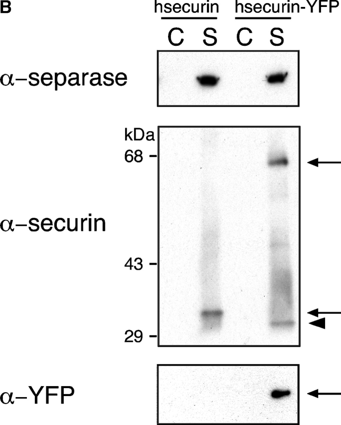
Securin–YFP is an appropriate marker for endogenous securin. (A) HeLa cells were microinjected in the nucleus with an expression construct for securin (left) or securin linked to YFP (right). After 3 h, the cells were fixed, and cells injected with the untagged securin were stained with antisecurin antibodies. Cells were analyzed by confocal fluorescence microscopy. A single z section is shown for each cell. (B) HeLa cells were transfected with an expression construct for securin tagged with a myc epitope (lanes 1 and 2) or an expression construct for securin–myc linked to YFP (lanes 3 and 4). Cells were arrested with nocodazole, and cell extracts were prepared and used to immunoprecipitate separase (S). An unrelated antibody was used as a control for immunoprecipitation (C). The precipitates were immunoblotted with specific antibodies against hseparase (top), hsecurin (middle), and YFP (bottom). Arrows indicate tagged forms of hsecurin; the arrowhead points to endogenous securin.
Securin–FP degradation is controlled by the spindle checkpoint
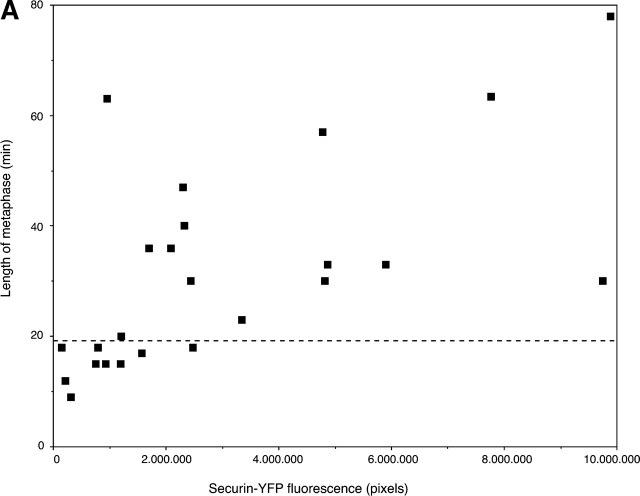
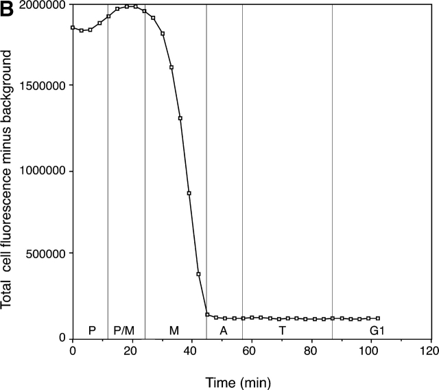
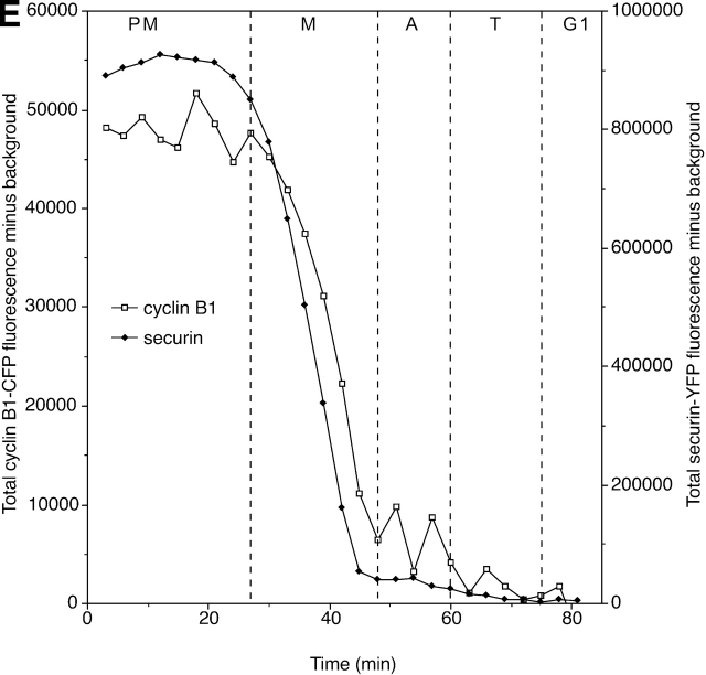
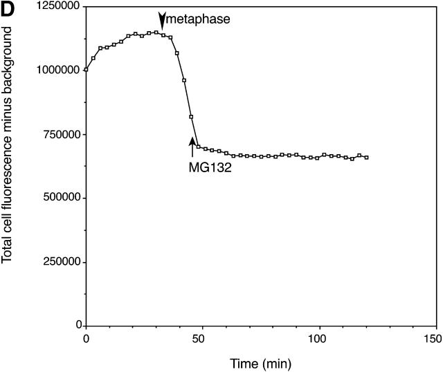
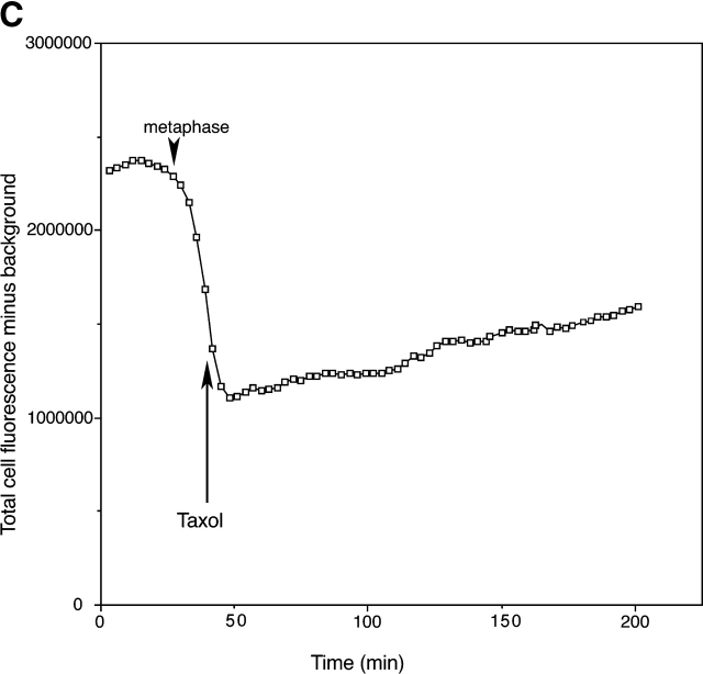
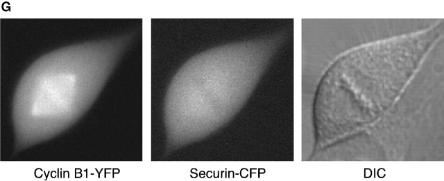
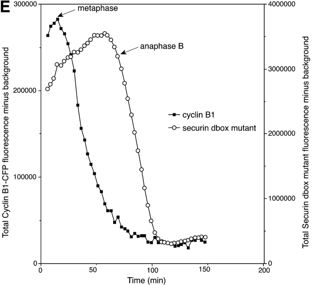
We injected cells with securin–FP constructs and assayed their progress through mitosis by time-lapse fluorescence and DIC microscopy (see Materials and methods). This showed that low to moderate levels of securin–FP made no significant difference to the length of any phase of mitosis, although high levels of securin–FP did extend metaphase (Fig. 2 A). Securin–FP fluorescence gradually increased through interphase and consistently began to fall just before all of the chromosomes aligned on the metaphase plate (Fig. 2 B). This indicated that securin might only have been recognized as a substrate for ubiquitination after the spindle checkpoint was satisfied. Consistent with this, disrupting proper spindle assembly by treating cells with nocodazole or taxol prevented securin–FP destruction (unpublished data), and spindle poisons were able to stop securin degradation even after it had begun (Fig. 2 C). Securin degradation was also blocked by the 26S proteasome inhibitor MG132, demonstrating that it was proteasome dependent (Fig. 2 D). In all of these properties, securin degradation strongly resembled that of cyclin B1 (Clute and Pines, 1999), and by quantifying the fluorescence levels of cyclin B1–CFP and securin–YFP expressed in the same cell we found their degradation consistently began at the same time (Fig. 2 E). However, cyclin B1 and securin only partially colocalized on the mitotic apparatus (Fig. 2 G).
Figure 2.
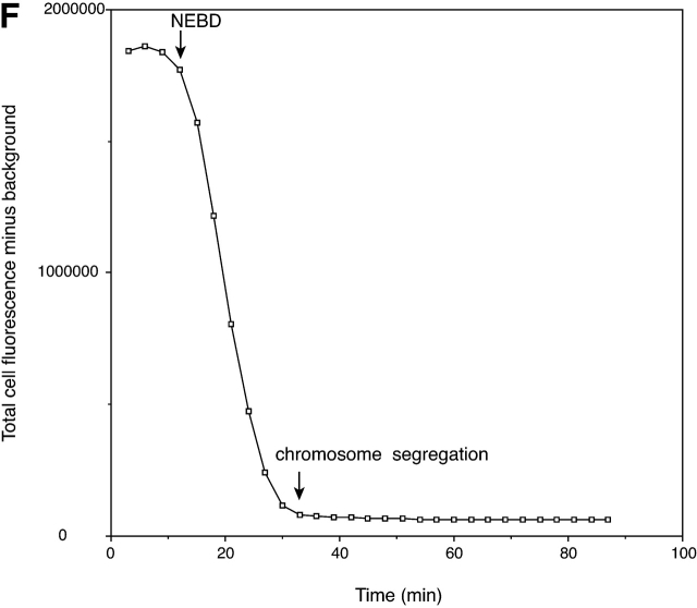
Securin is degraded in metaphase coincident with cyclin B1, and its proteolysis is controlled by the spindle checkpoint. (A) High levels of securin–FP extend metaphase. HeLa cells synchronized in late G2 phase were microinjected with an expression construct for securin–FP and followed by time-lapse fluorescence microscopy at 3-min intervals. The length of metaphase was determined and plotted against the level of securin–FP fluorescence at the beginning of metaphase. We defined metaphase as either the time between chromosome alignment and sister chromatid separation, or, in cells exhibiting chromosome nondisjunction, the time between chromosome alignment and cell elongation. The length of metaphase in uninjected cells was 9.9 ± 9.6 min (den Elzen and Pines, 2001). The dashed line indicates the maximum length of metaphase in control uninjected cells. (B) Securin degradation starts at metaphase. HeLa cells synchronized in late G2 phase were microinjected in the nucleus with an expression construct for securin–YFP and followed by time-lapse fluorescence and DIC microscopy at 3-min intervals. The total cell fluorescence minus background was quantified for each cell in successive images of a time series and plotted over time. The degradation profile of a single cell, representative of 10 cells analyzed, is shown. The stages of mitosis are indicated at the bottom of the figure. (C and D) Securin degradation is proteasome- and spindle checkpoint–dependent. HeLa cells were injected and analyzed as described in A. To reimpose the checkpoint or to stop proteasome-dependent degradation, cells in metaphase were treated with 10 μM taxol (C) or 100 μM MG132 (D), respectively. The arrow indicates the point at which taxol or MG132 was added, and the arrowhead indicates the start of metaphase. Graphs are of single cells, representative of at least four cells analyzed for each chemical. (E) Securin degradation coincides with cyclin B1 destruction. HeLa cells synchronized in late G2 phase were coinjected with cDNAs encoding cyclin B1–CFP and securin–YFP. Cells were analyzed as in A. The degradation profile of a single cell, representative of at least five cells analyzed, is shown. (F) In the absence of the spindle checkpoint, securin degradation starts in prometaphase. HeLa cells were synchronized in late G2 phase and coinjected with cDNAs encoding securin–YFP and a dominant negative mutant of Bub1. Cells were followed by time-lapse microscopy and analyzed as in A. The times of the completion of nuclear envelope breakdown and the start of chromosome segregation are indicated. The degradation profile of a single cell, representative of at least four cells analyzed, is shown. (G) Cyclin B1 and securin do not exactly colocalize. HeLa cells were coinjected with cDNAs encoding cyclin B1–YFP and securin–CFP, and fluorescence and DIC pictures were taken at the beginning of metaphase.
We and others showed that the difference in the timing of cyclin A2 and cyclin B destruction in mammalian cells was imposed by the spindle checkpoint in prometaphase (den Elzen and Pines, 2001; Geley et al., 2001). Cyclin A2 degradation was insensitive to the spindle checkpoint and began as soon as the nuclear envelope broke down, whereas cyclin B1 was only degraded once the spindle checkpoint was inactivated. Furthermore, disrupting the spindle checkpoint machinery with a dominant negative version of Bub1 caused cyclin B1 to be degraded prematurely in prometaphase (Geley et al., 2001). To determine whether this also applied to securin degradation, we coinjected securin–FP with a dominant negative mutant of Bub1 to disrupt the checkpoint. This showed that in the absence of the spindle checkpoint securin was degraded at the same time as cyclin A2, as soon as the nuclear envelope broke down (Fig. 2 F).
Securin degradation may not be sufficient for anaphase
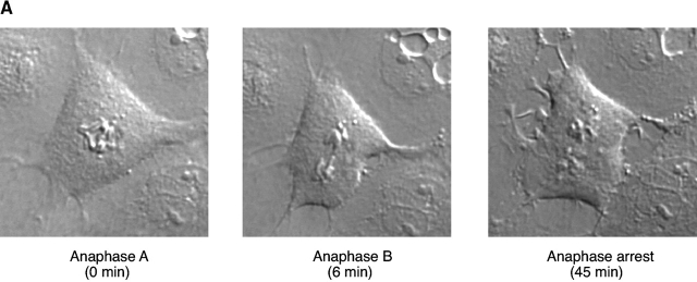
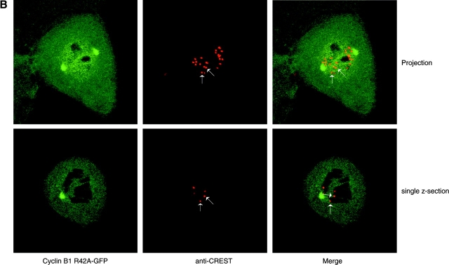
Introducing a dominant negative Bub1 mutant caused securin to be degraded prematurely, and cells rapidly began anaphase, but this did not show that securin degradation alone could cause sister chromatid separation because eliminating the spindle checkpoint would advance the degradation of other mitotic regulators, such as cyclin B1 (Geley et al., 2001). To determine the requirements for sister chromatid separation in mammalian cells, we coexpressed a dominant negative mutant of Bub1 with a nondegradable version of cyclin B1 and assayed sister chromatid separation by both DIC microscopy and by fixing and staining cells with anticentromere or anti–phospho-histone H3 antibodies. To analyze the behavior of all of the sister chromatids, we used PtK1 cells that only have 12 sets of chromosomes. In some experiments, we expressed a nondegradable cyclin B1 alone and analyzed cells after all of the securin should have been degraded (at least 1 h after metaphase began; on average, control PtK1 cells enter anaphase 23 min after chromosome alignment [Rieder et al., 1994]). We found that whether sister chromatids separated in the absence of securin depended on the level of cyclin B1. With low levels of cyclin B1, sister chromatids could separate (Fig. 3 A), but cells arrested in late anaphase as described previously (Wheatley et al., 1997); however, at moderate to high levels of cyclin B1 sister chromatids remained together for several hours after the disappearance of securin (Fig. 3, B and C). (We found that nondegradable cyclin B1 did not affect the degradation of securin [see Fig. 7 B; unpublished data]). We have shown previously that when expressed at moderate levels ectopic cyclin B1, or cyclin A2, is present at ∼50–100% of the level of the endogenous cyclin (Draviam et al., 2001; den Elzen and Pines, 2001). Thus, we estimate that a 1.5–2-fold overexpression of cyclin B1 is sufficient to block sister chromatid separation in the absence of securin. Although it is not clear whether cyclin B1 would ever normally reach these levels in vivo, our observations agree with the results of Stemmann et al. (2001) who found that a twofold excess of cyclin B1 blocked sister chromatid separation in Xenopus extracts through phosphorylating and inactivating separase.
Figure 3.
Cyclin B1 levels can affect sister chromatid separation. (A) Low levels of cyclin B1 at the end of metaphase cause a late anaphase arrest. PtK1 cells were injected with an expression construct for nondegradable cyclin B1–GFP (R42A mutation). Cells expressing low levels of R42A–cyclin B1–GFP (<106 pixels for GFP) were followed by time-lapse fluorescence and DIC microscopy at 3-min intervals. The cell shown is representative of more than four cells analyzed. (B and C) High levels of cyclin B1 at the end of metaphase prevent sister chromatid separation. PtK1 cells were injected with an expression construct for nondegradable cyclin B1–GFP as in A. Cells expressing moderate to high levels of R42A–cyclin B1–GFP (>106 pixels for GFP) were followed by time-lapse fluorescence and DIC microscopy at 3-min intervals. Once cells had been in metaphase for >1 h, they were fixed and stained with anti-CREST (B) or anti-CREST and phospho-histone H3 antibodies (C). Cells were analyzed by confocal laser scanning microscopy, and either a single z section (B) or a projection of several z sections is shown (B and C). The arrows in B indicate pairs of kinetochores. In the single z section, only one kinetochore of the pair is visible, and the chromosomes can be visualized by the negative stain for cyclin B. The cell shown is representative of more than four cells analyzed. The arrows in Fig. 3 C indicate unseparated sister chromatids. The cell shown is representative of more than three cells analyzed.
Figure 7.
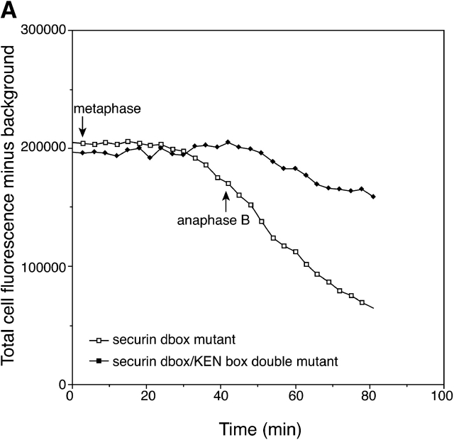
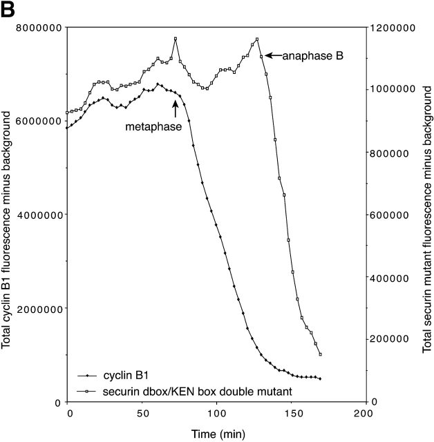
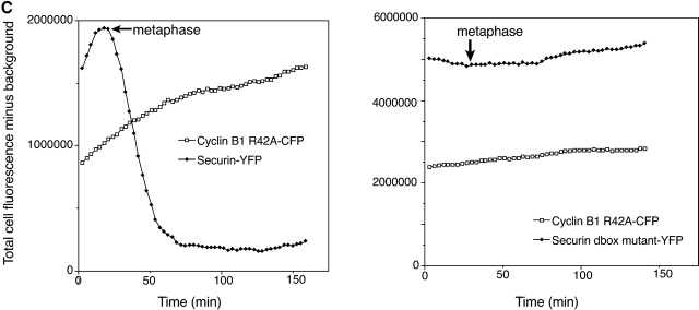
The degradation profile of a securin mutant dependent on a KEN box for destruction reveals when Cdh1 replaces Cdc20. (A) The securin D-box mutant is degraded in anaphase, and a D-box/KEN double box mutant is partially stabilized in mitosis. HeLa cells were coinjected with cDNAs encoding a securin D-box deletion mutant linked to CFP and a securin D-box/KEN box double mutant linked to YFP. Cells were followed through mitosis, and DIC and fluorescence pictures were taken at 3-min intervals and analyzed as in the legend to Fig 2. The times at which metaphase and anaphase B started are indicated. The graph of a single cell is shown and represents at least eight cells analyzed. (B) Coexpressing cyclin B1 enhances the degradation of the securin double mutant. HeLa cells were coinjected with expression constructs for a wild-type cyclin B1–CFP together with a securin D-box and KEN box double mutant linked to YFP. Cells were followed, and DIC and fluorescence pictures were taken every 3 min and analyzed as in the legend to Fig 2. The times at which metaphase and anaphase B started are indicated. The graph of a single cell is shown and represents at least five cells analyzed. (C) High cyclin B1/CDK1 activity prevents the degradation of a securin mutant that depends on a KEN box for its proteolysis. HeLa cells were coinjected with expression constructs for a nondegradable R42A–cyclin B1–CFP together with either securin–YFP (left) or a securin D-box mutant linked to YFP (right). Cells were followed, and DIC and fluorescence pictures were taken every 3 min and analyzed as in the legend to Fig 2. The times at which metaphase started are indicated. The graph of a single cell is shown and represents at least five cells analyzed for each securin mutant.
Nondegradable securin can cause complete chromosome nondisjunction
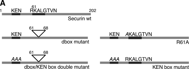
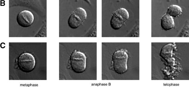
We wished to determine whether securin linked karyokinesis with cytokinesis in human cells. In budding yeast, nondegradable Pds1p arrested cells in metaphase, whereas in fission yeast nondegradable cut2 prevented sister chromatid separation but could not prevent cytokinesis. The reported effect of a nondegradable human securin was ambiguous: nondegradable hsecurin only induced a mild cut phenotype where a small amount of chromatin was trapped between the two daughter cells, and this was in only a minority (5%) of human cells (Zur and Brandeis, 2001). To explore the requirement for securin degradation in human cells, we made several mutations in the putative destruction motifs of hsecurin (Fig. 4 A). Initially, we made a point mutation in a conserved residue of the putative D-box or deleted the D-box altogether. When introduced into cells, both of these securin mutants failed to be degraded in metaphase (but were degraded later in mitosis [Fig. 4 E and see Fig. 6 B]) and caused a dramatic cut phenotype in 100% of the injected cells. This phenotype was observed with nondegradable securins with or without a GFP tag and routinely with high levels of wild-type securin–FP (Table I). In cells exhibiting a cut phenotype, all of the observable sister chromatids failed to separate before anaphase B and either remained in the center of the cell to be disrupted by the cytokinesis furrow (Fig. 4 B) or moved to one spindle pole to be inherited by one cell (Fig. 4 C). (We took cell elongation to be indicative of anaphase B.) Neither of these cut phenotypes appeared to be more severe than the other because there was no obvious correlation between the phenotype observed and the level of securin expressed (Table I).
Figure 4.
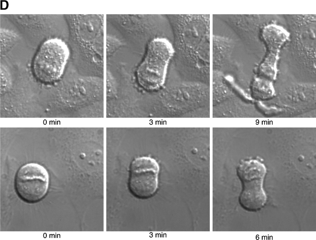
Nondegradable securin causes a cut phenotype. (A) Schematic diagram of securin constructs. The D-box (R61-N68) and KEN box (K9-N11) are indicated. (B and C) HeLa cells injected with an expression vector encoding a securin D-box deletion mutant–YFP fusion protein were followed through mitosis by time-lapse microscopy, and DIC pictures were taken every three min. These cells are representative of at least 18 cells analyzed. (D) Reversion of the sequence of anaphase A and B. HeLa cells were coinjected with cDNAs expressing cyclin B1–CFP and a securin D-box deletion mutant–YFP (top) or with cDNAs expressing securin D-box mutant–CFP and a securin D-box/KEN box double mutant–YFP (bottom). Cells were followed through mitosis by time-lapse microscopy, and DIC pictures were taken every three min. In a minority of cells, the sister chromatids separated after the cell elongated. (E) HeLa cells were coinjected with cDNAs expressing cyclin B1–CFP and a securin D-box deletion mutant–YFP. Cells were followed by time-lapse microscopy. Fluorescence and DIC pictures were taken at 3-min intervals and analyzed as described in the legend to Fig 2. The degradation profile of a single cell is shown and is representative of at least six cells analyzed. The times at which metaphase and anaphase B started are shown.
Figure 6.
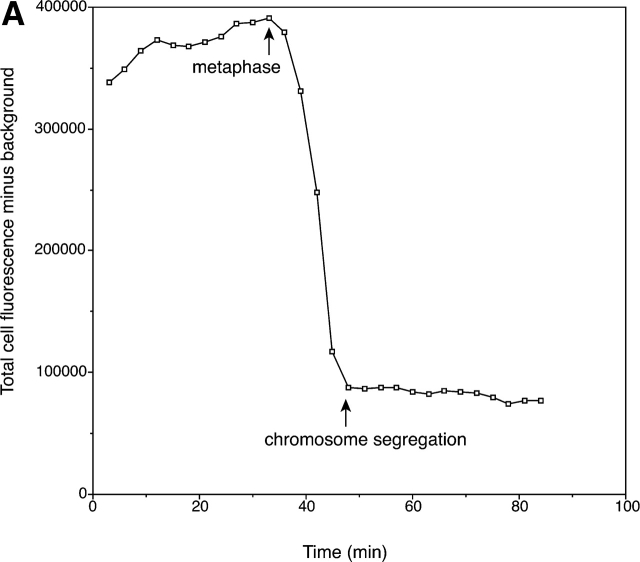
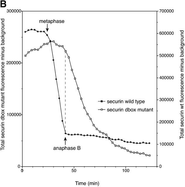
Securin destruction in metaphase requires only the D-box not the KEN box. (A) Degradation of the securin KEN box mutant starts at metaphase. HeLa cells were injected with an expression vector for a securin KEN box mutant–YFP fusion protein and followed through mitosis by time-lapse microscopy. Fluorescence and DIC pictures were taken at 3-min intervals and analyzed as described in the legend to Fig 2. The times of the beginning of metaphase and chromosome segregation are indicated. The graph is representative of at least five cells analyzed. (B) HeLa cells were coinjected with cDNAs encoding a securin D-box deletion mutant–CFP and the securin wildtype–YFP fusion proteins. The cells were analyzed as described in the legend to Fig. 2. The times at which metaphase and anaphase B started are indicated. The degradation profile of a single cell is shown and is representative of at least five cells analyzed. All cells had a cut phenotype.
Table I. Characterization of the effect of securin expression.
| Securin construct |
Coexpressed with |
Expression level |
Number of cells injected |
Number of cells with normal anaphase |
Number of cells with cut phenotype |
Cells with cut phenotype |
Number of cells with all chromosomes moving to one pole |
|---|---|---|---|---|---|---|---|
| % | |||||||
| Wild-type | YFP | 8 | 7 | 1 | 12.5 | 1 | |
| ΔD-box | YFP | 5 | 0 | 5 | 100 | 4 | |
| Wild-type–YFP | low | 11 | 8 | 3 | 27 | 0 | |
| high | 14 | 2 | 12 | 86 | 10 | ||
| ΔD-box–YFP | low | 8 | 0 | 8 | 100 | 4 | |
| high | 10 | 0 | 10 | 100 | 6 | ||
| R61A–YFP | low | 3 | 0 | 3 | 100 | 0 | |
| high | 2 | 0 | 2 | 100 | 2 | ||
| KEN box mutant–YFP | low | 6 | 5 | 1 | 16 | 1 | |
| high | 3 | 0 | 3 | 100 | 1 | ||
| ΔD-box/KEN box double mutant–YFP | low | 3 | 0 | 3 | 100 | 0 | |
| Wild-type–YFP | Cyclin B1–CFP | 7 | 5 | 2 | 28 | 1 | |
| ΔD-box–YFP | Cyclin B1–CFP | 6 | 0 | 6 | 100 | 3 | |
| KEN box mutant–YFP | Cyclin B1–CFP | 2 | 1 | 1 | 50 | 0 | |
| ΔD-box/KEN box double mutant–YFP | Cyclin B1–CFP | 5 | 0 | 5 | 100 | 4 | |
| Wild-type–YFP | Securin ΔD-box–CFP
|
5 | 0 | 5 | 100 | 0 | |
| ΔD-box/KEN box double mutant–YFP | Securin ΔD-box–CFP | 5 | 0 | 5 | 100 | 2 |
HeLa cells synchronized in late G2 phase were injected with securin expression constructs, with or without cyclin B1 constructs, and followed by time-lapse fluorescence and DIC microscopy. The total fluorescence per cell was quantified and categorized as either low (<2,000,000 pixels for YFP) or high (>2,000,000 pixels for YFP). Cells coexpressing securin constructs with high levels of cyclin B1 that arrested in metaphase were not included in this analysis. NB, A cut phenotype was never seen in control uninjected cells.
In those cells where all of the chromosomes moved to one pole, the sister chromatids occasionally separated in one half of a cell, reversing the normal sequence of anaphase A and B. Furthermore, in some of these cells a second cleavage furrow initiated between the separating chromatids (Fig. 4 D). Further observations showed that a cell that apparently failed to inherit chromosomes continued to bleb after cytokinesis and eventually either fused with the other daughter cells or died. The cell that inherited all of the chromosomes eventually reentered interphase. The daughter cells formed by either cut phenotype usually took a long time to reform their nuclear envelopes and often failed completely to separate. This was likely to be caused by chromatin that was trapped in the cleavage furrow, visualized by staining with Hoechst 33342 (unpublished data) as described previously (Mullins and Biesele, 1977; Hauf et al., 2001).
Based on our present understanding of mitosis, these cut phenotypes were indicative of cells in which securin was still present, preventing chromatid separation, but cyclin B1 had been degraded, enabling cytokinesis to occur. In agreement with this interpretation, nondegradable securin did not affect cyclin B1 degradation (Fig. 4 E).
Securin mutants may reveal when Cdh1 replaces Cdc20 in mitosis
Currently, APC/CCdc20 is thought to ubiquitinate proteins with a D-box, whereas APC/CCdh1 that is active later in mitosis is thought to recognize proteins with either a D-box or a KEN box (Pfleger and Kirschner, 2000). Thus, we considered that we might be able to use securin mutants to analyze when proteolysis switches from mediation by Cdc20 to Cdh1. Therefore, in addition to the D-box mutants we made hsecurin mutants in which we eliminated the KEN box by replacing the residues KEN with AAA.
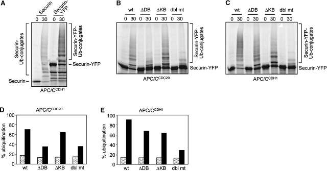
First, we tested which APC/C complexes were able to ubiquitinate the different securin mutants in vitro. We found that APC/CCdc20 and APC/CCdh1 complexes were able to ubiquitinate securin or securin–FP constructs with equal efficiency (Fig. 5 A; unpublished data). In accordance with the prevailing view, we found that APC/CCdc20 was only able efficiently to ubiquitinate securin or securin–FP that possessed a wild-type D-box, and it did not matter whether or not the mutant had a KEN box (Fig. 5, B and D). In contrast, APC/CCdh1 ubiquitinated securin with either a wild-type D-box or a KEN box (Fig. 5, C and E) but was unable efficiently to ubiquitinate a protein lacking both destruction motifs (Fig. 5, C and E).
Figure 5.
Securin can be ubiquitinated by APC/CCdc20 and APC/CCdh1. (A) APC/Ccdh1 can ubiquitinate securin and securin–YFP with equal efficiency in vitro. (B) In vitro ubiquitination of securin–YFP by APC/CCDC20 is dependent on an intact D-box. (C) APC/CCDH1 ubiquitinates securin–YFP containing either an intact D-box or KEN box. In A–C, APC/C was immunoprecipitated from Xenopus egg extracts and activated with recombinant CDH1 or CDC20. 35S-labeled in vitro–translated securin or securin–YFP wild-type (wt), D-box mutant (ΔDB), KEN box mutant (ΔKB), or D-box/KEN box double mutants were used as substrates. (D and E) Quantification of the data shown in B and C. The amount of securin–YFP conjugated to ubiquitin is shown as the percent of the total amount of securin–YFP per reaction. Time points 0 (hatched bars) and 30 min (black bars) indicate, respectively, the start and end points of the reaction.
When assayed in vivo, the KEN box mutant was degraded in metaphase at the same time as the wild-type protein, the sister chromatids separated on time and the cells exited mitosis normally (Fig. 6 A). (At high levels, this mutant also caused a cut phenotype in a similar manner to the wild-type protein [Table I].) This showed that the KEN box was dispensable for degradation in metaphase and apparently for mitosis. However, because we did not follow cells for a second cell cycle we do not know whether a mutation in the KEN box would give a phenotype in interphase or the next mitosis. As mentioned above, the D-box mutant caused a cut phenotype because it was not degraded in metaphase. However, just before the cell began to elongate, indicative of anaphase B, this mutant was degraded (Fig. 6 B).
One interpretation of these results was that proteolysis switched from mediation by APC/CCdc20 to APC/CCdh1 in anaphase, and at this point the D-box mutant could be degraded via its KEN box. To test this, we analyzed the degradation of a securin mutant that lacked both a D-box and a KEN box. We found that in most cells this protein was more stable than either the D-box or the KEN box mutants (Fig. 7 A). However, the double mutant was not completely stable after anaphase, indicating that there may be other motifs that target it for degradation (Fig. 7 A). As expected, this mutant blocked sister chromatid (giving cut phenotypes like those induced by the single D-box mutant) but had no effect on cyclin B1 degradation or cytokinesis. Interestingly, we found that coexpressing cyclin B1 with this mutant appeared to increase the efficiency with which the securin mutant was eventually degraded (Fig. 7 B).
Previous studies provided evidence that B-type cyclin–CDK activity inhibited APCCdh1-mediated proteolysis by phosphorylating Cdh1 (Zachariae et al., 1998; Jaspersen et al., 1999; Kramer et al., 2000). Thus, if APCCdh1 did mediate degradation of the D-box securin mutant this should have correlated with reduced cyclin B1–CDK activity. Consistent with this prediction, we found that D-box mutants began to be degraded at the time when the majority of cyclin B1–FP had been degraded (Fig. 4 E and Fig. 7 B). Furthermore, when we coexpressed the D-box mutant of securin with nondegradable cyclin B1 to maintain Cdh1 in its inactive (phosphorylated) form the mutant securin was now completely stable (Fig. 7 C).
Discussion
In this paper, we have studied the degradation of securin in dividing human cells. We have used securin FPs to follow the localization and proteolysis of securin in real time. A previous study using a securin–GFP fusion protein in living cells found that securin–GFP transfected into endothelial cells, inhibited mitosis, and promoted apoptosis (Yu et al., 2000). In those few cells that entered mitosis the protein was judged to disappear in anaphase, but this was based on a qualitative assessment of fluorescence. In contrast, we find that securin–GFP neither induces apoptosis nor inhibits entry into mitosis. Furthermore, we have quantified the disappearance of securin–FP and shown that its degradation begins at metaphase. The differences between our results and the previous study may lie in the cell type used or reflect an advantage of microinjection over transfection.
We have shown that the timing of securin destruction is controlled by the spindle checkpoint; destruction only begins at metaphase, and reimposing the checkpoint in metaphase rapidly inactivates securin proteolysis. Furthermore, disrupting the checkpoint machinery with a dominant negative mutant of Bub1 causes securin to be degraded prematurely, just after nuclear envelope breakdown. Thus, the properties of securin destruction resemble those of cyclin B1. However, securin and cyclin B1 do not have identical subcellular localization patterns in mitosis; unlike cyclin B1, securin does not stain the chromosomes in prometaphase. Therefore, if as suggested previously there is some spatial control on ubiquitination in mitosis (Clute and Pines, 1999; Huang and Raff, 1999) this might be able to discriminate between cyclin B1 and securin, although we have not yet found conditions that uncouple securin from cyclin B1 destruction. This is in contrast to budding yeast where securin (Pds1p) and a fraction of the major B-type cyclin, Clb2, are degraded before anaphase, but a significant proportion of Clb2 remains to be degraded later in mitosis (Lim et al., 1998; Baumer et al., 2000; Yeong et al., 2000).
Although securin destruction begins as soon as the spindle checkpoint is inactivated, sister chromatids often do not separate until much later (∼23 min later in PtK1 cells [Rieder et al., 1994]). Moreover, at anaphase all of the sister chromatids separate with a high degree of synchrony. It is difficult to reconcile this observation with a model in which sister chromatid separation is solely controlled by the activation of separase after securin is destroyed. If this is the case, then active separase could accumulate for ∼20 min before sister chromatid disjunction, and it is unlikely that all of the sister chromatids would separate at the same time. Furthermore, the majority of securin−/− mouse cells must also separate their chromosomes correctly as evidenced by their ability to proliferate in culture (Jallepalli et al., 2001) and to generate apparently normal animals (Mei et al., 2001). Thus, we favor a model in which there is a second step to sister chromatid separation, perhaps related to the recent demonstration that the budding yeast separase is only able to recognize the phosphorylated form of its cohesin substrate (Alexandru et al., 2001). In some cells injected with securin–FP we observe a cut phenotype even after the securin–GFP has fallen below detectable levels. This may be because there is still sufficient securin to inactivate the separase. However, the more interesting possibility is that there is insufficient time for the second step between securin destruction and the cleavage of cohesin.
The second step to sister chromatid separation may involve the inactivation of cyclin B1–CDK1. We find that moderate to high amounts of cyclin B1 (Fig. 3) will prevent sister chromatid separation in the absence of securin, and we have shown previously that cyclin A2 must also be degraded to allow anaphase (den Elzen and Pines, 2001). High amounts of cyclin B–CDK1 activity have been shown to prevent sister chromatid separation in Xenopus extracts because separase remains phosphorylated and its ability to cleave cohesin in vitro is significantly reduced (Stemmann et al. 2001). Thus, it appears that cyclin B1–CDK1, and possibly cyclin A2–CDKs, may directly or indirectly inactivate separase. This alternative means of regulating sister chromatid separation could explain why securin is not an essential protein in mammalian cells (Jallepalli et al., 2001; Mei et al., 2001).
Securin degradation is a key event in mitosis. We found that all of the securin mutants that could not be degraded in metaphase block sister chromatid separation, and unlike budding yeast human cells do not have a mechanism to prevent cytokinesis in this event. Thus, the daughter cells inherit an incorrect complement of chromosomes, and this may be the mechanism by which overexpressed human securin transforms 3T3 cells (Pei and Melmed, 1997). This emphasizes the importance of the spindle checkpoint both to normal progression through mammalian mitosis and for genomic stability. This is underscored by the observations that mice lacking spindle checkpoint components such as Mad2 (Dobles et al., 2000) and Bub3 (Kalitsis et al., 2000) die early in embryogenesis with gross mitotic abnormalities. Furthermore, a haplo insufficiency of Mad2 gives rise to chromosomal instability and eventually to tumorigenesis (Michel et al., 2001) so the level of the checkpoint proteins is likely to be crucial to the proper regulation of securin and cyclin B1 destruction.
Because all of the cells expressing securin mutants that could not be degraded in metaphase exhibit a cut phenotype, we conclude that degrading human securin is an essential prerequisite for sister chromatid separation. However, the phenotype we observed for the D-box mutant of securin differs from the cut phenotype observed by Zur and Brandeis (2001) in which only a small minority (5%) of HeLa cells expressing a nondegradable securin failed to complete cytokinesis and remained connected by chromatin threads. The difference may be due to the different experimental approaches; we microinjected cells and followed them by time-lapse microscopy, whereas Zur and Brandeis (2001) transfected cells and analyzed them after fixation.
The high rates of whole chromosome loss that we observe for cells with nondegradable securin are similar to the effects of the loss of securin in human cells (Jallepalli et al., 2001). This might appear paradoxical but can be explained by data indicating that securin is required fully to activate separase (Jallepalli et al., 2001). Thus, a nondegradable separase inhibitor would have the same effect as an inability to activate separase. Cells without securin also exhibit problems with sister chromatid separation, but these problems are only seen in about a third of the population, perhaps because the cells still have a low but detectable level of active separase.
The behavior of the various securin mutants in mitosis may give some clues to the changes in the machinery underlying progression through mitosis in somatic cells. A securin mutant with a defective D-box but an intact KEN box could be ubiquitinated by APC/CCdh1 but not APC/CCdc20 in vitro. Although this is an artificial substrate (because normally all the securin should be degraded in metaphase by APC/CCdc20) in vivo this mutant was stable throughout metaphase but became unstable just before the cell began to elongate at anaphase B. Thus, the switch from APC/CCdc20- to APC/CCdh1-dependent destruction appears to happen in anaphase. By extrapolation from results obtained with budding yeast, this could be explained by the disappearance of cyclin B1–CDK activity at the end of metaphase leading to the dephosphorylation of Cdh1. Dephosphorylated Cdh1 would then bind the APC (Kramer et al., 2000) and begin the degradation of Cdc20 and other KEN box substrates. In support of this model, we find that a nondegradable cyclin B1 mutant prevents the destruction of the securin D-box mutant with an intact KEN box. Alternatively, but less likely, APC/CCdc20 might alter its specificity to recognize the KEN box.
The question arises as to why somatic cells should switch from degradation mediated by APC/CCdc20 to APC/CCdh1 in anaphase. Clearly this is not required for exit from mitosis itself because degradation in embryonic cell cycles, such as those of Drosophila and Xenopus, is mediated solely by Cdc20. It could be that APC/CCdh1 is required because there are some late mitotic regulators that are only present in somatic cells that cannot be recognized by APC/CCdc20. Alternatively, there may be some proteins that must be degraded in somatic cells but not in embryonic cells. Such proteins could include the regulators or components of the prereplication complex because one of the major differences between somatic cell cycles and embryonic cell cycles is that somatic cells have an interval (G1) between mitosis and the next round of DNA replication. During G1 phase, somatic cells integrate intra- and extracellular signals before commitment to another round of DNA replication rather than the alternative fates of differentiation or quiescence. Thus, it may be important for somatic cells to ensure that components of the DNA replication machinery are not present until they commit to another round of proliferation. Equally, it is becoming clear that once somatic cells exit the cell cycle and differentiate the APC/C plays an important part in the physiology of postmitotic cells (Gieffers et al., 1999) where it must recognize substrates that are very different from those found in proliferating cells. However, this does not provide an answer to why Cdc20 itself should become a target for degradation upon exit from mitosis in somatic cells.
Lastly, our observation that a second cleavage furrow can form between the separating sister chromatids after all the chromosomes have moved to one pole indicates that the cytokinesis furrow can be very rapidly established, apparently by unseparated chromosomes. It will be interesting to determine which of the chromosomal passenger proteins implicated in cytokinesis are carried toward one spindle pole by the unseparated chromosomes and whether any are left behind at the first cleavage furrow.
Materials and methods
Cell culture and synchronization
HeLa cells and PtK1 cells were cultured and synchronized as described previously (Clute and Pines, 1999).
Construction of cDNA plasmids
Fusion proteins and point mutations were constructed by PCR using VENT polymerase (New England Biolabs, Inc.), cloned in the appropriate vectors, and confirmed by automated sequencing. Wild-type and mutant securin constructs were linked at their COOH terminus via a RDPPVAT linker to YFP or CFP in pEYFP/N1 or pECFP/N1 (CLONTECH Laboratories, Inc.). For the in vitro ubiquitination and transfections experiments, the securin constructs were subcloned into pCDNA3 (Invitrogen). The D-box of securin from R61 to N68 was deleted to generate the securin D-box deletion mutant, the sequence K9 E10 N11 was replaced by three alanines to obtain the KEN box mutant, and to generate the D-box/KEN box double mutant these two mutations were combined. To generate a point mutation in the D-box, R61 was replaced by alanine. pCMX/cyclin B1–YFP has been described previously (Hagting et al., 1999.) To generate cyclin B1–CFP, cyclin B1 was linked by its COOH terminus to CFP via a linker with the sequence LERDPPVAT using pECFP/N1. R42 of cyclin B1–CFP was replaced by an alanine to generate a nondegradable cyclin B1–CFP. R42A–B1–GFP has been described previously (Clute and Pines, 1999). pEF-Bub1 dominant negative (NH2-terminal fragment of mouse Bub1 [N334 was a gift from Stephan Geley and Tim Hunt, Cancer Research UK]) (Geley et al., 2001). All plasmids were resuspended in 10 mM Tris, pH 8.0, for microinjection (YFP constructs at 0.01 mg/ml, the CFP constructs at 0.05 mg/ml, and Bub1 DN at 0.1 mg/ml). Maps of all constructs and sequences used in this study are available on request.
Immunofluorescence
HeLa cells were seeded onto metasilicate-coated coverslips and then fixed and stained using paraformaldehyde/Triton as described (Pines, 1997). The antisecurin primary antibody (a gift from Hui Zou, Harvard Medical School, Cambridge, MA) was diluted 1:1,000 in 3% BSA/PBS, and washes were performed with PBS. Anti-CREST serum (a gift from Dr. Bill Earnshaw, University of Edinburgh, Edinburgh, UK) was used at 1:20,000, and anti–phospho-histone H3 Serine 28 (a gift from Dr. Inagaki, Aichi Cancer Center Research Institute, Nagoya, Japan) was used at 1:100. Secondary antibodies linked to Texas red (1:200; Jackson ImmunoResearch Laboratories) or Alexa dyes 594 or 647 (1:400; Molecular Probes) or Cy5 (1:200; Amersham Pharmacia Biotech) were diluted as indicated. Coverslips were mounted in 0.1% 1,4-phenylenediamine, 90% glycerol in PBS (pH 9.0). Cells were analyzed by confocal laser scanning microscopy.
Coimmunoprecipitation
HeLa cells grown to ∼60% confluency were transiently transfected with pcDNA3-myc securin and with pcDNA3-myc securin–YFP constructs. For transfection, LipofectAMINE PLUS™ (GIBCO BRL) was used according to the manufacturer's instructions. After 24 h of transfection, cells were treated with nocodazole for 18 h at a final concentration of 330 nM. Subsequently, cells were harvested, and cell extracts were prepared as described previously (Waizenegger et al., 2000). 30 μl of affi-prep protein A beads (Bio-Rad Laboratories, Inc.) coupled with 30 μg affinity purified rabbit antihseparase antibodies or with 30 μg affinity purified rabbit anti-CDC27 antibodies (a gift from Christian Gieffers, Research Institute of Molecular Pathology) were used for immunoprecipitation. 1.4 mg of a low speed supernatant of cell extracts was used for the immunoprecipitation. Proteins were allowed to bind to the antibodies for 3 h 45 min at 4°C. Beads were washed several times with 0.5 M NaCl in TBS supplemented with 0.5% Tween 20 and 1 mM DTT. Immunoprecipitates were eluted from beads by the addition of 40 μl of sample buffer. Immunoprecipitates were immunoblotted with mouse monoclonal antibody against hseparase (7A6; 1:1,000 diluted) with mouse monoclonal antibody against hsecurin (a gift from Claus Sørensen and Jiri Lukas, Danish Cancer Center, Copenhagen, Denmark; 1:500 diluted) and with mouse monoclonal antibodies against GFP (1814460; Roche).
Microinjection and time-lapse imaging and analysis
Cells were injected and analyzed by time-lapse DIC fluorescence microscopy as described previously (Clute and Pines, 1999; Furuno et al., 1999). For comparative quantitative analyses, all parameters were fixed; a fluorescence exposure time of 250 ms and a 40× oil objective with a numerical aperture of 1.0 were used. Images were saved in IP Lab Spectrum format as unsigned 16 data using a reference look up table with a preset linear pixel intensity scale. IP Lab Spectrum (Scanalytics) or a modified version of Image J software (NIH) was used to quantify the amount of fluorescence as described previously (Clute and Pines, 1999; Furuno et al., 1999). DIC images were used to determine mitotic phases. Images were converted to PICT format and exported to Adobe® PhotoShop.
In vitro ubiquitination
[35S]methionine- and [35S]cysteine-labeled wild-type or mutated securin and securin–YFP were prepared by coupled transcription-translation reactions in rabbit reticulocyte lysate (Promega). To obtain APC/CCDH1, APC/C was immunoprecipitated from Xenopus egg interphase extracts using anti-APC3/CDC27 antibody beads. To obtain mitotic APC/C appropriate for activation by CDC20, interphase egg extracts were driven into mitosis by addition of nondegradable sea urchin cyclin B Δ90 and used for immunoprecipitation with anti-APC3 antibody beads. The beads were subsequently washed three times with buffer XB (10 mM Hepes-KOH, pH 7.7, 100 mM KCl, 1 mM MgCl2, 0.1 mM CaCl2) supplemented with 0.2 μM okadaic acid and incubated with 100 ng recombinant purified CDH1 or CDC20 (Kramer et al., 2000) in buffer XB plus okadaic acid for 12 min at room temperature. After two more washing steps in XB plus okadaic acid, 5 μl beads were used in ubiquitination reactions containing purified E1 (80 μg/ml), UBC4 and UBCx (50 μg/ml each), ubiquitin (1.25 mg/ml), ATP regenerating system (7.5 mM creatine phosphate, 1 mM ATP, 1 mM MgCl2, 0.1 mM EGTA, 30 U/ml rabbit creatine phosphokinase type I [Sigma-Aldrich]), and 2 μl in vitro–translated substrate in a final volume of 15 μl in buffer QA (10 mM Hepes-KOH, pH 7.7, 100 mM KCl, 1 mM MgCl2, 0.1 mM CaCl2, 1 mM DTT). One half of the reaction was removed immediately and mixed with SDS-PAGE sample buffer, whereas the other half was incubated for 30 min at 25°C on a shaker before addition of sample buffer. The reaction was analyzed by SDS-PAGE and phosphorimaging. The ratio of polyubiquitinated substrate to total substrate was calculated after quantification using the ImageQuant software (Molecular Dynamics).
Acknowledgments
We are very grateful to Dr. Jean-Yves Thuret for modifying the Image J program to analyze time-lapse images. We also thank Mark Kirschner for the human securin cDNA, and he and Olaf Stemann for sharing unpublished results, Stephan Geley and Tim Hunt for dominant negative Bub1, Claus Sorensen and Jiri Lukas for the monoclonal securin antibodies, Hui Zou for securin antibodies, Dr. Inagaki for the anti–phospho-H3 antibody and Bill Earnshaw for anti-CREST serum and moral support. We are grateful to all the members of the laboratory for helpful discussions and especially to Catherine Lindon for helpful comments on the article.
Research in the lab of J.-M. Peters is supported by Boehringer Ingelheim and grants from the Austrian Science Fund and the Austrian Industrial Research Promotion Fund. A. Hagting was supported by a European Union Training and Mobility of Researchers Programme network grant and a Cancer Research Campaign project grant, and N. den Elzen was supported by the Association of Commonwealth Universities. This work was supported by a Cancer Research Campaign program grant to J. Pines.
N. den Elzen's present address is Peter MacCallum Cancer Institute, St. Andrew's Place, Melbourne, VIC 8006, Australia.
Footnotes
Abbreviations used in this paper: APC/C, anaphase-promoting complex/cyclosome; CDK, cyclin-dependent kinase; CFP, cyan fluorescent protein; D-box, destruction box; FP, fluorescent protein; GFP, green fluorescent protein; YFP, yellow fluorescent protein.
References
- Alexandru, G., F. Uhlmann, K. Mechtler, M.A. Poupart, and K. Nasmyth. 2001. Phosphorylation of the cohesin subunit Scc1 by Polo/Cdc5 kinase regulates sister chromatid separation in yeast. Cell. 105:459–472. [DOI] [PubMed] [Google Scholar]
- Baumer, M., G.H. Braus, and S. Irniger. 2000. Two different modes of cyclin clb2 proteolysis during mitosis in Saccharomyces cerevisiae. FEBS Lett. 468:142–148. [DOI] [PubMed] [Google Scholar]
- Burton, J.L., and M.J. Solomon. 2001. D box and KEN box motifs in budding yeast Hsl1p are required for APC-mediated degradation and direct binding to Cdc20p and Cdh1p. Genes Dev. 15:2381–2395. [DOI] [PMC free article] [PubMed] [Google Scholar]
- Ciosk, R., W. Zachariae, C. Michaelis, A. Shevchenko, M. Mann, and K. Nasmyth. 1998. An ESP1/PDS1 complex regulates loss of sister chromatid cohesion at the metaphase to anaphase transition in yeast. Cell. 93:1067–1076. [DOI] [PubMed] [Google Scholar]
- Clarke, D.J., M. Segal, G. Mondesert, and S.I. Reed. 1999. The Pds1 anaphase inhibitor and Mec1 kinase define distinct checkpoints coupling S phase with mitosis in budding yeast. Curr. Biol. 9:365–368. [DOI] [PubMed] [Google Scholar]
- Clute, P., and J. Pines. 1999. Temporal and spatial control of cyclin B1 destruction in metaphase. Nat. Cell Biol. 1:82–87. [DOI] [PubMed] [Google Scholar]
- Cohen-Fix, O., and D. Koshland. 1999. Pds1p of budding yeast has dual roles: inhibition of anaphase initiation and regulation of mitotic exit. Genes Dev. 13:1950–1959. [DOI] [PMC free article] [PubMed] [Google Scholar]
- Cohen-Fix, O., J.M. Peters, M.W. Kirschner, and D. Koshland. 1996. Anaphase initiation in Saccharomyces cerevisiae is controlled by the APC-dependent degradation of the anaphase inhibitor Pds1p. Genes Dev. 10:3081–3093. [DOI] [PubMed] [Google Scholar]
- den Elzen, N., and J. Pines. 2001. Cyclin A is destroyed in prometaphase and can delay chromosome alignment and anaphase. J. Cell Biol. 153:121–136. [DOI] [PMC free article] [PubMed] [Google Scholar]
- Dobles, M., V. Liberal, M.L. Scott, R. Benezra, and P.K. Sorger. 2000. Chromosome missegregation and apoptosis in mice lacking the mitotic checkpoint protein Mad2. Cell. 101:635–645. [DOI] [PubMed] [Google Scholar]
- Draviam, V.M., S. Orrechia, M. Lowe, R. Pardi, and J. Pines. 2001. The localization of human cyclins B1 and B2 determines CDK1 substrate specificity and neither enzyme requires MEK to disassemble the Golgi apparatus. J. Cell Biol. 152:–958. [DOI] [PMC free article] [PubMed] [Google Scholar]
- Evans, T., E.T. Rosenthal, J. Youngblom, D. Distel, and T. Hunt. 1983. Cyclin: a protein specified by maternal mRNA in sea urchin eggs that is destroyed at each cleavage division. Cell. 33:389–396. [DOI] [PubMed] [Google Scholar]
- Fang, G., H. Yu, and M.W. Kirschner. 1998. Direct binding of CDC20 protein family members activates the anaphase-promoting complex in mitosis and G1. Mol Cell. 2:163–171. [DOI] [PubMed] [Google Scholar]
- Funabiki, H., K. Kumada, and M. Yanagida. 1996. a. Fission yeast Cut1 and Cut2 are essential for sister chromatid separation, concentrate along the metaphase spindle and form large complexes. EMBO J. 15:6617–6628. [PMC free article] [PubMed] [Google Scholar]
- Funabiki, H., H. Yamano, K. Kumada, K. Nagao, T. Hunt, and M. Yanagida. 1996. b. Cut2 proteolysis required for sister-chromatid seperation in fission yeast. Nature. 381:438–441. [DOI] [PubMed] [Google Scholar]
- Furuno, N., N. den Elzen, and J. Pines. 1999. Human cyclin A is required for mitosis until mid prophase. J. Cell Biol. 147:295–306. [DOI] [PMC free article] [PubMed] [Google Scholar]
- Gardner, R., C.W. Putnam, and T. Weinert. 1999. RAD53, DUN1 and PDS1 define two parallel G2/M checkpoint pathways in budding yeast. EMBO J. 18:3173–3185. [DOI] [PMC free article] [PubMed] [Google Scholar]
- Geley, S., E. Kramer, C. Gieffers, J. Gannon, J.-M. Peters, and T. Hunt. 2001. APC/C-dependent proteolysis of human cyclin A starts at the beginning of mitosis and is not subject to the spindle assembly checkpoint. J. Cell Biol. 153:137–148. [DOI] [PMC free article] [PubMed] [Google Scholar]
- Gieffers, C., B.H. Peters, E.R. Kramer, C.G. Dotti, and J.M. Peters. 1999. Expression of the CDH1-associated form of the anaphase-promoting complex in postmitotic neurons. Proc. Natl. Acad. Sci. USA. 96:11317–11322. [DOI] [PMC free article] [PubMed] [Google Scholar]
- Hauf, S., I.C. Waizenegger, and J.M. Peters. 2001. Cohesin cleavage by separase required for anaphase and cytokinesis in human cells. Science. 293:1320–1323. [DOI] [PubMed] [Google Scholar]
- Hagting, A., M. Jackman, K. Simpson, and J. Pines. 1999. Translocations of cyclin B1 to the nucleus at prophase requires a phosphorylation-dependent nuclear import signal. Curr. Biol. 9:680–689. [DOI] [PubMed] [Google Scholar]
- Hilioti, Z., Y. Chung, Y. Mochizuki, C.F. Hardy, and O. Cohen-Fix. 2001. The anaphase inhibitor Pds1 binds to the APC/C-associated protein Cdc20 in a destruction box-dependent manner. Curr. Biol. 11:1347–1352. [DOI] [PubMed] [Google Scholar]
- Holloway, S.L., M. Glotzer, R.W. King, and A.W. Murray. 1993. Anaphase is initiated by proteolysis rather than by the inactivation of maturation-promoting factor. Cell. 73:1393–1402. [DOI] [PubMed] [Google Scholar]
- Huang, J., and J.W. Raff. 1999. The disappearance of cyclin B at the end of mitosis is regulated spatially in Drosophila cells. EMBO J. 18:2184–2195. [DOI] [PMC free article] [PubMed] [Google Scholar]
- Jager, H., A. Herzig, C.F. Lehner, and S. Heidmann. 2001. Drosophila separase is required for sister chromatid separation and binds to PIM and THR. Genes Dev. 15:2572–2584. [DOI] [PMC free article] [PubMed] [Google Scholar]
- Jallepalli, P.V., I.C. Waizenegger, F. Bunz, S. Langer, M.R. Speicher, J. Peters, K.W. Kinzler, B. Vogelstein, and C. Lengauer. 2001. Securin is required for chromosomal stability in human cells. Cell. 105:445–457. [DOI] [PubMed] [Google Scholar]
- Jaspersen, S.L., J.F. Charles, and D.O. Morgan. 1999. Inhibitory phosphorylation of the APC regulator Hct1 is controlled by the kinase Cdc28 and the phosphatase Cdc14. Curr. Biol. 9:227–236. [DOI] [PubMed] [Google Scholar]
- Jensen, S., M. Segal, D.J. Clarke, and S.I. Reed. 2001. A novel role of the budding yeast separin Esp1 in anaphase spindle elongation: evidence that proper spindle association of Esp1 is regulated by Pds1. J. Cell Biol. 152:27–40. [DOI] [PMC free article] [PubMed] [Google Scholar]
- Juang, Y.L., J. Huang, J.M. Peters, M.E. McLaughlin, C.Y. Tai, and D. Pellman. 1997. APC-mediated proteolysis of Ase1 and the morphogenesis of the mitotic spindle. Science. 275:1311–1314. [DOI] [PubMed] [Google Scholar]
- Kalitsis, P., E. Earle, K.J. Fowler, and K.H. Choo. 2000. Bub3 gene disruption in mice reveals essential mitotic spindle checkpoint function during early embryogenesis. Genes Dev. 14:2277–2282. [DOI] [PMC free article] [PubMed] [Google Scholar]
- Kramer, E.R., C. Gieffers, G. Holzl, M. Hengstschlager, and J.M. Peters. 1998. Activation of the human anaphase-promoting complex by proteins of the CDC20/Fizzy family. Curr. Biol. 8:1207–1210. [DOI] [PubMed] [Google Scholar]
- Kramer, E.R., N. Scheuringer, A.V. Podtelejnikov, M. Mann, and J.M. Peters. 2000. Mitotic regulation of the APC activator proteins CDC20 and CDH1. Mol. Biol. Cell. 11:1555–1569. [DOI] [PMC free article] [PubMed] [Google Scholar]
- Kumada, K., T. Nakamura, K. Nagao, H. Funabiki, T. Nakagawa, and M. Yanagida. 1998. Cut1 is loaded onto the spindle by binding to Cut2 and promotes anaphase spindle movement upon Cut2 proteolysis. Curr. Biol. 8:633–641. [DOI] [PubMed] [Google Scholar]
- Leismann, O., A. Herzig, S. Heidmann, and C.F. Lehner. 2000. Degradation of Drosophila PIM regulates sister chromatid separation during mitosis. Genes Dev. 14:2192–2205. [DOI] [PMC free article] [PubMed] [Google Scholar]
- Lim, H.H., P.Y. Goh, and U. Surana. 1998. Cdc20 is essential for the cyclosome-mediated proteolysis of both Pds1 and Clb2 during M phase in budding yeast. Curr. Biol. 8:231–234. [DOI] [PubMed] [Google Scholar]
- Mei, J., X. Huang, and P. Zhang. 2001. Securin is not required for cellular viability, but is required for normal growth of mouse embryonic fibroblasts. Curr. Biol. 11:1197–1201. [DOI] [PubMed] [Google Scholar]
- Michel, L.S., V. Liberal, A. Chatterjee, R. Kirchwegger, B. Pasche, W. Gerald, M. Dobles, P.K. Sorger, V.V. Murty, and R. Benezra. 2001. MAD2 haplo-insufficiency causes premature anaphase and chromosome instability in mammalian cells. Nature. 409:355–359. [DOI] [PubMed] [Google Scholar]
- Morgan, D.O. 1999. Regulation of the APC and the exit from mitosis. Nat. Cell Biol. 1:E47–E53. [DOI] [PubMed] [Google Scholar]
- Mullins, J.M., and J.J. Biesele. 1977. Terminal phase of cytokinesis in D-98s cells. J. Cell Biol. 73:672–684. [DOI] [PMC free article] [PubMed] [Google Scholar]
- Nasmyth, K., J.M. Peters, and F. Uhlmann. 2000. Splitting the chromosome: cutting the ties that bind sister chromatids. Science. 288:1379–1385. [DOI] [PubMed] [Google Scholar]
- Pei, L., and S. Melmed. 1997. Isolation and characterization of a pituitary tumor-transforming gene (PTTG). Mol. Endocrinol. 11:433–441. [DOI] [PubMed] [Google Scholar]
- Peters, J.M. 1998. SCF and APC: the Yin and Yang of cell cycle regulated proteolysis. Curr. Opin. Cell Biol. 10:759–768. [DOI] [PubMed] [Google Scholar]
- Pfleger, C.M., and M.W. Kirschner. 2000. The KEN box: an APC recognition signal distinct from the D box targeted by Cdh1. Genes Dev. 14:655–665. [PMC free article] [PubMed] [Google Scholar]
- Pfleger, C.M., E. Lee, and M.W. Kirschner. 2001. Substrate recognition by the Cdc20 and Cdh1 components of the anaphase-promoting complex. Genes Dev. 15:2396–2407. [DOI] [PMC free article] [PubMed] [Google Scholar]
- Pines, J. 1997. Localization of cell cycle regulators by immunofluorescence. Methods Enzymol. Vol. 283. W.G. Dunphy, editor. Academic Press, San Diego. 99–113. [DOI] [PubMed]
- Rieder, C.L., A. Schultz, R. Cole, and G. Sluder. 1994. Anaphase onset in vertebrate somatic cells is controlled by a checkpoint that monitors sister kinetochore attachment to the spindle. J. Cell Biol. 127:1301–1310. [DOI] [PMC free article] [PubMed] [Google Scholar]
- Sanchez, Y., J. Bachant, H. Wang, F. Hu, D. Liu, M. Tetzlaff, and S.J. Elledge. 1999. Control of the DNA damage checkpoint by chk1 and rad53 protein kinases through distinct mechanisms. Science. 286:1166–1171. [DOI] [PubMed] [Google Scholar]
- Schwab, M., A.S. Lutum, and W. Seufert. 1997. Yeast Hct1 is a regulator of Clb2 cyclin proteolysis. Cell. 90:683–693. [DOI] [PubMed] [Google Scholar]
- Schwab, M., M. Neutzner, D. Mocker, and W. Seufert. 2001. Yeast Hct1 recognizes the mitotic cyclin Clb2 and other substrates of the ubiquitin ligase APC. EMBO J. 20:5165–5175. [DOI] [PMC free article] [PubMed] [Google Scholar]
- Shirayama, M., A. Toth, M. Galova, and K. Nasmyth. 1999. APC(Cdc20) promotes exit from mitosis by destroying the anaphase inhibitor Pds1 and cyclin Clb5. Nature. 402:203–207. [DOI] [PubMed] [Google Scholar]
- Sigrist, S.J., and C.F. Lehner. 1997. Drosophila fizzy-related down-regulates mitotic cyclins and is required for cell proliferation arrest and entry into endocycles. Cell. 90:671–681. [DOI] [PubMed] [Google Scholar]
- Stemmann, O., H. Zhou, S.A. Gerber, S.P. Gygi, and M.W. Kirschner. 2001. Dual inhibition of sister chromatid separation at metaphase. Cell. 107:715–726. [DOI] [PubMed] [Google Scholar]
- Stratmann, R., and C.F. Lehner. 1996. Separation of sister chromatids in mitosis requires the Drosophila pimples product, a protein degraded after the metaphase/anaphase transition. Cell. 84:25–35. [DOI] [PubMed] [Google Scholar]
- Tinker-Kulberg, R.L., and D.O. Morgan. 1999. Pds1 and Esp1 control both anaphase and mitotic exit in normal cells and after DNA damage. Genes Dev. 13:1936–1949. [DOI] [PMC free article] [PubMed] [Google Scholar]
- Uhlmann, F., F. Lottspeich, and K. Nasmyth. 1999. Sister-chromatid separation at anaphase onset is promoted by cleavage of the cohesin subunit Scc1. Nature. 400:37–42. [DOI] [PubMed] [Google Scholar]
- Uhlmann, F., D. Wernic, M.-A. Poupart, E.V. Koonin, and K. Nasmyth. 2000. Cleavage of cohesin by the CD clan protease separin triggers anaphase in yeast. Cell. 103:375–386. [DOI] [PubMed] [Google Scholar]
- Visintin, R., S. Prinz, and A. Amon. 1997. CDC20 and CDH1: a family of substrate-specific activators of APC-dependent proteolysis. Science. 278:460–463. [DOI] [PubMed] [Google Scholar]
- Visintin, R., K. Craig, E.S. Hwang, S. Prinz, M. Tyers, and A. Amon. 1998. The phosphatase Cdc14 triggers mitotic exit by reversal of Cdk-dependent phosphorylation. Mol. Cell. 2:709–718. [DOI] [PubMed] [Google Scholar]
- Vodermaier, H.C. 2001. Cell cycle: waiters serving the destruction machinery. Curr. Biol. 11:R834–R837. [DOI] [PubMed] [Google Scholar]
- Waizenegger, I.C., S. Hauf, A. Meinke, and J.M. Peters. 2000. Two distinct pathways remove mammalian cohesin from chromosome arms in prophase and from centromeres in anaphase. Cell. 103:399–410. [DOI] [PubMed] [Google Scholar]
- Wang, H., D. Liu, Y. Wang, J. Qin, and S.J. Elledge. 2001. Pds1 phosphorylation in response to DNA damage is essential for its DNA damage checkpoint function. Genes Dev. 15:1361–1372. [DOI] [PMC free article] [PubMed] [Google Scholar]
- Wheatley, S.P., E.H. Hinchcliffe, M. Glotzer, A.A. Hyman, G. Sluder, and Y. Wang. 1997. CDK1 inactivation regulates anaphase spindle dynamics and cytokinesis in vivo. J. Cell Biol. 138:385–393. [DOI] [PMC free article] [PubMed] [Google Scholar]
- Yamamoto, A., V. Guacci, and D. Koshland. 1996. Pds1p, an inhibitor of anaphase in budding yeast, plays a critical role in the APC and checkpoint pathway(s). J. Cell Biol. 133:99–110. [DOI] [PMC free article] [PubMed] [Google Scholar]
- Yeong, F.M., H.H. Lim, C.G. Padmashree, and U. Surana. 2000. Exit from mitosis in budding yeast: biphasic inactivation of the Cdc28-Clb2 mitotic kinase and the role of Cdc20. Mol. Cell. 5:501–511. [DOI] [PubMed] [Google Scholar]
- Yu, R., S.G. Ren, G.A. Horwitz, Z. Wang, and S. Melmed. 2000. Pituitary tumor transforming gene (PTTG) regulates placental JEG-3 cell division and survival: evidence from live cell imaging. Mol. Endocrinol. 14:1137–1146. [DOI] [PubMed] [Google Scholar]
- Zachariae, W., and K. Nasmyth. 1999. Whose end is destruction: cell division and the anaphase-promoting complex. Genes Dev. 13:2039–2058. [DOI] [PubMed] [Google Scholar]
- Zachariae, W., M. Schwab, K. Nasmyth, and W. Seufert. 1998. Control of cyclin ubiquitination by CDK-regulated binding of Hct1 to the anaphase promoting complex. Science. 282:1721–1724. [DOI] [PubMed] [Google Scholar]
- Zou, H., T.J. McGarry, T. Bernal, and M.W. Kirschner. 1999. Identification of a vertebrate sister-chromatid separation inhibitor involved in transformation and tumorigenesis. Science. 285:418–422. [DOI] [PubMed] [Google Scholar]
- Zur, A., and M. Brandeis. 2001. Securin degradation is mediated by fzy and fzr, and is required for complete chromatid separation but not for cytokinesis. EMBO J. 20:792–801. [DOI] [PMC free article] [PubMed] [Google Scholar]


