Abstract
E-cadherin is a key cell–cell adhesion molecule at adherens junctions (AJs) and undergoes endocytosis when AJs are disrupted by the action of extracellular signals. To elucidate the mechanism of this endocytosis, we developed here a new cell-free assay system for this reaction using the AJ-enriched fraction from rat liver. We found here that non-trans-interacting, but not trans-interacting, E-cadherin underwent endocytosis in a clathrin-dependent manner. The endocytosis of trans-interacting E-cadherin was inhibited by Rac and Cdc42 small G proteins, which were activated by trans-interacting E-cadherin or trans-interacting nectins, which are known to induce the formation of AJs in cooperation with E-cadherin. This inhibition was mediated by reorganization of the actin cytoskeleton by Rac and Cdc42 through IQGAP1, an actin filament-binding protein and a downstream target of Rac and Cdc42. These results indicate the important role of the Rac/Cdc42-IQGAP1 system in the dynamic organization and maintenance of the E-cadherin–based AJs.
Keywords: cell-free assay; E-cadherin; endocytosis; Rho family small G proteins; IQGAP1
Introduction
Adherens junctions (AJs) are the principal mediators of cell–cell adhesion in epithelial cells and highly dynamic structures that turn over rapidly. This is exemplified during epithelial tissue morphogenesis and tumor cell invasion. E-cadherin is the major component of AJs responsible for homophillic cell–cell adhesion (Takeichi, 1988; Yap et al., 1997; Adams and Nelson, 1998; Gumbiner, 2000). E-cadherin first forms cis-dimers on the cell surface of the same cells, followed by formation of trans-dimers on the cell surface of two neighboring cells, causing cell–cell adhesion. As migrating epithelial cells do not always exhibit changes in the protein expression of E-cadherin (Miller and McClay, 1997), it is probable that the cellular processes that regulate the assembly/disassembly of AJs contribute to the acquisition of migratory potential. The endocytosis of E-cadherin represents one such cellular process that could regulate cellular migration (Kamei et al., 1999; Le et al., 1999; Fujita et al., 2002; Palacios et al., 2002). Various modes of the endocytosis for cellular control of cell surface events are known: EGF receptor and NGF receptor are internalized when their ligands bind, such as EGF for the EGF receptor and NGF for the NGF receptor (ligand-dependent endocytosis; Schmid, 1997; Brodsky et al., 2001). In contrast, the cell–matrix adhesion molecule integrin is constitutively internalized in the absence of binding partners, such as collagen for integrin (ligand-independent endocytosis; Lawson and Maxfield, 1995). In the case of E-cadherin, cell–cell contact-dependent regulation of endocytosis has potential implications for understanding the dynamics of the expression of E-cadherin at the cell surface. For example, cell–cell contact-dependent inhibition of endocytosis may contribute to the stabilization of E-cadherin at the cell surface that has been commonly documented to occur as cells grow to confluence (Le et al., 1999). Although the importance of the endocytosis of E-cadherin has been well documented, little is known about the molecular mechanisms of how cell–cell contact regulates the endocytosis of E-cadherin. It is attractive to speculate that the formation of trans-dimers (the trans interaction) of E-cadherin influences the cytoplasmic machinery responsible for the endocytosis of E-cadherin.
To elucidate this molecular mechanism, we have developed a new, general assay to measure the endocytosis of E-cadherin from the AJ fraction prepared from rat liver. It can be used to directly characterize the function of components involved in the endocytosis of E-cadherin. Here, we use a combination of biochemical and cell biological approaches to show that the endocytosis of E-cadherin is regulated by the activity state of E-cadherin through the activation of the Rac/Cdc42-IQGAP1 system induced by E-cadherin trans interaction.
Results
A cell-free assay system for the endocytosis of E-cadherin
We first attempted to develop a cell-free assay system for the endocytosis of E-cadherin based on in vitro clathrin-coated vesicle (CCV) formation assay (Schmid and Smythe, 1991; Miwako et al., 2003). For this purpose, we used the AJ-enriched fraction from rat liver. The major components of the AJ-enriched fraction are the belts of AJs (AJ plasma membranes), which are composed of the opposed plasma membranes and two rows of electron-dense undercoats (Tsukita and Tsukita, 1989). The AJ plasma membranes are the ideal materials for developing a cell-free assay system for the endocytosis of E-cadherin because E-cadherin is highly concentrated in these membranes (Tsukita and Tsukita, 1989) and the strong adhesion between the opposed plasma membranes gives tension to the membranes not easily rounded up and sealed.
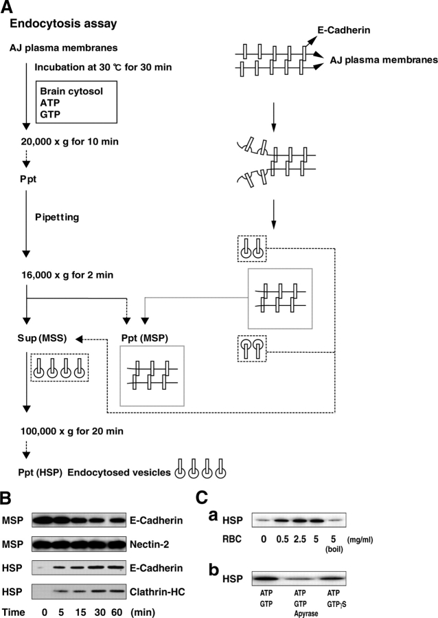
The AJ plasma membranes were first washed with 0.5 M Tris just to remove materials loosely associated with the membranes, and then incubated for various periods of time at 30°C in the presence of the rat brain cytosol, ATP, and GTP (a reaction buffer) to induce budding of vesicles. The vesicle budding has been proposed to be driven by the coordinated assembly of the cytoplasmic coat protein subunits during various cell-free transport reactions, such as ER-to-Golgi, trans-Golgi transport, and CCV formation (Malhotra et al., 1989; Schmid and Smythe, 1991; Barlowe et al., 1994; Miwako et al., 2003). This process requires cytosol, ATP, and GTP. Cytosol is required as the donor of the cytoplasmic coat protein subunits. ATP is required as an energy source. GTP is required for dynamin and small G proteins that regulate vesicle formation. The reaction was stopped by chilling each tube on ice, and the total membranes were collected by 20,000 g for 10 min to remove the excess reaction buffer. We expected that the vesicles carrying E-cadherin were budded from the collected AJ plasma membranes. The AJ plasma membranes were resuspended by trituration (vigorous pipetting) to mechanically pinch off the budded vesicles from the AJ plasma membranes. The sample was then centrifuged at 16,000 g for 2 min to recover the medium-speed supernatant (MSS), which might contain vesicles completely pinched off from the AJ plasma membranes, from the medium-speed pellet (MSP). To collect these vesicles, the MSS was centrifuged at 100,000 g for 20 min and the high-speed pellet (HSP) was obtained. The probable AJ plasma membrane structures in each fraction and procedures are schematically shown in Fig. 1 A. We examined the morphology of each fraction by electron microscopy and observed the coated bud structures (schematically shown in Fig. 1 A) that were formed on the AJ plasma membranes (Fig. S1 A, available at http://www.jcb.org/cgi/cotent/full/jcb.200401078/DC1).
Figure 1.
A cell-free assay system for the endocytosis of E-cadherin. (A) Flow diagram of the steps and schematic morphologies of the AJ plasma membranes. (B) Time-dependent endocytosis of E-cadherin. At the indicated times, the reactions were stopped by chilling each tube on ice and the MSP (16,000 g) and the HSP (100,000 g) were prepared by differential centrifugation as described in A. The amount of the MSP and the HSP (20 and 70% of total SDS solubilized membranes, respectively) were quantitated by immunoblotting with the anti–E-cadherin mAb, the anti-nectin-2 pAb, and the anti-clathrin HC mAb. (C) Requirement of the cytosol, ATP, and GTP for the endocytosis of E-cadherin. (Ca) The AJ plasma membranes were incubated in the presence of the indicated concentrations of the rat brain cytosol (RBC) or the boiled rat brain cytosol and assayed for the endocytosis of E-cadherin. (Cb) The AJ plasma membranes were incubated in the presence of 1 mM ATP and 100 μM GTP (control), 20U/ml of apyrase, 1 mM ATP, and 100 μM GTP (ATP depletion), or 1 mM ATP and 100 μM GTPγS (GTPγS), and assayed for the endocytosis of E-cadherin. All the reaction mixtures contained the rat brain cytosol. The results shown in all panels are representative of three independent experiments.
Two major stages are required to complete the endocytosis of E-cadherin (Schmid and Smythe, 1991). The first stage is the cargo protein selection and vesicle budding. The second stage is the fission of the vesicle from the membranes. We could recover the fully pinched off vesicles in the excess reaction buffer after an initial centrifugation (20,000 g for 10 min). The amount of recovered vesicles in this fraction was about half that of the HSP (Fig. S1 A). Thus, our assay system can be used to define separately these budding and fission stages. In this paper, we focused on the budding of endocytosed vesicles of E-cadherin, not the fissioned vesicles of E-cadherin, from the AJ plasma membranes.
We examined by immunoblotting whether or not E-cadherin was indeed recovered in the HSP. Before the incubation at 30°C in the presence of the reaction buffer, E-cadherin was confined to the MSP and undetectable in the HSP, whereas it was decreased in the MSP and conversely increased in the HSP time dependently (Fig. 1 B). Like E-cadherin, clathrin-HC was increased in the HSP time dependently. The maximal recovery of E-cadherin in the HSP was ∼17% of total E-cadherin after the 30-min incubation. The recovery of E-cadherin in the HSP required the rat brain cytosol in a dose-dependent manner (Fig. 1 Ca). The boiled rat brain cytosol was inactive in this reaction, demonstrating that the active component is a protein. ATP scavenger apyrase mostly inhibited the recovery of E-cadherin in the HSP (Fig. 1 Cb). Furthermore, GTPγS, a non-hydrolyzable analogue of GTP, reduced the recovery of E-cadherin in the HSP. These results indicate that the efficient recovery of E-cadherin in the HSP is dependent on the presence of the rat brain cytosol, ATP, and GTP.
Characteristics of the endocytosed vesicles of E-cadherin
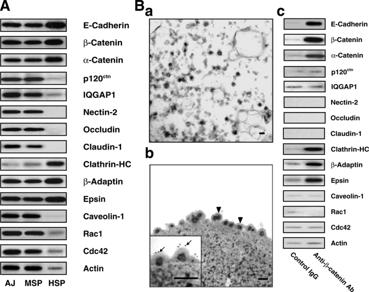
Next, we examined if other cell–cell adhesion molecules (CAMs) and their associated peripheral membrane proteins at AJs were recovered in the HSP. We compared the relative amount of each protein between the HSP and MSP (15 μg of protein in each fraction) by immunoblotting. The distribution of E-cadherin in the MSP and the HSP was compared with those of nectin-2, claudin-1, and occludin. Nectin-2 is an Ig-like CAM at AJs (Shimizu and Takai, 2003; Takai and Nakanishi, 2003; Takai et al., 2003). Claudin-1 and occludin are CAMs at TJs (Tsukita et al., 2001). Unlike E-cadherin, all of these CAMs were confined to the MSP and undetectable in the HSP even after the 30-min incubation in the presence of the reaction buffer (Figs. 1 B and 2 A). In addition, the distribution of proteins directly or indirectly associating with E-cadherin was examined in the MSP and the HSP. α- and β-Catenins were recovered in the HSP like E-cadherin. In contrast, p120ctn and IQGAP1 were confined to the MSP and undetectable in the HSP. β-Catenin and p120ctn are E-cadherin–binding proteins (Tsukita et al., 1992; Anastasiadis and Reynolds, 2000), whereas α-catenin is a β-catenin– and actin filament (F-actin)–binding protein (Tsukita et al., 1992). IQGAP1 is an F-actin cross-linking protein known to be a downstream target of Rac and Cdc42 small G proteins (Kuroda et al., 1996; Bashour et al., 1997; Fukata et al., 1997). Moreover, the relative amount of E-cadherin was higher in the HSP than that of the MSP or the original AJs. These results indicate that the recovery of E-cadherin in the HSP is not just due to nonspecific fragmentation of the AJ plasma membranes and suggest that E-cadherin and α- and β-catenins are selectively sorted into the endocytosed vesicles.
We confirmed by immunoelectron microscopy whether E-cadherin is indeed on the endocytosed vesicles budded from the AJ plasma membranes and recovered in the HSP. First, the HSP was observed by electron microscopy. We found that numerous vesicles showed heterogeneous sizes (50–400 nm in diameter) in this fraction, although most were 50–100 nm, as expected for endocytosed vesicles (Fig. 2 Ba). On the assumption that E-cadherin recovered in the HSP formed a complex with β-catenin, E-cadherin was immunoprecipitated from the MSS by the anti–β-catenin mAb-coated magnetic beads. The beads were fixed and examined by immunoelectron microscopy using the anti–E-cadherin mAb and the gold-conjugated secondary antibody. The beads were coated with vesicles on which the immunogolds for E-cadherin were accumulated (Fig. 2 Bb). All the proteins recovered in the HSP, including E-cadherin and α- and β-catenins, were also recovered in the immunopurified vesicles (Fig. 2 Bc). These results indicate that E-cadherin is indeed endocytosed from the AJ plasma membranes and recovered on the endocytosed vesicles.
Figure 2.
Characteristics of the endocytosed vesicles of E-cadherin. (A) Specific endocytosis of E-cadherin. The relative amount of the MSP and the HSP (15 μg of protein in each fraction) were analyzed by quantitative immunoblotting with various antibodies against the indicated proteins. The results were obtained from the same experiments and the same gels. (B) Isolation of endocytosed vesicles of E-cadherin. (Ba) Electron microscopic morphology of the HSP. The reaction for the formation of vesicles was performed as shown in Fig. 1 A. Bar, 100 nm. (Bb) Immunoelectron microscopic morphology of endocytosed vesicles of E-cadherin. The reaction for the formation of vesicles was performed as shown in Fig. 1 A. The vesicles were isolated from the MSS using the anti– β-catenin mAb-coated magnetic beads. The accumulated vesicles on the beads were processed for electron microscopy. The membranes bound to the beads consisted largely of a homogenous population of 60–80-nm vesicles (arrowheads). (inset) Immunogold labeling for E-cadherin. The immunoisolated beads were stained with the anti-pan-cadherin (cytoplasmic portion) pAb, followed by 10 nm gold-conjugated secondary antibody (arrows). Bars, 100 nm. (Bc) Composition of the endocytosed vesicles of E-cadherin. The vesicles were immunoisolated with the anti–β-catenin mAb or the anti-mouse IgG (control IgG)-coated magnetic beads from the MSS as shown in Bb. The bound proteins were analyzed by immunoblotting with various antibodies against the indicated proteins. The results were obtained from the same experiments and the same gels. The results shown in all panels are representative of three independent experiments.
We examined whether or not E-cadherin undergoes endocytosis in a clathrin-dependent manner. To perturb the clathrin-dependent endocytosis of E-cadherin from the AJ plasma membranes, we used the ENTH domain of epsin. Epsin is a regulatory component of the CCV formation (Brodsky et al., 2001), and its ENTH domain has been shown to be a potent inhibitor of the clathrin-dependent endocytosis (Nakashima et al., 1999). Recombinant GST-ENTH domain of epsin indeed reduced the amount of E-cadherin in the HSP in a dose-dependent manner (Fig. 3 A). Consistently, clathrin-HC, epsin, and β-adaptin were recovered in the HSP and the immunopurified vesicles (Fig. 2, A and Bc). β-Adaptin is a component of the CCV (Brodsky et al., 2001). The vesicles immunopurified by the anti–β-catenin mAb-coated beads showed diameters of ∼80 nm (Fig. 2 Bb), which is consistent with those of previously reported CCVs (Schmid and Smythe, 1991; Miwako et al., 2003). To further validate the results obtained in our assay system, we examined whether or not the ENTH domain of epsin affects the endocytosis of E-cadherin in MDCK cells. We confirmed that this epsin mutant inhibited both the HGF- and low Ca2+-induced endocytosis of E-cadherin in MDCK cells (Fig. 3 B, a and b). The inconsistency between our results and the former results using MCF7 cells (Paterson et al., 2003) may be due to the variation of mechanisms of endocytosis in different cell types.
Figure 3.
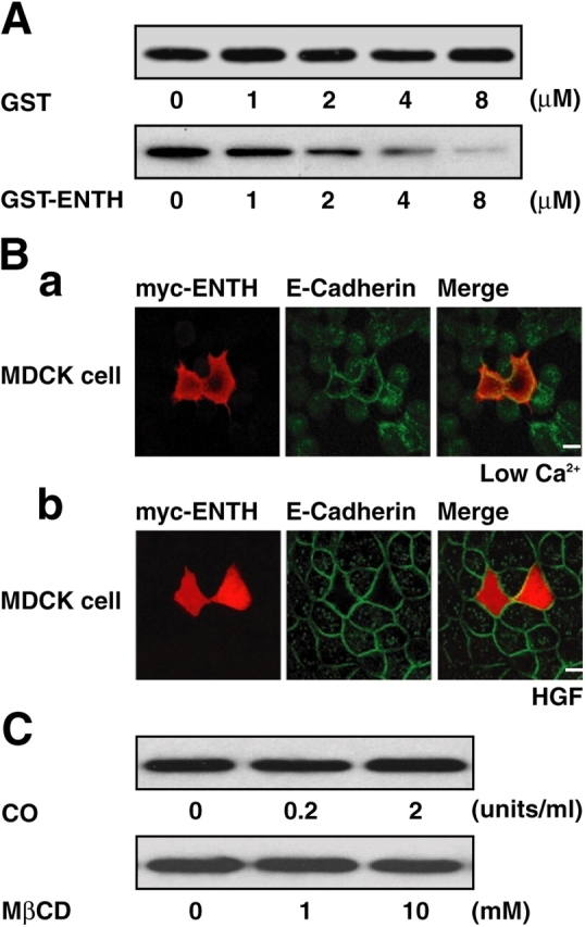
Clathrin-dependent endocytosis of E-cadherin. (A) Epsin-dependent endocytosis of E-cadherin in the cell-free assay system. The AJ plasma membranes were incubated in the presence of recombinant GST-ENTH or control GST at the indicated concentrations and assayed for the endocytosis of E-cadherin. (B) Epsin- dependent endocytosis of E-cadherin in MDCK cells. (Ba) MDCK cells were transfected with pEFBOS-myc-ENTH and cultured for 24 h. After the culture, the cells were incubated at 2 μM Ca2+ for 30 min. The cells were then fixed and stained for myc-ENTH and E-cadherin with the anti-myc pAb and the anti-E-cadherin mAb, respectively. Bar, 10 μm. (Bb) MDCK cells were transfected with pEFBOS-myc-ENTH and cultured for 24 h. After the culture, the cells were incubated with 10 ng/ml HGF for 3 h. The cells were then fixed and stained for myc-ENTH and E-cadherin with the anti-myc pAb and the anti-E-cadherin mAb, respectively. Bar, 10 μm. (C) Caveolin- independent endocytosis of E-cadherin. The AJ plasma membranes were incubated in the presence of cholesterol oxidase (CO) and cyclic heptasaccharide MβCD (MβCD) at the indicated concentrations and assayed for the endocytosis of E-cadherin. The results shown in all panels are representative of three independent experiments.
Next, we examined if the endocytosis of E-cadherin is caveolin-dependent in our assay system. Caveolin-dependent endocytosis has been shown to be selectively blocked by depletion of cholesterol from the plasma membranes (Ivanov et al., 2003). To deplete cholesterol, we used two reagents, cholesterol oxidase and cyclic heptasaccharide MβCD. We did not find any significant effect of these reagents on the endocytosis of E-cadherin (Fig. 3 C). Our results are consistent with the recent result that the low Ca2+-induced endocytosis of E-cadherin is caveolin independent in MDCK cells (Ivanov et al., 2003). Moreover, we examined the distribution of caveolin-1 in our assay system. Caveolin-1 was confined to the MSP, but not to the HSP (Fig. 2, A and Bc). Thus, the endocytosis of E-cadherin is likely to be caveolin independent in our assay system. Together, it is likely that E-cadherin undergoes endocytosis via CCVs in this newly developed cell-free assay system.
Regulation of the endocytosis of E-cadherin through Rac and Cdc42 activated by trans-interacting E-cadherin
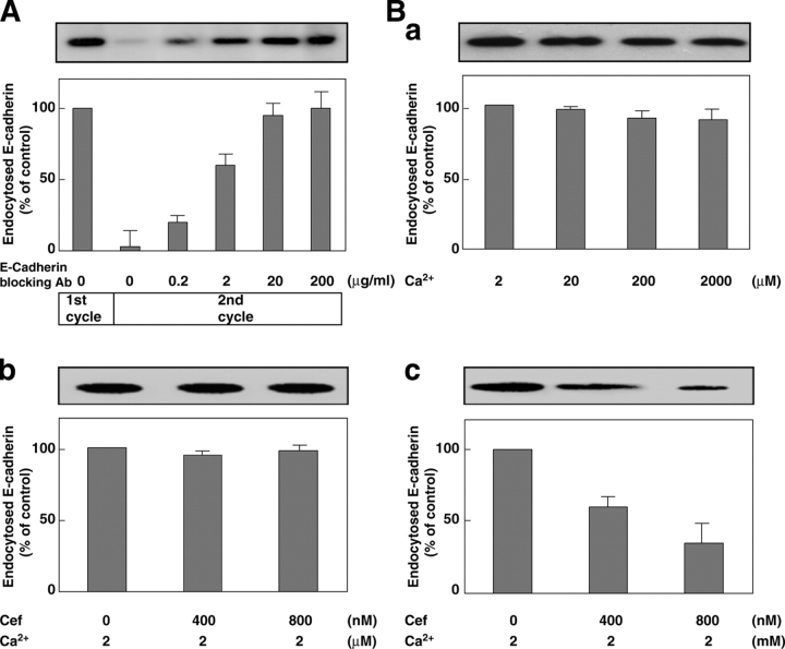
By use of the cell-free assay system, we first attempted to determine which form of E-cadherin, trans-interacting or non-trans interacting, is endocytosed. For this purpose, we first examined whether or not the rest of E-cadherin is trans interacting after the budding reaction. To examine if the rest of the E-cadherin is trans-interacting after the first cycle of the budding reaction, we added the E-cadherin–blocking antibody in the second cycle of the budding reaction. We found that the E-cadherin–blocking antibody enhanced the budding of E-cadherin in the second cycle reaction (Fig. 4 A). To confirm more directly that non-trans-interacting E-cadherin is preferentially endocytosed in our assay system, we examined if Cef, which is the extracellular fragment of E-cadherin fused to the Fc portion of human IgG, affects the endocytosis of E-cadherin in the presence of 2 mM or 2 μM Ca2+. Cef has been reported to trans interact with cellular E-cadherin in the presence of 2 mM Ca2+, but not in the presence of 2 μM Ca2+ (Kovacs et al., 2002a). To neglect a potential effect of Ca2+, we first determined the Ca2+ effect on the endocytosis of E-cadherin in our assay system because low Ca2+ enhances the dissociation of the trans interaction of E-cadherin. After the preincubation at various concentrations of Ca2+, the membranes were collected and assayed for the endocytosis of E-cadherin. Preincubation at 2 μM Ca2+ for 10 min did not affect the endocytosis of E-cadherin significantly in our assay system (Fig. 4 Ba). The inability of low Ca2+ to induce the endocytosis of E-cadherin under these conditions was just due to the short time incubation. We preincubated the AJ plasma membranes with Cef at 2 mM or 2 μM Ca2+ for 10 min. After the preincubation, the membranes were collected to remove excess Cef and assayed for the endocytosis of E-cadherin. Cef inhibited the endocytosis of E-cadherin in a dose-dependent manner at 2 mM Ca2+, but not at 2 μM Ca2+ (Fig. 4 B, b and c). These results indicate that only non-trans-interacting E-cadherin, but not trans-interacting one, undergoes endocytosis in our assay system.
Figure 4.
Regulation of endocytosis of E-cadherin by trans interaction of E-cadherin. (A) Enhancement of the endocytosis of E-cadherin by the E-cadherin–blocking antibody. The AJ plasma membranes were first assayed for the endocytosis of E-cadherin. The resulting membranes were incubated with the indicated concentrations of the E-cadherin–blocking antibody and assayed for the endocytosis of E-cadherin. (B) Inhibition of the endocytosis of non-trans-interacting E-cadherin by Cef. (Ba) After the preincubation at various concentrations of Ca2+, the membranes were collected and assayed for the endocytosis of E-cadherin. (Bb) The AJ plasma membranes were incubated with the indicated concentrations of Cef in the presence of 2 μM Ca2+ and assayed for the endocytosis of E-cadherin. (Bc) The AJ plasma membranes were incubated with the indicated concentrations of Cef in the presence of 2 mM Ca2+ and assayed for the endocytosis of E-cadherin. In all panels, the quantification of immunoblot is shown as the mean ± SD of duplicate assays in the bottom panel. The results shown in all panels are representative of three independent experiments.
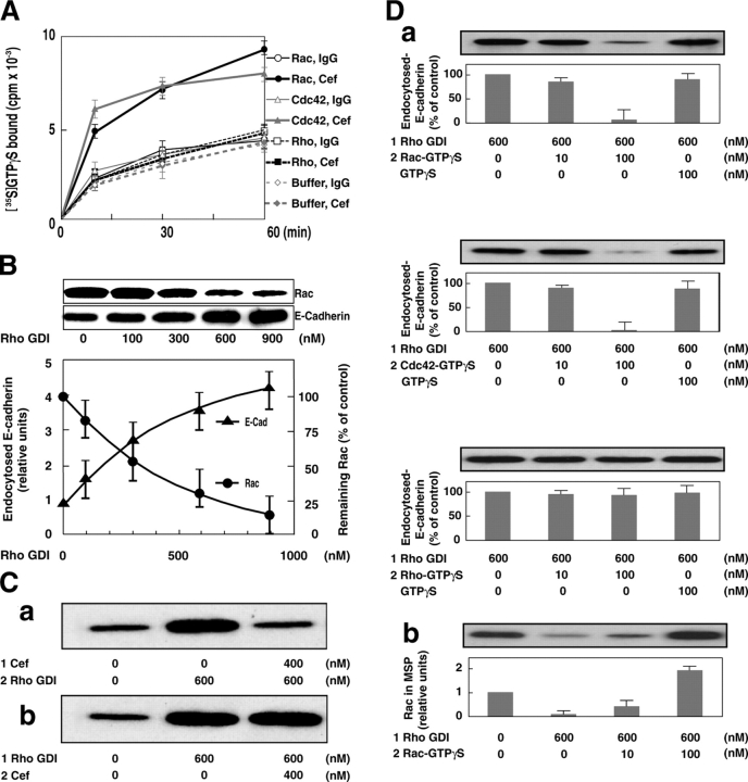
Then, we studied how the endocytosis of trans-interacting E-cadherin is inhibited. It has been shown that trans-interacting E-cadherin induces activation of Rac, but not Cdc42 or Rho small G protein (Van Aelst and Symons, 2002; Shimizu and Takai, 2003; Yap and Kovacs, 2003). Moreover, several studies have revealed that Rac might work positively or negatively in the endocytosis of E-cadherin, depending on both junctional maturity and cellular context (Takaishi et al., 1997; Akhtar and Hotchin, 2001; Palacios et al., 2002). However, the biological significance of Rac activated by trans-interacting E-cadherin is not thoroughly understood. We first examined, by use of Cef, if the trans interaction of E-cadherin induces activation of Rac, Cdc42, and Rho in our assay system. Cef activated Rac and Cdc42, but not Rho, time dependently (Fig. 5 A). Therefore, we examined if Rac and Cdc42 regulate the endocytosis of E-cadherin. For this purpose, we made advantage of Rho GDI, which forms a complex preferentially with the GDP-bound form to the GTP-bound form of the Rho family members, including Rho, Rac, and Cdc42, and extracts them from the membranes (Sasaki and Takai, 1998). Recombinant GST-tagged Rho GDI was added to the AJ plasma membranes in the presence of GDP and a high Mg2+, followed by incubation at 30°C for 20 min. High Mg2+ enhances the conversion of the GTP-bound form of the Rho family members to the GDP-bound form. The AJ plasma membranes were chilled on ice and recovered by centrifugation. Rac and Cdc42 were progressively extracted from the AJ plasma membranes by increasing amounts of GST-Rho GDI (Fig. 5 B and not depicted). However, the amount of Cdc42 in the AJ plasma membranes was 10 times lower than that of Rac by quantitative immunoblotting (unpublished data). Thus, Rac is likely to be the major substrate for Rho GDI in our assay system. After the 20 min preincubation of the AJ plasma membranes in the presence of Rho GDI, the amount of E-cadherin recovered in the HSP was measured. Increasing concentrations of Rho GDI increased the amount of E-cadherin in the HSP in proportion to the decreased amount of Rac in the AJ plasma membranes (Fig. 5 B). This Rho GDI-enhanced endocytosis of E-cadherin was inhibited by pretreatment of the AJ plasma membranes with Cef, and conversely the inhibitory effect of Cef was rescued by pretreatment of the AJ plasma membranes with Rho GDI (Fig. 5 C, a and b). These results indicate that the substrate small G proteins for Rho GDI inhibit the endocytosis of E-cadherin. We examined which substrate small G proteins show this effect. The GTPγS-bound form of recombinant Rac, Cdc42, or Rho was added to the AJ plasma membranes pretreated with Rho GDI, and the extent of the endocytosis of E-cadherin was measured. The GTPγS-bound form of Rac and Cdc42, but not that of Rho, inhibited the Rho GDI–enhanced endocytosis of E-cadherin (Fig. 5 Da). We confirmed that added GTPγS-bound form of Rac indeed bound to the AJ plasma membranes pretreated with Rho GDI (Fig. 5 Db). These results indicate that trans-interacting E-cadherin activates Rac and Cdc42, which then inhibit the endocytosis of E-cadherin in our assay system.
Figure 5.
Regulation of endocytosis of E-cadherin through Rac and Cdc42 activated by trans-interacting E-cadherin. (A) Activation of Rac and Cdc42 by Cef. The AJ plasma membranes were incubated with [35S]GTPγS and the GDP-bound form of recombinant Rac, Cdc42, or Rho in the presence or absence of Cef for the indicated periods of time. The bound [35S]GTPγS to Rac, Cdc42, or Rho was scintillation counted and plotted. The mean ± SD of duplicate assays is shown. (B) Enhancement of the endocytosis of E-cadherin by Rho GDI. The AJ plasma membranes were incubated with the indicated concentrations of GST-Rho GDI. The membranes were collected and assayed for the endocytosis of E-cadherin. The amount of remaining Rac on the AJ plasma membranes (10% of total) or the extent of the endocytosis of E-cadherin was determined by quantitative immunoblotting with the anti-Rac mAb or the anti–E-cadherin mAb, respectively. The mean ± SD of duplicate assays is shown. (C) Inhibition of the Rho GDI-enhanced endocytosis of E-cadherin by Cef. (Ca) The AJ plasma membranes were first incubated with Cef in the presence of 2 mM Ca2+ and then incubated with GST-Rho GDI. The membranes were collected and assayed for the endocytosis of E-cadherin. The numbers 1 and 2 indicate the sequence of the incubation. (Cb) Conversely, the AJ plasma membranes were first incubated with GST-Rho GDI and then incubated with Cef in the presence of 2 mM Ca2+. The membranes were collected and assayed for the endocytosis of E-cadherin. The numbers 1 and 2 indicate the sequence of the incubation. (D) Inhibition of the Rho GDI-enhanced endocytosis by Rac and Cdc42, but not by Rho. (Da) The AJ plasma membranes were first incubated with GST-Rho GDI and collected. The collected membranes were restored by adding the GTPγS-bound form of recombinant Rac, Cdc42, or Rho at the indicated concentrations and assayed for the endocytosis of E-cadherin. The numbers 1 and 2 indicate the sequence of the incubation. (Db) The membrane bound amount of added GTPγS-bound form of recombinant Rac in Da. (D, a and b) The quantification of immunoblot is shown as the mean ± SD of duplicate assays in the bottom panel. The numbers 1 and 2 indicate the sequence of the incubation. The results shown in all panels are representative of three independent experiments.
Involvement of IQGAP1 in the inhibitory effect of Rac and Cdc42 on the endocytosis of E-cadherin through the actin cytoskeleton
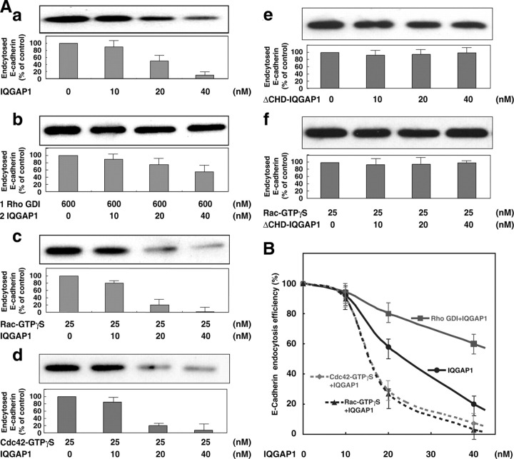
We attempted to search the Rac and Cdc42 target protein(s), which inhibits the endocytosis of E-cadherin. We fractionated the rat brain cytosol by successive column chromatographies. One peak fraction showed an inhibitory effect on the Rho GDI-enhanced endocytosis of E-cadherin. IQGAP1, a downstream target of Rac and Cdc42 (Kuroda et al., 1996; Bashour et al., 1997; Fukata et al., 1997), was enriched in this fraction as estimated by immunoblotting (unpublished data). To confirm this result, we examined the ability of recombinant His-IQGAP1 on the endocytosis of E-cadherin. Recombinant His-IQGAP1 potently inhibited the endocytosis of E-cadherin in a dose-dependent manner (Fig. 6, Aa and B). This inhibitory effect of IQGAP1 was reduced when the AJ plasma membranes pretreated with Rho GDI were used (Fig. 6, Ab and B). In contrast, the inhibitory effect of IQGAP1 was enhanced by the addition of the GTPγS-bound form of Rac and Cdc42 to the AJ plasma membranes (Fig. 6, Ac, Ad, and B). These results indicate that the inhibitory effect of Rac and Cdc42 on the endocytosis of E-cadherin is mediated through at least IQGAP1.
Figure 6.
Involvement of IQGAP1 in the inhibitory effect of Rac and Cdc42 on the endocytosis of E-cadherin. (A) Inhibition of the endocytosis of E-cadherin by IQGAP1. (Aa) The AJ plasma membranes were incubated with the indicated concentrations of recombinant His-IQGAP1 and assayed for the endocytosis of E-cadherin. (Ab) The AJ plasma membranes were first incubated with GST-Rho GDI and collected. The collected membranes were incubated with recombinant His-IQGAP1 at the indicated concentrations and assayed for the endocytosis of E-cadherin. The numbers 1 and 2 indicate the sequence of the incubation. (Ac) The AJ plasma membranes were incubated with the GTPγS-bound form of recombinant Rac and recombinant His-IQGAP1 at the indicated concentrations and assayed for the endocytosis of E-cadherin. (Ad) The AJ plasma membranes were incubated with the GTPγS-bound form of recombinant Cdc42 and recombinant His-IQGAP1 at the indicated concentrations and assayed for the endocytosis of E-cadherin. (Ae) The AJ plasma membranes were incubated with the indicated concentrations of recombinant His-ΔCHD-IQGAP1 and assayed for the endocytosis of E-cadherin. (Af) The AJ plasma membranes were incubated with the GTPγS-bound form of recombinant Rac and recombinant His-ΔCHD-IQGAP1 at the indicated concentrations and assayed for the endocytosis of E-cadherin. (A, a–f) The quantification of immunoblot is shown as the mean ± SD of duplicate assays in the bottom panel. (B) Endocytosis efficiency (%) is expressed relative to the controls in each experiment in A (a–d). The results shown in all panels are representative of three independent experiments.
IQGAP1 has been shown to cross-link F-actin into actin bundles (Bashour et al., 1997; Fukata et al., 1997). We generated ΔCHD-IQGAP1 as an IQGAP1 mutant, which had a defect in F-actin binding, but not in Rac binding (Fukata et al., 1997). We found that ΔCHD-IQGAP1 did not show any inhibitory effect on the endocytosis of E-cadherin in the presence or absence of the GTPγS-bound form of Rac (Fig. 6 A, e and f). Moreover, we confirmed that there is no difference between the Rac GTP-stabilizing activity of IQGAP1 and that of ΔCHD-IQGAP1 (Fukata et al., 1997; unpublished data). These results suggest that IQGAP1 is a target protein for Rac and Cdc42 and plays a role in the endocytosis of E-cadherin presumably through its F-actin cross-linking activity.
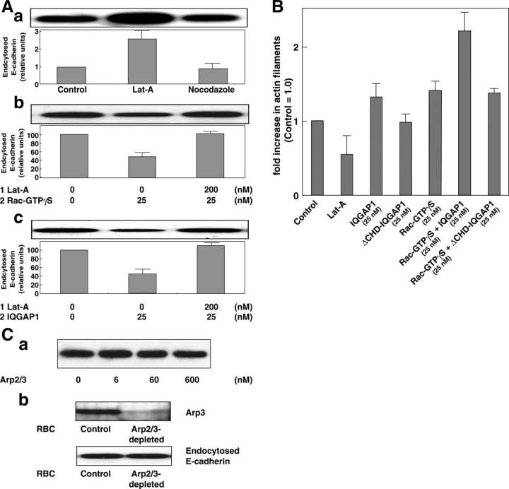
We next examined, by the use of an F-actin-disrupting reagent, latrunculin A (Lat-A), if the inhibitory effect of IQGAP1 on the endocytosis of E-cadherin is mediated through the actin cytoskeleton. Lat-A markedly enhanced the endocytosis of E-cadherin from the AJ plasma membranes (Fig. 7 Aa). In contrast, nocodazole, a microtubule-disrupting reagent, did not show any effect on the endocytosis of E-cadherin. The inhibitory effect of Rac and IQGAP1 on the endocytosis of E-cadherin was rescued by the Lat-A treatment (Fig. 7 A, b and c). Moreover, we measured F-actin levels. We found that Rac/IQGAP1 increased the amount of F-actin. In contrast, Rac/ΔCHD-IQGAP1 did not show any significant change in the F-actin level compared with that of the control (Fig. 7 B). These results indicate that IQGAP1 inhibits the endocytosis of E-cadherin through its Rac/Cdc42-dependent F-actin cross-linking activity.
Figure 7.
Involvement of the actin cytoskeleton in the inhibitory effect of IQGAP1 on the endocytosis of E-cadherin. (A) Prevention of the inhibitory effect of IQGAP1 on the endocytosis of E-cadherin by Lat-A. (Aa) The AJ plasma membranes were incubated with Lat-A or nocodazole and assayed for the endocytosis of E-cadherin. (Ab) The AJ plasma membranes were first incubated with Lat-A and collected. The collected membranes were incubated with the GTPγS-bound form of recombinant Rac and assayed for the endocytosis of E-cadherin. The numbers 1 and 2 indicate the sequence of the incubation. (Ac) The AJ plasma membranes were first incubated with Lat-A and collected. The collected membranes were incubated with recombinant His-IQGAP1 and assayed for the endocytosis of E-cadherin. (A, a–c) The quantification of immunoblot is shown as the mean ± SD of duplicate assays in the bottom panel. The numbers 1 and 2 indicate the sequence of the incubation. (B) F-Actin levels in the AJ plasma membranes. Increase in total F-actin in the AJ plasma membranes treated with the indicated factors was expressed as fold increase to the amount of F-actin in the nontreated control AJ plasma membranes. The mean ± SD of duplicate assays is shown. (C) Effect of the Arp2/3 complex on the endocytosis of E-cadherin. (Ca) The AJ plasma membranes were incubated with the indicated concentrations of the Arp2/3 complex and assayed for the endocytosis of E-cadherin. (Cb) The AJ plasma membranes were assayed for the endocytosis of E-cadherin in the presence of either control cytosol or Arp2/3-depleted cytosol. The results shown in all panels are representative of at least three independent experiments.
To neglect involvement of other actin regulator proteins, we examined the effect of the Arp2/3 complex on the endocytosis of E-cadherin in our assay system. The Arp2/3 complex has recently been reported to be recruited to the E-cadherin–based adhesion sites (Kovacs et al., 2002b). Moreover, the Arp2/3 complex–mediated actin polymerization has been reported to drive the vesicles away from the plasma membrane (Kaksonen et al., 2003). In our assay system, the Arp2/3 complex did not show any significant effect on the endocytosis of E-cadherin, even in the presence of the activators, such as the Cdc42-GTPγS-N-WASP complex and WAVE2 (Fig. 7 Ca and not depicted). Moreover, depletion of the Arp2/3 complex from the rat brain cytosol did not show any significant effect on the endocytosis of E-cadherin (Fig. 7 Cb). Thus, the Arp2/3 complex is unlikely to be involved in the endocytosis of E-cadherin in our assay system.
Inhibition of the endocytosis of E-cadherin by trans-interacting E-cadherin or trans-interacting nectin-1 in intact cells
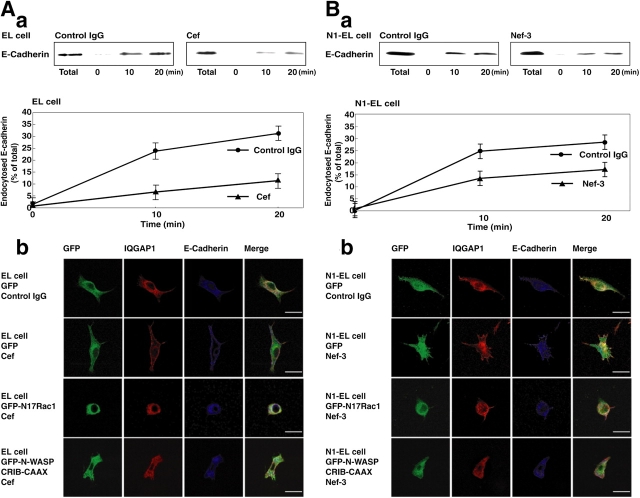
We finally examined if the endocytosis of trans-interacting E-cadherin is indeed inhibited in intact cells. For this purpose, we first used EL cells, L fibroblasts stably expressing E-cadherin, because E-cadherin is constitutively endocytosed and recycled when cells do not contact other cells and E-cadherin does not trans interact (Paterson et al., 2003). We assayed at 18°C to stop this recycling and to measure only the endocytosis of E-cadherin in EL cells. For mimicking the trans interaction of E-cadherin, we incubated EL cells with Cef. We quantified the effect of Cef on the endocytosis of E-cadherin by chasing surface-biotinylated E-cadherin (Le et al., 1999). Cef inhibited the constitutive endocytosis of E-cadherin (Fig. 8 Aa). Immunofluorescence analysis revealed that E-cadherin still remained on the cell surface in the presence of Cef and that IQGAP1 accumulated at the cell periphery where E-cadherin localized in the presence of Cef (Fig. 8 Ab). These results indicate that trans-interacting E-cadherin does not undergo endocytosis in intact cells. It has been shown that trans-interacting E-cadherin induces activation of Rac (Van Aelst and Symons, 2002; Shimizu and Takai, 2003; Yap and Kovacs, 2003). We examined if Rac or Cdc42, which is activated by trans-interacting E-cadherin, is required to induce IQGAP1 translocation to the plasma membrane in EL cells. We transfected cells with N17Rac1, a dominant-negative mutant of Rac, or N-WASP CRIB-CAAX, an inhibitor of activated Cdc42. Both N17Rac1 and N-WASP CRIB-CAAX inhibited the recruitment of IQGAP1 to the plasma membrane in EL cells (Fig. 8 Ab). Together, it is likely that both Rac and Cdc42 activated by trans-interacting E-cadherin inhibit its endocytosis through IQGAP1 also in intact cells.
Figure 8.
Inhibition of the endocytosis of E-cadherin by trans-interacting E-cadherin or trans-interacting nectin-1 in intact cells. (A) Inhibition of the constitutive endocytosis of E-cadherin by trans-interacting E-cadherin in intact EL cells. (Aa) EL cells were incubated in the medium with 400 nM Cef or 400 nM human IgG (control IgG) for 60 min. The cells were surface-biotinylated on ice and cultured at 18°C for the indicated periods of time to allow the endocytosis of E-cadherin. Biotinylated proteins on the plasma membrane were then stripped off by glutathione treatment, and biotinylated proteins inside the cells were recovered on streptavidin beads. The bound proteins were analyzed by immunoblotting with the anti-E-cadherin mAb. The relative amounts of endocytosed E-cadherin were expressed as percentage of total biotinylated E-cadherin in the bottom panel. The mean ± SD of duplicate assays is shown. (Ab) EL cells pretreated with the 400 nM Cef or 400 nM control IgG were incubated at 18°C for 20 min to allow the endocytosis of E-cadherin. EL cells overexpressing GFP, GFP-N17Rac1, or GFP-N-WASP-CRIB-CAAX were pretreated with the 400 nM Cef or 400 nM control IgG and then incubated at 18°C for 20 min to allow the endocytosis of E-cadherin. The cells were then fixed and immunostained with the anti–E-cadherin mAb and the anti-IQGAP1 pAb. Bars, 10 μm. (B) Inhibition of the constitutive endocytosis of E-cadherin by trans-interacting nectin-1 in intact nectin-1-EL cells. (Ba) Nectin-1-EL (N1-EL) cells were incubated in the medium with 400 nM Nef-3 or 400 nM control IgG for 60 min. The cells were surface-biotinylated on ice and cultured at 18°C for the indicated time to allow the endocytosis of E-cadherin. Biotinylated proteins on the plasma membranes were then stripped off by glutathione treatment, and biotinylated proteins inside the cells were recovered on streptavidin beads. The bound proteins were analyzed by immunoblotting with the anti–E-cadherin mAb. The relative amounts of endocytosed E-cadherin were expressed as percentage of total biotinylated E-cadherin in the bottom panel. The mean ± SD of duplicate assays is shown. (Bb) N1-EL cells pretreated with 400 nM Nef-3 or 400 nM control IgG were incubated at 18°C for 20 min to allow the endocytosis of E-cadherin. N1-EL cells overexpressing GFP, GFP-N17Rac1, or GFP-N-WASP-CRIB-CAAX were pretreated with the 400 nM Nef-3 or 400 nM control IgG and then incubated at 18°C for 20 min to allow the endocytosis of E-cadherin. The cells were then fixed and immunostained with the anti–E-cadherin mAb and the anti-IQGAP1 pAb. Bars, 10 μm. The results shown in all panels are representative of at least three independent experiments.
We have previously shown that nectins are involved in the formation of AJs in cooperation with E-cadherin (Takai et al., 2003). In the process of the formation of AJs, nectin-based cell–cell adhesion is first formed, followed by recruitment of E-cadherin there, finally forming AJs; and the nectin-based cell–cell adhesion induces activation of Cdc42 and Rac, which facilitates the formation of AJs. Nectins comprise a family consisting of four members: nectin-1, -2, -3, and -4. Like E-cadherin, each nectin forms cis-dimers, followed by formation of trans-dimers, causing cell–cell adhesion. Furthermore, nectin-3 forms hetero-trans-dimers with nectin-1 or -2, and of the many combinations of the trans-dimers, the hetero-trans-dimers between nectin-1 and -3 show the strongest cell–cell adhesion activity. Therefore, we examined whether or not the nectin-induced activation of Cdc42 and Rac is also involved in the inhibition of the endocytosis of E-cadherin. For this purpose, we used EL cells stably expressing nectin-1 (nectin-1-EL cells) and the extracellular fragment of nectin-3 fused to the Fc portion of human IgG (Nef-3; Honda et al., 2003). Nef-3, like Cef, inhibited the constitutive endocytosis of E-cadherin in nectin-1-EL cells (Fig. 8 Ba). Immunofluorescence analysis revealed that E-cadherin still remained on the cell surface in the presence of Nef-3 and that IQGAP1 accumulated at the cell periphery where E-cadherin localized in the presence of Nef-3 (Fig. 8 Bb). We examined whether Rac or Cdc42, which is activated by trans-interacting nectin-1, is required to induce IQGAP1 translocation to the plasma membrane in nectin-1-EL cells. We transfected cells with N17Rac1 or N-WASP CRIB-CAAX. Both N17Rac1 and N-WASP CRIB-CAAX inhibited the recruitment of IQGAP1 to the plasma membrane in nectin1-EL cells (Fig. 8 Bb). Therefore, it is likely that both Rac and Cdc42 activated by trans-interacting nectin-1 inhibits the endocytosis of E-cadherin.
Discussion
In this study, we have described a new biochemical assay that efficiently reconstitutes the endocytosis of E-cadherin using the AJ plasma membranes. We have found here that non-trans-interacting E-cadherin is constitutively endocytosed like integrin (ligand-independent endocytosis), that the formation of endocytosed vesicles of E-cadherin is clathrin dependent, and that E-cadherin, but not other CAMs at AJs and TJs including nectins, claudins, and occludin, is selectively sorted into the endocytosed vesicles. Recent cell-level studies using chemical inhibitors have shown that E-cadherin might be internalized by clathrin- or caveolin-dependent endocytosis (Le et al., 1999; Akhtar and Hotchin, 2001; Palacios et al., 2002; Thomsen et al., 2002; Paterson et al., 2003; Ivanov et al., 2003). However, the results obtained in this way are indirect and not substantial because the low resolution indirect immunofluorescence staining could only follow the appearance of E-cadherin in large vesicular structures (early or late endosomes) as a morphological measure for the endocytosis of E-cadherin. Thus, one of the main advantages of this assay method over previous cell-level assay methods is that it can be used to directly characterize the function of each component involved in the endocytosis of E-cadherin. It will be interesting to determine if and how other CAMs show different regulations to remove themselves from the cell surface, such as ectodomain shedding and proteolytic degradation (Arribas and Borroto, 2002), or other types of endocytosis, caveolin-dependent and clathrin- and caveolin-independent endocytosis (Le et al., 1999; Akhtar and Hotchin, 2001; Palacios et al., 2002; Thomsen et al., 2002; Paterson et al., 2003; Ivanov et al., 2003). We have reported here for the first time the immunoisolation of endocytosed vesicles of E-cadherin. This assay method will allow us not only to identify novel components of the endocytosed vesicles of E-cadherin but also to analyze the mechanisms of the endocytosis of other transmembrane proteins.
By the use of the new assay method, we have shown here that Rac and Cdc42 activated by trans-interacting E-cadherin inhibits the endocytosis of E-cadherin through IQGAP1 and F-actin. Because IQGAP1 is an F-actin cross-linking protein and a downstream target of Rac and Cdc42 (Kuroda et al., 1996; Bashour et al., 1997; Fukata et al., 1997), it is likely that IQGAP1 binds to Rac and Cdc42 activated by trans-interacting E-cadherin and reorganizes the actin cytoskeleton, resulting in inhibiting the endocytosis of E-cadherin and stabilizing E-cadherin on the cell surface. When migrating MDCK cells make nascent cell–cell contacts, E-cadherin is recruited specifically to the cell–cell contact sites (Adams et al., 1998). Furthermore , the inhibition of the endocytosis of E-cadherin by the Rac/Cdc42-IQGAP1 system may contribute to this site-specific accumulation of E-cadherin. p120ctn has recently been shown to be involved in the stabilization of cadherins on the cell surface, suggesting that p120ctn interacts with an unidentified endocytic machinery to inhibit the endocytosis of cadherins (Davis et al., 2003; Xiao et al., 2003). The p120ctn-binding site of E-cadherin is shown to be involved in the activation of Rac (Goodwin et al., 2003). Together, p120ctn and/or other unknown proteins may be upstream regulators of the Rac/Cdc42-IQGAP1 system that inhibits the endocytosis of E-cadherin.
We have previously proposed that nectins, but not E-cadherin, play an important role in recruiting IQGAP1 to very primordial nectin-based cell–cell adhesion sites through the actin cytoskeleton (Katata et al., 2003), in contrast to previous observation that E-cadherin is required for the accumulation of IQGAP1 at AJs solely by binding to β-catenin (Fukata and Kaibuchi, 2001). Nectins are associated with the actin cytoskeleton through afadin, an F-actin– and nectin-binding protein, as E-cadherin is associated with the actin cytoskeleton through α- and β-catenins (Shimizu and Takai, 2003; Takai and Nakanishi, 2003; Takai et al., 2003). We have shown here that Rac and Cdc42 activated by trans-interacting nectin-1 also has a potency to inhibit the endocytosis of E-cadherin. Therefore, together, IQGAP1 would be first recruited through the actin cytoskeleton to the cell–cell adhesion sites where Rac and Cdc42 are activated by trans-interacting nectins. IQGAP1 bound to Rac and Cdc42 then induces reorganization of the actin cytoskeleton. In contrast, E-cadherin is recruited to the nectin-based cell–cell adhesion sites and induces the activation of Rac and Cdc42, which then facilitates the recruitment of IQGAP1 and the subsequent reorganization of the actin cytoskeleton. These sequential reorganizations of the actin cytoskeleton are likely to inhibit the endocytosis of E-cadherin on one hand and strengthen the cell–cell adhesion activity of E-cadherin on the other hand.
It has been shown in cultured MDCK cells that HGF induces cell–cell disruption and cell scattering, which is accompanied with endocytosis of E-cadherin. However, it remains unknown that the endocytosis of E-cadherin alone is sufficient for the disruption of the cell–cell adhesion because previous studies have shown that a small pool of E-cadherin (20% of total surface E-cadherin) is endocytosed during the HGF actions in MDCK cells (Kamei et al., 1999; Fujita et al., 2002). The endocytosis of a specific portion of E-cadherin may be necessary and sufficient for the HGF action. Another possibility is that an unidentified factor(s), which is essential for the maintenance of the E-cadherin–based cell–cell adhesion, is coendocytosed with E-cadherin. The exact roles and the mechanisms of the endocytosis of E-cadherin in cell migration are important issues to be addressed in the future for our understanding of the signal switching from disruption of cell–cell adhesion to cell scattering.
Materials and methods
Cell-free assay for the endocytosis of E-cadherin
The AJ-enriched fraction was prepared from rat livers as described previously (Tsukita and Tsukita, 1989), washed with 0.5 M Tris/HCl, pH 7.5, resuspended in buffer A (20 mM Hepes, pH 7.4, and 125 mM KOAc), and stored at −80°C until use. Thawed AJ plasma membranes (20 μg of protein) were incubated at 30°C in a reaction buffer (36 mM Hepes, pH 7.4, 0.25 M sorbitol, 70 mM KOAc, 5 mM EGTA, 1.8 mM CaCl2, 2.5 mM Mg(OAc)2, an ATP-regenerating system [1 mM ATP, 5 mM creatine phosphate, and 0.2 IU creatine phosphate kinase], 100 μM GTP, and 2.5 mg/ml rat brain cytosol). The method for the preparation of the rat brain cytosol is described in the following section. The reaction was stopped by chilling the tube on ice and the membranes were collected by centrifugation at 20,000 g for 10 min. The membranes were resuspended by tritulation (20 times pipetting) in 50 μl of 20 mM Hepes, pH 7.2, and 0.25 M sorbitol, and then supplemented with KOAc and Mg(OAc)2 to final concentrations of 150 and 2.5 mM, respectively (final volume of 60.6 μl). Immediately after addition of KOAc and Mg(OAc)2, differential centrifugation was performed at a medium speed (16,000 g) for 2 min. The top 42 μl supernatant fraction (designated MSS) was harvested and centrifuged at a high speed (100,000 g) for 20 min. Membrane pellets from the MSP and HSP spins were solubilized in an SDS sample buffer at RT for 30 min with vigorously shaking, and proteins were separated by 10% SDS-PAGE. Proteins were transferred to nitrocellulose membrane sheets and immunoblotted with the anti–E-cadherin mAb, followed by the HRP-conjugated secondary antibody (Amersham Biosciences). Blots were developed using the ECL kit and quantitated using a molecular imaging system (Personal Densitometer SI; Amersham Biosciences).
Preparation of the rat brain cytosol
Rat brains were thoroughly homogenized in a homogenizing buffer (20 mM Hepes, pH 7.4, 85 mM sucrose, 100 mM KOAc, and 1 mM Mg(OAc)2). The homogenate was centrifuged at 15,000 g for 10 min and the supernatant was centrifuged at 100,000 g for 1 h. The supernatant was subsequently dialyzed for 4 h in buffer A and frozen at −80°C until use. Arp2/3 complex was depleted from rat brain cytosol using GST-N-WASP CA affinity column.
Assay for activation of Rho, Rac, and Cdc42
Activation of small G proteins was assayed as described previously (Yamamoto et al., 1990). The AJ plasma membranes (25 μg) and the GDP-bound form of recombinant RhoA, Rac1, or Cdc42 (5 pmol) were treated with Cef or IgG in the presence of [35S]GTPγS. After the incubation for the indicated periods of time, the radioactivity of [35S]GTPγS bound to RhoA, Rac1, or Cdc42 was measured.
Assay for quantification of F-actin
F-Actin was quantified as described previously (Vouret-Craviari et al. 1999). After incubation with various reagents, the AJ plasma membranes were collected and incubated with F-actin buffer (20 mM KPO4, 10 mM Pipes, 5 mM EGTA, 2 mM MgCl2, 0.1% Triton X-100, 3.7% formaldehyde, and 2 μM FITC-phalloidin) at RT for 1 h. The FITC-phalloidin bound membranes were collected by centrifugation 16,000 g for 2 min. The membranes were washed twice (0.1% saponin, 20 mM KPO4, 10 mM Pipes, 5 mM EGTA, and 2 mM MgCl2) and then resuspended in 1.5 ml of ice-cold methanol to extract phalloidin bound to F-actin. After rotating at RT for 1 h, the methanol suspension was collected and the amount of FITC was determined in each sample by using a fluorimeter (fluorescence emission at 520 nm and excitation at 492 nm).
Immunoisolation of the endocytosed vesicles of E-cadherin
Immunoisolation of vesicles was performed as described previously (Saucan and Palade, 1994). In brief, dynabeads M-500 (Dynal) magnetic beads were covalently coupled with the goat anti–mouse IgG (Fc specific; Sigma-Aldrich) secondary antibody at a density of ∼10 μg of antibody per 107 beads according to manufacturer's instructions. The beads were coated with the anti–β-catenin mAb or the control IgG at a twofold molar excess over the secondary antibody and then washed four times with PBS plus 1 mg/ml BSA. The anti–β-catenin mAb-coated beads (1 mg beads) were added to the MSS (0.15–0.35 ml) and incubated in the presence of buffer B (20 mM Hepes, pH 7.2, 0.25 M sorbitol, 0.15 M KOAc, 5% FBS, and 2 mM EDTA; final volume of 0.4 ml), with continuous rotation at 4°C for 2 h. The immunoisolated beads were washed five times with buffer B using a magnet and transferred to fresh tubes. The beads were finally resuspended in 50 μl of the SDS sample buffer for SDS-PAGE, immunoblotting, or EM as described in the following section.
Immunoelectron microscopy
EM was performed to visualize the immunoisolated vesicles on the magnetic beads. The samples were fixed in 4% PFA and 0.1% glutaraldehyde in PBS for 45 min, and then in 1% osmium tetroxide in the same buffer for 1 h. The samples were stained in black in 2% uranyl acetate, dehydrated in ethanol and propylene oxide, and embedded in Epon. Thin sections were cut, stained, and examined in a TEM. For immunogold labeling, the vesicles on the beads were first reacted with the anti-pan-cadherin rabbit pAb for 1 h, followed by the goat anti–rabbit IgG conjugated to 10-nm gold (Jackson ImmunoResearch Laboratories) for 1 h. The beads were washed, fixed, and examined in a TEM.
Biotinylation assay for the endocytosis of E-cadherin
The assay was performed as described previously with minor modifications (Le et al., 1999). In brief, EL cells were incubated with 0.5 mg/ml sulfo-NHS-ss-biotin on ice, and the cells were then incubated at 18°C to allow the endocytosis of E-cadherin for the indicated periods of time in the presence of IgG or Cef. The cells were incubated in several washes with a glutathione buffer (60 mM glutathione, 83 mM NaCl, 83 mM NaOH, and 10% FCS) on ice to remove bound biotinyl groups from remaining cell surface biotinylated proteins. The cells were then lysed in a RIPA buffer. The cell extracts were incubated with streptavidin beads (Sigma-Aldrich) to collect bound biotinylated proteins. Bound proteins were then analyzed by SDS-PAGE and immunoblotting with the anti–E-cadherin mAb. For cell staining, the cells were transfected with pEGFP, pEGFP-N-WASP CRIB-CAAX, or pEGFP-N17Rac1 using LipofectAMINE 2000 Reagent. After a 24-h culture, the cells were incubated at 18°C to allow the endocytosis of E-cadherin for 20 min in the presence of IgG or Cef. The cells were then fixed, followed by immunostaining with the anti–E-cadherin mAb (ECCD-2) and the anti-IQGAP1 pAb. Images were captured using a confocal laser scanning microscope (Bio-Rad Laboratories) using a 60× oil immersion objective lens (model Radiance 2000; Bio-Rad Laboratories). Collected data were exported as 8-bit TIFF files and processed using Adobe Photoshop software.
Online supplemental material
Fig. S1 is available in the online supplemental material. Fig. S1 shows the stage specific assay for the endocytosis of E-cadherin and EM pictures of all fractions. Materials and methods for antibodies, reagents, and immunofluorescence microscopic assay for the endocytosis of E-cadherin are available in the online supplemental material. Online supplemental material is available at http://www.jcb.org/cgi/cotent/full/jcb.200401078/DC1.
Acknowledgments
This work was supported by grants-in-aid for Scientific Research and for Cancer Research from the Ministry of Education, Culture, Sports, Science and Technology, Japan (2003). T. Sakisaka is a recipient of a Human Frontier Science Program Career Development Award (2003).
Abbreviations used in this paper: AJ, adherens junction; CAMs, cell–cell adhesion molecules; CCV, clathrin-coated vesicle; HSP, high-speed pellet; Lat-A, latrunculin A; MSP, medium-speed pellet; MSS, medium-speed supernatant.
References
- Adams, C.L., and W.J. Nelson. 1998. Cytomechanics of cadherin-mediated cell-cell adhesion. Curr. Opin. Cell Biol. 10:572–577. [DOI] [PubMed] [Google Scholar]
- Adams, C.L., Y.T. Chen, S.J. Smith, and W.J. Nelson. 1998. Mechanisms of epithelial cell-cell adhesion and cell compaction revealed by high-resolution tracking of E-cadherin-green fluorescent protein. J. Cell Biol. 142:1105–1119. [DOI] [PMC free article] [PubMed] [Google Scholar]
- Akhtar, N., and N.A. Hotchin. 2001. RAC1 regulates adherens junctions through endocytosis of E-cadherin. Mol. Biol. Cell. 12:847–862. [DOI] [PMC free article] [PubMed] [Google Scholar]
- Anastasiadis, P.Z., and A.B. Reynolds. 2000. The p120 catenin family: complex roles in adhesion, signaling and cancer. J. Cell Sci. 113:1319–1334. [DOI] [PubMed] [Google Scholar]
- Arribas, J., and A. Borroto. 2002. Protein ectodomain shedding. Chem. Rev. 102:4627–4638. [DOI] [PubMed] [Google Scholar]
- Barlowe, C., L. Orci, T. Yeung, M. Hosobuchi, S. Hamamoto, N. Salama, M.F. Rexach, M. Ravazzola, M. Amherdt, and R. Schekman. 1994. COPII: a membrane coat formed by Sec proteins that drive vesicle budding from the endoplasmic reticulum. Cell. 77:895–907. [DOI] [PubMed] [Google Scholar]
- Bashour, A.M., A.T. Fullerton, M.J. Hart, and G.S. Bloom. 1997. IQGAP1, a Rac- and Cdc42-binding protein, directly binds and cross-links microfilaments. J. Cell Biol. 137:1555–1566. [DOI] [PMC free article] [PubMed] [Google Scholar]
- Brodsky, F.M., C.Y. Chen, C. Kneuhl, M.C. Towler, and D.E. Wakeham. 2001. Biological basket weaving: formation and function of clathrin-coated vesicles. Annu. Rev. Cell Dev. Biol. 17:517–568. [DOI] [PubMed] [Google Scholar]
- Davis, M.A., R.C. Ireton, and A.B. Reynolds. 2003. A core function for p120-catenin in cadherin turnover. J. Cell Biol. 163:525–534. [DOI] [PMC free article] [PubMed] [Google Scholar]
- Fujita, Y., G. Krause, M. Scheffner, D. Zechner, H.E. Leddy, J. Behrens, T. Sommer, and W. Birchmeier. 2002. Hakai, a c-Cbl-like protein, ubiquitinates and induces endocytosis of the E-cadherin complex. Nat. Cell Biol. 4:222–231. [DOI] [PubMed] [Google Scholar]
- Fukata, M., and K. Kaibuchi. 2001. Rho-family GTPases in cadherin-mediated cell-cell adhesion. Nat. Rev. Mol. Cell Biol. 2:887–897. [DOI] [PubMed] [Google Scholar]
- Fukata, M., S. Kuroda, K. Fujii, T. Nakamura, I. Shoji, Y. Matsuura, K. Okawa, A. Iwamatsu, A. Kikuchi, and K. Kaibuchi. 1997. Regulation of cross-linking of actin filament by IQGAP1, a target for Cdc42. J. Biol. Chem. 272:29579–29583. [DOI] [PubMed] [Google Scholar]
- Goodwin, M., E.M. Kovacs, M.A. Thoreson, A.B. Reynolds, and A.S. Yap. 2003. Minimal mutation of the cytoplasmic tail inhibits the ability of E-cadherin to activate Rac but not phosphatidylinositol 3-kinase: direct evidence of a role for cadherin-activated Rac signaling in adhesion and contact formation. J. Biol. Chem. 278:20533–20539. [DOI] [PubMed] [Google Scholar]
- Gumbiner, B.M. 2000. Regulation of cadherin adhesive activity. J. Cell Biol. 148:399–404. [DOI] [PMC free article] [PubMed] [Google Scholar]
- Honda, T., K. Shimizu, T. Kawakatsu, M. Yasumi, T. Shingai, A. Fukuhara, K. Ozaki-Kuroda, K. Irie, H. Nakanishi, and Y. Takai. 2003. Antagonistic and agonistic effects of an extracellular fragment of nectin on formation of E-cadherin-based cell-cell adhesion. Genes Cells. 8:51–63. [DOI] [PubMed] [Google Scholar]
- Ivanov, A.I., A. Nusrat, and C.A. Parkos. 2003. Endocytosis of epithelial apical junctional proteins by a clathrin-mediated pathway into a unique storage compartment. Mol. Biol. Cell. 15:176–188. [DOI] [PMC free article] [PubMed] [Google Scholar]
- Kaksonen, M., Y. Sun, and D.G. Drubin. 2003. A pathway for association of receptors, adaptors, and actin during endocytic internalization. Cell. 115:475–487. [DOI] [PubMed] [Google Scholar]
- Kamei, T., T. Matozaki, T. Sakisaka, A. Kodama, S. Yokoyama, Y.F. Peng, K. Nakano, K. Takaishi, and Y. Takai. 1999. Coendocytosis of cadherin and c-Met coupled to disruption of cell-cell adhesion in MDCK cells–regulation by Rho, Rac and Rab small G proteins. Oncogene. 18:6776–6784. [DOI] [PubMed] [Google Scholar]
- Katata, T., K. Irie, A. Fukuhara, T. Kawakatsu, A. Yamada, K. Shimizu, and Y. Takai. 2003. Involvement of nectin in the localization of IQGAP1 at the cell-cell adhesion sites through the actin cytoskeleton in Madin-Darby canine kidney cells. Oncogene. 22:2097–2109. [DOI] [PubMed] [Google Scholar]
- Kovacs, E.M., R.G. Ali, A.J. McCormack, and A.S. Yap. 2002. a. E-cadherin homophilic ligation directly signals through Rac and phosphatidylinositol 3-kinase to regulate adhesive contacts. J. Biol. Chem. 277:6708–6718. [DOI] [PubMed] [Google Scholar]
- Kovacs, E.M., M. Goodwin, R.G. Ali, A.D. Paterson, and A.S. Yap. 2002. b. Cadherin-directed actin assembly: E-cadherin physically associates with the Arp2/3 complex to direct actin assembly in nascent adhesive contacts. Curr. Biol. 12:379–382. [DOI] [PubMed] [Google Scholar]
- Kuroda, S., M. Fukata, K. Kobayashi, M. Nakafuku, N. Nomura, A. Iwamatsu, and K. Kaibuchi. 1996. Identification of IQGAP as a putative target for the small GTPases, Cdc42 and Rac1. J. Biol. Chem. 271:23363–23367. [DOI] [PubMed] [Google Scholar]
- Lawson, M.A., and F.R. Maxfield. 1995. Ca2+- and calcineurin-dependent recycling of an integrin to the front of migrating neutrophils. Nature. 377:75–79. [DOI] [PubMed] [Google Scholar]
- Le, T.L., A.S. Yap, and J.L. Stow. 1999. Recycling of E-cadherin: a potential mechanism for regulating cadherin dynamics. J. Cell Biol. 146:219–232. [PMC free article] [PubMed] [Google Scholar]
- Malhotra, V., T. Serafini, L. Orci, J.C. Shepherd, and J.E. Rothman. 1989. Purification of a novel class of coated vesicles mediating biosynthetic protein transport through the Golgi stack. Cell. 58:329–336. [DOI] [PubMed] [Google Scholar]
- Miller, J.R., and D.R. McClay. 1997. Characterization of the role of cadherin in regulating cell adhesion during sea urchin development. Dev. Biol. 192:323–339. [DOI] [PubMed] [Google Scholar]
- Miwako, I., T. Schroter, and S.L. Schmid. 2003. Clathrin- and dynamin-dependent coated vesicle formation from isolated plasma membranes. Traffic. 4:376–389. [PubMed] [Google Scholar]
- Nakashima, S., K. Morinaka, S. Koyama, M. Ikeda, M. Kishida, K. Okawa, A. Iwamatsu, S. Kishida, and A. Kikuchi. 1999. Small G protein Ral and its downstream molecules regulate endocytosis of EGF and insulin receptors. EMBO J. 18:3629–3642. [DOI] [PMC free article] [PubMed] [Google Scholar]
- Palacios, F., J.K. Schweitzer, R.L. Boshans, and C. D'Souza-Schorey. 2002. Arf6-GTP recruits NM23-H1 to facilitate dynamin-mediated endocytosis during adherens junctions disassembly. Nat. Cell Biol. 4:929–936. [DOI] [PubMed] [Google Scholar]
- Paterson, A.D., R.G. Parton, C. Ferguson, J.L. Stow, and A.S. Yap. 2003. Characterization of E-cadherin endocytosis in isolated MCF-7 and chinese hamster ovary cells: the initial fate of unbound E-cadherin. J. Biol. Chem. 278:21050–21057. [DOI] [PubMed] [Google Scholar]
- Sasaki, T., and Y. Takai. 1998. The Rho small G protein family-Rho GDI system as a temporal and spatial determinant for cytoskeletal control. Biochem. Biophys. Res. Commun. 245:641–645. [DOI] [PubMed] [Google Scholar]
- Saucan, L., and G.E. Palade. 1994. Membrane and secretory proteins are transported from the Golgi complex to the sinusoidal plasmalemma of hepatocytes by distinct vesicular carriers. J. Cell Biol. 125:733–741. [DOI] [PMC free article] [PubMed] [Google Scholar]
- Schmid, S.L. 1997. Clathrin-coated vesicle formation and protein sorting: an integrated process. Annu. Rev. Biochem. 66:511–548. [DOI] [PubMed] [Google Scholar]
- Schmid, S.L., and E. Smythe. 1991. Stage-specific assays for coated pit formation and coated vesicle budding in vitro. J. Cell Biol. 114:869–880. [DOI] [PMC free article] [PubMed] [Google Scholar]
- Shimizu, K., and Y. Takai. 2003. Roles of the intercellular adhesion molecule nectin in intracellular signaling. J. Biochem. (Tokyo). 134:631–636. [DOI] [PubMed] [Google Scholar]
- Takai, Y., and H. Nakanishi. 2003. Nectin and afadin: novel organizers of intercellular junctions. J. Cell Sci. 116:17–27. [DOI] [PubMed] [Google Scholar]
- Takai, Y., K. Irie, K. Shimizu, T. Sakisaka, and W. Ikeda. 2003. Nectins and nectin-like molecules: roles in cell adhesion, migration, and polarization. Cancer Sci. 94:655–667. [DOI] [PMC free article] [PubMed] [Google Scholar]
- Takaishi, K., T. Sasaki, H. Kotani, H. Nishioka, and Y. Takai. 1997. Regulation of cell-cell adhesion by rac and rho small G proteins in MDCK cells. J. Cell Biol. 139:1047–1059. [DOI] [PMC free article] [PubMed] [Google Scholar]
- Takeichi, M. 1988. The cadherins: cell-cell adhesion molecules controlling animal morphogenesis. Development. 102:639–655. [DOI] [PubMed] [Google Scholar]
- Thomsen, P., K. Roepstorff, M. Stahlhut, and B. van Deurs. 2002. Caveolae are highly immobile plasma membrane microdomains, which are not involved in constitutive endocytic trafficking. Mol. Biol. Cell. 13:238–250. [DOI] [PMC free article] [PubMed] [Google Scholar]
- Tsukita, S., and S. Tsukita. 1989. Isolation of cell-to-cell adherens junctions from rat liver. J. Cell Biol. 108:31–41. [DOI] [PMC free article] [PubMed] [Google Scholar]
- Tsukita, S., S. Tsukita, A. Nagafuchi, and S. Yonemura. 1992. Molecular linkage between cadherins and actin filaments in cell-cell adherens junctions. Curr. Opin. Cell Biol. 4:834–839. [DOI] [PubMed] [Google Scholar]
- Tsukita, S., M. Furuse, and M. Itoh. 2001. Multifunctional strands in tight junctions. Nat. Rev. Mol. Cell Biol. 2:285–293. [DOI] [PubMed] [Google Scholar]
- Van Aelst, L., and M. Symons. 2002. Role of Rho family GTPases in epithelial morphogenesis. Genes Dev. 16:1032–1054. [DOI] [PubMed] [Google Scholar]
- Vouret-Craviari, V., D. Grall, G. Flatau, J. Pouyssegur, P. Boquet, and E. Van Obberghen-Schilling. 1999. Effects of cytotoxic necrotizing factor 1 and lethal toxin on actin cytoskeleton and VE-cadherin localization in human endothelial cell monolayers. Infect. Immun. 67:3002–3008. [DOI] [PMC free article] [PubMed] [Google Scholar]
- Xiao, K., D.F. Allison, K.M. Buckley, M.D. Kottke, P.A. Vincent, V. Faundez, and A.P. Kowalczyk. 2003. Cellular levels of p120 catenin function as a set point for cadherin expression levels in microvascular endothelial cells. J. Cell Biol. 163:535–545. [DOI] [PMC free article] [PubMed] [Google Scholar]
- Yamamoto, T., K. Kaibuchi, T. Mizuno, M. Hiroyoshi, H. Shirataki, and Y. Takai. 1990. Purification and characterization from bovine brain cytosol of proteins that regulate the GDP/GTP exchange reaction of smg p21s, ras p21-like GTP-binding proteins. J. Biol. Chem. 265:16626–16634. [PubMed] [Google Scholar]
- Yap, A.S., and E.M. Kovacs. 2003. Direct cadherin-activated cell signaling: a view from the plasma membrane. J. Cell Biol. 160:11–16. [DOI] [PMC free article] [PubMed] [Google Scholar]
- Yap, A.S., W.M. Brieher, and B.M. Gumbiner. 1997. Molecular and functional analysis of cadherin-based adherens junctions. Annu. Rev. Cell Dev. Biol. 13:119–146. [DOI] [PubMed] [Google Scholar]


