Abstract
Small ubiquitin-related modifier (SUMO) processing and deconjugation are mediated by sentrin-specific proteases/ubiquitin-like proteases (SENP/Ulps). We show that SUMO-specific protease 1 (SUSP1), a mammalian SENP/Ulp, localizes within the nucleoplasm. SUSP1 depletion within cell lines expressing enhanced green fluorescent protein (EGFP) fusions to individual SUMO paralogues caused redistribution of EGFP-SUMO2 and -SUMO3, particularly into promyelocytic leukemia (PML) bodies. Further analysis suggested that this change resulted primarily from a deficit of SUMO2/3-deconjugation activity. Under these circumstances, PML bodies became enlarged and increased in number. We did not observe a comparable redistribution of EGFP-SUMO1. We have investigated the specificity of SUSP1 using vinyl sulfone inhibitors and model substrates. We found that SUSP1 has a strong paralogue bias toward SUMO2/3 and that it acts preferentially on substrates containing three or more SUMO2/3 moieties. Together, our findings argue that SUSP1 may play a specialized role in dismantling highly conjugated SUMO2 and -3 species that is critical for PML body maintenance.
Introduction
Small ubiquitin-related modifier (SUMO) proteins have been implicated in a wide variety of processes (Johnson, 2004). Although budding yeast has a single SUMO, called Smt3p, there are three commonly expressed mammalian SUMO paralogues, called SUMO1, -2, and -3 (Johnson, 2004). SUMO2 and -3 are 96% identical, whereas SUMO1 is ∼45% identical to either SUMO2 or -3. (Where they are not distinguishable, SUMO2 and -3 are referred to jointly as SUMO2/3 in this paper.) Newly synthesized SUMO proteins are proteolytically processed to expose a C-terminal diglycine motif. Mature SUMO proteins are linked to their substrates through an amide bond between their C-terminal carboxyl group and an ɛ-amino group of target lysine residues within the substrate. This linkage is accomplished by a pathway that requires an activating enzyme (E1), a conjugating enzyme (E2), and SUMO protein ligases (E3s; Melchior et al., 2003; Johnson, 2004). The linkage between SUMO proteins and their substrates can be hydrolyzed by SUMO proteases (Melchior et al., 2003; Johnson, 2004) and may therefore be dynamic in vivo. Individual SUMO paralogues appear to play distinct functions in vertebrate cells (Saitoh and Hinchey, 2000; Ayaydin and Dasso, 2004), and many substrates are modified in a paralogue-specific fashion (Saitoh and Hinchey, 2000; Azuma et al., 2003). Because all paralogues share the same E1 and E2 (Johnson, 2004), the selectivity of E3 enzymes and proteases is likely to play key roles in regulating paralogue-specific conjugation patterns.
Ubiquitin forms polymeric chains through the linkage of additional ubiquitin moieties to internal lysines of previously conjugated ubiquitins. The biological roles of ubiquitin chains depend upon the lysines chosen as acceptors during their extension (Pickart and Fushman, 2004). Although the prevalence and physiological role of SUMO chains have not been established, it has been shown that Smt3p, SUMO2, and SUMO3 can form chains in vitro and in vivo (Tatham et al., 2001; Bencsath et al., 2002; Bylebyl et al., 2003). The major acceptor lysines used in these chains are Lys15 in Smt3p and Lys11 in SUMO2 and -3. Although SUMO1 does not have a conserved lysine at the equivalent residue, it can also form chains in vitro through an uncharacterized linkage (Pichler and Melchior, 2002). There are a limited number of reports indicating that chain formation by SUMO2 or -3 is required in vivo for correct regulation of substrate function (Li et al., 2003; Fu et al., 2005). The promyelocytic leukemia protein (PML) is a major SUMO-conjugation substrate and the defining constituent of PML bodies, which are nuclear structures of undefined function. It has been reported that the formation of SUMO3 chains may be particularly important for regulation of PML body structure and dynamics (Fu et al., 2005).
Ulp1p (ubiquitin-like protease 1p) and Ulp2p/Smt4p are budding yeast Smt3p proteases that share a conserved catalytic domain (Li and Hochstrasser, 1999, 2000). These enzymes are not functionally redundant. Ulp1p is likely to have an important role in posttranslational processing of Smt3p; overexpression of mature Smt3p weakly suppresses ulp1Δ mutants, whereas nonprocessed forms of Smt3p do not (Li and Hochstrasser, 1999). In contrast, Ulp2p has been implicated in the deconjugation of Smt3p from its substrates (Schwienhorst et al., 2000) and, specifically, in preventing the formation of poly-Smt3p chains (Bylebyl et al., 2003). ulp2Δ cells accumulate high-molecular-weight Smt3p-containing species, which are lost when conjugatable lysine residues within Smt3p are mutated (Bylebyl et al., 2003). Additionally, Smt3p mutants that do not form chains suppress some ulp2Δ phenotypes (Bylebyl et al., 2003), consistent with the notion that those phenotypes arise from inappropriate accumulation of Smt3p chains.
Mammalian proteins related to Ulp1p and -2p have been called sentrin-specific proteases (SENPs; Yeh et al., 2000). Mammals have seven distinct genes encoding SENP/Ulp family members (Yeh et al., 2000; Melchior et al., 2003). Notably, some of these gene products act on other ubiquitin-like proteins (Gan-Erdene et al., 2003; Wu et al., 2003). Moreover, there are distinctions even among the SENP/Ulps that have been verified as SUMO proteases. First, they show distinct localizations (Melchior et al., 2003). Second, in vitro studies suggest that SENP/Ulps have specialized enzymatic activities. For example, SENP2 is significantly more efficient in processing SUMO2 than SUMO1 or -3 (Reverter and Lima, 2004); although SENP1 processes SUMO-1 and -2 efficiently, it is ineffective for processing of SENP3 (Xu and Au, 2005). In addition, SUMO-specific protease 1 (SUSP1, also known as SENP6; Yeh et al., 2000) was reported to act effectively in vitro as a processing enzyme but not as a deconjugating enzyme for SUMO1 (Kim et al., 2000). The enzymatic specificities of individual SENP/Ulps have not been systematically evaluated, nor have in vitro observations on individual SENP/Ulps been well correlated to their in vivo roles.
We have examined the localization, biological function, and specificity of SUSP1, the largest human SENP/Ulp. We found that SUSP1 localizes to the nucleoplasm. Suppression of SUSP1 synthesis in cell lines stably expressing EGFP fusions to individual SUMO paralogues caused redistribution of EGFP-SUMO2 and -SUMO3 into nuclear foci. A similar redistribution was not observed in cells expressing EGFP-SUMO1. Immunofluorescence studies showed that the majority of EGFP-SUMO2 and -SUMO3 foci in the SUSP1-depleted cells corresponded to PML bodies. Notably, both the size and number of PML bodies increased under these circumstances. Fusion protein maturation was not required for this redistribution, suggesting that it resulted primarily from a deficit of deconjugation activity. We investigated the enzymatic specificity of SUSP1 using vinyl sulfone (VS) inhibitors and model substrates. We found that SUSP1 has a strong paralogue preference for SUMO2/3, and particularly for substrates containing three or more SUMO2/3 moieties. Our findings suggest that SUSP1 may play a highly specialized role in dismantling SUMO2 and -3 chains that is critical for PML body maintenance.
Results
Localization of SUSP1
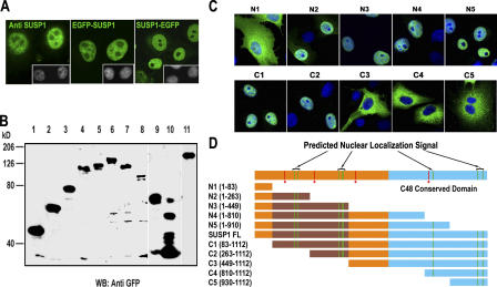
It has been reported that SUSP1 localizes primarily in the cytoplasm (Kim et al., 2000). Because all human SUMO paralogues are predominately nuclear (Ayaydin and Dasso, 2004), we reexamined the localization of SUSP1 in HeLa cells (Fig. 1 A) using both immunofluorescence staining and N- or C-terminal fusions between SUSP1 and EGFP. All of these approaches clearly indicated that SUSP1 is a nucleoplasmic protein, with minimal distribution to the cytoplasm, nucleolus, or nuclear envelope. To map the domain responsible for its nuclear localization, different fragments of SUSP1 were expressed as C-terminal EGFP fusions (Fig. 1 B). When we examined their distribution in fixed cells, we found that sequences between residues 84 and 448 of SUSP1 are required for its nuclear localization (Fig. 1, C and D). SUSP1's localization sequence is notable with respect to targeting sequences of other SENP/Ulps: the catalytic domains of all SENP/Ulps are localized toward their C termini (Melchior et al., 2003). In every case where the targeting requirements of SENP/Ulps have been determined, correct localization requires sequences within their N-terminal domains (Fig. S1, available at http://www.jcb.org/cgi/content/full/jcb.200510103/DC1). Our results for SUSP1 are consistent with this pattern, suggesting that the N-terminal domains of SENP/Ulps generally mediate their subcellular targeting.
Figure 1.
SUSP1 requires an N-terminal domain for its nucleoplasmic localization. (A) HeLa cells were fixed, permeabilized, and stained for immunofluorescence with anti-SUSP1 antibodies (left). Alternatively, HeLa cells were transfected with pEGFP-C1-SUSP1 (middle) or pEGFP-N1-SUSP1 (right). In all cases, DNA was simultaneously visualized with Hoechst 33258 dye (insets). (B) HeLa cells were transfected with plasmids expressing EGFP fusions to fragments of SUSP1. After 24 h, cell lysates were analyzed by Western blotting with anti-GFP antibodies. Lanes were as follows: 1, SUSP11-84-EGFP (N1); 2, SUSP11-263-EGFP (N2); 3, SUSP11-449-EGFP (N3); 4, SUSP11-810-EGFP (N4); 5, SUSP11-930-EGFP (N5); 6, SUSP184-1112-EGFP (C1); 7, SUSP1263-1112-EGFP (C2); 8, SUSP1449-1112-EGFP (C3); 9, SUSP1810-1112-EGFP (C4); 10, SUSP1930-1112-EGFP (C5); and 11, SUSP11-1112-EGFP (FL). (C) HeLa cells were transfected with GFP fusion constructs N1-N5 and C1-C5 (green). After 24 h, the cells were fixed and counterstained with Hoechst 33258 (blue). (D) Schematic summary of the SUSP1 sequences. Predicted NLSs within the full-length protein are indicated by green lines. The conserved catalytic domain is indicated in blue. The brown region of bottom bars indicates the nuclear targeting domain. Red lines indicate consensus SUMO-binding motifs (Song et al., 2004).
SUSP1 depletion causes redistribution of SUMO2 and -3 but not SUMO1
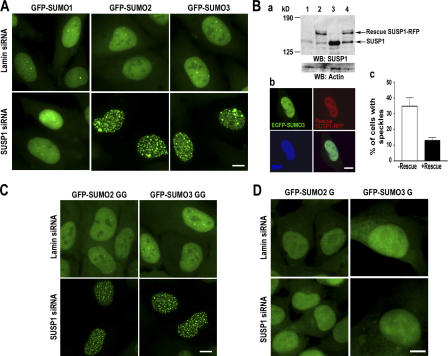
To examine the biological role of SUSP1, its expression was suppressed by siRNA-mediated gene silencing in human osteosarcoma-derived cells (U2OS cells) that stably express N-terminal EGFP fusions to SUMO1, -2, or -3 (Fig. 2 A). 48 h after transfection, cells that were treated with siRNAs against SUSP1 mRNA showed substantially lower levels of SUSP1 protein (<10%) than control cells that were treated with siRNAs directed against Lamin A/C mRNA (Fig. 2 B). In SUSP1-depleted cells, EGFP-SUMO1 distribution was indistinguishable from Lamin-depleted controls (Fig. 2 A). In contrast, EGFP-SUMO2 and -SUMO3 showed striking accumulation within nuclear foci in ∼30% of the SUSP1-depleted cells, although this redistribution was not observed in the control cells. Although the overall spectrum of EGFP-SUMO2– or EGFP-SUMO3–conjugated targets detected by Western blotting was not grossly different after depletion of SUSP1, we observed a moderate but consistent increase in very high molecular weight GFP-containing species (Fig. S2, available at http://www.jcb.org/cgi/content/full/jcb.200510103/DC1). No comparable accumulation of high molecular weight GFP-containing species occurred in EGFP-SUMO1–expressing cells (unpublished data). With prolonged incubations after siRNA treatment, we observed a higher level of cell death in the SUSP1-depleted cells expressing EGFP-SUMO2 and -SUMO3 than in Lamin-depleted controls (unpublished data), possibly suggesting that the accumulation of such SUMO2/3-conjugated species is detrimental to cell survival.
Figure 2.
Deficit of SUSP1 deconjugating activity causes focal concentration of EGFP-SUMO2 or -SUMO3. (A) U2OS cells stably expressing full-length EGFP-SUMO1, -SUMO2, or -SUMO3 were transfected with Lamin or SUSP1 siRNA. After 48 h, live cell images were acquired. Approximately 30% of EGFP-SUMO2– or EGFP-SUMO3–expressing cells showed the speckled phenotype after SUSP1 depletion. This phenomenon was never observed in the Lamin-depleted cells or in the EGFP-SUMO1–expressing cells. (B) Cells stably expressing EGFP–SUMO3 were transfected with a plasmid for the synthesis of SUSP1-RFP (pDSRed-N1-Mono-SUSP1), encoding by an mRNA with point mutations that rendered it impervious to RNAi. (a) Western blotting analysis with anti-SUSP1 antibodies (top). Lane 1 shows SUSP1 siRNA without rescue, lane 2 shows SUSP1 siRNA with rescue, lane 3 shows Lamin siRNA without rescue, and lane 4 shows Lamin siRNA with rescue. The same membrane was reprobed with anti-actin antibodies (bottom), to show protein loading. (b) A representative picture showing SUSP1-RFP in rescued cells. EGFP-SUMO3 is in green, SUSP1-RFP is in red, and DNA is in blue. (c) The percentage of cells showing the EGFP-SUMO3 speckles after SUSP1 siRNA and transfection with a control plasmid, pDSRed-N1 (−Rescue) or with pDSRed-N1-Mono-SUSP1 (+Rescue). Error bars represent SD for two separate experiments counting >200 total cells. n = 2. (C) U2OS cells stably expressing mature EGFP-SUMO2 or -SUMO3, ending in C-terminal diglycine motifs were transfected with Lamin siRNA or SUSP1 siRNA as in A. (D) U2OS cells stably expressing nonconjugatable EGFP-SUMO2 or -SUMO3, ending in a single C-terminal glycine were transfected with Lamin or SUSP1 siRNA, as in A. Bars, 10 μM.
We did not observe redistribution of endogenous SUMO2 and -3 after SUSP1 depletion by immunofluorescence in U2OS cells without EGFP-SUMO fusions (unpublished data). The most straightforward explanation for this difference is that there is sufficient SENP activity remaining in SUSP1-depleted cells, either from residual SUSP1 or from the redundant activity of other SENP/Ulps, to prevent the inappropriate accumulation of conjugated species when SUMO2 and -3 are present at physiological concentrations. In this case, the amount of SUSP1 activity may become limiting only when the concentration of conjugated species is elevated, as in cells expressing EGFP-SUMO2 or -SUMO3.
Importantly, redistribution of EGFP-SUMO2 and -SUMO3 was not observed when siRNAs were cotransfected with a plasmid that expresses a fusion between SUSP1 and the red fluorescent protein (RFP), encoded by a mutant mRNA that is not degraded by the siRNAs (Fig. 2 B). This finding substantiates the conclusion that altered EGFP-SUMO2 and -SUMO3 distributions are a direct result of SUSP1 depletion. Together, these findings indicate that SUSP1 plays a paralogue-specific role in the regulation of nucleoplasmic SUMO2/3.
SUSP1 acts as an isopeptidase for SUMO2/3 deconjugation
To determine whether the processing function of SUSP1 was crucial for the redistribution of EGFP-SUMO2 or -SUMO3, we repeated the siRNA experiment in U2OS-derived cell lines stably expressing the processed forms of EGFP-SUMO2 and -SUMO3, which terminate with the mature diglycine motif (Fig. 2 C). Similar to cells expressing the unprocessed forms, EGFP-SUMO2(GG) or -SUMO3(GG) became concentrated strongly into nuclear foci. This result implies that accumulation of EGFP-SUMO2 or -SUMO3 into foci after SUSP1 depletion results from the inability to deconjugate these fusion proteins from their substrates rather than insufficient processing capacity. Consistent with this conclusion, we observed no redistribution after siRNA of nonconjugatable EGFP-SUMO2 or -SUMO3 that had only a single C-terminal glycine (Fig. 2 D).
EGFP-SUMO2 and -SUMO3 accumulate in PML bodies after SUSP1 depletion
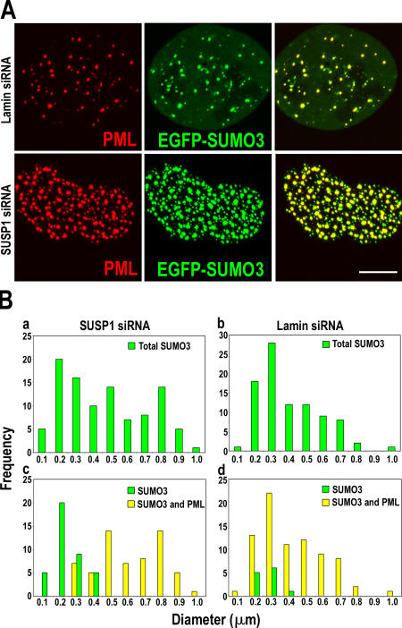
Together, our results suggested that insufficient levels of SUSP1 deconjugation activity caused the accumulation of EGFP-SUMO2 and -SUMO3 in nuclear foci. It was therefore of interest to characterize these structures. First, we examined the size of EGFP-SUMO2– or EGFP-SUMO3–labeled foci in cells depleted of SUSP1 or Lamin A/C (Fig. 3). We observed a bimodal distribution of foci size in the SUSP1-depleted cells, with a substantial peak of larger foci centered ∼0.8 μm in diameter. There were very few foci of this size in the Lamin-depleted cells.
Figure 3.
Large EGFP-SUMO3 speckles in SUSP1-depleted cells colocalize with PML. (A) U2OS cells expressing EGFP-SUMO3 (green) were treated with Lamin or SUSP1 siRNA as indicated. The cells were fixed, permeabilized, and stained with anti-PML antibodies (red). Merged images are shown on right. (B) The diameters of EGFP-SUMO3 foci in SUSP1-depleted (a) and Lamin-depleted (b) cells were measured. These distributions were compared with the size distribution of foci containing (yellow bars) or lacking (green bars) colocalized PML in SUSP1-depleted (c) and Lamin-depleted (d) cells. Bar, 10 μM.
We reasoned that these foci might correspond to nuclear subcompartments where SUMO2/3 normally play a physiological role. To test this idea, we stained SUSP1-depleted cells with a variety of antibodies that recognize antigens characteristic of splicing foci (SC35; Huang and Spector, 1991), pericentric heterochromatin (trimethyl-Histone H3 [Lys9]; Peters et al., 2003), and centromeres (CREST sera; Earnshaw and Rothfield, 1985). We did not observe colocalization of EGFP-SUMO2/3 with either SC35 or trimethyl-Histone H3 (unpublished data). We observed some colocalization with CREST sera staining, but the extent of this accumulation was not substantially different between SUSP1-depleted and control cells (unpublished data). We also stained the cells with antibodies directed against a variety of SUMO substrates, including Bloom's antigen (BLM), Wilms' tumor 1 (WT1), proliferating cell nuclear antigen (PCNA), p300, and PML (Johnson, 2004; Smolen et al., 2004). We did not find redistribution of BLM, WT1, PCNA, or p300, nor their accumulation within the EGFP-SUMO2/3 foci (unpublished data). However, we saw a substantial increase in the number of PML bodies after SUSP1 depletion and extensive colocalization of PML with EGFP-SUMO2 or -SUMO3 (Fig. 3 A).
Comparison of PML bodies versus EGFP-SUMO3 foci (Fig. 3 B) showed that the size distribution of PML bodies after SUSP1 depletion mirrored the change in EGFP-SUMO3 foci. Indeed, the larger EGFP-SUMO3 foci in SUSP1-depleted cells almost universally correlated to PML-containing structures (Fig. 3 A), suggesting that enlarged PML bodies result from insufficient SUMO2/3 deconjugation after SUSP1 depletion. The smaller, non–PML-containing bodies may analogously represent other structures to which SUMO2/3 conjugates associate under normal circumstances.
SUSP1 acts specifically as a SUMO2/3 protease in vitro
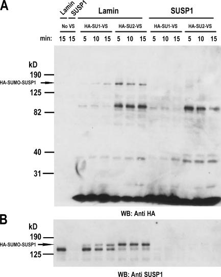
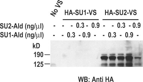
To directly determine the paralogue specificity of SUSP1, we used VS derivatives of tagged SUMO1 and -2 (HA-SU1-VS and HA-SU2-VS; Hemelaar et al., 2004). VS reagents derived from ubiquitin-like proteins covalently react with the nucleophilic active site residues of their respective modifying enzymes, showing considerable preference toward deconjugation enzymes over E1 and E2 enzymes. To test the reactivity of SUSP1 for SUMO1 and -2, extracts of control and SUSP1-depleted U2OS cells were incubated with HA-SU1-VS or HA-SU2-VS. Samples were taken at different times and subjected to SDS-PAGE and Western blotting with anti-HA antibodies (Fig. 4 A). We observed a band in HA-SU2-VS–treated control extracts that migrated with an apparent molecular mass of ∼160 kD. This band was absent in the SUSP1-depleted extracts. Both the molecular mass of this band and its depletion through RNAi indicate that it was derived from a covalent attachment of HA-SU2-VS and SUSP1. Moreover, when we immunoblotted the same samples with anti-SUSP1 antibodies, we observed that SUSP1 was quantitatively shifted into a higher molecular mass band within 5 min of incubation with HA-SU2-VS, confirming this conclusion.
Figure 4.
SUSP1 paralogue preference for SUMO2. Cell lysates from SUSP1- or Lamin-depleted cells were incubated with HA-SU1-VS or HA-SU2-VS for the indicated times. The reactions were terminated with SDS sample buffer and analyzed by Western blotting using anti-HA (A) or anti-SUSP1 antibodies (B).
Notably, an HA-reactive band of the same size was only weakly seen in the reaction containing HA-SU1-VS, suggesting that it has a significantly lower affinity for SUSP1. Although some SUSP1 migrated at a higher molecular weight after incubation with HA-SU1-VS, this conversion was significantly slower than in reactions containing HA-SU2-VS and did not proceed to completion within 30 min. Together, these results suggest that SUSP1 has a strong preference for SUMO2 over SUMO1. To test this conclusion using more stringent criterion, we preblocked U2OS cell lysates with a threefold molar excess (over the VSs) of untagged aldehyde derivatives of SUMO1 or -2, which bind reversibly to the active sites of SUMO proteases (Pickart and Rose, 1986). After blocking, the extracts were allowed to react with HA-SU2-VS and analyzed by Western blotting using anti-HA antibodies (Fig. 5). In these experiments, SUMO2 aldehyde, but not SUMO1 aldehyde, competed for the binding of HA-SU2-VS to SUSP1 and caused reduction in the intensity of the HA-reactive band at 160 kD, rigorously supporting the conclusion that SUSP1 has higher affinity for SUMO2/3 paralogues.
Figure 5.
SUSP1 preferentially binds to SUMO2 aldehyde. U2OS cell lysates were incubated with 0.3 or 0.9 ng/μl of SUMO1 or SUMO2 aldehyde. The samples were then allowed to react with HA-SU1-VS or HA-SU2-VS, as indicated, for 5 min. The reactions were terminated with sample buffer and analyzed by Western blotting with anti-HA antibodies.
SUSP1 preferentially cleaves substrates with multiple SUMO2 moieties
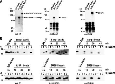
We developed model substrates to further evaluate the enzymatic activity of SUSP1 in processing and deconjugation assays. To examine SUSP1 activity as a processing enzyme, we expressed SUMO1, -2, and -3 in bacteria, fused at their C termini to a T7 tag (SU1-, SU2-, and SU3-T7). We purified these substrates by affinity chromatography. We immunoprecipitated SUSP1 from HeLa extracts using anti-SUSP1 antibodies and confirmed its activity through reactivity with HA-SU2-VS (Fig. 6 A). We then tested whether the immunoprecipitated SUSP1 fraction could release the T7 tag from the model processing substrates. We observed negligible cleavage of the SU1-, SU2-, and SU3-T7 during the course of a 45-min reaction at 37°C (Fig. 6 B, bottom), indicating that SUSP1 works poorly as a processing enzyme under these conditions.
Figure 6.
SUMO maturation activity of SUSP1. (A) SUSP1, Senp1, and control IgG beads were used to immunoprecipitate the respective enzymes from HeLa cell lysate. The enzymes bound to the beads were then reacted with HA-SUMO2-VS for 15 min. Western blot was performed with the indicated antibodies. (B) Full-length SUMO1, -2, or -3 proteins fused to a T7 tag at their C terminus (SU1-, SU2-, and SU3-T7) were expressed in bacteria and purified. These substrates were incubated with IgG, Senp1, or SUSP1 beads for the indicated times, and Western blotting was performed with the indicated antibodies.
As a positive control, we performed the same experiment using immunoprecipitated SENP1 protein (Fig. 6 A), which has been shown to be an efficient processing enzyme for SUMO1 but inefficient in its action against SUMO3 (Xu and Au, 2005). We judged that comparable concentrations of active enzyme were added to both reactions through equivalent reactivity with HA-SU2-VS, which would irreversibly label the active sites of both enzymes during the course of a 15-min reaction. Consistent with the earlier findings, we observed efficient cleavage of SU1- and SU2-T7 by SENP1, with minimal activity toward SU3-T7 (Fig. 6 B, top). Although these results do not strictly rule out the possibility that SUSP1 is ever involved in processing of any paralogue, they argue that SUSP1 is unlikely to be a major processing enzyme.
To examine deconjugation, we produced a purified, recombinant C-terminal fragment containing the primary acceptor site Ran GTPase-activating protein 1 (RanGAP1; His6-T7-RanGAP1-C2). We incubated this fragment in vitro with purified E1 and E2 enzymes plus SUMO1 or -2 (Azuma et al., 2001), resulting primarily in monoconjugated species containing each paralogue. We again found that immunoprecipitated SUSP1 showed little activity against these model substrates (unpublished data), leading us to conclude that SUSP1 works poorly as a deconjugating enzyme against substrates containing single SUMO moieties.
These results suggested that SUSP1 might not act as a general processing or deconjugation enzyme but might act on a much more specialized subset of SUMO2/3-containing substrates. To test whether it might act on substrates that are multiply conjugated, we used polycistronic vectors for bacterial expression of His6-T7-RanGAP1-C2 with SUMO E1 and E2 enzymes, along with SUMO1 or -2 (Uchimura et al., 2004). In this system, His6-T7-RanGAP1-C2 becomes highly conjugated with the coexpressed SUMO proteins (Fig. 7), as isopeptidases are absent. Formation of these conjugates requires the diglycine motif at the C termini of SUMO proteins and the primary SUMO acceptor lysine of RanGAP1 (Lys517 in Xenopus laevis), indicating that SUMO conjugation in Escherichia coli occurs through an isopeptide bond between Lys517 of RanGAP1 and glycine at the C terminus of SUMO1 (Uchimura et al., 2004). To this extent, conjugation in E. coli and mammalian cells are similar, as both occur specifically through the single, conserved primary acceptor lysine (Mahajan et al., 1998; Matunis et al., 1998). This specificity is likely to reflect the strong and specific binding of Ubc9 to this region of RanGAP1 (Bernier-Villamor et al., 2002; Reverter and Lima, 2005). It is formally possible that conjugation of Lys517 in bacteria promotes the subsequent recognition and conjugation of other lysine residues within His6-T7-RanGAP1-C2. However, we consider this scenario to be improbable because none of the other lysine residues lie within an optimal sequence context, nor do they become conjugated under any other circumstances, including in vitro assays with high concentrations of all conjugation pathway enzymes. Thus, it is most likely that the conjugates from E. coli bearing multiple SUMO polypeptides are configured in chains or branched structures.
Figure 7.
Preferential SUMO2 chain cleavage by SUSP1. T7-tagged RanGAP-C2 conjugated to either SUMO1 or -2 in E. coli was purified and incubated with SUSP1 beads for the indicated times. The samples were separated by SDS-PAGE and assayed by Western blotting with anti-T7 (A), anti-SUMO1 (B), or anti-SUMO2 (C) antibodies. Single and double asterisks indicate conjugated species bearing two and three SUMO moieties, respectively. More highly conjugated species are indicated with a bracket.
After bacterial lysis and affinity purification of His6-T7-RanGAP1-C2–containing species, we obtained a preparation containing His6-T7-RanGAP1-C2 conjugated to different numbers of SUMO moieties. We assayed whether immunoprecipitated SUSP1 would effectively deconjugate any or all of the species contained in this mixture (Fig. 7). We observed that the immunoprecipitated SUSP1 fraction cleaved high-molecular-weight SUMO2 conjugates very efficiently. Interestingly, conjugation products containing only one or two SUMO2 moieties were not efficiently deconjugated, even with relatively long incubations (Fig. 7, A and C, 60 min). Remarkably, we observed minimal deconjugation of any SUMO1-containing substrates (Fig. 7 B), indicating that SUSP1 is also not an efficient isopeptidase for SUMO1 conjugates, even those that linked to high numbers of SUMO1 moieties.
Overall, this pattern indicates that SUSP1 acts preferentially against substrates bearing three or more SUMO2/3 moieties. This conclusion is consistent with the limited change in GFP-SUMO2–conjugated substrates after SUSP1 depletion (Fig. S2), where we see accumulation of very high molecular weight species rather than a global change in the pattern of conjugated targets, supporting the notion that SUSP1 dismantles SUMO2/3 chains both in vitro and in vivo.
Discussion
We have used a coordinated approach to investigate in vivo function and in vitro biochemical properties of SUSP1. Our observations demonstrate that SUSP1 is a nucleoplasmic SUMO2/3 isopeptidase. The activity of this enzyme is required to maintain the distribution of SUMO-conjugated species between subnuclear compartments, and we observe the inappropriate accumulation of SUMO2/3-modified species in PML bodies and other structures in its absence. Moreover, in vitro analysis of paralogue specificity through VS derivatives of SUMO proteins strongly indicate that SUSP1 acts selectively on SUMO2. Further experiments with other model substrates revealed that SUSP1 has minimal activity as a processing enzyme or in the deconjugation of single SUMO moieties. On the other hand, assays of its activity against SUMO-conjugated substrates that were prepared using an E. coli–based expression system showed that SUSP1 could act effectively against species conjugated with multiple SUMO2 moieties. Collectively, these observations suggest that SUSP1 plays a highly specialized role in vertebrate cells in the dismantling of highly conjugated SUMO2/3 species and that the appropriate control of these species is critical for the accurate maintenance of nuclear structures, particularly PML bodies.
An earlier report indicated that SUSP1 acts as a processing enzyme for SUMO1 in vitro but that it does not efficiently deconjugate SUMO1 from RanGAP1, leading to the conclusion that SUSP1 is primarily involved in SUMO1 processing (Kim et al., 2000). In contrast, our observations imply that SUSP1 has little processing activity (Fig. 6) but is important for SUMO2/3 deconjugation, especially from species containing three or more SUMO moieties (Fig. 7). This apparent conflict may reflect the fact that Kim et al. (2000) examined neither processing of SUMO2/3 nor deconjugation of monomeric or polymeric SUMO2/3 species. As a result, SUMO1 processing may have appeared to be the most robust activity of this enzyme, even though this activity is not substantial in comparison to SUSP1's capacity to dismantle highly conjugated SUMO2/3 species. Kim et al. (2000) also reported that SUSP1 localizes to the cytoplasm, whereas we find that the majority of this protein resides within nuclei of HeLa and U2OS cells (Fig. 1). We are confident in the conclusion that SUSP1 is a nuclear protein, because we have used multiple independent methods to assay its localization. Mapping of the sequences sufficient for targeting of SUSP1 defines an N-terminal domain that contains multiple putative nuclear localization signals (Fig. 1 D), providing some suggestion that SUSP1 gains entry to the nucleus through classical nuclear import pathways.
Interestingly, we find that the depletion of SUSP1 alters the distribution of EGFP-SUMO2 and -SUMO3, causing marked accumulation of these fusion proteins in PML-containing bodies (Figs. 2 and 3). It has previously been reported that RNAi-mediated depletion of SUMO3 specifically redistributes PML and causes loss of PML body integrity (Fu et al., 2005). This defect could be rescued by the expression of exogenous wild-type SUMO3, but not by a mutant lacking lysine 11, the primary residue implicated in SUMO2/3 chain formation. In combination with our findings, these data suggest that the integrity of PML bodies may require the formation of SUMO2/3 chains. Because SUSP1 acts to dismantle multiply conjugated species (Fig. 7), its absence after RNAi-mediated depletion promotes not only the accumulation of such species within the PML bodies of cells expressing EGFP-SUMO2 or -SUMO3, but also the assembly of PML bodies of remarkably increased size and number.
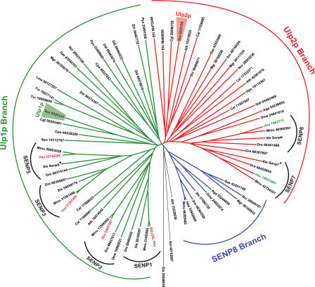
We have performed genomic searches across eukaryotic species to identify SENP/Ulp family members, to perform a comparison of their protein sequences (Fig. 8). The SENP8/Deneddylase 1–related branch of this family tree was clearly distinct, consistent with the finding that these enzymes act specifically on another ubiquitin-like protein, Nedd8 (Gan-Erdene et al., 2003; Wu et al., 2003). More notable, we found that another branch point divided the SUMO-specific SENP/Ulp family members into two distinct subsets, which contained budding yeast Ulp1p and -2p, respectively. Vertebrates possess four enzymes within the Ulp1p branch (SENP1, -2, -3, and -5), but only two within the Ulp2p branch (SUSP1/SENP6 and SENP7). Given this evolutionary relationship, it is interesting to note that our data indicate some conservation of function within one of these two branches: elegant genetic and biochemical studies suggest that the critical function of Ulp2p in budding yeast involves regulation of Smt3p chain elongation (Bylebyl et al., 2003). Our findings indicate that SUSP1 acts preferentially against multiply conjugated SUMO2/3 species (Fig. 7), which are likely to contain chained or branched SUMO structures (Uchimura et al., 2004). Moreover, we find that changes in the profile of EGFP-SUMO2– or EGFP-SUMO3–conjugated substrates upon SUSP1 depletion are most pronounced among very high molecular mass species (Fig. S2), consistent with the possibility that polymerized EGFP-SUMO2 and -SUMO3 structures form in its absence, whereas other aspects of SUMO2 and -3 metabolism are largely unaltered.
Figure 8.
Evolutionary relationship of SENP/Ulp family members. The eukaryotic genome databases of National Center for Biotechnology Information were searched using psi-BLAST using human SENP protein sequences and query with e-value inclusion cutoff of 0.001, for 6–10 cycles. Whenever any specific genome of interest failed to give any hit, the nucleotide genome sequence of corresponding organism was searched using TBLASTN with human Senp protein sequences as query. The collected protein sequences were aligned using MUSCLE (Edgar, 2004) with 100 iterations. The generated multiple alignments were manually corrected using the conserved C48 peptidase domain as anchor. Phylogenetic analyses were performed using the minimum evolution (least-square) method as implemented in MEGA3.1 (Kumar et al., 2004), with Poisson correction model and pairwise deletion of gaps and 1,000 bootstrap replicates. For each protein sequence, the letters indicate the shortened binomial nomenclature, and the numbers indicate the protein GenBank accession nos. Asterisks indicate sequences that are not in GenBank.
SENP/Ulps appear to share both the conserved catalytic region and a similar overall arrangement, wherein the catalytic domains are localized toward the C terminus of each protein and targeting sequences are found within their N termini (Melchior et al., 2003). Our finding that sequence features required for nucleoplasmic targeting of SUSP1 lie within its N-terminal domain is clearly consistent with this pattern (Fig. S1). Two other features of SUSP1's sequence may be further notable in light of its strong preference for multiply conjugated SUMO2 and -3 species. First, SUSP1 possesses four sequence motifs that conform to a previously identified SUMO-binding domain (Fig. 1 D; Song et al., 2004). It is interesting to speculate that these motifs may confer a higher affinity of SUSP1 for multimeric SUMO2/3 chains or orient SUSP1 during the process of substrate recognition. Second, SUSP1 also contains a 195-residue insertion that splits the conserved region corresponding to the catalytic domain (Fig. S1). This insertion appears to be unique to SUSP1, although SENP7 possesses a smaller inserted sequence in a closely equivalent configuration. We do not yet know the function of this inserted sequence, although it is possible to speculate that it may either enhance SUSP1's activity against polymeric SUMO2/3 or restrict SUSP1's activity against other targets, including single SUMO conjugates or processing substrates.
The most extensive analysis of SENP/Ulp functions have been performed in budding yeast (Li and Hochstrasser, 1999, 2000, 2003; Bylebyl et al., 2003); these studies have conclusively shown that Ulp1p and -2p are not functionally redundant, as elimination of either protein results in highly distinct phenotypic consequences. We believe that vertebrate SENP/Ulps may possess an even higher degree of specialization with respect to their enzymatic activity, paralogue preference, and targeted localization within the nucleus. In this case, it will be vital to characterize each of these enzymes accurately and completely, in order to interpret experiments involving their manipulation correctly and to understand fully their role within the whole SUMO pathway.
Materials and methods
Antibodies and reagents
Monoclonal mouse antibody (PG-M3) against PML was obtained from Santa Cruz Biotechnology, Inc. SUSP1 antibody was raised in rabbit against the N-terminal 499 amino acid of SUSP1 and was affinity purified using an antigen column. Anti-HA 3F10 rat monoclonal antibody was purchased from Roche Diagnostics Corporation. Alexa Fluor–labeled secondary antibodies were obtained from Invitrogen, HRP-conjugated mouse and rabbit secondary antibodies and N-hydroxysuccinimide–Sepharose (FF) were obtained from GE Healthcare, HRP-conjugated anti-rat antibody was obtained from Pierce Chemical Co., and HRP-conjugated anti-T7 tag antibody was obtained from Novagen (EMD Biosciences). Anti-SUMO1, -SUMO2, and -Senp1 polyclonal antibodies were raised in rabbit and affinity purified (provided by M. Matunis, Johns Hopkins University School of Public Health, Baltimore, MD). All other reagents were obtained from Sigma-Aldrich unless otherwise stated.
DNA constructs
pEGFP and pDsRed vectors were obtained from CLONTECH Laboratories, Inc. Restriction enzymes were purchased from New England Biolabs, Inc. DNA primers and Platinum Taq Hi Fi DNA polymerase were obtained from Invitrogen.
Full-length (unprocessed) human SUMO1, -2, and -3; mature SUMO1GG, SUMO2GG, and SUMO3GG (with diglycine at amino acid positions 96–97, 92–93, and 90–91, respectively); and nonconjugatable SUMO1G, SUMO2G, and SUMO3G (with a single C-terminal glycine at amino acid positions 96, 92, and 90, respectively) coding sequences were subcloned into HindIII and BamHI restriction sites of pEGFP-C3. Full-length SUSP1 was cloned into XhoI and BamHI sites of either pEGFP-NI or -C2 by RT-PCR from HeLa cell total RNA. Deletion fragments of SUSP1 were amplified by PCR using specific primers and cloned into XhoI and BamHI sites of pEGFP-N1.
The rescue SUSP1 plasmid was prepared by sense-antisense method of site-directed mutagenesis using the primer set 5′-GAAGAATCTGAAGGGGATACA-3′, 5′-TTCATGATCAAAGCTCCAATT-3′, which incorporates three sense point mutations at 124 bp (A to T), 125 bp (G to C), and 132 bp (A to G) from the start of the open reading frame and generates a BciVI restriction site. This was then subcloned into XhoI and BamHI restriction sites of pDSRed-Mono-N1 vector. The absence of any unintended mutations was confirmed for all constructs by DNA sequencing (SeqWright DNA Technology Services).
Cell culture
HeLa cells, U2OS cells, and their derivative stable lines were grown at 37°C in a humidified atmosphere of 5% CO2 in DME (Biosource International) with 2 mM glutamine supplemented with 10% fetal bovine serum (Gemini Bio-Products), 100 U penicillin/ml, and 100 μg/ml streptomycin. Cells were transfected with plasmids using Effectene reagent (QIAGEN) according to the manufacturer's instructions.
siRNA-mediated depletion of SUSP1
For transient transfections, cells were grown to 50% confluency and Effectene reagent (QIAGEN) was used according to the manufacturer's instructions. Duplex siRNA directed against SUSP1 and Lamin A/C messages were obtained from QIAGEN. SUSP1 and Lamin A/C siRNA sequences were 5′-AAGAAAGTGAAGGAGATACAG-3′ and 5′-AACTGGACTTCCAGAAGAACA-3′, respectively, with standard dTdT 3′ extension.
siRNA experiments were performed in 24-well culture plates, coverslip-bottomed 4-well chambers (LabTekII; Fisher Scientific), or 6-cm dishes. Cells were allowed to attach overnight at 40% confluency and washed once with DME without antibiotics immediately before transfection of siRNA. 3 μl siRNA (20 μM) and 3 μl of oligofectamine (Invitrogen) were suspended in 50 and 12 μl of OptiMEM (Invitrogen), respectively, in separate tubes. The tubes were gently tapped and kept at RT for 5 min, and the contents of the two tubes were mixed, kept at RT for a further 20 min, and added to a single well of 24-well plate or a well of 4-well chamber containing 0.5 ml complete DME without antibiotics. For the 6-cm dish, the same procedure was followed but the reagents were scaled up to four times.
Production and maintenance of stable cell lines
For stable cell line selection, 0.5 mg/ml Geneticin was added to culture medium 24 h after transfection. Cells were incubated in Geneticin-containing culture medium that was refreshed daily for a period of 1 wk after resistant colonies were reseeded sparsely to culture single cell–derived colonies. Uniformly fluorescent colonies derived from single cells were marked and isolated under an inverted fluorescence microscope. Stably transgenic cells were maintained thereafter in medium containing 0.25 mg/ml Geneticin.
Immunofluorescence
Cells were grown either on poly-l-lysine–coated coverslips or LabTekII coverslip-bottomed chambers. For immunofluorescence, the cells were washed with PBS and fixed for 12 min at ambient temperature with 4% paraformaldehyde in PME buffer (PBS supplemented with 5 mM each of MgCl2 and EGTA). The cells were permeabilized with 0.5% Triton X-100 for 10 min. After washing with PME, the cells were blocked for 10 min in 1% normal horse serum (Vector Laboratories) in PME and incubated for 1 h with anti-PML antibodies diluted 1:200 in blocking solution. The coverslips were washed and incubated for 45 min with Alexa Fluor 568–conjugated secondary antibodies (Invitrogen) diluted 1:400 in blocking solution. Unbound antibodies were removed by washing, and the cells were briefly incubated in 100 ng/ml Hoechst 33258 DNA stain. Finally, the coverslips were mounted in Vectashield mounting medium (Vector Laboratories).
Microscopy
Fluorescence microscopy was performed on a confocal microscope (LSM510 META; Carl Zeiss MicroImaging, Inc.) equipped with 40× Plan Neofluar objective. LabTekII coverslip-bottomed chambers were used for live analyses. Temperature (37°C) and CO2 concentration (5%) were maintained using a humidified environmental chamber. Confocal microscopy software (SP2 version 3.2; Carl Zeiss MicroImaging, Inc.) was used for capturing images, which were then analyzed by Photoshop 7.0 (Adobe). In all figures, scale bars represent 10 μm.
Illumination of samples for fluorescent imaging
We used a 543-nm HeNe laser (5 mW output; detection LP560 nm) for detection of RFP-SUSP1-rescue signal and indirect immunolocalization of PML by Alexa Fluor 568–labeled antibodies. The 488-nm line of an Argon laser (25 mW nominal output; detection BP 505–530 nm) were used for analysis of GFP-conjugated proteins. Hoechst 33258 images were captured using the 364-nm line of an ion laser (Enterprise II ML UV [Coherent, Inc.]; 80 mW nominal output; detection BP 385–470 nm).
Synthesis of SUMO derivatives
Expression of SUMO-intein-CBD fusion proteins in E. coli was as described in Hemelaar et al., (2004). Bacterial lysates were prepared as described previously (Hemelaar et al., 2004) and bound to chitin bead columns (New England Biolabs, Inc.). The columns were washed with five volumes of lysis buffer (50 mM Hepes, 100 mM sodium acetate, pH 6.5, and 50 μM PMSF), followed by three volumes of lysis buffer containing 50 mM β-mercaptoethanesulfonic acid (MESNa; Sigma-Aldrich). The column was incubated overnight at 37°C in the buffer with MESNa to allow on-column cleavage. The SUMO-MESNa thiolesters were eluted with 1.5 column volumes of lysis buffer, and the fractions containing SUMO-MESNa products were concentrated on a Centriprep (3,000-molecular-weight cutoff; Millipore). To convert SUMO-MESNa products to their VS derivatives, a large excess of Glycine VS (final concentration 0.25 M) was added to concentrated SUMO-MESNa (1–3 mg/ml; 500 μl), followed by addition of 75 μl of 2 M N-hydroxysuccinimide and 30 μl of 2 M NaOH (provided by H. Ovaa, Netherlands Cancer Institute, Amsterdam, Netherlands; Hemelaar et al., 2004). The mixture was incubated for 1–2 h at 37°C, and the reaction was terminated by the addition of 30 μl of 2 M HCl.
To obtain SUMO1 and -2 aldehydes, SUMO1 and -2 acetals were synthesized by reacting SUMO-MESNa thiolesters (1–3 mg/ml; 500 μl) with 0.2 ml of 4 M aminoacetaldehyde diethyl acetal, pH 8.5, and 0.14 ml of N-hydroxysuccinimide, pH 7.0, at 37°C for 2 h. To obtain SUMO1 and -2 aldehydes, SUMO1 and -2 acetals were separately incubated with 0.15 M HCl at 37°C for 30 min. All steps of derivative syntheses were monitored by HPLC and by SDS-PAGE.
SUMO derivatives were purified by ion exchange chromatography, desalted on PD10 columns, and dialyzed into 50 mM Triethanolamine, pH 6.5. SUMO1 and -2 aldehydes were further purified by adsorbing on an anion exchange column (Mono Q HR; Pharmacia) equilibrated with 50 mM TEA, pH 6.5, and eluted with a linear gradient of NaCl from 0 to 0.5 M (flow rate of 0.75 ml/min; fraction size = 1 ml). For purification of HA-SUMO1-VS and HA-SUMO2-VS on Mono Q, 50 mM Bis-Tris, pH 5.5, was used, with a gradient from 0 to 0.15 M NaCl. In all cases, proteins were monitored by absorbance at 205 nm. Individual Mono Q fractions were assayed by HPLC, and appropriate fractions were pooled and stored at −80°C.
HA-SUMO-VS experiments
VS and aldehyde derivatives of SUMO1 and -2 were prepared as described previously (Hemelaar et al., 2004). 48 h after transfection with siRNAs directed against Lamin or SUSP1, as indicated, U2OS cells were harvested and washed twice in PBS containing 1 mM AEBSF. The cells were resuspended in reaction buffer (10 mM Hepes, pH 7.4, 150 mM NaCl, 5 mM EDTA, 1 mM DTT, and 25 μg/ml each of leupeptin, aprotinin, and pepstatin) and sonicated. Cell lysates were clarified by centrifugation at 16,000 g for 10 min. The protein concentration of the total cell lysate was maintained between 500 μg/ml and 2 mg/ml. SUMO-VS was added to a final concentration of 0.3 ng/μl and allowed to react for the indicated intervals. The reactions were terminated by the addition of SDS sample buffer and analyzed by Western blotting with the indicated antibodies.
SUMO deconjugation assay
SUMO1- and SUMO2-conjugated (His)6-T7-RanGAP-C2 fragment were produced in bacteria (Uchimura et al., 2004) and purified using Ni-NTA beads (provided by Y. Uchimura and H. Saitoh, Kumamoto University, Kumamoto, Japan). SUSP1 was immunoprecipitated from HeLa cell lysate using antibody conjugated to N-hydroxysuccinimide–Sepharose (2 μg/ml of beads). 10 μl SUSP1 beads were incubated with 500 ng of SUMO-conjugated T7-RanGAP-C2 in a total reaction volume of 30 μl at 23°C for the indicated times. The reaction was terminated with sample buffer and analyzed by Western blotting with antibodies against T7, SUMO1, or SUMO2/3.
Preparation of SUMO processing substrates
Full-length SUMO1, -2, and -3 cDNAs were subcloned into pET28b expression plasmids that had been cut with NcoI–NdeI, NcoI–NdeI, and BspH1–NdeI restriction endonucleases, respectively. These plasmids were transfected into E. coli, and the expression of the fusion proteins was induced with 0.4 mM IPTG under standard conditions. The expressed C-terminally tagged SUMO-T7-His proteins were purified on a Ni-NTA column followed by Q-Sepharose and MonoQ columns to generate pure tagged substrate for the processing reactions.
SUMO processing assay
HeLa cells from confluent 15-cm dishes were harvested by trypsinization and washed twice with ice-cold PBS. The cells were snap frozen in liquid nitrogen and then sonicated in 500 μl lysis buffer (10 mM Hepes, pH 7.5, 150 mM NaCl, 1 mM DTT, 1 mM AEBSF, and 5 mg/ml each of leupeptin, pepstatin, and aprotinin). The cell lysates were centrifuged at 120,000 rpm for 5 min at 2°C. The cleared supernatants were incubated with 5 μg of rabbit IgG, affinity-purified anti-SENP1 antibody, or affinity-purified anti-SUSP1 antibody for 1 h at 4°C. 50 μl preblocked protein A beads were added to the lysates and incubated for another hour and subsequently washed three times in lysis buffer. The beads were reacted with 0.3 ng/μl HA-SUMO2-VS in lysis buffer containing 100 μg/ml BSA for 15 min at RT, and the reactions were terminated with sample buffer. Using the different dilutions of the HA-SUMO-VS adducts thus obtained, beads having an equivalent enzymatic activity for SENP1 and SUSP1 were determined by anti-HA Western blot. Beads having equivalent amount of HA-SUMO2-VS reactivity and control beads were then incubated with 40 ng/μl SUMO1, -2 or -3 T7 for different time points at 23°C in 20 μl reaction volume. The reactions were stopped by the addition of sample buffer, and Western blots were performed using anti-T7, -SUMO1, or -SUMO2/3 antibodies.
Evolutionary relationship of SENP/Ulp family members
The eukaryotic genome databases of the National Center for Biotechnology Information were searched using psi-BLAST using human Senp protein sequence and query with e-value inclusion cutoff of 0.001, for 6–10 cycles. Whenever any specific genome of interest failed to give any hit, the nucleotide genome sequence of corresponding organism was searched using TBLASTN with human Senp protein sequences as query. The collected protein sequences were aligned using MUSCLE (Edgar, 2004) with 100 iterations. The generated multiple alignments were manually corrected using the conserved C48 peptidase domain as anchor. Phylogenetic analyses were performed using minimum evolution (least-square) method as implemented in MEGA3.1 (Kumar et al., 2004), with Poisson correction model and pairwise deletion of gaps and 1,000 bootstrap replicates.
Online supplemental material
Fig. S1 shows aligned sequences of Ulp1p and Ulp2p/Smt4p from S. cerevisiae and mammalian SENP/Ulp family members. Fig. S2 shows EGFP-SUMO3 in SUSP1-depleted cells. Online supplemental material is available at http://www.jcb.org/cgi/content/full/jcb.200510103/DC1.
Supplementary Material
Acknowledgments
We would like to thank Dr. M.K. Basu (National Library of Medicine, National Institutes of Health) for his help in construction of the SENP evolutionary tree. We would like to thank Dr. M. Matunis for providing anti-BLM antibody and H. Ovaa (Netherlands Cancer Institute) for a gift of gylcyl VS tosylate. We would like to thank Drs. Y. Uchimura and H. Saitoh (Kumamoto University) for providing constructs and strains required for production of substrates used in Fig. 7.
D. Mukhopadhyay, F. Ayaydin, S.-H. Tan, and M. Dasso were supported throughout this work by National Institute of Child Health and Human Development Intramural funds. T. Anan was the recipient of a fellowship from the Japan Society for the Promotion of Science. K.D. Wilkinson and N. Kolli were supported by National Institutes of Health grant GM66355. Studies on PML were initiated with support from Human Frontier Science Program research grant RG0229/1999-M.
F. Ayaydin, N. Kolli, and S.-H. Tan contributed equally to this paper.
F. Ayaydin's present address is Hungarian Academy of Sciences, H-6701 Szeged, Hungary.
S.-H. Tan's present address is Department of Cancer Biology, Kimmel Cancer Center, Thomas Jefferson University, Philadelphia, PA 19107.
T. Anan's present address is Department of Pediatrics, Kumamoto University School of Medicine, Kumamoto 860-0811, Japan.
Abbreviations used in this paper: BLM, Bloom's antigen; PML, promyelocytic leukemia protein; RanGAP1, Ran GTPase-activating protein 1; RFP, red fluorescent protein; SENP, sentrin-specific protease; SUMO, small ubiquitin-related modifier; SUSP1, SUMO-specific protease 1; Ulp, ubiquitin-like protease; VS, vinyl sulfone.
References
- Ayaydin, F., and M. Dasso. 2004. Distinct in vivo dynamics of vertebrate SUMO paralogues. Mol. Biol. Cell. 15:5208–5218. [DOI] [PMC free article] [PubMed] [Google Scholar]
- Azuma, Y., S.H. Tan, M.M. Cavenagh, A.M. Ainsztein, H. Saitoh, and M. Dasso. 2001. Expression and regulation of the mammalian SUMO-1 E1 enzyme. FASEB J. 15:1825–1827. [DOI] [PubMed] [Google Scholar]
- Azuma, Y., A. Arnaoutov, and M. Dasso. 2003. SUMO-2/3 regulates topoisomerase II in mitosis. J. Cell Biol. 163:477–487. [DOI] [PMC free article] [PubMed] [Google Scholar]
- Bencsath, K.P., M.S. Podgorski, V.R. Pagala, C.A. Slaughter, and B.A. Schulman. 2002. Identification of a multifunctional binding site on Ubc9p required for Smt3p conjugation. J. Biol. Chem. 277:47938–47945. [DOI] [PubMed] [Google Scholar]
- Bernier-Villamor, V., D.A. Sampson, M.J. Matunis, and C.D. Lima. 2002. Structural basis for E2-mediated SUMO conjugation revealed by a complex between ubiquitin-conjugating enzyme Ubc9 and RanGAP1. Cell. 108:345–356. [DOI] [PubMed] [Google Scholar]
- Bylebyl, G.R., I. Belichenko, and E.S. Johnson. 2003. The SUMO isopeptidase Ulp2 prevents accumulation of SUMO chains in yeast. J. Biol. Chem. 278:44113–44120. [DOI] [PubMed] [Google Scholar]
- Earnshaw, W.C., and N. Rothfield. 1985. Identification of a family of human centromere proteins using autoimmune sera from patients with scleroderma. Chromosoma. 91:313–321. [DOI] [PubMed] [Google Scholar]
- Edgar, R.C. 2004. MUSCLE: multiple sequence alignment with high accuracy and high throughput. Nucleic Acids Res. 32:1792–1797. [DOI] [PMC free article] [PubMed] [Google Scholar]
- Fu, C., K. Ahmed, H. Ding, X. Ding, J. Lan, Z. Yang, Y. Miao, Y. Zhu, Y. Shi, J. Zhu, et al. 2005. Stabilization of PML nuclear localization by conjugation and oligomerization of SUMO-3. Oncogene. 24:5401–5413. [DOI] [PubMed] [Google Scholar]
- Gan-Erdene, T., K. Nagamalleswari, L. Yin, K. Wu, Z.Q. Pan, and K.D. Wilkinson. 2003. Identification and characterization of DEN1, a deneddylase of the ULP family. J. Biol. Chem. 278:28892–28900. [DOI] [PubMed] [Google Scholar]
- Hemelaar, J., A. Borodovsky, B.M. Kessler, D. Reverter, J. Cook, N. Kolli, T. Gan-Erdene, K.D. Wilkinson, G. Gill, C.D. Lima, et al. 2004. Specific and covalent targeting of conjugating and deconjugating enzymes of ubiquitin-like proteins. Mol. Cell. Biol. 24:84–95. [DOI] [PMC free article] [PubMed] [Google Scholar]
- Huang, S., and D.L. Spector. 1991. Nascent pre-mRNA transcripts are associated with nuclear regions enriched in splicing factors. Genes Dev. 5:2288–2302. [DOI] [PubMed] [Google Scholar]
- Johnson, E.S. 2004. Protein modification by SUMO. Annu. Rev. Biochem. 73:355–382. [DOI] [PubMed] [Google Scholar]
- Kim, K.I., S.H. Baek, Y.J. Jeon, S. Nishimori, T. Suzuki, S. Uchida, N. Shimbara, H. Saitoh, K. Tanaka, and C.H. Chung. 2000. A new SUMO-1-specific protease, SUSP1, that is highly expressed in reproductive organs. J. Biol. Chem. 275:14102–14106. [DOI] [PubMed] [Google Scholar]
- Kumar, S., K. Tamura, and M. Nei. 2004. MEGA3: integrated software for molecular evolutionary genetics analysis and sequence alignment. Brief. Bioinform. 5:150–163. [DOI] [PubMed] [Google Scholar]
- Li, S.J., and M. Hochstrasser. 1999. A new protease required for cell-cycle progression in yeast. Nature. 398:246–251. [DOI] [PubMed] [Google Scholar]
- Li, S.J., and M. Hochstrasser. 2000. The yeast ULP2 (SMT4) gene encodes a novel protease specific for the ubiquitin-like Smt3 protein. Mol. Cell. Biol. 20:2367–2377. [DOI] [PMC free article] [PubMed] [Google Scholar]
- Li, S.J., and M. Hochstrasser. 2003. The Ulp1 SUMO isopeptidase: distinct domains required for viability, nuclear envelope localization, and substrate specificity. J. Cell Biol. 160:1069–1081. [DOI] [PMC free article] [PubMed] [Google Scholar]
- Li, Y., H. Wang, S. Wang, D. Quon, Y.W. Liu, and B. Cordell. 2003. Positive and negative regulation of APP amyloidogenesis by sumoylation. Proc. Natl. Acad. Sci. USA. 100:259–264. [DOI] [PMC free article] [PubMed] [Google Scholar]
- Mahajan, R., L. Gerace, and F. Melchior. 1998. Molecular characterization of the SUMO-1 modification of RanGAP1 and its role in nuclear envelope association. J. Cell Biol. 140:259–270. [DOI] [PMC free article] [PubMed] [Google Scholar]
- Matunis, M.J., J. Wu, and G. Blobel. 1998. SUMO-1 modification and its role in targeting the Ran GTPase-activating protein, RanGAP1, to the nuclear pore complex. J. Cell Biol. 140:499–509. [DOI] [PMC free article] [PubMed] [Google Scholar]
- Melchior, F., M. Schergaut, and A. Pichler. 2003. SUMO: ligases, isopeptidases and nuclear pores. Trends Biochem. Sci. 28:612–618. [DOI] [PubMed] [Google Scholar]
- Peters, A.H., S. Kubicek, K. Mechtler, R.J. O'Sullivan, A.A. Derijck, L. Perez-Burgos, A. Kohlmaier, S. Opravil, M. Tachibana, Y. Shinkai, et al. 2003. Partitioning and plasticity of repressive histone methylation states in mammalian chromatin. Mol. Cell. 12:1577–1589. [DOI] [PubMed] [Google Scholar]
- Pichler, A., and F. Melchior. 2002. Ubiquitin-related modifier SUMO1 and nucleocytoplasmic transport. Traffic. 3:381–387. [DOI] [PubMed] [Google Scholar]
- Pickart, C.M., and I.A. Rose. 1986. Mechanism of ubiquitin carboxyl-terminal hydrolase. Borohydride and hydroxylamine inactivate in the presence of ubiquitin. J. Biol. Chem. 261:10210–10217. [PubMed] [Google Scholar]
- Pickart, C.M., and D. Fushman. 2004. Polyubiquitin chains: polymeric protein signals. Curr. Opin. Chem. Biol. 8:610–616. [DOI] [PubMed] [Google Scholar]
- Reverter, D., and C.D. Lima. 2004. A basis for SUMO protease specificity provided by analysis of human Senp2 and a Senp2-SUMO complex. Structure. 12:1519–1531. [DOI] [PubMed] [Google Scholar]
- Reverter, D., and C.D. Lima. 2005. Insights into E3 ligase activity revealed by a SUMO-RanGAP1-Ubc9-Nup358 complex. Nature. 435:687–692. [DOI] [PMC free article] [PubMed] [Google Scholar]
- Saitoh, H., and J. Hinchey. 2000. Functional heterogeneity of small ubiquitin-related protein modifiers SUMO-1 versus SUMO-2/3. J. Biol. Chem. 275:6252–6258. [DOI] [PubMed] [Google Scholar]
- Schwienhorst, I., E.S. Johnson, and R.J. Dohmen. 2000. SUMO conjugation and deconjugation. Mol. Gen. Genet. 263:771–786. [DOI] [PubMed] [Google Scholar]
- Smolen, G.A., M.T. Vassileva, J. Wells, M.J. Matunis, and D.A. Haber. 2004. SUMO-1 modification of the Wilms' tumor suppressor WT1. Cancer Res. 64:7846–7851. [DOI] [PubMed] [Google Scholar]
- Song, J., L.K. Durrin, T.A. Wilkinson, T.G. Krontiris, and Y. Chen. 2004. Identification of a SUMO-binding motif that recognizes SUMO-modified proteins. Proc. Natl. Acad. Sci. USA. 101:14373–14378. [DOI] [PMC free article] [PubMed] [Google Scholar]
- Tatham, M.H., E. Jaffray, O.A. Vaughan, J.M. Desterro, C.H. Botting, J.H. Naismith, and R.T. Hay. 2001. Polymeric chains of SUMO-2 and SUMO-3 are conjugated to protein substrates by SAE1/SAE2 and Ubc9. J. Biol. Chem. 276:35368–35374. [DOI] [PubMed] [Google Scholar]
- Uchimura, Y., M. Nakao, and H. Saitoh. 2004. Generation of SUMO-1 modified proteins in E. coli: towards understanding the biochemistry/structural biology of the SUMO-1 pathway. FEBS Lett. 564:85–90. [DOI] [PubMed] [Google Scholar]
- Wu, K., K. Yamoah, G. Dolios, T. Gan-Erdene, P. Tan, A. Chen, C.G. Lee, N. Wei, K.D. Wilkinson, R. Wang, and Z.Q. Pan. 2003. DEN1 is a dual function protease capable of processing the C terminus of Nedd8 and deconjugating hyper-neddylated CUL1. J. Biol. Chem. 278:28882–28891. [DOI] [PubMed] [Google Scholar]
- Xu, Z., and S.W. Au. 2005. Mapping residues of SUMO precursors essential in differential maturation by SUMO-specific protease, SENP1. Biochem. J. 386:325–330. [DOI] [PMC free article] [PubMed] [Google Scholar]
- Yeh, E.T., L. Gong, and T. Kamitani. 2000. Ubiquitin-like proteins: new wines in new bottles. Gene. 248:1–14. [DOI] [PubMed] [Google Scholar]
Associated Data
This section collects any data citations, data availability statements, or supplementary materials included in this article.


