Abstract
In plants, silencing of mRNA can be transmitted from cell to cell and also over longer distances from roots to shoots. To investigate the long-distance mechanism, WT and mutant shoots were grafted onto roots silenced for an mRNA. We show that three genes involved in a chromatin silencing pathway, NRPD1a encoding RNA polymerase IVa, RNA-dependent RNA polymerase 2 (RDR2), and DICER-like 3 (DCL3), are required for reception of long-distance mRNA silencing in the shoot. A mutant representing a fourth gene in the pathway, argonaute4 (ago4), was also partially compromised in the reception of silencing. This pathway produces 24-nt siRNAs and resulted in decapped RNA, a known substrate for amplification of dsRNA by RDR6. Activation of silencing in grafted shoots depended on RDR6, but no 24-nt siRNAs were detected in mutant rdr6 shoots, indicating that RDR6 also plays a role in initial signal perception. After amplification of decapped transcripts, DCL4 and DCL2 act hierarchically as they do in antiviral resistance to produce 21- and 22-nt siRNAs, respectively, and these guide mRNA degradation. Several dcl genotypes were also tested for their capacity to transmit the mobile silencing signal from the rootstock. dcl1–8 and a dcl2 dcl3 dcl4 triple mutant are compromised in micro-RNA and siRNA biogenesis, respectively, but were unaffected in signal transmission.
Keywords: epigenetics, long-distance signaling, RNA interference
RNA silencing is a highly conserved process across eukaryotic organisms and is triggered when DICER-like (DCL) proteins process dsRNA into siRNAs or micro-RNAs (miRNAs), 21–24 nt in length. These siRNAs and miRNAs guide Argonaute-like (AGO) proteins to mediate either degradation of homologous mRNA and viral RNA, translational repression, or transcriptional silencing of transposons and repetitive DNA (1–3).
In Arabidopsis, there are 4 DCL and 10 AGO proteins (4, 5). The dsRNA template for DCLs can be produced by transcription of endogenous or transgenic sequences, or alternatively, by the action of RNA-dependent RNA polymerases (RDRs). The miRNA pathway involves the transcription of endogenous, noncoding sequences to form stem-loop RNA structures that are processed into 21-nt miRNAs via the action of the first member of the DICER family, DCL1, and miRNAs guide AGO1 to cleave homologous transcripts (6). siRNAs are produced by DCL2, DCL3, or DCL4 from fully complementary dsRNA, and combine with AGO proteins to bring about sequence-specific gene silencing. DCL4 and DCL2 process dsRNA precursor into 21- and 22-nt siRNAs, respectively, and these guide degradation of homologous RNA (4, 7–10). The process of RNA-mediated chromatin silencing in Arabidopsis is thought to involve the production of dsRNA substrate by the combined action of RNA polymerase IVa (Pol IVa) and RDR2 (11–13). DCL3 then processes this substrate into 24-nt siRNAs (11), which combine with AGO4 (or AGO6) to direct DNA methylation, histone modification, and transcriptional silencing of transposons and repetitive DNA (14–16). Interestingly, both pol IVa (nrpd1a) and rdr2 mutants display delayed onset of posttranscriptional transgene silencing induced by a potato virus X amplicon, particularly in flowers, illustrating a degree of cross talk between the chromatin silencing and posttranscriptional RNA silencing pathways (12).
When dsRNA is expressed or injected in one tissue of plants and nematodes, mRNA silencing can spread to other tissues (17, 18). In plants, spreading of silencing can be limited to as few as 10–15 cells (7, 19, 20), or in other cases, it can involve long-distance transmission between tissues (19, 21, 22). Cell-to-cell spreading of silencing is thought to occur via the plasmodesmata, whereas long-distance transmission of silencing is thought to occur via the phloem (21, 22). Recent work has identified some of the molecular events responsible for short-distance, cell-to-cell spreading, with a key component being 21-nt siRNAs produced by DCL4 (7), but also requiring Pol IVa and RDR2 (23, 24). Further cell-to-cell spreading of silencing beyond 10–15 cells has only been demonstrated when the target for silencing is a transgene, and requires the production of secondary RDR6-dependent siRNAs (20).
The mechanism underlying the long-distance transmission of mRNA silencing between tissues has remained more elusive. Grafting experiments in Nicotiana benthamiana have shown that the RDR6 homologue is required for shoots to respond to the long-distance silencing signal but not for transmission of the silencing signal from the rootstock (25). It has also been shown that the homologues of AGO1 and AGO4 are required for long-distance mRNA silencing in N. benthamiana (26). The nature of the mobile signal responsible for triggering silencing remains to be discovered. Although some lines of evidence have indicated that the signal may not be an siRNA (27, 28), other work has shown that the presence of 24-nt siRNAs correlates with long-distance transmission of RNA silencing (19).
We have adapted a system of grafting Arabidopsis seedlings (29), and used GFP as a reporter to further investigate long-distance transmission of mRNA silencing. We show that, in addition to RDR6, the Pol IVa–RDR2–DCL3–AGO4 chromatin silencing pathway is involved in the reception of long-distance mRNA silencing in the scion. The detection of decapped RNAs and associated siRNAs downstream of the initiating mobile signal, provide further insights into the mechanism of the reception of long-distance mRNA silencing. We also demonstrate that mutants defective in various forms RNA silencing, in particular, dcl1-8 and a dcl2 dcl3 dcl4 triple mutant, were not compromised in transmission of the mobile signal from the rootstock.
Results and Discussion
Graft-Transmissible mRNA Silencing Is Executed on Sequences Downstream of the Mobile Signal.
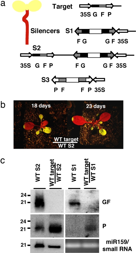
To investigate long-distance mRNA silencing, scions expressing high levels of a target GFP were grafted onto rootstocks of three silencer plant lines, S1, S2, and S3 (Fig. 1a). S1 expressed a hairpin RNA homologous to nucleotides 9–400 of the GFP coding sequence, referred to as GF. The remaining 317-nt downstream GFP sequence is referred to as P (Fig. 1a). S2 expressed the same RNAi transgene as S1 plus an intact, albeit silenced, GFP transgene [supporting information (SI) Fig. 6]. S3 expressed a RNAi transgene targeting nucleotides 300–700 of the GFP coding sequence, referred to as FP (Fig. 1a). When WT target scions (expressing GFP) were grafted onto WT silencer rootstocks, GFP silencing was only induced in newly formed leaves and did not spread into older tissue (Fig. 1b). This phenotype distinguishes our signaling pathway from short-distance, cell-to-cell spreading of transgene silencing (20). Southern analysis with HpaII and bisulfite sequencing demonstrated there was no cytosine methylation of the GFP transgene in silenced scions (SI Fig. 7 and SI Table 1), confirming earlier reports in tobacco (28). As expected, high levels of GF-specific siRNAs were detected in both S1 and S2 lines, and P-specific siRNAs were also detected in S2 plants (Fig. 1c). In scions silenced by S1 or S2 rootstocks, however, we could detect only P-specific siRNAs, and no GF-specific siRNAs (Fig. 1c). The fact that S1 lacked P-specific siRNAs but when used as a rootstock induced only P-specific siRNAs in the scion indicates that silencing in the scion is initiated outside of the sequence homologous to the dsRNA expressed in the rootstock.
Fig. 1.
Graft-transmissible mRNA silencing in WT Arabidopsis. (a) Schematic representation of seedling grafting and the transgene constructs used to produce the target and silencer (S1, S2, and S3) plant lines. (b) Phenotype of WT target scion grafted onto S2 rootstocks at 18 and 23 days after grafting. (c) Small RNA analysis from silencers (S1 and S2) and from target scions grafted onto S1 and S2 rootstocks, using GF and P DNA probes; identical results were obtained by using sense and antisense riboprobes (data not shown).
No G-specific siRNAs were detected in scions silenced by grafting onto S3 (FP-silenced) rootstocks (SI Fig. 8). FP-specific siRNAs were only barely detectable in this scion tissue compared with the S3 rootstock, and most of the detected siRNAs were homologous to the downstream ocs 3′-UTR (SI Fig. 8). Collectively, these results indicate that graft-transmissible mRNA silencing is executed primarily on sequences downstream of those homologous to the mobile signal.
Transmission of Silencing Is Not Compromised in a dcl2 dcl3 dcl4 Triple Mutant or dcl1–8 Rootstock.
To test candidate genes for their potential involvement in long-distance mRNA silencing, we combined GFP-expressing scions and silenced rootstocks in various mutant backgrounds. Nuclear-localized DCL3 is known to generate 24-nt siRNAs (11), and these have previously been correlated with systemic silencing in N. benthamiana (19). However, these experiments were based on Agrobacterium infiltration of N. benthamiana leaves rather than grafting, and as such, a possible role for 24-nt siRNA as the mobile signal, could not be distinguished from an alternative role in responding to the signal in newly silenced tissue. When we used dcl3 S2 rootstocks, which lack 24-nt siRNAs (SI Fig. 9a), the silencing signal was efficiently transmitted to WT scions (SI Fig. 9b and SI Table 2). This clearly shows that, in our Arabidopsis grafting system, 24-nt siRNA is not the long-distance signaling molecule.
Additional dcl genotypes were also tested for their capacity to transmit the mobile silencing signal from the rootstock. DCL2, DCL3, and DCL4 have previously been shown to be largely responsible for 21- to 24-nt siRNA production (8–10, 30). However, when two independent dcl2 dcl3 dcl4 triple-mutant S2 transgenic lines were used as rootstocks, although the levels of 21- to 24-nt siRNAs were greatly reduced (Fig. 2a), silencing was still transmitted (Fig. 2b). Additionally, a dcl1-8 mutant that is compromised in miRNA biogenesis (11, 31, 32) was unaffected in signal transmission (SI Table 2).
Fig. 2.
Genetic requirements for long-distance mRNA silencing. (a) Small RNA analysis of WT S2 and the triple mutant dcl2 dcl3 dcl4 (dcl234) S2 lines. miRNA159 serves as a loading control. (b) Phenotypes of grafts using WT S2 and dcl234 S2 as the rootstock. (c) Phenotypes of dcl3, nrpd1a, rdr2, rdr6, and ago4 target scions grafted onto WT S2 rootstocks. Some ago4 scions, ago4(B), showed delayed silencing. (d) Northern blot analysis of GFP mRNA levels in various grafted and nongrafted plants.
The observations that a dcl2 dcl3 dcl4 triple mutant and dcl1-8 are compromised in siRNA and miRNA biogenesis, respectively, but not in signal transmission from the rootstock, may indicate a redundant role for these small RNA as mobile long-distance signals. Alternatively, the lack of correlation between accumulation of 21- to 24-nt siRNA and the capacity for these dcl mutant rootstocks to transmit silencing, suggests the mobile signal may be another class of RNA.
Nuclear Gene Silencing Directs Reception of mRNA Silencing in Scions.
When grafts were performed by using dcl3 scions expressing GFP and WT silencer rootstocks, no reduction in GFP fluorescence or transcript levels was observed in the scions (Fig. 2 c and d), thus demonstrating that DCL3 is essential for the silencing response in the scion. DCL3 is known to play a role in silencing of transposons and repetitive DNA, and together with NRPD1a (Pol IVa) and RDR2, is involved in the production of endogenous 24-nt siRNA from AtSN1 retroelements, 5S rDNA repeats, and from other less repetitive loci (11, 12). Nuclear-localized AGO4 (11) also plays a role in some components of this pathway, including both the production of siRNA from, and the RNA-directed DNA methylation of, specific loci including the AtSN1 retroelement (14).
Because DCL3, Pol IVa, RDR2, and AGO4 are in the same transcriptional silencing pathway, we decided to graft nrpd1a (pol IV1a), rdr2, and ago4 mutant scions onto silencer rootstocks. We found that Pol IVa and RDR2 were also required for scions to respond to the long-distance silencing signal (Fig. 2c and SI Table 2). When ago4 was used as a scion, 5 of 13 showed no silencing and most of the remaining 8 displayed a delayed onset of silencing (Fig. 2c and SI Table 2). With the exception of ago4 grafts that showed delayed silencing, no decrease in fluorescence (Fig. 2c) or GFP transcript levels (Fig. 2d) was observed in mutant scions, indicating that the long-distance silencing signal is recognized, presumably in the nucleus, by the Pol IVa–RDR2–DCL3–AGO4 pathway. This is consistent with recent work in N. benthamiana that has shown that AGO4 is involved in long-distance mRNA silencing (26). However, our results demonstrate that, although the Pol IVa–RDR2–DCL3–AGO4 pathway is not required for the transmission of the silencing signal from the rootstock (SI Table 2), it is essential for signal reception. As expected from other work in N. benthamiana (25), induction of silencing in scions (Fig. 2 c and d), but not signal transmission from rootstocks (SI Table 2), also depended on RDR6 function.
Pol IVa and RDR2, but not DCL3 or AGO4, are also required for cell-to-cell spreading of mRNA silencing (23, 24). Collectively, these results indicate that there are both overlapping and unique components of long-distance, compared with cell-to-cell, spreading of mRNA silencing. In contrast to our grafting results on long-distance mRNA silencing, the role of Pol IVa and RDR2 in transmission versus reception of cell-to-cell mRNA silencing is yet to be resolved (23, 24).
Transcriptional Down-Regulation Induced by Long-Distance mRNA Silencing.
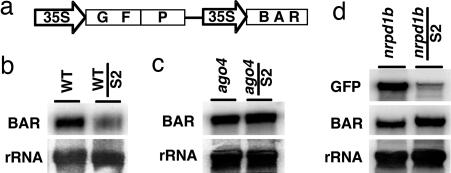
A potential consequence of the involvement of the Pol IVa–RDR2–DCL3–AGO4 pathway in the nuclear reception and initiation of silencing would be some level of transcriptional down-regulation because of chromatin compaction. Transcriptional down-regulation has been shown to spread outside the initiating region to affect expression of adjacent genes (33, 34). Northern blot analysis and quantitative RT-PCR showed that the steady-state transcript levels from the 35S:BAR gene, a selectable marker flanking 35S:GFP in the target line (Fig. 3a and SI Fig. 10), were down-regulated in GFP-silenced scions compared with ungrafted controls (Fig. 3b and SI Fig. 11a). No BAR-specific siRNAs were detected in silenced scions (SI Fig. 11b), indicating that the decrease in BAR transcript level may not have been due to posttranscriptional regulation. BAR transcript levels did not decrease when GFP silencing occurred in ago4 scions (Fig. 3c), providing evidence for transcriptional down-regulation of BAR expression in WT GFP-silenced scions. In addition, the GFP transcript was detectable in ago4-silenced scions but not in WT counterparts (Fig. 2d).
Fig. 3.
Long-distance down-regulation of transcription. (a) Target GFP transgene and flanking 35S:BAR selectable marker. (b) Northern blot analysis of BAR transcript levels in WT GFP-silenced scions grafted onto WT S2 rootstocks. (c) Northern blot analysis of BAR transcript levels in WT and ago4 GFP-silenced scions grafted onto WT S2 rootstocks. (d) Northern blot analysis of GFP and BAR mRNA levels in nrpd1b silenced scions grafted onto WT S2 rootstock.
To further investigate the possibility of the transcriptional down-regulation associated with long-distance mRNA silencing, a nrpd1b mutant (35) was used in grafting experiments. Pol IVb (NRPD1b) is a functionally diverse homologue of Pol IVa (NRPD1a) that is involved in RNA-directed DNA methylation and transcriptional silencing, acting downstream of the generation of siRNAs by the Pol IVa pathway (35, 36). Unlike Pol IVa, Pol IVb has not been implicated in posttranscriptional gene silencing (12, 36). When nrpd1b scions were grafted onto WT S2 rootstocks, normal reception of GFP silencing was observed (SI Table 2 and SI Fig. 12), but not down-regulation of the linked BAR transgene (Fig. 3d). As was the case for ago4-silenced scions (Fig. 2d), the level of GFP transcript was considerably higher in nrpd1b than in WT scions (Fig. 3d).
Together, these results indicate that subtle transcriptional down-regulation of homologous DNA can be associated with long-distance mRNA silencing. The subtle nature of transcriptional down-regulation in GFP-silenced WT scions is reflected in the lack of both DNA methylation (ref. 28; SI Fig. 7) and detectable histone modification of the GFP coding sequence (data not shown), and the lack of inheritance of the silencing phenotype in progeny of silenced scions (data not shown).
In contrast to ago4 scions that showed either no silencing or a delayed onset of silencing (Fig. 2c and SI Table 2), nrpd1b scions were unaffected in their ability to initiate mRNA silencing of GFP (SI Table 2 and SI Fig. 12). This implies the partially redundant role that AGO4 plays in the reception of silencing, is in RNA slicing (15), rather than chromatin modification.
Decapped RNA Is Detected in Silenced Scions.
The presence of only P-specific siRNAs in S1- or S2-silenced scions (Fig. 1c) suggests that the initiation of silencing via the Pol IVa–RDR2–DCL3–AGO4 pathway could result in the production of a decapped RNA substrate (37–39) for the amplification of P-specific dsRNA. In an effort to identify decapped RNAs, we performed 5′-RACE on RNA extracted from S2-silenced scions (Fig. 4a). The size of 5′-RACE products confirmed the presence of P-specific polyadenylated RNA, with 5′-ends mapping between 37 and 79 nt into the P region (SI Fig. 13). The 5′ heterogeneity among P-specific RNAs (Fig. 4a) can be explained by a population of siRNAs being responsible for the cleavage, and is in contrast to a single-sized 5′-RACE product detected after miR171-facilitated cleavage of the Scarecrow6-like IV (SCL6-IV) transcript (40).
Fig. 4.
Molecular genetic analysis of graft-transmissible mRNA silencing. (a Upper) Agarose gel electrophoresis of 5′-RACE products from GFP-silenced target scions grafted onto S2 rootstocks. The white bracket indicates the region cloned and sequenced (see SI Fig. 9). (a Lower) 5′-RACE amplification of the miR171-cleaved SCL6-IV transcript. (b) Target GFP transgene showing the regions used as probes for small RNA analysis. (c) Small RNA analysis of mutant scions grafted onto WT S2 rootstocks. (d) Small RNA analysis of WT scions within the first 70 nt of P. (e) Small RNA analysis of dcl4 target scions grafted onto WT S2 rootstocks.
To correlate the decapped RNA with a population of siRNAs, we decided to perform a more detailed analysis of siRNAs within the P region (Fig. 4 b–d). The majority of 24-nt siRNAs were detected within the first 33 nt of P (probe 1a, Fig. 4d), and siRNAs detected by other P-specific probes (probes 1b and 2, representing nucleotides 36–70 and 73–317 of P, respectively) were almost exclusively 21 nt in size (Fig. 4 b–d). There were no detectable P-specific siRNAs in nonsilenced mutant scions (Fig. 4c). Similar siRNA profiles relative to the rootstock silencer were seen for S3-silenced scions, although there appeared to be more spreading of 24-nt siRNAs downstream of the FP silencer sequence, into the ocs terminator (SI Fig. 8).
The presence of 24-nt siRNAs with sequence homology to the start of P-specific decapped RNAs in S2-silenced scions, combined with dependence of silencing on Pol IVa, RDR2, DCL3, and AGO4, raises the possibility that the 24-nt siRNAs are responsible for guiding the production of these P-specific RNAs. This is also consistent with AGO4's capacity to slice RNA (15), and decapped RNA serving as a substrate for RDR6 to induce RNA silencing (37). In this scenario, the Pol IVa–RDR2–DCL3–AGO4 pathway precedes the RDR6/DCL4 production of 21-nt siRNAs, and provides the prediction that 24-nt but not 21-nt siRNAs, should be present in rdr6 scions grafted on S2 rootstocks. However, testing rdr6 scions failed to detect either 21- or 24-nt siRNAs (Fig. 4c). This suggests that the 24-nt siRNAs detected in silenced scions require the synergistic action of RDR6 and DCL3, and implies that RDR6 is involved at multiple points in the pathway.
DCL4 and DCL2 Act Hierarchically as in Antiviral Resistance and Processing Hairpin RNAs.
DCL4 has been shown to process dsRNA produced by RDR6 into 21-nt trans-acting siRNAs (30, 38, 39). It is also required for short-distance, cell-to-cell spreading of silencing (7). However, revealing another distinction between the two known non-cell-autonomous RNA silencing pathways, a normal silencing phenotype was observed when dcl4 mutant scions were grafted onto WT silencer rootstocks (data not shown). The siRNA profiles of S2 silenced dcl4 scions did nevertheless reveal a shift in the size of siRNAs from 21 to 22 nt along the entire P region (Fig. 4e). The change in siRNA size is in accordance with the previously reported redundant nature of DCL proteins in Arabidopsis (30). Our results also demonstrate that, in the absence of DCL4, 22-nt siRNAs can functionally substitute for 21-nt siRNAs in degrading homologous mRNA transcripts. These classes of siRNAs have been recently demonstrated to act in the same hierarchical nature to confer resistance to RNA viruses (8, 9), and in processing hairpin RNA expressed from a transgene (10).
Conclusions
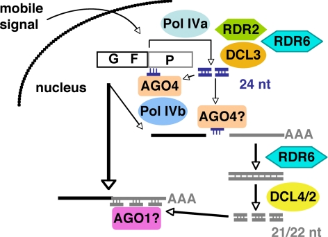
A model summarizing the crucial molecular events in reception of the long-distance silencing of the GFP mRNA is presented in Fig. 5. In the case of the S1 and S2 silencers, a mobile GF-specific signal is delivered into the shoot apex, where it stimulates the Pol IVa pathway in the nucleus to produce 24-nt siRNAs from adjacent P-specific DNA or RNA template. This process also depends on RDR6. AGO4, in association with these 24-nt siRNAs, then mediates cleavage of some mRNA transcripts. The decapped transcripts, are then converted to dsRNA by RDR6, and subsequently processed into 21-nt siRNAs by DCL4, or in its absence, into 22-nt siRNAs by DCL2. The 21- or 22-nt siRNAs then direct silencing. The precise nature of the mobile signal remains to be elucidated; however, our work indicates that it is not exclusively produced by any of the four Arabidopsis DCLs. This raises the possibility that the signal is either dsRNA, or it is produced from dsRNA by a DCL-independent process.
Fig. 5.
Model for long-distance mRNA silencing in Arabidopsis. In the case of silencer S1 and S2, the mobile GF signal is delivered into nuclei in the shoot apex and stimulates Pol IVa–RDR2–DCL3 to produce P-specific 24-nt siRNAs (blue) from either the GFP transgene or mRNA. This process also depends on RDR6. AGO4 uses the 24-nt siRNAs to mediate cleavage of some GFP transcript to produce P-specific polyadenylated RNAs, which become a substrate for RDR6. DCL4 or DCL2 then produce 21- or 22-nt siRNAs, respectively, and these direct mRNA silencing.
The biochemical pathway we have discovered may be of broader biological significance. Translocated RNA has been shown to direct key developmental processes such as leaf development (41). From a silencing perspective, long-distance movement of RNA silencing has long been thought to be an adaptive measure by which plants can protect themselves from viruses (17, 42), but it has the potential to play other roles in systemic gene regulation. Our work has not only identified components of long-distance gene silencing, but it emphasizes further the modular nature of gene-silencing pathways in Arabidopsis (23) and the importance of cross talk between the pathways (12).
Methods
Plant Material.
The rdr6 (sde1) (43) and ago4-1 (14) mutants used were as previously described. The rdr6-1 and nrpd1a (pol IV1a; sde4; Salk_128428) mutants were kindly provided by David Baulcombe (Sainsbury Laboratory, JIC, Norwich, U.K.). The rdr2-1 (Salk_059661), dcl2-1 (Salk_064627), dcl3-1 (Salk_005512), and nrpd1b-12 (Salk_033852) mutants were obtained from The Salk Institute Genome Analysis Laboratory (La Jolla, CA) and have been described previously (11, 35). The dcl4-2 mutant was from the GABI-Kat collection (GABI160A04) (38). The dcl2 dcl3 dcl4 triple mutant has been described previously (10). rdr6, ago4-1, dcl3-1, dcl4-2, and nrpd1b-12 lines were crossed to the WT Colombia line transformed with binary vector pUQC214 (SI Fig. 10) to generate mutant lines expressing a 35S:GFP transgene. nrpd1a and rdr2 target plants expressing 35S:GFP were generated by transformation with the binary vector pUQC214 (SI Fig. 10). The primers used to genotype mutants are shown in SI Table 3 or are published elsewhere (14, 35). To produce lines expressing dsRNA homologous to GFP, WT and mutants were usually transformed directly with binary vectors (SI Fig. 10).
Plasmid Construction and Transformation.
The GFP (S65T) coding region (GenBank accession no. U43284) was cloned as a 35S:GFP:ocs cassette into pUQC477 to form the binary construct called pUQC214 (SI Fig. 10). pUQC477 is a modified version of the binary vector pNB96, obtained from Hong-Gil Nam (POSTECH, Pohang, Republic of Korea) and carries 35S:BAR:nos as a plant selectable marker. For the GF-specific RNAi transgene, nucleotides 9–400 of GFP (S65T) were amplified and cloned as an intron-splicible inverted repeat into pHannibal (44). The resulting 35S-driven, GF-specific RNAi transgene was then cloned into pUQC214 to produce the binary vector pUQC218, or into the modified version of pUQC477 to produce the binary vector pUQC251 (SI Fig. 10). The FP-specific RNAi transgene (pUQC868) was created as described for pUQC251 except by using nucleotides 304–701 of GFP. Binary vectors were introduced into Agrobacterium tumefaciens GV3101. Floral dip transformation of Arabidopsis (45) with pUQC214 produced the GFP-expressing target line, whereas transformation with pUQC251, pUQC218, and pUQC868, produced the GFP silencer lines S1, S2, and S3, respectively. T-DNA loci were introduced into mutant backgrounds by crossing to a WT transgenic line, or by direct transformation (SI Table 2). When mutant genotypes were directly transformed, at least two independent transgenic lines were produced and analyzed in grafting experiments. When crosses were used to introduce the GFP or silencer transgenes into mutant backgrounds, the silencing phenotype was scored for grafted mutant versus WT F2 segregants (SI Table 2). The pUQC214 WT line used in grafting and crosses carried two T-DNAs but never showed silencing except when grafted onto silencer rootstocks (data not shown). pUQC1081 was used to produce the BAR S2 silencer line expressing BAR-specific siRNAs (SI Fig. 10).
Grafting of Arabidopsis.
Grafting was performed by using the butt grafting method described in ref. 29, with some modifications. Seedlings were germinated on Murashige and Skoog medium (46) with the plates orientated vertically. The grafting procedure was carried out on a single 0.45-μm nitrocellulose filter (Millipore, Bedford, MA) on top of two pieces of moist Whatman (Maidstone, U.K.) no. 1 filter paper in a 90-mm Petri dish. Scions were produced by using a no. 15 scalpel blade, slicing within about a millimeter of the apex of the seedling. When necessary, one cotyledon was removed to orientate the scion as close to the membrane as possible. Rootstocks were generated by the same cutting procedure that was used to produce scions. Grafts were aligned by using a dissecting microscope, and plates were sealed with parafilm and incubated vertically at 21°C for 7 days. Grafted plants were then transferred to soil and grown under long-day length at 21°C.
GFP Imaging.
Plants were viewed under blue light by using a Dark Reader Spot Lamp (Clare Chemical Research, Dolores, CO) and photographed by using a Canon (Tokyo, Japan) EOS digital camera.
RNA Extraction and Analysis.
Total RNA was isolated by using TRIzol reagent (Invitrogen, Carlsbad, CA) and used for high- and low-molecular-weight RNA blot analysis, as well as RT-PCR analysis. Enrichment for low-molecular-weight RNA and Northern blot analysis was performed as described previously (47). Riboprobes were generated by using the Riboprobe Combination System, SP6/T7 kit (Promega, Madison, WI). Random-labeled DNA probes were generated by using the MegaPrime DNA labeling system (Amersham Biosciences, Piscataway, NJ). Oligonucleotide probes, GFPsiRNA1 (probe 1a), GFPsiRNA2 (probe 1b), and miR159 (SI Table 3), were labeled with 32P by using T4 polynucleotide kinase (New England Biolabs, Beverly, MA). Probe 1a and 1b detected GFP antisense siRNAs. Detection of decapped 5′ ends was performed by using the First Choice RLM-RACE kit (Ambion, Austin, TX) (40). First-strand cDNA synthesis was primed by using oligo(dT) or random hexamers, and both yielded identical results. Primary PCR was performed by using the RLM-RACE outer primer and a gene-specific primer (OCS-R), and nested PCR was performed by using the primers RLM-RACE inner and GFPs65TBamHI (SI Table 3). Amplification of the SCL6-IV decapped RNA was performed as a control, as described previously (40).
DNA Extraction and Analysis.
DNA extractions (48) and alkali treatment of tissue for PCR analysis (49) were performed as described previously.
Supplementary Material
Acknowledgments
We thank David Baulcombe for providing nrpd1a and rdr6 seed and for discussion; Jean Finnegan, Adriana Fusaro, and Ralf Dietzgen for technical advice, critical comments on the manuscript, and/or stimulating discussions; and Siân Curtis and Mark Curtis for editorial comments on the manuscript. This work was supported by an Australian Research Council grant (to B.J.C.), a Queensland Government grant (to B.J.C.), a University of Queensland Research Development grant (to B.J.C.), a Sugar Research and Development Corporation scholarship (to C.A.B.), and a Grains Research and Development Corporation scholarship (to M.C.).
Abbreviations
- DCL
DICER-like
- miRNA
micro-RNA
- AGO
Argonaute-like
- RDR
RNA-dependent RNA polymerase
- Pol IV
RNA polymerase IV.
Footnotes
The authors declare no conflict of interest.
This article contains supporting information online at www.pnas.org/cgi/content/full/0706701104/DC1.
References
- 1.Bartel DP. Cell. 2004;116:281–297. doi: 10.1016/s0092-8674(04)00045-5. [DOI] [PubMed] [Google Scholar]
- 2.Baulcombe D. Nature. 2004;431:356–363. doi: 10.1038/nature02874. [DOI] [PubMed] [Google Scholar]
- 3.Lippman Z, Martienssen R. Nature. 2004;431:364–370. doi: 10.1038/nature02875. [DOI] [PubMed] [Google Scholar]
- 4.Bernstein E, Caudy AA, Hammond SM, Hannon GJ. Nature. 2001;409:363–366. doi: 10.1038/35053110. [DOI] [PubMed] [Google Scholar]
- 5.Qi Y, Hannon GJ. FEBS Lett. 2005;579:5899–5903. doi: 10.1016/j.febslet.2005.08.035. [DOI] [PubMed] [Google Scholar]
- 6.Jones-Rhoades MW, Bartel DP, Bartel B. Annu Rev Plant Biol. 2006;57:19–53. doi: 10.1146/annurev.arplant.57.032905.105218. [DOI] [PubMed] [Google Scholar]
- 7.Dunoyer P, Himber C, Voinnet O. Nat Genet. 2005;37:1356–1360. doi: 10.1038/ng1675. [DOI] [PubMed] [Google Scholar]
- 8.Bouche N, Lauressergues D, Gasciolli V, Vaucheret H. EMBO J. 2006;25:3347–3356. doi: 10.1038/sj.emboj.7601217. [DOI] [PMC free article] [PubMed] [Google Scholar]
- 9.Deleris A, Gallego-Bartolome J, Bao J, Kasschau KD, Carrington JC, Voinnet O. Science. 2006;313:68–71. doi: 10.1126/science.1128214. [DOI] [PubMed] [Google Scholar]
- 10.Fusaro AF, Matthew L, Smith NA, Curtin SJ, Dedic-Hagan J, Ellacott GA, Watson JM, Wang MB, Brosnan C, Carroll BJ, Waterhouse PM. EMBO Rep. 2006;7:1168–1175. doi: 10.1038/sj.embor.7400837. [DOI] [PMC free article] [PubMed] [Google Scholar]
- 11.Xie ZX, Johansen LK, Gustafson AM, Kasschau KD, Lellis AD, Zilberman D, Jacobsen SE, Carrington JC. PLoS Biol. 2004;2:642–652. doi: 10.1371/journal.pbio.0020104. [DOI] [PMC free article] [PubMed] [Google Scholar]
- 12.Herr AJ, Jensen MB, Dalmay T, Baulcombe DC. Science. 2005;308:118–120. doi: 10.1126/science.1106910. [DOI] [PubMed] [Google Scholar]
- 13.Pontes O, Li CF, Nunes PC, Haag J, Ream T, Vitins A, Jacobsen SE, Pikaard CS. Cell. 2006;126:79–92. doi: 10.1016/j.cell.2006.05.031. [DOI] [PubMed] [Google Scholar]
- 14.Zilberman D, Cao XF, Jacobsen SE. Science. 2003;299:716–719. doi: 10.1126/science.1079695. [DOI] [PubMed] [Google Scholar]
- 15.Qi Y, He X, Wang XJ, Kohany O, Jurka J, Hannon GJ. Nature. 2006;443:1008–1012. doi: 10.1038/nature05198. [DOI] [PubMed] [Google Scholar]
- 16.Zheng X, Zhu J, Kapoor A, Zhu JK. EMBO J. 2007;26:1691–1701. doi: 10.1038/sj.emboj.7601603. [DOI] [PMC free article] [PubMed] [Google Scholar]
- 17.Voinnet O. FEBS Lett. 2005;579:5858–5871. doi: 10.1016/j.febslet.2005.09.039. [DOI] [PubMed] [Google Scholar]
- 18.Feinberg EH, Hunter CP. Science. 2003;301:1545–1547. doi: 10.1126/science.1087117. [DOI] [PubMed] [Google Scholar]
- 19.Hamilton A, Voinnet O, Chappell L, Baulcombe D. EMBO J. 2002;21:4671–4679. doi: 10.1093/emboj/cdf464. [DOI] [PMC free article] [PubMed] [Google Scholar]
- 20.Himber C, Dunoyer P, Moissiard G, Ritzenthaler C, Voinnet O. EMBO J. 2003;22:4523–4533. doi: 10.1093/emboj/cdg431. [DOI] [PMC free article] [PubMed] [Google Scholar]
- 21.Palauqui JC, Elmayan T, Pollien JM, Vaucheret H. EMBO J. 1997;16:4738–4745. doi: 10.1093/emboj/16.15.4738. [DOI] [PMC free article] [PubMed] [Google Scholar]
- 22.Voinnet O, Baulcombe DC. Nature. 1997;389:553. doi: 10.1038/39215. [DOI] [PubMed] [Google Scholar]
- 23.Smith LM, Pontes O, Searle I, Yelina N, Yousafzai FK, Herr AJ, Pikaard CS, Baulcombe DC. Plant Cell. 2007;19:1507–1521. doi: 10.1105/tpc.107.051540. [DOI] [PMC free article] [PubMed] [Google Scholar]
- 24.Dunoyer P, Himber C, Ruiz-Ferrer V, Alioua A, Voinnet O. Nat Genet. 2007;39:848–856. doi: 10.1038/ng2081. [DOI] [PubMed] [Google Scholar]
- 25.Schwach F, Vaistij FE, Jones L, Baulcombe DC. Plant Physiol. 2005;138:1842–1852. doi: 10.1104/pp.105.063537. [DOI] [PMC free article] [PubMed] [Google Scholar]
- 26.Jones L, Keining T, Eamens A, Vaistij FE. Plant Physiol. 2006;141:598–606. doi: 10.1104/pp.105.076109. [DOI] [PMC free article] [PubMed] [Google Scholar]
- 27.Mallory AC, Ely L, Smith TH, Marathe R, Anandalakshmi R, Fagard M, Vaucheret H, Pruss G, Bowman L, Vance VB. Plant Cell. 2001;13:571–583. doi: 10.1105/tpc.13.3.571. [DOI] [PMC free article] [PubMed] [Google Scholar]
- 28.Mallory AC, Mlotshwa S, Bowman LH, Vance VB. Plant J. 2003;35:82–92. doi: 10.1046/j.1365-313x.2003.01785.x. [DOI] [PubMed] [Google Scholar]
- 29.Turnbull CGN, Booker JP, Leyser HMO. Plant J. 2002;32:255–262. doi: 10.1046/j.1365-313x.2002.01419.x. [DOI] [PubMed] [Google Scholar]
- 30.Gasciolli V, Mallory AC, Bartel DP, Vaucheret H. Curr Biol. 2005;15:1494–1500. doi: 10.1016/j.cub.2005.07.024. [DOI] [PubMed] [Google Scholar]
- 31.Park W, Li J, Song R, Messing J, Chen X. Curr Biol. 2002;12:1484–1495. doi: 10.1016/s0960-9822(02)01017-5. [DOI] [PMC free article] [PubMed] [Google Scholar]
- 32.Reinhart BJ, Weinstein EG, Rhoades MW, Bartel B, Bartel DP. Genes Dev. 2002;16:1616–1626. doi: 10.1101/gad.1004402. [DOI] [PMC free article] [PubMed] [Google Scholar]
- 33.Finnegan EJ, Kovac KA, Jaligot E, Sheldon CC, Peacock WJ, Dennis ES. Plant J. 2005;44:420–432. doi: 10.1111/j.1365-313X.2005.02541.x. [DOI] [PubMed] [Google Scholar]
- 34.Huettel B, Kanno T, Daxinger L, Aufsatz W, Matzke AJ, Matzke M. EMBO J. 2006;25:2828–2836. doi: 10.1038/sj.emboj.7601150. [DOI] [PMC free article] [PubMed] [Google Scholar]
- 35.Pontier D, Yahubyan G, Vega D, Bulski A, Saez-Vasquez J, Hakimi MA, Lerbs-Mache S, Colot V, Lagrange T. Genes Dev. 2005;19:2030–2040. doi: 10.1101/gad.348405. [DOI] [PMC free article] [PubMed] [Google Scholar]
- 36.Kanno T, Huettel B, Mette MF, Aufsatz W, Jaligot E, Daxinger L, Kreil DP, Matzke M, Matzke AJ. Nat Genet. 2005;37:761–765. doi: 10.1038/ng1580. [DOI] [PubMed] [Google Scholar]
- 37.Gazzani S, Lawrenson T, Woodward C, Headon D, Sablowski R. Science. 2004;306:1046–1048. doi: 10.1126/science.1101092. [DOI] [PubMed] [Google Scholar]
- 38.Allen E, Xie ZX, Gustafson AM, Carrington JC. Cell. 2005;121:207–221. doi: 10.1016/j.cell.2005.04.004. [DOI] [PubMed] [Google Scholar]
- 39.Yoshikawa M, Peragine A, Park MY, Poethig RS. Genes Dev. 2005;19:2164–2175. doi: 10.1101/gad.1352605. [DOI] [PMC free article] [PubMed] [Google Scholar]
- 40.Llave C, Xie ZX, Kasschau KD, Carrington JC. Science. 2002;297:2053–2056. doi: 10.1126/science.1076311. [DOI] [PubMed] [Google Scholar]
- 41.Kim M, Canio W, Kessler S, Sinha N. Science. 2001;293:287–289. doi: 10.1126/science.1059805. [DOI] [PubMed] [Google Scholar]
- 42.Mlotshwa S, Voinnet O, Mette MF, Matzke M, Vaucheret H, Ding SW, Pruss G, Vance VB. Plant Cell. 2002;14:S289–S301. doi: 10.1105/tpc.001677. [DOI] [PMC free article] [PubMed] [Google Scholar]
- 43.Dalmay T, Hamilton A, Rudd S, Angell S, Baulcombe DC. Cell. 2000;101:543–553. doi: 10.1016/s0092-8674(00)80864-8. [DOI] [PubMed] [Google Scholar]
- 44.Wesley SV, Helliwell CA, Smith NA, Wang MB, Rouse DT, Liu Q, Gooding PS, Singh SP, Abbott D, Stoutjesdijk PA, et al. Plant J. 2001;27:581–590. doi: 10.1046/j.1365-313x.2001.01105.x. [DOI] [PubMed] [Google Scholar]
- 45.Clough SJ, Bent AF. Plant J. 1998;16:735–743. doi: 10.1046/j.1365-313x.1998.00343.x. [DOI] [PubMed] [Google Scholar]
- 46.Murashige T, Skoog F. Physiol Plant. 1962;15:473–497. [Google Scholar]
- 47.Mitter N, Sulistyowati E, Dietzgen RG. Mol Plant Microbe Interact. 2003;16:936–944. doi: 10.1094/MPMI.2003.16.10.936. [DOI] [PubMed] [Google Scholar]
- 48.Carroll BJ, Klimyuk VI, Thomas CM, Bishop GJ, Harrison K, Scofield SR, Jones JDG. Genetics. 1995;139:407–420. doi: 10.1093/genetics/139.1.407. [DOI] [PMC free article] [PubMed] [Google Scholar]
- 49.Klimyuk VI, Carroll BJ, Thomas CM, Jones JD. Plant J. 1993;3:493–494. doi: 10.1111/j.1365-313x.1993.tb00169.x. [DOI] [PubMed] [Google Scholar]
Associated Data
This section collects any data citations, data availability statements, or supplementary materials included in this article.


