Abstract
Small interfering RNAs (siRNAs), the guides that direct RNA interference (RNAi), provide a powerful tool to reduce the expression of a single gene in human cells. Ideally, dominant, gain-of-function human diseases could be treated using siRNAs that specifically silence the mutant disease allele, while leaving expression of the wild-type allele unperturbed. Previous reports suggest that siRNAs can be designed with single nucleotide specificity, but no rational basis for the design of siRNAs with single nucleotide discrimination has been proposed. We systematically identified siRNAs that discriminate between the wild-type and mutant alleles of two disease genes: the human Cu, Zn superoxide dismutase (SOD1) gene, which contributes to the progression of hereditary amyotrophic lateral sclerosis through the gain of a toxic property, and the huntingtin (HTT) gene, which causes Huntington disease when its CAG-repeat region expands beyond approximately 35 repeats. Using cell-free RNAi reactions in Drosophila embryo lysate and reporter assays and microarray analysis of off-target effects in cultured human cells, we identified positions within an siRNA that are most sensitive to mismatches. We also show that purine:purine mismatches imbue an siRNA with greater discriminatory power than other types of base mismatches. siRNAs in which either a G:U wobble or a mismatch is located in the “seed” sequence, the specialized siRNA guide region responsible for target binding, displayed lower levels of selectivity than those in which the mismatch was located 3′ to the seed; this region of an siRNA is critical for target cleavage but not siRNA binding. Our data suggest that siRNAs can be designed to discriminate between the wild-type and mutant alleles of many genes that differ by just a single nucleotide.
Synopsis
First discovered in nematodes, RNA interference (RNAi) has become an essential tool in the study of mammalian gene function. RNAi directed by small interfering RNAs (siRNAs), 21 nt, double-stranded RNAs target complementary mRNAs for destruction. siRNAs can be introduced into mammalian cells grown in culture, or even administered intravenously to rodents or primates, where they repress production of the targeted gene product. Thus, siRNA-directed RNAi has tremendous potential as a human therapeutic strategy. Dominant genetic disorders, in which a mutant allele of a gene causes disease in the presence of a second, normal copy, might be treated with therapeutic siRNAs, provided that the siRNAs could be designed to destroy the mutant, disease-causing mRNA, while leaving the normal mRNA intact. Here, Schwarz and colleagues describe an experimentally validated strategy for the design of such siRNAs. Their design strategy should facilitate the design of siRNAs targeting dominant genetic disorders such as amyotrophic lateral sclerosis and Huntington disease.
Introduction
In the RNA interference (RNAi) pathway, small interfering RNAs (siRNAs), 21- to 23-nucleotide double-stranded RNAs, target a corresponding mRNA for post-transcriptional destruction. siRNAs act as guides for a protein complex, RISC (RNA-induced silencing complex), which mediates target RNA destruction [1–3]. Synthetic siRNAs provide a straightforward means to knock-down gene expression in vitro in cultured human cells [4] and in vivo in mice [5–8] and primates [9]. When an siRNA is complementary to its mRNA target, the siRNA directs endonucleolytic cleavage of the mRNA at a single phosphate across from nucleotides 10 and 11 of the siRNA “guide” strand, the strand loaded into the RNA-induced silencing center (RISC), triggering mRNA destruction [1,10–12]. Both strands of an siRNA can act as guides [3,10,13,14], but siRNAs are most effective when specifically designed to load the antisense strand into RISC and concomitantly destroy the sense, or “passenger,” strand [14,15]. Which siRNA strand serves as the guide reflects the relative thermodynamic stability of the 5′ ends of the two siRNA strands [14,15]. siRNAs that exhibit near absolute asymmetry, with only one strand of the siRNA capable of entry into the RISC, are said to be functionally asymmetric [14].
Some mismatches between an siRNA and its target RNA block target cleavage by RISC [13,16–22]. Thus, siRNAs can discriminate between mRNAs that differ at a single base-pair, suggesting the potential application of this mechanism to suppress mutant genes in dominant human diseases including amyotrophic lateral sclerosis (ALS) [23–26], Huntington disease (HD) [27], Alzheimer disease [28], human immunodeficiency virus infection [29,30], slow channel congenital myasthenic syndrome [31], spinocerebellar ataxia type 3 [32], sickle cell anemia [33], and cancer [34]. Because siRNAs to treat these and similar diseases would need to target single nucleotide polymorphisms (SNPs), their design is limited to the region surrounding the mutation.
ALS is an age-dependent neurodegenerative disease that can be caused by sporadic or inherited dominant point mutations in the Cu, Zn superoxide dismutase gene (SOD1) [35]. Point mutations in SOD1 have been linked to the acquisition of a toxic property by the mutant protein, rather than loss of the wild-type function of SOD1 in preventing cellular damage by destroying free oxygen radicals released from metabolic processes [36]. With many point mutations clinically identified, one possible therapy to ameliorate the symptoms of ALS would be allele-specific therapy to selectively eliminate expression of the mutant copy of the SOD1 gene. Ideally, such a therapy would target only the mutant allele, because loss of the wild-type gene in mice causes many abnormalities, including degeneration of motor neuron axons [37,38], liver cancer, and shortened life span [39], as well as reduced fertility [40,41]. HD likewise is a neurodegenerative disorder caused by a dominant, toxic gene product. HD patients suffer cell death in the cortex and striatum of the brain, causing motor and cognitive symptoms [42]. The cause of the disease is an expansion of the CAG repeat in exon 1 of the huntingtin gene (HTT), which leads to a protein containing an extended polyglutamine tract [43]. The time of onset of HD correlates with the length of the expanded CAG repeat [44,45].
To develop a general strategy for the design of single nucleotide–specific siRNAs, we examined the effect of mismatch identity and position on the ability of an siRNA to discriminate between two target RNAs that differ by only a single base. As a model system, we used siRNAs that silence the G85R point mutation in human SOD1, an allele that causes a familial form of ALS. We then tested the generality of our findings by designing siRNAs to discriminate between two alleles of HTT mRNA that differ at a SNP. Although specific-SNP isoforms are not known to correlate with HD, previous attempts to distinguish between wild-type and disease-causing HTT alleles using siRNAs targeting the disease-causing CAG-repeat expansion have proved unsuccessful [46], suggesting that allele-specific therapy may be required.
Here, we show that maximal discrimination is achieved when the siRNA:target RNA pairing is disrupted by a purine:purine mismatch; little or no discrimination is achieved by other classes of mismatches. Surprisingly, mismatches in the “seed” region of the siRNA—the six-nucleotide siRNA region that contributes the bulk of target-binding energy—do not ensure effective single nucleotide discrimination in cultured cells. In contrast, mismatches in the central and 3′ regions of the siRNA provide a high degree of single nucleotide discrimination, consistent with previous proposals that target cleavage requires that these regions pair with their target RNAs to form an A-form RNA:RNA helix [47,48]. Remarkably, a mismatch at siRNA nucleotide 16 provided more than 4-fold discrimination between two alleles for all ten siRNA:target RNA pairs examined and robust discrimination (at least 20-fold) for seven of the ten sequences tested.
Results
A Tiled Set of Functionally Asymmetric siRNAs Targeting Mutant SOD1
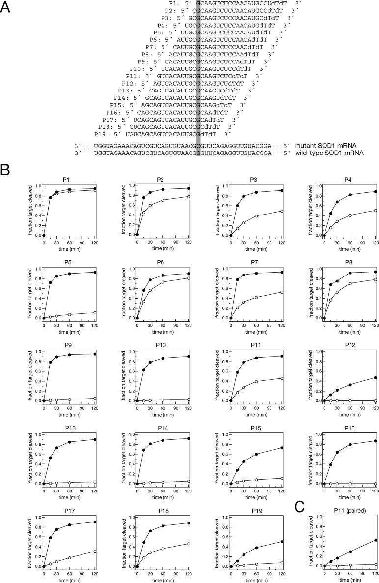
We synthesized a set of 19 siRNAs tiling across the G85R point mutation of human SOD1 (Figure 1A). The G85R mutant contains a cytosine at position 323 of the mRNA, whereas the wild-type mRNA bears a guanosine at that position. Each siRNA fully matched the mutant SOD1 but contained a G:G mismatch with wild-type. To ensure that the antisense strand of each siRNA served as the guide in RISC, each siRNA had an unpaired, antisense-strand 5′ end, a design strategy that imparts “functional asymmetry” to an siRNA [14,15]. In vitro RNAi experiments in Drosophila embryo lysate demonstrated that all 19 siRNAs effectively targeted their fully matched targets, the mutant G85R allele of SOD1, allowing us to assess how well each siRNA discriminated against the wild-type SOD1 allele (Figure 1B). The importance of this strategy can be seen by comparing the conventionally designed—i.e., fully paired—siRNA position 11 (P11) from our previous study of allele-specific siRNAs [23] with the functionally asymmetric version used here (Figure 1B and 1C). The conventionally designed P11 siRNA showed considerable discrimination against the wild-type SOD1 allele (Figure 1C). The functionally asymmetric P11 siRNA (Figure 1B) revealed that the source of this discrimination was the poor activity of the original siRNA against the fully matched target, rather than a large difference in its activities against the two SOD1 alleles.
Figure 1. Analysis in Drosophila Embryo Lysate of a Tiled Set of siRNAs Targeting Human SOD1.
(A) Sequences of the guide siRNA strands used, indicating the site of the G:G mismatch. The third nucleotide from the 3′ end of the sense strand of the siRNA (not shown) was mismatched with the guide, creating an unpaired 5′ end to facilitate entry of the guide strand into RISC. For example, where the first nucleotide of the guide strand was U, the sense strand was changed to C, and vice versa. Where the first guide nucleotide was G, the corresponding sense strand was designed to be A, and vice versa. The mutant (matched) and wild-type (mismatched) SOD1 mRNA sequences targeted by the siRNAs are shown below.
(B) Rate of target cleavage in Drosophila embryo lysate of mutant (filled circle) or wild-type target (open circle) for each siRNA in the tiled set.
(C) Comparison of a 5′ paired and a 5′ unpaired P11 siRNA (B) reveals that the 5′ paired siRNA exaggerates the inherent discrimination of this siRNA between matched and mismatched target RNAs.
Analysis of the tiled set of functionally asymmetric siRNAs showed that the P5, P9, P10, P13, P14, P15, and P16 siRNAs all discriminated between G85R mutant and wild-type SOD1 (Figure 1B). Additionally, the P12 and P19 siRNAs displayed some discrimination against the mismatched wild-type target, but these two siRNAs did not show robust silencing of the perfectly matched mutant target in the cell-free RNAi reaction, consistent with the idea that an unpaired guide strand 5′ end is not the sole determinant of siRNA efficacy [15,49]. To provide a more quantitative measure of siRNA efficacy, we also determined for each siRNA in the tiled set its initial rate of cleavage in single-turnover reaction conditions (Figure S1). The initial rate of cleavage reflects the concentration of active RISC containing the antisense strand of the siRNA duplex and the inherent catalytic rate of cleavage of the targeted sequence but not the rate of product dissociation from RISC. Four siRNAs exhibited surprisingly slow initial rates of reaction: P12, P15, P16, and P19. Of these siRNAs, P12 and P19 also showed a low extent of cleavage over a longer time course (Figure 1B). In contrast, the P15 and P16 siRNAs performed well over the 2-h time course, although they showed a slow rate of initial cleavage.
A Stringent Biochemical Test for siRNA Selectivity
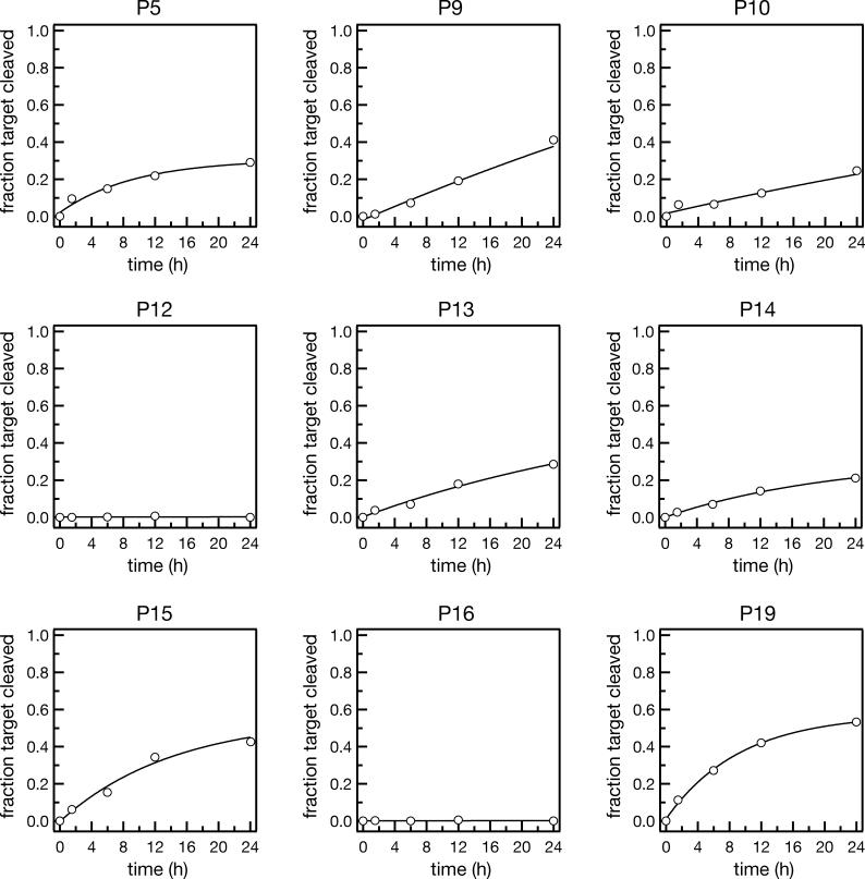
While some of the siRNAs in this study exhibited high levels of discrimination during a 2-h reaction, a more rigorous test of the ability of an siRNA to discriminate against a mismatched RNA target is to examine cleavage over a 24-h period. We performed 24-h cleavage reactions using a high concentration of siRNA and a low concentration of target RNA, so as to detect even a small degree of activity of the siRNA against the mismatched target (Figure 2). Under these intentionally artificial conditions, many of the siRNAs which originally showed complete discrimination against the wild-type SOD1 RNA target showed detectable levels of cleavage of the wild-type, mismatched RNA. In contrast, the P12 and P16 siRNAs showed no cleavage of the wild-type target, suggesting that the mismatch at these positions effectively blocked RISC activity, under these experimental conditions.
Figure 2. Mismatched Target Cleavage after 24-h Incubation in Drosophila Embryo Lysate.
To detect low levels of target cleavage, long-time scale reactions were performed with the concentration of RISC greater than that of the RNA target for those siRNAs (P5, P9, P10, P12, P13, P14, P15, P16, and P19) that showed high levels of discrimination between the matched and mismatched targets in Figure 1.
Analysis of Tiled siRNAs in Cultured Human Cells
To what extent does the cell-free analysis using Drosophila embryo lysate predict the behavior of an siRNA in a human cell? We assessed the efficacy and discriminatory power of each siRNA by co-transfecting it into HEK 293T cells with a plasmid expressing a firefly (Photinus pyralis [Pp]) luciferase bearing either the relevant region of the wild-type or the G85R mutant SOD1 sequence cloned into its 3′-untranslated region. Silencing efficiency was determined by measuring firefly luciferase activity, relative to an untargeted Renilla (Rr) luciferase control, 24 h after transfection with either 2 nM or 20 nM siRNA (Figure 3). Wild-type SOD1 contains a G at position 323; in the G85R mutant, this position is a C. The siRNAs were also evaluated using a Pp-luciferase-SOD1 fusion target containing a uridine residue at position 323 of the SOD1 mRNA sequence. G:U wobbles were previously reported to be poorly tolerated in the seed sequence of a miRNA-like siRNA [50], suggesting that G:U mismatches in this region might provide single nucleotide specificity, although data in flies suggest that extensive pairing outside the seed can compensate for a G:U wobble pair within the seed [51].
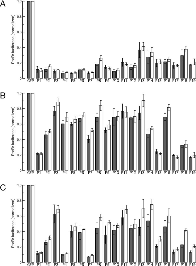
Figure 3. Analysis of Tiled siRNAs against an SOD1-Luciferase Fusion in Cultured Human Cells.
(A) Relative firefly luciferase expression for siRNAs (Figure 1A) co-transfected at either 2 nM (gray bars) or 20 nM (white bars) with a reporter plasmid containing the mutant (matched) SOD1 sequence fused to the luciferase coding sequence.
(B) The same set of siRNAs was analyzed by co-transfection with a reporter plasmid containing the wild-type (mismatched) SOD1 sequence, creating a G:G clash, or (C) a reporter plasmid encoding a U at the same position, creating a G:U wobble.
Each experiment was performed in triplicate and the data were normalized to luciferase expression measured using a GFP siRNA. Average ± standard deviation is shown.
The 19 siRNAs examined using the fully matched target RNA (the G85R mutant SOD1) silenced the reporter by at least 60%; of these, 15 silenced the reporter by 80% or more (Figure 3A). When the same set was examined using the mismatched, wild-type SOD1 reporter, ten of the 19 siRNAs effectively discriminated against the mismatched target RNA (Figure 3B). siRNAs P3, P4, P5, P6, P8, P10, P11, P12, P13, and P16 repressed wild-type reporter expression by less than 40%. Thus, most of the siRNAs that exhibited high levels of discrimination in the cell-free Drosophila RNAi system also discriminated in cultured human cells, including siRNAs P5, P10, P12, P13, and P16.
Next, the same set of siRNAs was co-transfected with a reporter designed to create a G:U wobble instead of a G:G mismatch. Only five siRNAs of the 19 showed effective discrimination against the wild-type SOD1 reporter (i.e., less than a 40% reduction in expression) (Figure 3C). In theory, the “seed” region of an siRNA, which mediates siRNA binding to a target RNA, should be highly sensitive to mismatches, but siRNA P3 was the only one of the six siRNAs to show more than 2-fold allele specificity when a G:U wobble was placed within the seed sequence. Rather, mismatches 3′ to the seed—siRNAs P8, P11, P13, P14, and P16—best retained the ability to discriminate against the G:U wobble. Perhaps seed mismatches are ineffective at destabilizing the binding of siRNAs bearing extensive complementarity to their targets, because base-pairs outside the seed region compensate for mismatches with the seed [51], whereas mutations 3′ to the seed disrupt the A-form helical geometry required for target cleavage [47,48].
Off-Target Effects Reveal the Strand Specificity of siRNAs against Mutant SOD1
We tested our siRNA design strategy—unpairing the 5′ end of the antisense strand to promote its incorporation into RISC—by examining the nature of the “off-target” silencing profile of each siRNA: the constellation of mRNAs whose steady-state concentration was decreased in response to that particular siRNA. Such off-target silencing is thought to reflect seed sequence-directed binding of the siRNA to mRNAs other than its intended mRNA target [52–54]. Because the siRNA seed sequence is the primary determinant of siRNA binding, off-target mRNAs contain six-nucleotide sequences (“hexamers”) complementary to the seed sequence of the siRNA strand—sense or antisense—that directed RISC to destroy them. Thus, determining which strand gives the greatest enrichment of seed region hexamer matches to the off-target expression signatures is a measure of which siRNA strand is preferentially loaded into RISC.
We transfected the siRNAs into cultured human HeLa cells at 100 nM final concentration. This siRNA concentration was selected to facilitate detection of silencing of unintended transcripts, in order to reveal the identity of the siRNA strand loaded into RISC. Total RNA was isolated from the siRNA transfected cells and analyzed by microarray transcription profiling (Figure 4A and Table 1). Each of the 18 siRNAs that we were able to evaluate contains a different seed sequence, so each should have a diagnostic off-target signature. In addition, this experiment poses a stringent test for siRNA specificity, in that (1) the transfected siRNA concentration was 5 times greater than the highest standard concentration [55] and 50 times greater than the lower effective concentration (2 nM) used in Figure 3; and (2) endogenous, wild-type SOD1 is the human mRNA most complementary to each of the siRNAs that target mutant SOD1. (Human cell lines expressing the G85R allele of SOD1 are not available.)
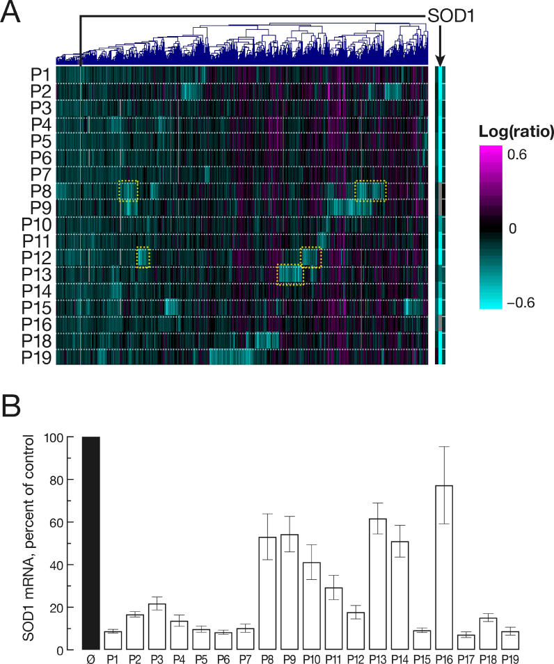
Figure 4. Microarray Analysis of 5′ Unpaired SOD1 siRNAs.
siRNAs from Figure 1A were transfected at 100 nM into HeLa cells and total cellular RNA isolated 24 h later.
(A) Microarray analysis of genes down-regulated by siRNAs. Gene expression profiles were determined by competitive hybridization of amplified mRNA from siRNA-treated versus mock-treated cells. Shown is a heat map depiction of mRNAs whose expression decreased following siRNA transfection (p < 0.01, in one or two experiments, except for SOD1). Regulated genes are in columns, experiments in rows. The experiment using P17 was lost. Teal, genes having decreased mRNA levels compared to mock transfected cells, magenta, genes having increased mRNA levels. The color bar indicates log10 expression ratio transfected/mock transfected cells, −0.6 (teal) to +0.6 (magenta; i.e., 4-fold). Genes in yellow boxes are representative groups of transcripts enriched for seed region hexamer matches (see Table 1). The arrow indicates the position of the wild-type SOD1 mRNA, compared to a mock HeLa cell transfection.
(B) Endogenous SOD1 mRNA levels were determined by quantitative RT-PCR for each siRNA transfection. Shown is the mean ± standard deviation of three replicate determinations for each siRNA. Endogenous wild-type SOD1 mRNA is mismatched to the siRNAs used; taller bars imply greater discrimination against the mismatched target RNA.
Table 1.
Off-Target Analysis Shows that Most of the SOD1 siRNAs Designed to Be Functionally Asymmetric Load the Guide Strand into RISC
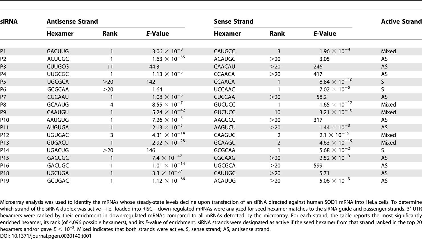
Analysis of the off-target mRNAs down-regulated by the 18 siRNAs suggests that eight loaded predominantly their antisense strand into RISC and five loaded both strands to some degree (Table 1). siRNAs P5, P6, and P14 predominantly loaded their sense strands into RISC, suggesting that for these three siRNAs, our design strategy was not effective. Both siRNA P13, which appeared to load both strands into RISC, and siRNA P14, which loaded predominantly its sense strand, also showed little activity against their fully matched SOD1 mutant mRNA target (Figure 3A). The low activity of the antisense strands of these two siRNAs precluded further analysis of their ability to discriminate between mutant and wild-type SOD1 mRNA. The microarray data show that three siRNAs, P8, P9, and P16, triggered no detectable down-regulation of endogenous wild-type SOD1 (Figure 4A). Quantitative RT-PCR (qRT-PCR) corroborated the microarray analysis (Figure 4B). The P16 siRNA detectably incorporated only the antisense strand into RISC, whereas both the P8 and P9 siRNAs loaded both strands into RISC. The P9 and P16 siRNAs were also highly active in both Drosophila embryo lysate (Figures 1, 2, and S1) and HEK 293T cells (Figure 3) against the perfectly matched G85R mutant mRNA.
Notably, all of the siRNAs bearing one G:G mismatch between the siRNA seed sequence and the endogenous wild-type SOD1 gene—P2, P3, P4, P5, P6, and P7—targeted the SOD1 mRNA for destruction at this high siRNA concentration (Figure 4A and 4B). That is, none of these siRNAs retained its ability to discriminate against wild-type SOD1 when the siRNA was transfected at 100 nM. These data are consistent with the view that mismatches between the seed and its target compromise only RISC binding, not catalysis, and can therefore be overcome by increasing the concentration of the siRNA [48]. However, we cannot exclude the possibility that, at this high siRNA concentration, other small RNA-directed mechanisms account for mRNA destruction in cultured cells and that disrupting seed pairing inherently blocks Ago2-catalyzed mRNA cleavage.
Nature of the Mismatch Determines Discrimination
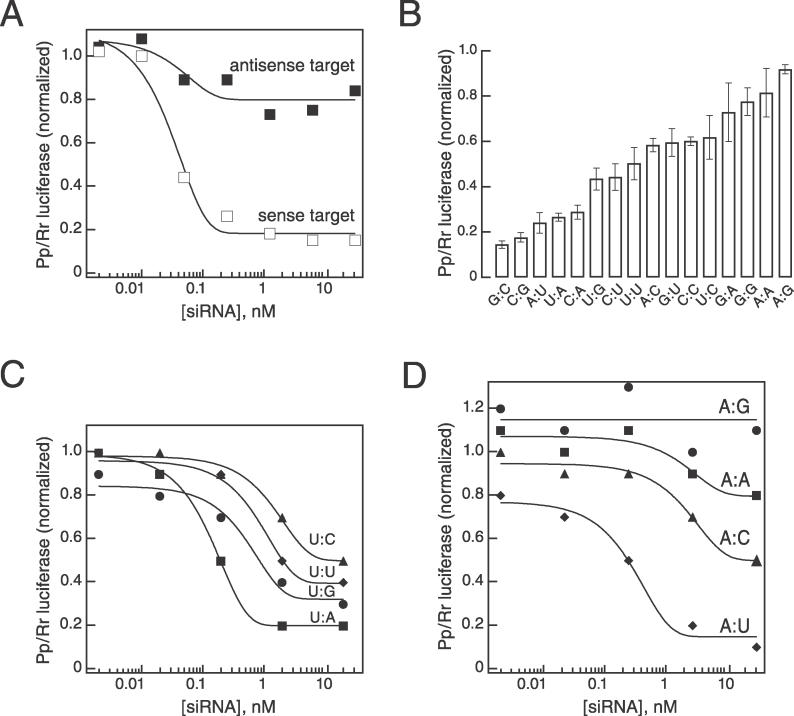
Our examination of the tiled set of siRNAs targeting the G85R SOD1 mutation compared a G:C base-pair with a G:G mismatch and a G:U wobble. To extend this analysis to other types of mismatches, we synthesized four siRNAs based on the P10 siRNA sequence (Figure 1A), placing a G, C, U, or A at position 10 of the siRNA and constructed four corresponding reporter constructs expressing Pp-luciferase target RNAs containing each possible nucleotide across from siRNA position 10. Combining these four siRNAs with the four reporter constructs allowed us to examine all possible position 10 matches and mismatches between the siRNA and the target. The siRNA sequence used in these studies was intrinsically asymmetric, silencing a reporter complementary to the siRNA antisense strand greater than 8-fold more effectively than a reporter complementary to the siRNA sense strand (Figure 5A).
Figure 5. Purine:Purine Mismatches Provide the Greatest Discrimination for mRNAs Differing at a Single Nucleotide.
(A) The asymmetry of a fully base-paired P10 siRNA was measured using a firefly luciferase reporter containing sense or antisense SOD1 sequences. Even at high concentrations of siRNA, only the sense target (open squares) was efficiently silenced, compared to the antisense target (filled squares). Thus, the guide strand of the P10 siRNA was predominantly the antisense strand, consistent with the off-target analysis in Table 1.
(B) All possible single nucleotide pairs were examined for the P10 siRNA. Among the perfectly complementary siRNA:mRNA pairs, a position 10 G:C pair triggered greater silencing than an A:U pair. Purine:pyrimidine and pyrimidine:pyrimidine mismatches displayed intermediate levels of silencing. The least silencing, i.e., greatest discrimination, was observed with purine:purine mismatches.
(C) Over a range of concentrations, the pyrimidine:purine mismatches show moderate levels of discrimination compared to the perfectly matched siRNA:mRNA pair. U:C mismatches, triangles; U:U mismatches, diamonds; U:G mismatches, circles; U:A matched pair, squares.
(D) Purine:purine mismatches cannot be overcome by high concentration of siRNAs and therefore the observed discrimination is not an artefact of low concentration. A:G mismatches, circles; A:A mismatches, squares; A:C mismatches, triangles; A:U matched pair, diamonds. Mismatches are reported as siRNA nucleotide:target RNA nucleotide.
Analysis of all possible siRNA:target pairs using 2 nM siRNA concentration revealed that the strength of pairing and compatibility with an A-form RNA:RNA helix between the siRNA and its target at siRNA position 10 correlated with silencing efficacy. At least one full A-form helical turn is required for an siRNA to direct cleavage of its RNA target [47,48]. All of the perfectly matched siRNAs (G:C, C:G, A:U, and U:A) effectively silenced the reporter, with G:C and C:G pairs being the most active. Mismatches expected to be well accommodated in an A-form RNA:RNA helix (pyrimidine:pyrimidine, pyrimidine:purine, or purine:pyrimidine) displayed intermediate levels of discrimination, whereas purine:purine mismatches, expected either to destabilize the helix or to promote a stable, but nonhelical, conformation, silenced the reporter least (Figure 5B). Increasing the siRNA concentration increased the extent of silencing—i.e., decreased single nucleotide discrimination—for all siRNA:target combinations, except for the A:G mismatch, which maintained its ability to discriminate against the mismatched reporter at 20 nM siRNA (Figure 5C and 5D).
Analysis of Purine:Purine Mismatches across the siRNA Sequence
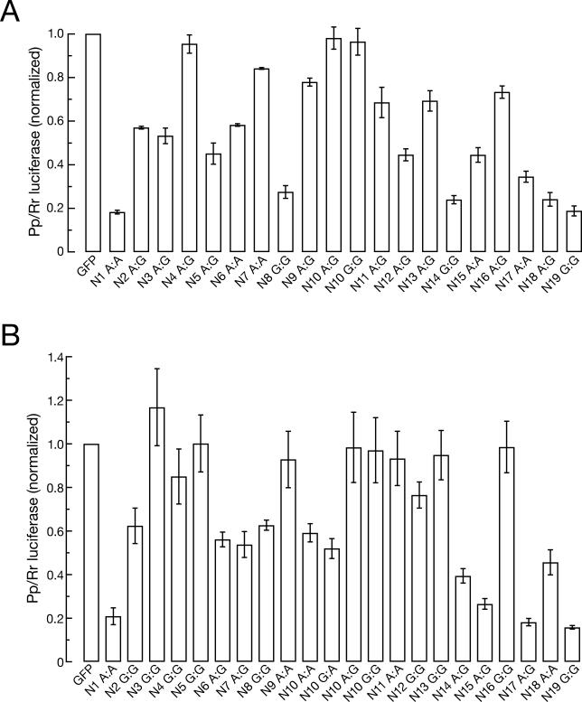
Our data suggest that purine:purine mismatches provide the highest level of discrimination against mismatched targets. To corroborate these findings, we examined the effect of a purine:purine mismatch at the 19 positions (N1 to N19) of a single siRNA sequence, the P10 siRNA. For each purine position in the P10 siRNA, a reporter was constructed that expressed a Pp luciferase mRNA with a purine at the corresponding target position. For pyrimidine positions in the P10 siRNA, a variant siRNA was synthesized substituting a single pyrimidine with a purine so as to create a purine:purine clash with the reporter mRNA. Seven siRNAs reduced expression of the mismatched reporter to less than 40% of the unsilenced level: N4, N7, N9, N10, N11, N13, and N16 (Figure 6A).
Figure 6. Purine:Purine Mismatches Are Tolerated at Some, but Not Other, siRNA Positions.
(A) Purine residues were placed at each position, N1 to N19, along the P10 siRNA. Targets were constructed so that siRNA:mRNA target pairs would result in a purine:purine mismatch. Luciferase activity was measured for each pair.
(B) The P4 siRNA (Figure 1A) was used as a scaffold for the analysis of the effect of purine:purine mismatches on reporter silencing. Taller bars correspond to greater single nucleotide discrimination.
The same method of analysis was applied to the P4 siRNA (Figure 6B). Luciferase silencing was disrupted the least by siRNA:target mRNA combinations that placed a single purine:purine mismatch at siRNA guide position 3, 4, 5, 9, 10, 11, 12, 13, or 16. Intriguingly, seed sequence mismatches—at positions 3, 4, and 5—were strongly discriminatory for this siRNA, which has the most thermodynamically stable seed sequence pairing of all the siRNAs in this study.
For the P4 siRNA scaffold, G:G and A:G mismatches at position 10 were more selective than A:A or G:A mismatches. While single nucleotide base-pairs can either stabilize or destabilize a helix, depending on the identity of both the mismatch and the adjacent base-pairs [56], the effect of G:G, A:G, A:A, and G:A mismatches flanked by U:A base-pairs has not been experimentally determined.
Position 16 Mismatches Generally Discriminate Well
Throughout our analyses—including cell-free RNAi reactions, reporter transfections, and microarray and quantitative-PCR analysis of endogenous mRNA—purine:purine mismatches at siRNA position 16 consistently discriminated against the mismatched target. We therefore sought to test the generality of an siRNA position 16 mismatch as a strategy for designing allele-specific siRNA. We synthesized ten distinct siRNA-mRNA pairs bearing purine:purine mismatches: five targeting an SOD1 point mutation and five targeting an HTT SNP. For comparison, a fully matched siRNA was synthesized for each target. Each pair of mismatched and matched siRNAs targeted a site inserted into the 3′ untranslated region of Renilla or firefly luciferase (Table 2). Reporter silencing, relative to a cotransfected luciferase control, was determined for each siRNA over a concentration range from 0.001 nM to 20 nM. The siRNA concentration producing half-maximal silencing (IC50) was calculated for the match or mismatched siRNAs (Table 2).
Table 2.
Mismatches at Position 16 Confer a High Level of Single Nucleotide Discrimination
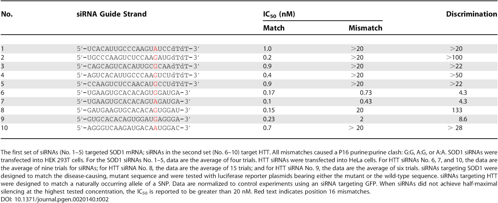
For all ten siRNA pairs tested, the IC50 for the siRNA:target combination with the position 16 mismatch was greater than that for the fully matched. For seven of the ten siRNA pairs, the IC50 was at least 20-fold greater for the mismatched siRNA:target combination.
Discussion
The use of single nucleotide–specific siRNA to reduce expression of mutant, disease-causing alleles holds promise for the treatment of dominantly inherited human diseases caused by point mutations that lead to a gain of function. Moreover, human disease alleles often differ from their wild-type counterparts by SNPs that do not themselves cause the disease phenotype. By targeting the SNP isoform present in the disease allele, single nucleotide–specific siRNA might be used to reduce selectively expression of the disease-causing allele without altering expression of the wild-type allele. It is therefore critical to understand the parameters for designing siRNAs with optimal discrimination between mRNAs that differ at a single nucleotide. Previous studies hinted that purine:purine mismatches between an siRNA and its target mRNA provide the greatest level of discrimination [23,33,57], probably reflecting greater disruption of the central A-form helix between the siRNA and mRNA that is required for siRNA-directed cleavage of the mRNA target [47,48]. Our systematic analysis here demonstrates the generality of this principle. Therefore, a point mutation or SNP in which a purine is changed to a pyrimidine increases the probability that an siRNA can be designed to discriminate between two alleles.
Because the last two nucleotides of an siRNA appear not to contribute to binding specificity [20] and may in fact slow RISC-catalyzed target cleavage by slowing the release of products [48], there are 19 siRNAs that, in theory, could be specific for any given point mutation. Here, we show that the extent of single nucleotide discrimination varies considerably among the 19 different positions. Using a combination of cell-free RNAi reactions and cultured human cell transfection experiments, we found that placing a purine:purine mismatch at specific siRNA positions, particularly P10 and P16, predisposes the siRNA to discriminate effectively between two alleles that differ at a single nucleotide. P5, P9, P10, P12, P13, and P16 mismatches consistently discriminated between two alleles, as tested with three different experimental strategies. Cell-free RNAi reactions using Drosophila embryo lysate provided an initial screen for candidate siRNAs capable of distinguishing between target alleles. Subsequent studies in cultured human cells assessed these siRNAs for both potency against the matched target and effective discrimination against the mismatched target (Figure 3), as well as determining siRNA strand choice by off-target profiling (Figure 4). In general, mismatches in the 5′ seed region of the siRNA, which is responsible for binding and recognition [48,50,53,54,58–60], provided only moderate single nucleotide discrimination, which was overwhelmed by high siRNA concentrations. In contrast, mismatches 3′ to the seed sequence promoted robust single nucleotide discrimination. This discrimination was most apparent for mismatches at positions 10 and 16, but also P9, P12, P13, and P14. In fact, in each assay used here, a mismatch at siRNA guide position 16 consistently showed high levels of discrimination against a mismatched RNA target, even at 100 nM siRNA concentration. Intriguingly, the function of plant miRNAs, which direct siRNA-like cleavage of their mRNA targets, also appears to be sensitive to position 16 mismatches between the small RNA and target: no position 16 mismatches and few G:U wobble pairs have been identified between plant miRNAs and their experimentally validated mRNA targets [61]. Thus, the sixteenth nucleotide of small RNA guides in both plants and animals may play a biochemically distinct role in directing target RNA cleavage.
A clear implication of our results is that siRNAs best discriminate between alleles when the conformation of the siRNA with the mismatch target acts to block catalysis, rather than binding, and is therefore less sensitive to siRNA concentration. Although we do not understand the detailed biochemical basis for this effect, our data suggest that the conformation of the helix formed between the small RNA and its target is monitored by some protein factor—perhaps even the core RISC protein Argonaute2 itself, the siRNA-directed RNA endonuclease that destroys the target mRNA.
Materials and Methods
General methods.
Preparation of Drosophila embryo lysate and target RNAs, siRNA annealing, and in vitro RNAi reactions were as described [12,62,63]. SOD1 mutant and wild-type RNAs were transcribed from BamHI-linearized plasmids [64] with recombinant histidine-tagged T7 RNA polymerase. Approximately 5 nM target RNA and 50 nM siRNA were used in Figure 1; approximately 0.5 nM target and 100 nM siRNA were used in Figure S1. Gels were dried and exposed to PhosphorImager (Fuji, Tokyo, Japan) plates, analyzed using an FLA-5000 PhosphorImager, analyzed, and quantified using ImageGuage 3.45 (Fuji), Excel X (Microsoft, Redmond, Washington, United States), and Igor Pro 5.01 (Wavemetrics, Lake Oswego, Oregon, United States).
Cell culture, transfection, and luciferase assays.
HeLa cells were propagated and maintained as described [65]. HEK 293T cells were maintained in Dulbecco's modified Eagle medium (Invitrogen, Carlsbad, California, United States), supplemented with 10% fetal bovine serum, 100 U/ml penicillin, and 100 μg/ml streptomycin. HTT sequences were engineered into the 3′ UTR of the pRLTK Renilla luciferase vector (Promega, Madison, Wisconsin, United States) using 55-bp DNA oligonucleotides (IDT, Coralville, Iowa, United States) designed to create 5′ overhangs when annealed, allowing their insertion into the plasmid XbaI site. Plasmid constructs were verified by bidirectional sequencing. SOD1 sequences were cloned into the 3′ UTR of the firefly luciferase mRNA (pGL2 control, Promega) into NdeI and SpeI sites engineered into the plasmid by annealing two 39-nucleotide DNA oligos and ligating them into the vector. Transfections were carried out using LipofectAMINE 2000 (Invitrogen) in 24-well plates using 0.25 μg of pGL2 firefly luciferase (Promega) and 0.1 μg of Renilla-HTT constructs or in 96-well plates using 2 μg/ml firefly fusion vector and 0.1 μg/ml Renilla vector. Cells were washed in 1× PBS (Invitrogen) and harvested 24 h after transfection in 1× passive lysis buffer (Promega). Luciferase levels were determined using the Dual Luciferase kit (Promega) and a Veritas Microplate Luminometer (Turner Biosystems, Sunnyvale, California, United States). Renilla luciferase/firefly luciferase ratios were normalized to a transfection with a GFP siRNA (Qiagen, Valencia, California, United States). IC50 values were determined by fitting the data to the Hill equation with n = 1. For siRNAs in which the half-maximal concentration for silencing was not reached at the highest concentration tested, IC50 values were reported as greater than the highest concentration tested.
Microarray and quantitative PCR analysis.
HeLa cells were from the American Type Culture Collection (Rockville, Maryland, United States). Cells were plated 24 h prior to transfection with OligofectAMINE (Invitrogen). Duplexes were used at a final concentration of 100 nM. Cells were transfected in six-well plates and RNA was isolated 24 h following transfection. Total RNA was purified using the RNeasy kit (Qiagen). Microarray analysis was performed as described [53,66,67]. Amplified cRNA from siRNA-transfected cells was hybridized against cRNA from mock-transfected cells (treated with transfection reagent in the absence of RNA duplex). Ratio hybridizations were performed with fluorescent label reversal to eliminate dye bias. Error models have been described previously [53,66,67]. Data were analyzed using Rosetta Resolver (Rosetta Biosoftware, Seattle, Washington, United States). mRNA levels were also measured by qRT-PCR using an ABI PRISM 7900HT Sequence Detection System and Assays-on-Demand gene expression products (Applied Biosystems, Foster City, California, United States). SOD1 mRNA was measured using ABI assay No. Hs00533490_A1 and normalized to β-glucuronidase mRNA, measured using ABI assay No. 4310888E.
Supporting Information
Reactions were performed using an excess of RISC over substrate RNA in order to ensure single-turnover conditions. The initial rate of reaction was determined using data from the first 120 s. Filled circles, mutant siRNA with mutant (matched) target; open circles, mutant siRNA with wild-type (mismatched) target.
(401 KB PDF)
Accession Numbers
Unprocessed microarray data have been deposited in the NCBI Gene Expression Omnibus database (http://www.ncbi.nlm.nih.gov/geo) under the accession number GSE5291.
Acknowledgments
We thank members of the Zamore, Xu, and Aronin laboratories and Nelson Lau for encouragement, helpful discussions, and comments on the manuscript. PDZ is a W.M. Keck Foundation Young Scholar in Medical Research.
Abbreviations
- ALS
amyotrophic lateral sclerosis
- RISC
RNA-induced silencing complex
- RNAi
RNA interference
- siRNA
small interfering RNA
- SNP
single nucleotide polymorphism
Footnotes
¤ Current address: Department of Molecular and Cellular Biology, Harvard University, Cambridge, Massachusetts, United States of America
Competing interests. PDZ is a co-founder and advisor for Alnylam Pharmaceuticals, Inc., an siRNA therapeutics company.
A previous version of this article appeared as an Early Online Release on July 24, 2006 (DOI: 10.1371/journal.pgen.0020140.eor).
Author contributions. DSS, NA, ZX, and PDZ conceived the experiments. DSS, HD, LK, PSL, ZX, and PDZ designed the experiments. DSS, HD, LK, JTM, JS, and JB performed the experiments. DSS, LK, JTM, JS, JB, and ZX analyzed the results. DSS, HD, JB, JS, and PSL contributed reagents/materials/analysis tools. DSS, ZX, and PDZ prepared the figures. DSS and PDZ wrote the manuscript.
Funding. This work was supported, in part, by grants from the National Institutes of Health to PDZ (R01 GM62862), ZX (R01 NS048145), ZX and PDZ (R21 NS44952–01), and NA and PDZ (R01 NS38194) and by grants to ZX from The Robert Pachard Center for ALS Research at Johns Hopkins University and from the ALS Association, and to NA and PDZ from the HiQ Foundation.
References
- Hammond SM, Bernstein E, Beach D, Hannon GJ. An RNA-directed nuclease mediates post-transcriptional gene silencing in Drosophila cells. Nature. 2000;404:293–296. doi: 10.1038/35005107. [DOI] [PubMed] [Google Scholar]
- Martinez J, Patkaniowska A, H HU, Lührmann R, Tuschl T. Single stranded antisense siRNA guide target RNA cleavage in RNAi. Cell. 2002;110:563–574. doi: 10.1016/s0092-8674(02)00908-x. [DOI] [PubMed] [Google Scholar]
- Nykanen A, Haley B, Zamore PD. ATP requirements and small interfering RNA structure in the RNA interference pathway. Cell. 2001;107:309–321. doi: 10.1016/s0092-8674(01)00547-5. [DOI] [PubMed] [Google Scholar]
- Elbashir SM, Harborth J, Lendeckel W, Yalcin A, Weber K, et al. Duplexes of 21-nucleotide RNAs mediate RNA interference in cultured mammalian cells. Nature. 2001;411:494–498. doi: 10.1038/35078107. [DOI] [PubMed] [Google Scholar]
- Palliser D, Chowdhury D, Wang QY, Lee SJ, Bronson RT, et al. An siRNA-based microbicide protects mice from lethal herpes simplex virus 2 infection. Nature. 2006;439:89–94. doi: 10.1038/nature04263. [DOI] [PubMed] [Google Scholar]
- Song E, Lee SK, Wang J, Ince N, Ouyang N, et al. RNA interference targeting Fas protects mice from fulminant hepatitis. Nat Med. 2003;9:347–351. doi: 10.1038/nm828. [DOI] [PubMed] [Google Scholar]
- Song E, Zhu P, Lee SK, Chowdhury D, Kussman S, et al. Antibody mediated in vivo delivery of small interfering RNAs via cell-surface receptors. Nat Biotechnol. 2005;23:709–717. doi: 10.1038/nbt1101. [DOI] [PubMed] [Google Scholar]
- Soutschek J, Akinc A, Bramlage B, Charisse K, Constien R, et al. Therapeutic silencing of an endogenous gene by systemic administration of modified siRNAs. Nature. 2004;432:173–178. doi: 10.1038/nature03121. [DOI] [PubMed] [Google Scholar]
- Zimmermann TS, Lee AC, Akinc A, Bramlage B, Bumcrot D, et al. RNAi-mediated gene silencing in non-human primates. Nature. 2006;441:111–114. doi: 10.1038/nature04688. [DOI] [PubMed] [Google Scholar]
- Elbashir SM, Lendeckel W, Tuschl T. RNA interference is mediated by 21- and 22-nucleotide RNAs. Genes Dev. 2001;15:188–200. doi: 10.1101/gad.862301. [DOI] [PMC free article] [PubMed] [Google Scholar]
- Hamilton AJ, Baulcombe DC. A species of small antisense RNA in posttranscriptional gene silencing in plants. Science. 1999;286:950–952. doi: 10.1126/science.286.5441.950. [DOI] [PubMed] [Google Scholar]
- Zamore PD, Tuschl T, Sharp PA, Bartel DP. RNAi: Double-stranded RNA directs the ATP-dependent cleavage of mRNA at 21 to 23 nucleotide intervals. Cell. 2000;101:25–33. doi: 10.1016/S0092-8674(00)80620-0. [DOI] [PubMed] [Google Scholar]
- Harborth J, Elbashir SM, Bechert K, Tuschl T, Weber K. Identification of essential genes in cultured mammalian cells using small interfering RNAs. J Cell Sci. 2001;114:4557–4565. doi: 10.1242/jcs.114.24.4557. [DOI] [PubMed] [Google Scholar]
- Schwarz DS, Hutvágner G, Du T, Xu Z, Aronin N, et al. Asymmetry in the assembly of the RNAi enzyme complex. Cell. 2003;115:199–208. doi: 10.1016/s0092-8674(03)00759-1. [DOI] [PubMed] [Google Scholar]
- Khvorova A, Reynolds A, Jayasena SD. Functional siRNAs and miRNAs exhibit strand bias. Cell. 2003;115:209–216. doi: 10.1016/s0092-8674(03)00801-8. [DOI] [PubMed] [Google Scholar]
- Amarzguioui M, Holen T, Babaie E, Prydz H. Tolerance for mutations and chemical modifications in a siRNA. Nucleic Acids Res. 2003;31:589–595. doi: 10.1093/nar/gkg147. [DOI] [PMC free article] [PubMed] [Google Scholar]
- Boutla A, Delidakis C, Livadaras I, Tsagris M, Tabler M. Short 5′-phosphorylated double-stranded RNAs induce RNA interference in Drosophila . Curr Biol. 2001;11:1776–1780. doi: 10.1016/s0960-9822(01)00541-3. [DOI] [PubMed] [Google Scholar]
- Brummelkamp T, Bernards R, Agami R. Stable suppression of tumorigenicity by virus-mediated RNA interference. Cancer Cell. 2002;2:243. doi: 10.1016/s1535-6108(02)00122-8. [DOI] [PubMed] [Google Scholar]
- Brummelkamp TR, Bernards R, Agami R. A system for stable expression of short interfering RNAs in mammalian cells. Science. 2002;296:550–553. doi: 10.1126/science.1068999. [DOI] [PubMed] [Google Scholar]
- Elbashir SM, Martinez J, Patkaniowska A, Lendeckel W, Tuschl T. Functional anatomy of siRNAs for mediating efficient RNAi in Drosophila melanogaster embryo lysate. EMBO J. 2001;20:6877–6888. doi: 10.1093/emboj/20.23.6877. [DOI] [PMC free article] [PubMed] [Google Scholar]
- Holen T, Amarzguioui M, Wiiger MT, Babaie E, Prydz H. Positional effects of short interfering RNAs targeting the human coagulation trigger Tissue Factor. Nucleic Acids Res. 2002;30:1757–1766. doi: 10.1093/nar/30.8.1757. [DOI] [PMC free article] [PubMed] [Google Scholar]
- Yu JY, DeRuiter SL, Turner DL. RNA interference by expression of short-interfering RNAs and hairpin RNAs in mammalian cells. Proc Natl Acad Sci U S A. 2002;99:6047–6052. doi: 10.1073/pnas.092143499. [DOI] [PMC free article] [PubMed] [Google Scholar]
- Ding H, Schwarz DS, Keene A, Affar el B, Fenton L, et al. Selective silencing by RNAi of a dominant allele that causes amyotrophic lateral sclerosis. Aging Cell. 2003;2:209–217. doi: 10.1046/j.1474-9728.2003.00054.x. [DOI] [PubMed] [Google Scholar]
- Maxwell MM, Pasinelli P, Kazantsev AG, Brown RHJ. RNA interference-mediated silencing of mutant superoxide dismutase rescues cyclosporin A-induced death in cultured neuroblastoma cells. Proc Natl Acad Sci U S A. 2004;101:3178–3183. doi: 10.1073/pnas.0308726100. [DOI] [PMC free article] [PubMed] [Google Scholar]
- Ralph GS, Radcliffe PA, Day DM, Carthy JM, Leroux MA, et al. Silencing mutant SOD1 using RNAi protects against neurodegeneration and extends survival in an ALS model. Nat Med. 2005;11:429–433. doi: 10.1038/nm1205. [DOI] [PubMed] [Google Scholar]
- Raoul C, Abbas-Terki T, Bensadoun JC, Guillot S, Haase G, et al. Lentiviral-mediated silencing of SOD1 through RNA interference retards disease onset and progression in a mouse model of ALS. Nat Med. 2005;11:423–428. doi: 10.1038/nm1207. [DOI] [PubMed] [Google Scholar]
- Harper SQ, Staber PD, He X, Eliason SL, Martins IH, et al. RNA interference improves motor and neuropathological abnormalities in a Huntington's disease mouse model. Proc Natl Acad Sci U S A. 2005;102:5820–5825. doi: 10.1073/pnas.0501507102. [DOI] [PMC free article] [PubMed] [Google Scholar]
- Miller VM, Gouvion CM, Davidson BL, Paulson HL. Targeting Alzheimer's disease genes with RNA interference: An efficient strategy for silencing mutant alleles. Nucleic Acids Res. 2004;32:661–668. doi: 10.1093/nar/gkh208. [DOI] [PMC free article] [PubMed] [Google Scholar]
- Lee SK, Dykxhoorn DM, Kumar P, Ranjbar S, Song E, et al. Lentiviral delivery of short hairpin RNAs protects CD4 T cells from multiple clades and primary isolates of HIV. Blood. 2005;106:818–826. doi: 10.1182/blood-2004-10-3959. [DOI] [PMC free article] [PubMed] [Google Scholar]
- Li Z, Xiong Y, Peng Y, Pan J, Chen Y, et al. Specific inhibition of HIV-1 replication by short hairpin RNAs targeting human cyclin T1 without inducing apoptosis. FEBS Lett. 2005;579:3100–3106. doi: 10.1016/j.febslet.2005.04.074. [DOI] [PubMed] [Google Scholar]
- Abdelgany A, Wood M, Beeson D. Allele-specific silencing of a pathogenic mutant acetylcholine receptor subunit by RNA interference. Hum Mol Genet. 2003;12:2637–2644. doi: 10.1093/hmg/ddg280. [DOI] [PubMed] [Google Scholar]
- Miller VM, Xia H, Marrs GL, Gouvion CM, Lee G, et al. Allele-specific silencing of dominant disease genes. Proc Natl Acad Sci U S A. 2003;100:7195–7200. doi: 10.1073/pnas.1231012100. [DOI] [PMC free article] [PubMed] [Google Scholar]
- Dykxhoorn DM, Schlehuber LD, London IM, Lieberman J. Determinants of specific RNA interference-mediated silencing of human beta-globin alleles differing by a single nucleotide polymorphism. Proc Natl Acad Sci U S A. 2006;103:5953–5958. doi: 10.1073/pnas.0601309103. [DOI] [PMC free article] [PubMed] [Google Scholar]
- Martinez LA, Naguibneva I, Lehrmann H, Vervisch A, Tchenio T, et al. Synthetic small inhibiting RNAs: Efficient tools to inactivate oncogenic mutations and restore p53 pathways. Proc Natl Acad Sci U S A. 2002;99:14849–14854. doi: 10.1073/pnas.222406899. [DOI] [PMC free article] [PubMed] [Google Scholar]
- Kunst CB. Complex genetics of amyotrophic lateral sclerosis. Am J Hum Genet. 2004;75:933–947. doi: 10.1086/426001. [DOI] [PMC free article] [PubMed] [Google Scholar]
- Xu Z. Mechanism and treatment of motoneuron degeneration in ALS: What have SOD1 mutants told us? Amyotroph Lateral Scler Other Motor Neuron Disord. 2000;1:225–234. doi: 10.1080/14660820050515052. [DOI] [PubMed] [Google Scholar]
- Flood DG, Reaume AG, Gruner JA, Hoffman EK, Hirsch JD, et al. Hindlimb motor neurons require Cu/Zn superoxide dismutase for maintenance of neuromuscular junctions. Am J Pathol. 1999;155:663–672. doi: 10.1016/S0002-9440(10)65162-0. [DOI] [PMC free article] [PubMed] [Google Scholar]
- Shefner JM, Reaume AG, Flood DG, Scott RW, Kowall NW, et al. Mice lacking cytosolic copper/zinc superoxide dismutase display a distinctive motor axonopathy. Neurology. 1999;53:1239–1246. doi: 10.1212/wnl.53.6.1239. [DOI] [PubMed] [Google Scholar]
- Elchuri S, Oberley TD, Qi W, Eisenstein RS, Jackson Roberts L, et al. CuZnSOD deficiency leads to persistent and widespread oxidative damage and hepatocarcinogenesis later in life. Oncogene. 2005;24:367–380. doi: 10.1038/sj.onc.1208207. [DOI] [PubMed] [Google Scholar]
- McFadden SL, Ding D, Salvi R. Anatomical, metabolic and genetic aspects of age-related hearing loss in mice. Audiology. 2001;40:313–321. [PubMed] [Google Scholar]
- Matzuk MM, Dionne L, Guo Q, Kumar TR, Lebovitz RM. Ovarian function in superoxide dismutase 1 and 2 knockout mice. Endocrinology. 1998;139:4008–4011. doi: 10.1210/endo.139.9.6289. [DOI] [PubMed] [Google Scholar]
- Vonsattel JP, DiFiglia M. Huntington disease. J Neuropathol Exp Neurol. 1998;57:369–384. doi: 10.1097/00005072-199805000-00001. [DOI] [PubMed] [Google Scholar]
- The Huntington's Disease Collaborative Research Group. A novel gene containing a trinucleotide repeat that is expanded and unstable on Huntington's disease chromosomes. Cell. 1993;72:971–983. doi: 10.1016/0092-8674(93)90585-e. [DOI] [PubMed] [Google Scholar]
- Andrew SE, Goldberg YP, Kremer B, Telenius H, Theilmann J, et al. The relationship between trinucleotide (CAG) repeat length and clinical features of Huntington's disease. Nat Genet. 1993;4:398–403. doi: 10.1038/ng0893-398. [DOI] [PubMed] [Google Scholar]
- Snell RG, MacMillan JC, Cheadle JP, Fenton I, Lazarou LP, et al. Relationship between trinucleotide repeat expansion and phenotypic variation in Huntington's disease. Nat Genet. 1993;4:393–397. doi: 10.1038/ng0893-393. [DOI] [PubMed] [Google Scholar]
- Caplen NJ, Taylor JP, Statham VS, Tanaka F, Fire A, et al. Rescue of polyglutamine-mediated cytotoxicity by double-stranded RNA-mediated RNA interference. Hum Mol Genet. 2002;11:175–184. doi: 10.1093/hmg/11.2.175. [DOI] [PubMed] [Google Scholar]
- Chiu YL, Rana TM. RNAi in human cells: Basic structural and functional features of small interfering RNA. Mol Cell. 2002;10:549–561. doi: 10.1016/s1097-2765(02)00652-4. [DOI] [PubMed] [Google Scholar]
- Haley B, Zamore PD. Kinetic analysis of the RNAi enzyme complex. Nat Struct Mol Biol. 2004;11:599–606. doi: 10.1038/nsmb780. [DOI] [PubMed] [Google Scholar]
- Reynolds A, Leake D, Boese Q, Scaringe S, Marshall WS, et al. Rational siRNA design for RNA interference. Nat Biotechnol. 2004;22:326–330. doi: 10.1038/nbt936. [DOI] [PubMed] [Google Scholar]
- Doench JG, Sharp PA. Specificity of microRNA target selection in translational repression. Genes Dev. 2004;18:504–511. doi: 10.1101/gad.1184404. [DOI] [PMC free article] [PubMed] [Google Scholar]
- Brennecke J, Stark A, Russell RB, Cohen SM. Principles of microRNA-target recognition. PLoS Biol. 2005;3:e85. doi: 10.1371/journal.pbio.0030085. DOI: 10.1371/journal.pbio.0030085. [DOI] [PMC free article] [PubMed] [Google Scholar]
- Birmingham A, Anderson EM, Reynolds A, Ilsley-Tyree D, Leake D, et al. 3′ UTR seed matches, but not overall identity, are associated with RNAi off-targets. Nat Methods. 2006;3:199–204. doi: 10.1038/nmeth854. [DOI] [PubMed] [Google Scholar]
- Jackson AL, Bartz SR, Schelter J, Kobayashi SV, Burchard J, et al. Expression profiling reveals off-target gene regulation by RNAi. Nat Biotechnol. 2003;21:635–637. doi: 10.1038/nbt831. [DOI] [PubMed] [Google Scholar]
- Lim LP, Lau NC, Garrett-Engele P, Grimson A, Schelter JM, et al. Microarray analysis shows that some microRNAs downregulate large numbers of target mRNAs. Nature. 2005;433:769–773. doi: 10.1038/nature03315. [DOI] [PubMed] [Google Scholar]
- Semizarov D, Frost L, Sarthy A, Kroeger P, Halbert DN, et al. Specificity of short interfering RNA determined through gene expression signatures. Proc Natl Acad Sci U S A. 2003;100:6347–6352. doi: 10.1073/pnas.1131959100. [DOI] [PMC free article] [PubMed] [Google Scholar]
- Kierzek R, Burkard ME, Turner DH. Thermodynamics of single mismatches in RNA duplexes. Biochemistry. 1999;38:14214–14223. doi: 10.1021/bi991186l. [DOI] [PubMed] [Google Scholar]
- Du Q, Thonberg H, Wang J, Wahlestedt C, Liang Z. A systematic analysis of the silencing effects of an active siRNA at all single-nucleotide mismatched target sites. Nucleic Acids Res. 2005;33:1671–1677. doi: 10.1093/nar/gki312. [DOI] [PMC free article] [PubMed] [Google Scholar]
- Lai EC. Micro RNAs are complementary to 3′ UTR sequence motifs that mediate negative post-transcriptional regulation. Nat Genet. 2002;30:363–364. doi: 10.1038/ng865. [DOI] [PubMed] [Google Scholar]
- Lewis BP, Burge CB, Bartel DP. Conserved seed pairing, often flanked by adenosines, indicates that thousands of human genes are microRNA targets. Cell. 2005;120:15–20. doi: 10.1016/j.cell.2004.12.035. [DOI] [PubMed] [Google Scholar]
- Lewis BP, Shih IH, Jones-Rhoades MW, Bartel DP, Burge CB. Prediction of mammalian microRNA targets. Cell. 2003;115:787–798. doi: 10.1016/s0092-8674(03)01018-3. [DOI] [PubMed] [Google Scholar]
- Mallory AC, Reinhart BJ, Jones-Rhoades MW, Tang G, Zamore PD, et al. MicroRNA control of PHABULOSA in leaf development: Importance of pairing to the microRNA 5′ region. EMBO J. 2004;23:3356–3364. doi: 10.1038/sj.emboj.7600340. [DOI] [PMC free article] [PubMed] [Google Scholar]
- Haley B, Tang G, Zamore PD. In vitro analysis of RNA interference in Drosophila melanogaster . Methods. 2003;30:330–336. doi: 10.1016/s1046-2023(03)00052-5. [DOI] [PubMed] [Google Scholar]
- Tuschl T, Zamore PD, Lehmann R, Bartel DP, Sharp PA. Targeted mRNA degradation by double-stranded RNA in vitro. Genes Dev. 1999;13:3191–3197. doi: 10.1101/gad.13.24.3191. [DOI] [PMC free article] [PubMed] [Google Scholar]
- Crow JP, Sampson JB, Zhuang Y, Thompson JA, Beckman JS. Decreased zinc affinity of amyotrophic lateral sclerosis-associated superoxide dismutase mutants leads to enhanced catalysis of tyrosine nitration by peroxynitrite. J Neurochem. 1997;69:1936–1944. doi: 10.1046/j.1471-4159.1997.69051936.x. [DOI] [PubMed] [Google Scholar]
- Schwarz DS, Hutvágner G, Haley B, Zamore PD. Evidence that siRNAs function as guides, not primers, in the Drosophila and human RNAi pathways. Molecular Cell. 2002;10:537–548. doi: 10.1016/s1097-2765(02)00651-2. [DOI] [PubMed] [Google Scholar]
- Hughes TR, Mao M, Jones AR, Burchard J, Marton MJ, et al. Expression profiling using microarrays fabricated by an ink-jet oligonucleotide synthesizer. Nat Biotechnol. 2001;19:342–347. doi: 10.1038/86730. [DOI] [PubMed] [Google Scholar]
- Jackson AL, Burchard J, Schelter J, Chau BN, Cleary M, et al. Widespread siRNA “off-target” transcript silencing mediated by seed region sequence complementarity. RNA. 2006;12:1179–1187. doi: 10.1261/rna.25706. [DOI] [PMC free article] [PubMed] [Google Scholar]
Associated Data
This section collects any data citations, data availability statements, or supplementary materials included in this article.
Supplementary Materials
Reactions were performed using an excess of RISC over substrate RNA in order to ensure single-turnover conditions. The initial rate of reaction was determined using data from the first 120 s. Filled circles, mutant siRNA with mutant (matched) target; open circles, mutant siRNA with wild-type (mismatched) target.
(401 KB PDF)


