Abstract
Human bocavirus (HBoV), a newly cloned human virus of the genus Bocavirus, was detected by PCR from nasopharyngeal swab samples (8 of 318; 5.7%) collected from children with lower respiratory tract infections. HBoV may be one of the causative agents of lower respiratory tract infections in young children.
The family Parvoviridae contains two subfamilies: Parvovirinae, which infects vertebrates, and Densovirinae, which infects insects. The subfamily Parvovirinae consists of five genera: Parvovirus, Erythrovirus, Dependovirus, Amdovirus, and Bocavirus (12). Parvovirus B19, which belongs to the genus Erythrovirus, is a well-known human pathogen (3, 12). A new human virus of the genus Bocavirus, provisionally named human bocavirus (HBoV), was recently cloned from pooled human respiratory tract samples and is considered to be pathogenic to humans (1). In this study, nasopharyngeal swab samples obtained from children with lower respiratory tract infections were investigated for the presence of HBoV.
From October 2002 to September 2003 and from January 2005 to July 2005, a total of 318 nasopharyngeal swab samples were collected at four hospitals in Sapporo, Japan, from 318 children with lower respiratory tract infections. All of the samples were collected after the possibility of infection with human respiratory syncytial virus or influenza A or B virus was excluded by rapid antigen detection tests and after the possibility of infection with human metapneumovirus was excluded by a reverse transcription-PCR test (6). The median age of the children was 21.3 months. The male-to-female ratio was 1.4 to 1. All samples were collected after obtaining informed consent from the children's parents. RNA and DNA were extracted from each sample by using Chomczynski's protocol (5). RNA was used for the detection of human metapneumovirus (6) and DNA was used for the detection of HBoV as described below. The PCR primers and conditions used for detection of HBoV have been described previously (1). A forward primer with a sequence of 5′-GAGCTCTGTAAGTACTATTAC-3′ and a reverse primer with a sequence of 5′-CTCTGTGTTGACTGAATACAG-3′ were used for both PCR and sequencing. The PCR mixture consisted of 100 μmol of each deoxyribonucleotide, 1.0 U of AmpliTaq Gold, 50 mmol of potassium chloride/liter, 10 mmol of Tris-HCl (pH 8.3)/liter, 1.5 mmol of magnesium chloride/liter, 0.01% (wt/vol) gelatin, 10 pmol of each primer, and DNA in a volume of 25 μl. The PCR conditions were as follows: 94°C for 9 min, followed by 35 cycles of 94°C for 1 min, 54°C for 1 min, and 72°C for 2 min. Sense and antisense strands of the PCR products were sequenced directly by using a BigDye terminator cycle sequencing ready reaction kit (PerkinElmer Applied Biosystems, Tokyo, Japan) with an ABI Prism 310 genetic analyzer (PerkinElmer Applied Biosystems).
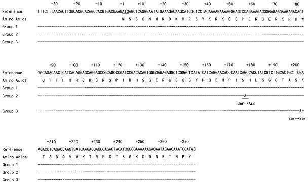
DNA sequences of HBoV were detected in samples from 18 (5.7%) of the 318 patients with lower respiratory tract infections. Seventeen of the 18 HBoV-positive samples were collected during the period from January to May (one sample in January, three in February, one in March, four in April, eight in May, and one in July). The ages of patients with HBoV-positive samples ranged from 7 months to 3 years (one patient was 7 to 9 months of age, five were 10 to 12 months of age, ten were 1 to 2 years of age, and two were 2 to 3 years of age). Direct sequencing of PCR products of the 18 samples showed that 14 of the 18 sequences were completely identical to the published sequence of HBoV (GenBank accession numbers DQ000495 and DQ000496) (Fig. 1, Group 1). Three of the 18 sequences had a single-base-pair substitution in the NP-1 gene, resulting in an amino acid exchange (Fig. 1, Group 2). One of the 18 sequences had a single-base-pair substitution in the NP-1 gene without an amino acid exchange (Fig. 1, Group 3). The 18 new sequences were deposited in GenBank.
FIG. 1.
Sequences of PCR products of HBoV-positive samples. A of the ATG initiator methionine codon of NP-1 protein is denoted as nucleotide +1. The sequences of the 18 samples were divided into three groups and compared with that of the published sequence of HBoV (GenBank accession number DQ000495) as a reference. Group 1 (JPBS03-112, JPBS03-217, JPBS03-219, JPBS03-261, JPBS03-298, JPBS05-7, JPBS05-18, JPBS05-28, JPBS05-29, JPBT03-5, JPBT03-12, JPBT03-43, JPBT05-46, and JPBT05-52) was completely identical to the reference. Group 2 (JPBS03-182, JPBS03-207, and JPBS03-208) had a single-base-pair substitution (176G to A) in the NP-1 gene, resulting in an amino acid exchange (Ser to Asn). Group 3 (JPBS03-98) had a single-base-pair substitution (201G to A) in the NP-1 gene without an amino acid exchange.
Clinical and laboratory features of the 18 HBoV-positive patients are shown in Table 1. All of the 18 patients suffered from fever, cough, and various degrees of respiratory distress. Maximum body temperatures ranged from 37.5 to 40.2°C. The duration of fever (temperature of >37.5°C) ranged from 1 to 8 days. Eight of the 18 patients showed abnormal findings on a chest X ray (four patients with lung infiltration, three patients with peribronchial infiltration, and one patient with hyperinflation). The clinical diagnoses of the HBoV-positive patients were pneumonia (six patients), wheezy bronchitis (six patients), bronchitis (two patients), bronchiolitis (two patients), asthma attack (one patient), and laryngotracheitis (one patient). Sixteen of the 18 patients were admitted to the hospital for 3 to 9 days.
TABLE 1.
Clinical characteristics of 18 patients positive for HBoV DNA by PCRa
| Case no. | Sample no. | Sex | Age | Diagnosis | Max. temp (°C) | Duration of fever (days)b | Cough | Wheezing | Chest X ray result | Max. WBC/μl | Max. CRP (mg/dl) | No. of days hospitalized |
|---|---|---|---|---|---|---|---|---|---|---|---|---|
| 1 | JPBS03-98 | F | 1 yr, 6 mo | Bronchitis | 39.4 | 8 | + | − | No abnormality | 14,300 | <0.20 | 4 |
| 2 | JPBS03-112 | F | 10 mo | Bronchiolitis | 37.9 | 3 | + | + | Peribronchial infiltration | 6,400 | <0.20 | 7 |
| 3 | JPBS03-182 | M | 1 yr, 3 mo | Pneumonia | 40.2 | 2 | + | + | Bilateral lung infiltration | 21,900 | 3.80 | 8 |
| 4 | JPBS03-207 | M | 1 yr, 10 mo | Wheezy bronchitis | 39.1 | 2 | + | + | No abnormality | 13,980 | 0.20 | 5 |
| 5 | JPBS03-208 | M | 1 yr, 0 mo | Pneumonia | 40.0 | 4 | + | + | Right lung infiltration | 11,800 | 1.58 | 7 |
| 6 | JPBS03-217 | M | 1 yr, 3 mo | Wheezy bronchitis | 38.8 | 3 | + | + | No abnormality | 21,980 | 2.40 | 5 |
| 7 | JPBS03-219 | M | 10 mo | Pneumonia | 40.0 | 6 | + | − | Peribronchial infiltration | 20,260 | 1.80 | 5 |
| 8 | JPBS03-261 | F | 1 yr, 1 mo | Wheezy bronchitis | 38.0 | 3 | + | + | No abnormality | 6,590 | 0.40 | 7 |
| 9 | JPBS03-298 | F | 1 yr, 9 mo | Wheezy bronchitis | 38.1 | 1 | + | + | No abnormality | 15,190 | <0.20 | 4 |
| 10 | JPBS05-7 | M | 9 mo | Bronchiolitis | 37.9 | 3 | + | + | Hyperinflation | 12,400 | 0.20 | 9 |
| 11 | JPBS05-18 | M | 1 yr, 11 mo | Laryngotracheitis | 39.6 | 5 | + | − | No abnormality | 14,000 | 1.02 | 7 |
| 12 | JPBS05-28 | M | 11 mo | Pneumonia | 38.8 | 3 | + | − | Right lung infiltration | 17,000 | 4.48 | 7 |
| 13 | JPBS05-29 | F | 11 mo | Pneumonia | 37.7 | 3 | + | − | Right lung infiltration | 10,220 | 0.38 | 6 |
| 14 | JPBT03-5 | M | 2 yr, 7 mo | Asthma attack | 37.5 | 1 | + | + | No abnormality | 15,000 | 2.57 | 3 |
| 15 | JPBT03-12 | M | 2 yr, 4 mo | Pneumonia | 39.0 | 2 | + | + | Peribronchial infiltration | 4,800 | 0.94 | 0 |
| 16 | JPBT03-43 | M | 1 yr, 3 mo | Bronchitis | 39.5 | 3 | + | + | No abnormality | 9,400 | 0.77 | 4 |
| 17 | JPBT05-46 | M | 1 yr, 3 mo | Wheezy bronchitis | 38.8 | 3 | + | + | No abnormality | 11,700 | 0.27 | 7 |
| 18 | JPBT05-52 | M | 1 yr, 9 mo | Wheezy bronchitis | 39.0 | 1 | + | + | No abnormality | 10,800 | 0.23 | 0 |
F, female; M, male; +, symptom present; −, symptom not present; Max. WBC, maximum white blood cell count; Max. CRP, maximum C-reactive protein level.
Fever, temperature of >37.5°C.
Bovine parvovirus and canine minute virus (MVC) are members of the family Parvoviridae, the subfamily Parvovirinae, and the genus Bocavirus. Bovine parvovirus caused mild diarrhea in calves inoculated per os, and it caused diarrhea and mild respiratory symptoms in calves inoculated intranasally (11). MVC was first described as an isolate from a healthy dog in the United States (2). Although it is likely that most infections with MVC are subclinical, diseases associated with virus infection include fetal infections leading to reproductive failure and neonatal respiratory disease (4, 7, 8). The virus may also be associated with some cases of enteritis in puppies or older dogs (2). HBoV was first cloned from pooled human respiratory tract samples collected in Sweden and was provisionally classified into the genus Bocavirus based on sequence resemblance (1). HBoV has recently been found in Australian children with respiratory tract infections (10). HBoV was detected from samples collected during the period from winter to spring (1, 10). The detection rate of HBoV in respiratory tract infections has been reported to be 3.1% to 5.6% (1, 10), which is consistent with our data (5.7%). It should be noted that the possibility of infection with other viruses (parainfluenza viruses, rhinoviruses, and coronaviruses) in patients with bocavirus-positive specimens could not be excluded and that the possibility of influenza virus infection could not be totally excluded because of the limited sensitivity (60 to 80%) of rapid antigen tests for influenza virus infection (9). The sequences of the amplified NP-1 region showed limited variation (1) as did our data (Table 1). The HBoV genome has been detected in samples from patients aged between 5 and 17 months (1) and aged between 6 months and 2 years (10). In our study, the ages of HBoV-positive patients ranged from 9 months to 2 years, 7 months (Table 1). The antibody against HBoV derived from the mother might protect infants under 5 months of age from HBoV infection, and primary HBoV infection might occur early in life. Serological study is needed to validate this hypothesis. Clinical findings for HBoV-positive patients are indistinguishable from those for patients with other respiratory viruses. Our study showed that pneumonia and wheezy bronchitis were the major diagnoses for HBoV-positive patients. Carefully controlled studies are needed to clarify the full spectrum of diseases associated with HBoV. Five nasopharyngeal samples from five patients were inoculated on LLC-MK2 cells. However, the HBoV genome was not detected in DNA extracted from cells after a 3-week culture period (data not shown), suggesting that LLC-MK2 cells might not be appropriate for the initial isolation of HBoV.
To our knowledge, this is the first report of detection of HBoV in Asia in patients with lower respiratory tract infections. This study suggests that HBoV may be widespread throughout the world and that it is one of the causative agents of lower respiratory tract infections in young children. To clarify the clinical impact of HBoV, further surveillance of various age groups and clinical groups is needed.
Nucleotide sequence accession numbers.
The sequences described in this paper were deposited in GenBank under accession numbers DQ296618 to DQ296635.
Acknowledgments
This research was supported in part by a Grant-in-Aid for Scientific Research (C), 2005 (17591065), from the Ministry of Education, Science, Sports and Culture of Japan.
Nasopharyngeal swab samples were kindly provided by Yutaka Takahashi of Kohnan Hospital; Hiroyuki Sawada and Tsuguyo Nakayama of Hokkaido Social Insurance Hospital; Yachiyo Ohta, Yasutsugu Koga, Takashi Iwai, and Koji Okuhara of Tenshi Hospital; and Mutsuko Konno of Sapporo Kosei General Hospital. We thank Stewart Chisholm for proofreading the manuscript.
REFERENCES
- 1.Allander, T., M. T. Tammi, M. Eriksson, A. Bjerkner, A. Tiveljung-Lindell, and B. Andersson. 2005. Cloning of a human parvovirus by molecular screening of respiratory tract samples. Proc. Natl. Acad. Sci. USA 102:12891-12896. [DOI] [PMC free article] [PubMed] [Google Scholar]
- 2.Binn, L. N., E. C. Lazar, G. A. Eddy, and M. Kajima. 1970. Recovery and characterization of a minute virus of canines. Infect. Immun. 1:503-508. [DOI] [PMC free article] [PubMed] [Google Scholar]
- 3.Bloom, M. E., and N. S. Young. 2001. Parvovirus B19, p. 2364-2373. In D. M. Knipe, P. M. Howley, D. E. Griffin, R. A. Lamb, M. A. Martin, B. Roizman, and S. E. Straus (ed.), Fields virology, 4th ed., vol. 2. Lippincott Williams & Wilkins, Philadelphia, Pa. [Google Scholar]
- 4.Carmichael, L. E., D. H. Schlafer, and A. Hashimoto. 1991. Pathogenicity of minute virus of canines (MVC) for the canine fetus. Cornell Vet. 81:151-171. [PubMed] [Google Scholar]
- 5.Chomczynski, P. 1993. A reagent for the single-step simultaneous isolation of RNA, DNA and proteins from cell and tissue samples. BioTechniques 15:532-534, 536-537. [PubMed] [Google Scholar]
- 6.Ebihara, T., R. Endo, H. Kikuta, N. Ishiguro, H. Ishiko, M. Hara, Y. Takahashi, and K. Kobayashi. 2004. Human metapneumovirus infection in Japanese children. J. Clin. Microbiol. 42:126-132. [DOI] [PMC free article] [PubMed] [Google Scholar]
- 7.Harrison, L. R., E. L. Styer, A. R. Pursell, L. E. Carmichael, and J. C. Nietfeld. 1992. Fatal disease in nursing puppies associated with minute virus of canines. J. Vet. Diagn. Investig. 4:19-22. [DOI] [PubMed] [Google Scholar]
- 8.Jarplid, B., H. Johansson, and L. E. Carmichael. 1996. A fatal case of pup infection with minute virus of canines (MVC). J. Vet. Diagn. Investig. 8:484-487. [DOI] [PubMed] [Google Scholar]
- 9.Ruest, A., S. Michaud, S. Deslandes, and E. H. Frost. 2003. Comparison of the Directigen flu A+B test, the QuickVue influenza test, and clinical case definition to viral culture and reverse transcription-PCR for rapid diagnosis of influenza virus infection. J. Clin. Microbiol. 41:3487-3493. [DOI] [PMC free article] [PubMed] [Google Scholar]
- 10.Sloots, T. P., P. McErlean, D. J. Speicher, K. E. Arden, M. D. Nissen, and I. M. Mackay. 2006. Evidence of human coronavirus HKU1 and human bocavirus in Australian children. J. Clin. Virol. 35:99-102. [DOI] [PMC free article] [PubMed] [Google Scholar]
- 11.Spahn, G. J., S. B. Mohanty, and F. M. Hetrick. 1966. Experimental infection of calves with hemadsorbing enteric (HADEN) virus. Cornell Vet. 56:377-386. [PubMed] [Google Scholar]
- 12.Tattersall, P., M. Bergoin, M. E. Bloom, K. E. Brown, R. M. Linden, N. Muzyczka, C. R. Parrish, and P. Tijsses. 2005. Family Parvoviridae, p. 353-369. In C. M. Fauquet, M. A. Mayo, J. Maniloff, U. Desselberger, and L. A. Ball (ed.), Virus taxonomy: classification and nomenclature of viruses. Eighth report of the International Committee on the Taxonomy of Viruses. Elsevier Academic Press, London. United Kingdom.


