Abstract
Mammalian cells and viruses encode inhibitors of programmed cell death that localize to mitochondria and suppress apoptosis initiated by a wide variety of inducers. Mutagenesis was used to probe the role of a predicted α-helical region within the hydrophobic antiapoptotic domain (AAD) of cytomegalovirus vMIA, the UL37x1 gene product. This region was found to be essential for cell death suppression activity. A screen for proteins that interacted with the AAD of functional vMIA but that failed to interact with mutants identified growth arrest and DNA damage 45 (GADD45α), a cell cycle regulatory protein activated by genotoxic stress, as a candidate cellular binding partner. GADD45α interaction required the AAD α-helical character that also dictated GADD45α-mediated enhancement of death suppression. vMIA mutants that failed to interact with GADD45α were completely nonfunctional in cell death suppression, and any of the three GADD45 family members (GADD45α, GADD45β/MyD118, or GADD45γ/OIG37/CR6/GRP17) was able to cooperate with vMIA; however, none influenced cell death when introduced into cells alone. GADD45α was found to increase vMIA protein levels comparably to treatment with protease inhibitors MG132 and ALLN. Targeted short interfering RNA knockdown of all three GADD45 family members maximally reduced vMIA activity, and this reduction was abrogated by additional GADD45α. Interestingly, GADD45 family members were also able to bind and enhance cell death suppression by Bcl-xL, a member of the Bcl-2 family of cell death suppressors, suggesting a direct cooperative link between apoptosis and the proteins that regulate the DNA damage response.
Programmed cell death, or apoptosis, is an evolutionarily conserved cellular process that controls the destruction of cells during tissue development (34) and immune selection (51), and it is considered to be one of the ancient mechanisms for eliminating cells infected by obligate intracellular pathogens, such as viruses (12). Cellular components of apoptosis machinery include tumor necrosis factor family death receptors that engage soluble or cell-associated ligands to initiate an extrinsic pathway and intracellular stress sensors that initiate an intrinsic pathway of cell death. These share enzymes involved in common steps, including cytosolic protease zymogen caspases that initiate and execute essential steps in the pathway as well as Bcl-2 family proteins that localize to mitochondria and enhance or inhibit apoptosis. Many viruses encode proteins that modulate critical steps in apoptosis, thereby promoting an environment commensurate with viral replication (6).
DNA viruses, including adenoviruses, poxviruses, and herpesviruses, express antiapoptotic proteins that imitate cellular functions (12, 19, 22) targeting two major apoptosis checkpoints. One, carried out by FLICE (an acronym for caspase 8) inhibitory proteins, prevents autocatalytic activation of procaspase 8 in the extrinsic pathway of apoptosis that follows activation of death receptors at the plasma membrane. The other, carried out by antiapoptotic Bcl-2 family proteins, blocks mitochondrial membrane permeabilization, a common checkpoint for apoptotic signals originating within or outside the cell. By blocking apoptosis at these points, viruses may prevent cell death occurring as a result of cytokine or cellular immune surveillance as well as the intracellular antiviral response.
Human cytomegalovirus (CMV) encodes a range of functions that modulate interaction with host cells (35). This includes two potent cell death suppressors that lack sequence homology with cellular proteins even though they interfere with commonly targeted steps, inhibitor of caspase 8 activation (vICA) and mitochondrial-localized inhibitor of apoptosis (vMIA). Both of these were initially identified through a functional screen of CMV open reading frames in a HeLa cell death suppression assay (23, 48). vICA homologs are broadly distributed in all characterized betaherpesviruses infecting primates as well as rodents (32). Human CMV vICA is dispensable for viral replication in cultured cells (23, 48). vMIA sequence homologs are found only in chimpanzee CMV, rhesus macaque CMV, and African green monkey CMV (32). Murine CMV encodes a newly annotated gene, m38.5, whose position is conserved relative to UL37 homologs in primate CMVs (9, 31). This murine CMV gene product localizes to mitochondria and suppresses apoptosis (31). Although UL37 was initially reported to be critical for viral growth (15, 43, 58), replication is minimally disrupted in human CMV strains that retain a functional copy of vICA (31).
The mitochondrial checkpoint of apoptosis that connects external and internal cell death induction signals to downstream nuclear and cytoskeletal events is tightly regulated by the Bcl-2 family of proteins (5). The vMIA family of antiapoptotic proteins (23, 32) functions at a step that appears analogous to Bcl-xL despite the lack of any sequence homology. Human CMV vMIA is a potent suppressor of cell death induced by mitochondrial membrane-permeabilizing agents, reactive oxygen species, and preapoptotic chromatin condensation (3, 7, 55), providing insight into apoptotic signaling and cellular pathways integrating DNA damage with apoptosis checkpoints. Highly conserved regions within vMIA (26) coincide with an amino-terminal mitochondrial targeting domain (amino acids [aa] 1 to 34) and a carboxyl-terminal antiapoptotic domain (AAD) (115 to 147) (32), both of which are contained within a functional 69-aa deletion mutant (Δ35-112/Δ148-163) of the natural protein (26), a derivative that is remarkably similar to some natural primate CMV vMIA homologs (23, 32). The mitochondrial targeting domain is required for activity, which strongly suggests that vMIA-mediated cell death suppression requires proper localization; however, the role of the AAD is not understood.
The antiapoptotic Bcl-2 family members Bcl-2 and Bcl-xL bind proapoptotic family members, such as Bax, Bak, and Bid, through a hydrophobic pocket formed by conserved hydrophobic and amphipathic α-helices (36, 40). Viral Bcl-2 homologs are thought to form similar structures and interactions (40). vMIA, though not homologous to Bcl-2 or Bcl-xL, contains a conserved amphipathic α-helical motif within the AAD that we found to be critical in cell death suppression. We identified cellular AAD-dependent binding proteins in order to better characterize the mechanism of cell death suppression and found a common role for GADD45 family members in cell death suppression by vMIA and Bcl-xL. This work suggests that vMIA and Bcl-2 family members share a common cellular partner.
MATERIALS AND METHODS
Structural predictions.
The amino acid sequence of vMIA was analyzed using DNASTAR Protean 5.03 sequence analysis software. The Garnier-Robson secondary structure prediction method was applied with calculated decision constants, and the Chou-Fasman secondary structure prediction method was applied based on the 64-structure parameter set, with standard thresholds for Pα and Pβ of 103 and 105, respectively.
Cells, cell death suppression, immunofluorescence, and siRNA knockdown.
HeLa cells and 293T cells were maintained and transfected by calcium phosphate transfection as described previously (44), and Polyfect (QIAGEN, Valencia CA) was used as recommended by the manufacturer. Cell death suppression was assayed in HeLa cells as reported previously (23). Transfected cells were incubated with 40 ng/ml anti-Fas mouse monoclonal antibody (Ab) (7C11; Immunotech Beckman-Coulter, Fullerton CA) and 10 μg/ml cycloheximide (Sigma, St. Louis, MO) for 18 h (23), washed with phosphate-buffered saline (PBS), fixed for 10 min in a 3.7% formaldehyde PBS solution, and washed three times with PBS. By 18 h after starting this treatment, dying cells within the culture detached from the substratum and disintegrated into fragments, whereas surviving cells remained attached to the substratum. The degree of cell death in the culture was determined by counting surviving cells in five representative fields at magnification ×200 (about 2,000 cells on control wells) and calculating the standard deviation (SD). Immunofluorescence staining was performed as described previously (33) using anti-c-Myc mouse monoclonal Ab (9E10; SC-40; Santa Cruz Biotechnology, Santa Cruz, CA) diluted 1:1,000 and fluorescein isothiocyanate-conjugated anti-mouse immunoglobulin G horse polyclonal Ab diluted 1:1,000. Short interfering RNA (siRNA) targeted against GADD45α, GADD45β, GADD45γ, and glyceraldehyde-3-phosphate dehydrogenase (GAPDH) transcripts was generated by transcribing RNA from both strands of a cDNA product, annealing, and cleaving with a dicer enzyme according to kit instructions (Silencer siRNA cocktail kit [Dicer]; Ambion, Austin, TX). Thirty-nanomolar concentrations of siRNA were sufficient to reduce specific, transfected GADD45 family protein levels by approximately 90%, as determined by indirect fluorescent-antibody assay. Student's t test was used to determine the statistical significance of the siRNA-dependent change in vMIA activity.
Yeast two-hybrid interaction.
Saccharomyces cerevisiae strain EGY48 (MATα his3 trp1 ura3 LexAop-LEU2) containing the pSH18-34 lacZ reporter plasmid was cotransformed with bait and prey fusion plasmids by a high-efficiency lithium acetate transformation procedure (1). Two-hybrid interaction screening was carried out using the LexA system as described previously (24). The pLexA-vMIA fusion protein did not show intrinsic activation and did not allow growth on medium lacking leucine, but fusion protein synthesis and nuclear translocation were confirmed by repression assays (24). Positive interactions between LexA-vMIA and prey constructs led to transcriptional activation of the LEU2 gene, identified by selective growth and activation of the lacZ reporter gene (24).
The two-hybrid screen employed a HeLa cell cDNA library fused to the carboxyl terminus of the B42 acidic activator domain in the pJG4-5 prey plasmid (25). Colonies (1.3 × 10⁶) were screened for interaction. Positively interacting prey plasmids were isolated from yeast and transformed into new yeast cells to confirm the specificity of interaction, minimizing false positives by separate transformations of candidate-interacting prey plasmids with pRFHM1 bait negative control (24).
Plasmids and cDNAs.
The cDNAs for GADD45α, GADD45β, GADD45γ, ATPIF, EID1, and Bcl-xL were generated by PCR of an infected cell cDNA library (28) using primers with 5′ EcoRI, MfeI, XhoI, or SalI restriction endonuclease recognition sites (GADD45α primer 1 [gene sequence in lowercase letters], GCCAATTGatgactttggaggaattctcg; GADD45α primer 2, CGCTCGAGtcaccgttcaggg; GADD45β primer 1, GCGAATTCatgacgctggaagagc; GADD45β primer 2, CGCTCGAGtcagcgttcctgaagag; GADD45γ primer 1, GCGAATTCatgactctggaagaagtcc; GADD45γ primer 2, CGCTCGAGtcactcggggagggtgatgc; ATPIF primer 1, GCGAATTCatggcagtgacggcg; ATPIF primer 2, CGCTCGAGttaatcatcatgttttagc; EID1 primer 1, GCGAATTCatgtcggaaatggctgagttg; EID1 primer 2, CGGTCGACctactctctatcaataatctc; Bcl-xL primer 1, GCGAATTCatgtctcagagcaacc; Bcl-xL primer 2, CGCTCGAGtcatttccgactgaagag) and subcloned into the pcDNA3 vector or a pcDNA3-Flag vector derived from pcDNA3, containing in its polylinker section a DNA sequence (atggattacaaggacgatgacgacaag) encoding one copy of the flag epitope amino terminal to the restriction cloning site. Mutations in the vMIA AAD were generated through QuikChange PCR mutagenesis (Stratagene). All constructs were confirmed by nucleotide sequencing (Stanford PANS facility and Sequetech).
Cellular fractionation, immunoprecipitation, and immunoblotting.
Cells were resuspended in resuspension buffer (10 mM NaCl, 1.5 mM MgCl2, 10 mM Tris-HCl, pH 7.5, protease inhibitor cocktail [Complete EDTA-free; Roche]) on ice for 10 min to swell. Cell samples were Dounce homogenized on ice 15 times each (1-ml glass Dounce homogenizer, tight-fitting pestol) and then diluted in 525 mM mannitol, 175 mM sucrose, 12.5 mM Tris-HCl, pH 7.5, 2.5 mM EDTA, protease inhibitor cocktail. Samples were subjected to centrifugation at 1,000 × g for 5 min, 3,000 × g for 10 min, 15,000 × g for 15 min, and sometimes 100,000 × g for 45 min, and the pellet from each step was resuspended in 1× sodium dodecyl sulfate (SDS) lysis buffer (50 mM Tris-HCl, pH 6.8, 2.5% sucrose, 2% SDS, 0.1% bromophenol blue, 5% β-mercaptoethanol). Supernatant from the 100,000 × g step was combined with concentrated SDS lysis buffer so that the final concentration was 1×. After resuspension in SDS lysis buffer, samples were boiled for 5 min and then resolved by SDS-10% polyacrylamide gel electrophoresis (PAGE) and transferred to nitrocellulose membranes (Immobilon-NC; Millipore) for immunoblot analysis.
For immunoprecipitation, cells were resuspended in lysis buffer (50 mM Tris-HCl, pH 7.4, 150 mM NaCl, 1 mM EDTA, 1% Triton X-100, protease inhibitor cocktail) for 20 min before being centrifuged at 12,000 × g for 10 min. The supernatant was transferred to a microcentrifuge tube containing 40 μl anti-c-Myc agarose conjugate (A7470; Sigma) washed three times with PBS. After gentle agitation overnight at 4°C, the supernatant was removed and the beads were washed three times. Finally, the beads were boiled in SDS lysis buffer for 10 min, analyzed by SDS-PAGE, and subjected to immunoblotting.
RESULTS
Cellular binding partners and function of the amphipathic α-helix of the vMIA AAD in cell death suppression.
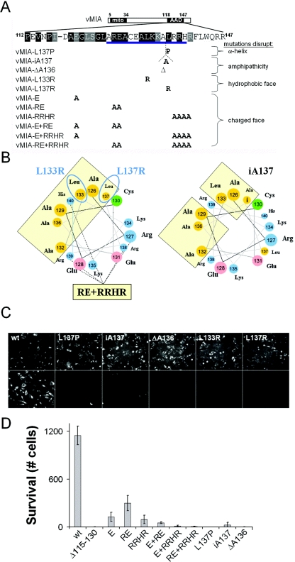
To identify cellular proteins involved in cell death suppression, we screened a human HeLa cell cDNA library for vMIA binding partners through a lexA-based yeast two-hybrid interaction trap (25). Thirty-two clones were found to bind wild-type (wt) vMIA (Table 1). In order to identify interactions likely to be important during vMIA function, we used a bioinformatic analysis to guide directed mutagenesis of the AAD, identifying a conserved α-helix motif from the overlap of Garnier-Robson (21) and Chou-Fasman (11) secondary structure predictions. We evaluated the functional role of this predicted α-helix (aa 126 to 140) by perturbing its amphipathicity, hydrophobicity, and hydrophilicity. We expected the vMIA amphipathic α-helix motif might have an important binding partner, because interactions between pro- and antiapoptotic Bcl-2 family members involve amphipathic α-helical domains (45, 56). Substitution of Pro at aa 137 (L137P) (Fig. 1A), which was predicted to perturb the AAD α-helix (2), abrogated vMIA antiapoptotic function in a standard HeLa cell death assay (23) (Fig. 1C and D). In order to alter AAD amphipathicity without perturbing this α-helical character, we inserted one or two helix-stable Ala residues (37) at aa 137 (iA137, iAA137; Fig. 1A; also data not shown) or deleted one (aa 136) or two (aa 136 to 137) residues (ΔA136, ΔAL136-137; Fig. 1A; also data not shown). These mutations failed to function (Fig. 1C and D), presumably due to shifting charge onto the hydrophobic face and hydrophobicity onto the hydrophilic face (Fig. 1B). Hence, amphipathicity of the AAD appeared critical for vMIA function. Next, we tested the contribution of the hydrophobic and charged faces of the AAD. We substituted the helix-stable basic residue Arg (37) at aa 133 or aa 137 (L133R, L137R) (Fig. 1A) to alter the hydrophobic face (Fig. 1B) and disrupted vMIA function. However, when we altered the hydrophilic face by substituting Ala for charged residues at aa 113, aa 118, aa 120 (E), aa 127 to 128 (RE), aa 131, aa 134 to 135, aa 138 to 141 (RRHR), or aa 146 to 147 (Fig. 1A; also data not shown), each retained the ability to inhibit apoptosis (Fig. 1D; also data not shown). Only when five or six Ala substitutions were combined (E+RRHR or RE+RRHR) (Fig. 1A) was vMIA rendered nonfunctional (Fig. 1B and D). The basic nature of the AAD (pI = 10.79) is conserved in vMIA homologs (32), and any combination of mutations that destroyed this character reduced function. Taken together, mutagenesis revealed that vMIA was critically dependent on an intact α-helical domain or the integrity of its hydrophobic face, suggesting that this might be a site of contact with other proteins.
TABLE 1.
Cellular vMIA interacting partners by determined two-hybrid screen
| No. of hitsa | Gene (abbreviation) | Binding of gene product(s) to vMIA with the following AAD mutation(s):
|
|||
|---|---|---|---|---|---|
| None | E+RRHR | RE+RRHR | L137P | ||
| 1 | ATPase inhibitory factor 1 (ATPIF) | + | + | + | |
| 1 | BC007384—unknown | + | + | + | |
| 1 | cAMP response element modulator 2β (CREM2β) | + | + | + | |
| 2 | Cell division cycle 10 (CDC10) | + | + | + | |
| 1 | Creatine kinase, brain type (CKB) | + | + | + | |
| 2 | E1a-like inhibitor of differentiation 1 (EID1) | + | − | − | − |
| 1 | Endothelial-derived gene 1 (EG1) | + | + | + | |
| 2 | Eukaryotic translation initiation factor 4 gamma, 3 (EIF4G3) | + | + | + | |
| 2 | Ferritin heavy chain 1 (FTH1) | + | + | + | |
| 1 | Ferritin light chain (FTL) | + | + | + | |
| 1 | Galanin (GAL) | + | + | + | |
| 1 | Growth arrest and DNA damage gene 45 (GADD45α) | + | − | − | − |
| 3 | Heterogeneous nuclear ribonucleoprotein A3 (HNRPA3) | + | + | + | |
| 2 | Keratin 17 (KRT17) | + | − | − | − |
| 1 | Kinesin family member 2C (KIF2C) | + | + | + | |
| 1 | Lactate dehydrogenase A (LDHA) | + | + | + | |
| 2 | Nonmetastatic cells 2, protein expressed (NME2) | + | + | + | |
| 1 | Papillary renal cell carcinoma (PRCC) | + | + | + | |
| 1 | Peroxisomal farnesylated protein (PxF) | + | + | + | |
| 1 | Rabaptin 5 (RABPT5) | + | + | + | |
| 1 | Ras-associated protein 20 (RAB20) | + | + | + | |
| 1 | Ribosomal phosphoprotein, large, P1 (RPLP1) | + | + | + | |
| 1 | S100 calcium-binding protein A11 (S100A11) | + | + | + | |
| 1 | S100 calcium-binding protein (S100P) | + | + | + | |
Number of clones resulting for each gene.
FIG. 1.
Structural analysis and function of vMIA AAD. (A) Predicted structure of AAD. (Top) A vMIA protein schematic is with the positions of the mitochondrial targeting domain (mito, aa 5 to 34) and AAD (aa 118 TO 147) indicated. In the expanded region, conserved amino acids (boxed in black when ≥75% identical or in gray when ≥75% similar to primate CMV homologs) are depicted. Mutations within the predicted α-helix (underlined, aa 126 to 140) are indicated below the sequence. These mutations are predicted to disrupt the α-helix's structure by Pro substitution (vMIA-L137P), its amphipathicity by Ala insertion (vMIA-iA137) or deletion (vMIA-ΔA136), its hydrophobic face by Arg substitution (vMIA-L133R, vMIA-L137R), and its charged face by multiple Ala substitutions (vMIA-E, vMIA-RE, vMIA-RRHR, vMIA-E+RE, vMIA-E+RRHR, vMIA-RE+RRHR). (B) Helical wheel models of AAD α-helix. Basic (blue), acidic (pink), hydrophobic (yellow), and polar (green) residues are indicated, with the clusters of hydrophobic residues outlined and shaded light yellow. Positions of Arg substitution point mutations vMIA-L133R and vMIA-L137R (blue ellipses, L133R and L137R) in the hydrophobic face and Ala substitution mutant vMIA-RE+RRHR (dashed lines to filled yellow rectangle; RE+RRHR) are shown at left. The position of the Ala insertion point mutation vMIA-iA137 is displayed at right with the inserted Ala (marked “i”) and predicted hydrophobic face disruption outlined (shaded light yellow). (C) Localization and cell death suppression assay with wt and mutant vMIA expression. HeLa cells transfected with wt vMIA (wt), vMIA-L137P (L137P), vMIA-iA137 (iA137), vMIA-ΔA136 (ΔA136), vMIA-L133R (L133R), or vMIA-L137R (L137R) are shown. Apoptosis was induced 24 h after transfection with cycloheximide control (top row) or anti-Fas Ab and cycloheximide (bottom row) for 18 h. Surviving cells were fixed and stained for c-Myc epitope tag expression by immunofluorescence (magnification, ×16.5). (D) Cell survival after apoptosis induction. HeLa cells transfected with wt vMIA (vMIA), vMIA-Δ115-130 (Δ115-130), vMIA-E (E), vMIA-RE (RE), vMIA-RRHR (RRHR), vMIA-E+RE (E+RE), vMIA-E+RRHR (E+RRHR), vMIA-RE+RRHR (RE+RRHR), vMIA-L137P (L137P), vMIA-iA137 (iA137), or vMIA-ΔA136 (ΔA136) were treated with anti-Fas Ab and cycloheximide as in C. Counts of five fields at ×200 (about 2,000 cells on control wells) were graphed with SD indicated (error bars).
GADD45α-mediated enhancement of vMIA.
We next tested candidate proteins for their ability to bind the specific nonfunctional vMIA AAD mutants vMIA-L137P, vMIA-E+RRHR, and vMIA-RE+RRHR. Only 3 of the 32 candidate proteins failed to bind the nonfunctional AAD mutants: E1a-like inhibitor of differentiation (EID1), growth arrest and DNA damage 45α (GADD45α), and keratin 17 (KRT17) (Table 1). A full-length version of KRT17 did not bind vMIA and so was not pursued. Thus, EID1 and GADD45α each bound specifically to vMIA but not to nonfunctional vMIA mutants.
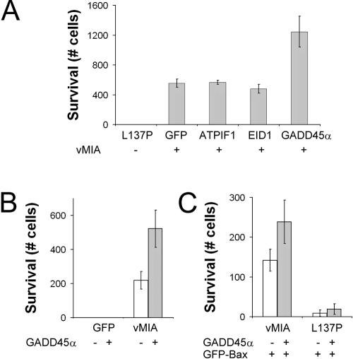
When we investigated whether interaction of these proteins with the AAD had any impact on vMIA function following cotransfection, we found that GADD45α enhanced vMIA suppression of Fas-induced apoptosis by two- to threefold (Fig. 2A). In contrast, neither EID1 nor ATPase inhibitory factor 1, another vMIA binding protein that has been implicated in the regulation of mitochondrial ATPase activity in response to pH stress (46), influenced vMIA function following cotransfection into cells (Fig. 2A). GADD45α failed to influence apoptosis when expressed alone (Fig. 2B) but also enhanced vMIA function when apoptosis was induced with green fluorescent protein (GFP)-tagged Bax (Fig. 2C). The ability of GADD45α to enhance vMIA-mediated cell death suppression from both external (Fas) and internal (Bax) stimuli was consistent with an effect at the mitochondria where vMIA is known to localize rather than at a step upstream of the mitochondria. As expected for the ability to bind GADD45α, enhancement was observed only with wt vMIA and not with the vMIA-L137P mutant (Fig. 2C). Thus, GADD45α enhanced the antiapoptotic activity of vMIA in a pattern that reflected its ability to associate with vMIA.
FIG. 2.
vMIA-interacting proteins and cell death suppression. (A) Screen using vMIA and vMIA-interacting proteins. HeLa cells were transfected with vMIA-L137P (L137P), or wt vMIA together with GFP, ATPase inhibitory factor 1 (ATPIF1), EID1, or GADD45α. Cell death suppression assay was as described in the legend to Fig. 1. (B) Effect of GADD45α on vMIA suppression of extrinsic apoptosis. HeLa cells transfected with GFP or vMIA together with dsRed2 control (white bars) or GADD45α (gray bars) were subjected to Fas-mediated cell death. (C) Effect of GADD45α on vMIA suppression of Bax-mediated cell death. HeLa cells transfected with GFP-Bax along with wt vMIA or vMIA-L137P (L137P) and either dsRed2 control (white bars) or GADD45α (gray bars) were fixed 24 h after transfection. Counts of five fields of GFP-Bax-positive cells were graphed with SD as described in Fig. 1.
GADD45α increases the amount of vMIA in cells.
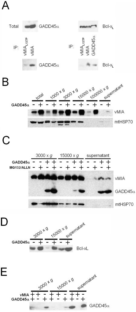
As expected from the results of the two-hybrid interaction screen, GADD45α was found to associate with vMIA in mammalian cells, demonstrated by coimmunoprecipitation with epitope-tagged vMIA but not with the AAD mutant vMIA-L137P (Fig. 3A). Although there were slightly higher amounts of GADD45α in the total lysate of wt vMIA-transfected cells than in vMIA-L137P-cotransfected cells, this did not account for the significantly greater amount of GADD45α found to coimmunoprecipitate with wt vMIA, suggesting that GADD45α enhancement of cell death suppression is mediated by a direct interaction with the AAD of vMIA. Somewhat surprisingly, the cellular mitochondrium-localized cell death suppressor Bcl-xL was also found to coimmunoprecipitate with epitope-tagged GADD45α (Fig. 3A). In addition, Bcl-xL coimmunoprecipitated with wt vMIA but not with vMIA-L137P (Fig. 3A). This result suggests that the cellular proteins Bcl-xL and GADD45α may form a complex into which vMIA infiltrates.
FIG. 3.
Fractionation of vMIA and GADD45α-transfected cells. (A) Immunoprecipitation of Myc-tagged vMIA-L137P, wt vMIA, and GADD45α from 293T cell lysates with anti-c-Myc-conjugated agarose beads and immunoblot detection of coimmunoprecipitated Flag-tagged GADD45α and Bcl-xL after SDS-PAGE. (B) GADD45α effect on vMIA expression and mitochondrial accumulation. Cells transfected with DNA encoding vMIA and GFP or vMIA and GADD45α. Total cell lysates and 1,000 × g, 3,000 × g, 15,000 × g, 100,000 × g, and remaining supernatant fractions were subjected to immunoblot analysis for Myc-tagged vMIA (anti-c-Myc Ab) and mtHSP70 (anti-mtHSP70 Ab). (C) GADD45α effect on mitochondrial accumulation of vMIA. Cells transfected with vMIA and GFP or vMIA and GADD45α were treated 20 h posttransfection for 4 h with control dimethyl sulfoxide (0.1%) or protease inhibitors (20 μM MG132 and 100 μM ALLN). After fractionation, the 3,000 × g and 15,000 × g pellets and remaining supernatant were immunoblotted for Myc-tagged vMIA (anti-c-Myc Ab), Flag-tagged GADD45α (anti-Flag Ab), and mtHSP70 (anti-mtHSP70 Ab). (D) GADD45α effect on mitochondrial levels of Bcl-xL. Cells transfected with Bcl-xL and GFP or Bcl-xL and GADD45α were fractionated into 3,000 × g, 15,000 × g, and remaining supernatant for analysis by immunoblotting for Flag-tagged Bcl-xL (anti-Flag Ab). (E) vMIA effect on mitochondrial accumulation of GADD45α. Cells transfected with vMIA and GADD45α, vMIA and GFP, or GFP and GADD45α were fractionated into 3000 × g, 15,000 × g, and remaining supernatant for analysis by immunoblotting for Flag-tagged GADD45α (anti-Flag Ab).
GADD45α expression increased the total cellular levels of vMIA protein relative to those of the mitochondrial heat shock protein (mtHSP70) control (Fig. 3B). This resulted in increased amounts of vMIA detected in the nuclear (1,000 × g pellet), heavy mitochondrial (3,000 × g pellet), light mitochondrial (15,000 × g pellet), lighter membrane (100,000 × g pellet, including endoplasmic reticulum, lysosomes, and endosomes), and cytosolic (100,000 × g supernatant) cellular fractions (Fig. 3B). This GADD45α-dependent difference was most striking for vMIA associated with light mitochondrial and lighter membrane fractions but did not affect the amount of mitochondria present in each fraction based on the lack of change in mtHSP70 staining (Fig. 3B). The alteration of mitochondrial organization from reticular (predominantly heavy mitochondria) to punctate (predominantly light mitochondria) is one of the consequences of vMIA activity in transfected cells as well as during viral infection (33), although it is not clear how this relates to cell death suppression activity.
GADD45α stabilizes vMIA from degradation.
To determine whether enhancement of vMIA activity was associated with protein stabilization, we evaluated the impact of a 4-h treatment with protease inhibitors MG132 (20 μM) and ALLN (100 μM) on both vMIA and GADD45α protein levels. Treatment with MG132/ALLN increased vMIA protein levels comparably to GADD45α coexpression, whereas the combination of GADD45α coexpression and MG132/ALLN treatment did not further increase the amount of vMIA found in heavy and light mitochondrial (3,000 × g and 15,000 × g pellet) fractions relative to mtHSP70 control (Fig. 3C). Thus, protease inhibition abrogated the relative increase in mitochondrial vMIA associated with GADD45α coexpression, suggesting that GADD45α may have an impact on proteasome-dependent degradation of vMIA, but this area requires further investigation.
In keeping with the coimmunoprecipitation of Bcl-xL with GADD45α (Fig. 3A), Bcl-xL protein levels in heavy and light mitochondrial (3,000 × g and 15,000 × g pellet) as well as lighter membrane (15,000 × g supernatant) fractions were increased by GADD45α coexpression (Fig. 3D). The similarity in effect of GADD45α on both vMIA and Bcl-xL suggests a common role of GADD45α in cell death suppression by either protein and predicts the potential importance of a complex containing all three.
Although much of GADD45α localized to the nucleus in this setting (53), association with heavy and light mitochondrial (3,000 × g and 15,000 × g pellet) and lighter membrane (15,000 × g supernatant) fractions was clearly demonstrated in our analysis of cellular fractions (Fig. 3C and E). In contrast to the increase in vMIA protein levels with GADD45α coexpression, GADD45α protein levels in heavy and light mitochondrial fractions were reduced by coexpression of vMIA (Fig. 3E). This decrease appears to be dependent on protein turnover, since GADD45α protein levels in mitochondrial fractions were increased by a brief MG132/ALLN treatment (Fig. 3C).
GADD45 family members enhance Bcl-xL as well as vMIA activity.
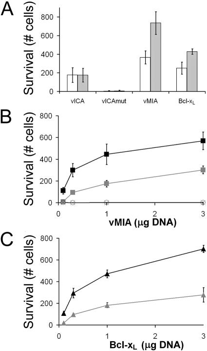
To determine whether GADD45α affected steps of apoptosis upstream of mitochondria, we tested its impact on vICA, the CMV-encoded procaspase 8 inhibitor, and compared this to a nonfunctional vICA mutant (32, 48). Coexpression of GADD45α had no impact on the cell death suppression activity of vICA, although it enhanced the activity of the mitochondrial cell death suppressor Bcl-xL (Fig. 4A). Thus, GADD45α did not broadly alter cell death but appeared to enhance cell death suppression specifically at the mitochondrial checkpoint. GADD45α was able to enhance the activity of Bcl-xL across a range of doses in a pattern similar to that with vMIA (Fig. 4B and C). Thus, despite the lack of sequence homology, vMIA and Bcl-xL were each shown to cooperate with GADD45α in cell death suppression (Fig. 4), coimmunoprecipitate with GADD45α (Fig. 3A), and exhibit increased association with mitochondria following coexpression with GADD45α (Fig. 3B and D).
FIG. 4.
GADD45α influence on mitochondrial cell death suppressors. (A) HeLa cells were transfected with GADD45α (gray bars) or GFP (white bars) together with vICA, vICAmut, vMIA, or Bcl-xL. A cell death suppression assay was performed and graphed with SD as in Fig. 1. (B) HeLa cells were transfected with vMIA and GADD45α (black filled squares), vMIA and GFP (gray filled squares), vMIA-L137P and GADD45α (open circles), and vMIA-L137P and GFP (black crosses), varying the input amount of vMIA or vMIA-L137P plasmid DNA as indicated in the presence of a constant 3.0-μg GADD45α or GFP plasmid DNA. (C) HeLa cells were transfected with Bcl-xL and GADD45α (black triangles) or Bcl-xL and GFP (gray triangles), varying the input amount of Bcl-xL plasmid DNA as indicated in the presence of a constant 3.0 μg GADD45α or GFP plasmid DNA.
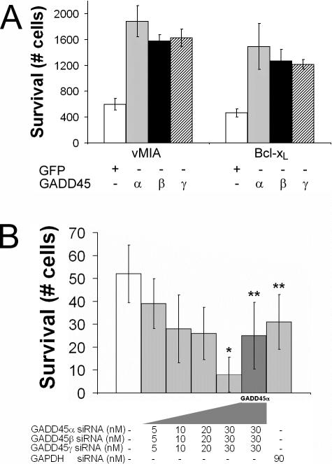
Since GADD45α has 60% amino acid identity and many activities in common with GADD45β and GADD45γ, we tested the other GADD45 family proteins for their effect on vMIA and Bcl-xL activity. Remarkably, each manifested a consistent two- to threefold enhancement of cell death suppression by either vMIA or Bcl-xL (Fig. 5A). Endogenous expression of GADD45 family members depends on a variety of overlapping stimuli (54, 57), and all three are expressed in a wide variety of cell types. Thus, enhancement of mitochondrial inhibitors of apoptosis is a shared characteristic of all three GADD45 family members.
FIG. 5.
GADD45 family members cooperate with mitochondrial cell death suppressors. (A) HeLa cells transfected with plasmids encoding vMIA (left) or Bcl-xL (right) and either GFP (white bars), GADD45α (α, gray bars), GADD45β (β, black bars), or GADD45γ (γ, hatched bars) were subjected to the cell death suppression assay and graphed with SD as in Fig. 1. (B) HeLa cells transfected with plasmids encoding vMIA and indicated concentrations of GADD45α siRNA, GADD45β siRNA, GADD45γ siRNA, or control GAPDH siRNA (light-gray bars) and with plasmid encoding GADD45α (dark-gray bar) were subjected to a cell death suppression assay and graphed with SD as in Fig. 1. A single asterisk indicates a P value of <0.05 (t test) for surviving cell counts relative to the well that did not receive siRNA (white bar). A double asterisk indicates a P value of <0.05 (t test) for surviving cell counts relative to the well that received 30 nM (each) GADD45α siRNA, GADD45β siRNA, and GADD45γ siRNA.
Finally, to determine the role of endogenous GADD45 family proteins in vMIA function, we employed a siRNA-mediated expression knockdown approach, evaluating vMIA cell death suppression activity when GADD45α, GADD45β, and GADD45γ were inhibited. vMIA-dependent cell survival was significantly reduced after cotransfection of increasing amounts of siRNA directed against the three GADD45 family members (Fig. 5B). Maximal specific inhibition was observed with a 30 nM dose of each based on the ability of additional, exogenous GADD45α to restore cell death suppression activity to GAPDH control siRNA-treated levels (Fig. 5B). Thus, not only do GADD45 family members enhance vMIA activity through exogenous expression, but natural GADD45 appears to be required for vMIA activity. In summary, these data demonstrate that the hydrophobic character of the AAD is critical for vMIA cell death suppression, GADD45α binding to vMIA requires an intact AAD, and GADD45 family members both enhance and are required for cell death suppression activity of vMIA.
DISCUSSION
We have shown that vMIA and Bcl-xL exhibit a common interaction with the DNA damage response protein GADD45α and that GADD45 family members play direct roles in increasing the levels and activity of vMIA and Bcl-xL. Our work has revealed a previously unsuspected common component of mitochondrial cell death suppression pathways. This work raises the possibility that GADD45 family members, expressed in response to DNA damage, virus infection, or various types of cell stress, collaborate with all mitochondrial inhibitors in protection from apoptosis. Enhancement of cell death suppression depends on a direct interaction of GADD45α with vMIA via the AAD. GADD45α impedes MG132/ALLN-inhibitable degradation of vMIA. Many of these characteristics extend to Bcl-xL. Bcl-xL contains five amphipathic α-helices centered on two hydrophobic α-helices (36, 45) that may provide an interaction surface with GADD45α analogous to the vMIA AAD amphipathic α-helix. Although structural similarity more than primary sequence homology may dictate function in these regions, more comparative work will need to be completed before this becomes clear.
There have been a few candidate interaction partners implicated in vMIA function. When the activity was first described, the mitochondrial inner membrane protein Ade nucleotide translocase was suggested to be a binding partner (23), but when investigated, this interaction was not associated with vMIA-mediated cell death suppression (4). Bax recently emerged as a candidate binding partner and provided a link with mitochondria as well as with Bcl-2 family proteins (42), but this interaction did not emerge in our studies. The unbiased approach we used relied on assays where specific point mutations in the hydrophobic face of a predicted AAD α-helix known to be essential for vMIA function were employed to qualify binding partners that were identified in a yeast two-hybrid screen. The nonfunctional E128P substitution mutant of vMIA previously characterized (4), as well as other mutants described in Results, all perturb the putative α-helix. E128 itself is not critical for vMIA function, as evidenced by the partial death suppression activity of the helix-stabilizing Ala substitution mutant vMIA-RE: R127A E128A mutations affecting the same region (Fig. 1D). Although a Bcl-2 homology domain 3-like Bax-binding domain was predicted to exist within the AAD (aa 122 to 131) (8), the fact that the vMIA-Δ131-147 deletion mutant (42) does not disrupt this domain and yet is nonfunctional argues that this prediction is not accurate. This prediction is also confounded by the lack of Bax binding to a vMIA peptide representing aa 114 to 141, although aa 114 to 150 bound to Bax (4). Furthermore, vMIA point mutants located outside of the proposed BH3 motif that disrupt the hydrophobic face (L133R and L137R) or amphipathicity (iA137 and ΔA136) of the AAD α-helix completely inactivate vMIA function. Finally, the weak BH domain-like characteristics of aa 122 to aa 131 are not conserved in a fully functional primate CMV vMIA homolog (32), so the appearance of weak similarity in amino acid sequence of this region more likely reflects a requirement for hydrophobic spacing within the α-helix, a structural component shared by the AAD and BH domains. By establishing that vMIA binds GADD45α through its AAD, identifying potentially important interactions by two-hybrid assay, confirming the interaction in mammalian cells, and demonstrating cofractionation of the two proteins with mitochondria, we believe our data have revealed a physiologically significant functional partnership.
Connections between the DNA damage response and apoptosis pathways have been suggested from investigations of the adenovirus E1b 19-kDa and 55-kDa proteins, two proteins that block the cellular proapoptotic response to infection and are believed to act by targeting and binding Bax and p53, respectively (10). The tumor suppressor p53 upregulates expression of several genes, including those encoding Bax and GADD45α, in response to DNA damage (16), and in separate work, p53 has been reported to translocate to mitochondria and activate Bax and Bak (39). GADD45α, GADD45β, and GADD45γ are upregulated as part of the DNA damage response that would be expected to deliver proapoptotic signals to cells via the mitochondria and contribute as well to adenovirus-mediated inhibition of apoptosis. Likewise, the growing number of Bcl-2 homologs identified in gammaherpesviruses (41) may function in similar ways.
GADD45α was originally identified as a gene transcribed in response to DNA damage by UV irradiation (20). GADD45 family members are expressed in response to other genotoxic stress mediators, such as γ-irradiation and methylmethanesulphonate treatment (54, 57), and each family member induces cell cycle arrest at G2/M in a p53-dependent fashion (57). Although initially implicated in growth suppression (61), DNA repair (49, 50), and G2/M cell cycle arrest (57, 60), GADD45 family members have been ascribed seemingly conflicting roles in apoptosis (30, 47) depending on the context. All three GADD45 family members have been reported to induce apoptosis under some settings (52). GADD45β has also been ascribed a role in blocking tumor necrosis factor alpha- or Fas-mediated apoptosis in some cell lines (13, 59). In contrast to these previous analyses, GADD45 family members neither induced nor blocked apoptosis induced by Fas or Bax in the p53-independent HeLa cell assay system used here. We have not observed a change in apoptosis following introduction of any GADD45 family member into HeLa cells and have confirmed cDNA sequence and expression of our constructs. GADD45 family members exhibited potent enhancement of cell death suppression only when coexpressed with vMIA or Bcl-xL. Control cells into which GADD45 family siRNA was introduced did not apoptose but were much less responsive to vMIA-mediated cell death suppression. Thus, the GADD45 family of proteins influences apoptosis in association with mitochondrial cell death suppressors.
The role of GADD45α in vMIA or Bcl-xL function was accompanied by increased association with mitochondrial membranes and stability that could be paralleled by a short treatment with protease inhibitors. Thus, our work is most consistent with a GADD45 family-mediated inhibition of proteasome-dependent degradation of vMIA or Bcl-xL that is itself dependent on GADD45 family turnover (27). Turnover of GADD45α (29) appeared to increase upon association with vMIA. It is possible that mitogen-activated protein kinase signaling pathways may also control proteasome-dependent turnover (14). GADD45 family members have been reported to bind and modulate mitogen-activated protein kinase signaling constituents MEKK4 (52) and MKK7 (38). Thus, GADD45 family members may control additional activities beyond vMIA or Bcl-xL interaction.
GADD45 family and Bcl-2 family members play important roles for many herpesviruses, suggesting a relationship between DNA damage response pathways and intrinsic apoptosis induced by viral infection. Consistent with a role for GADD45 family members in increasing the efficacy of cell death suppressors, cells lacking GADD45α show increased sensitivity to proapoptotic stimuli (49, 50). In addition, GADD45β is one of a few host mRNAs that is not degraded but becomes upregulated specifically during herpes simplex virus 1 infection (17, 18) and may be expected to enhance Bcl-xL through the mechanism we have identified here. CMV and herpes simplex virus 1 may both exploit this common property to delay cell death and facilitate increased viral replication. Given that bona fide Bcl-2 homologs are found in all gammaherpesviruses (41), the relationship between DNA damage response pathways and Bcl-2 family members may well be exploited very broadly across the herpesvirus family.
Acknowledgments
We thank Victor Goldmacher for information and reagents that facilitated our investigation, A. Louise McCormick for advice and critical review of the manuscript, and Yin Dong for cell culture assistance.
This work was supported by PHS research grant AI20211, Medical Scientist Training grant GM07365, and NRSA F30 grant NS051109.
REFERENCES
- 1.Agatep, R., R. D. Kirkpatrick, D. L. Parchaliuk, R. A. Woods, and R. D. Gietz. 1998. Transformation of Saccharomyces cerevisiae by the lithium acetate/single-stranded carrier DNA/polyethylene glycol (LiAc/ss-DNA/PEG) protocol. Tech. Tips Online [Online.] http://www.bio.com/protocolstools/protocol.jhtml?id=p1657.
- 2.Altmann, K. H., J. Wojcik, M. Vasquez, and H. A. Scheraga. 1990. Helix-coil stability constants for the naturally occurring amino acids in water. XXIII. Proline parameters from random poly(hydroxybutylglutamine-co-L-proline). Biopolymers 30:107-120. [DOI] [PubMed] [Google Scholar]
- 3.Andreau, K., M. Castedo, J. L. Perfettini, T. Roumier, E. Pichart, S. Souquere, S. Vivet, N. Larochette, and G. Kroemer. 2004. Preapoptotic chromatin condensation upstream of the mitochondrial checkpoint. J. Biol. Chem. 279:55937-55945. [DOI] [PubMed] [Google Scholar]
- 4.Arnoult, D., L. M. Bartle, A. Skaletskaya, D. Poncet, N. Zamzami, P. U. Park, J. Sharpe, R. J. Youle, and V. S. Goldmacher. 2004. Cytomegalovirus cell death suppressor vMIA blocks Bax- but not Bak-mediated apoptosis by binding and sequestering Bax at mitochondria. Proc. Natl. Acad. Sci. USA 101:7988-7993. [DOI] [PMC free article] [PubMed] [Google Scholar]
- 5.Barnhart, B. C., E. C. Alappat, and M. E. Peter. 2003. The CD95 type I/type II model. Semin. Immunol. 15:185-193. [DOI] [PubMed] [Google Scholar]
- 6.Barry, M., and G. McFadden. 1998. Apoptosis regulators from DNA viruses. Curr. Opin. Immunol. 10:422-430. [DOI] [PubMed] [Google Scholar]
- 7.Belzacq, A. S., C. El Hamel, H. L. Vieira, I. Cohen, D. Haouzi, D. Metivier, P. Marchetti, C. Brenner, and G. Kroemer. 2001. Adenine nucleotide translocator mediates the mitochondrial membrane permeabilization induced by lonidamine, arsenite and CD437. Oncogene 20:7579-7587. [DOI] [PubMed] [Google Scholar]
- 8.Boya, P., A. L. Pauleau, D. Poncet, R. A. Gonzalez-Polo, N. Zamzami, and G. Kroemer. 2004. Viral proteins targeting mitochondria: controlling cell death. Biochim. Biophys. Acta 1659:178-189. [DOI] [PubMed] [Google Scholar]
- 9.Brocchieri, L., T. N. Kledal, S. Karlin, and E. S. Mocarski. 2005. Predicting coding potential from genome sequence: application to betaherpesviruses infecting rats and mice. J. Virol. 79:7570-7596. [DOI] [PMC free article] [PubMed] [Google Scholar]
- 10.Burgert, H. G., Z. Ruzsics, S. Obermeier, A. Hilgendorf, M. Windheim, and A. Elsing. 2002. Subversion of host defense mechanisms by adenoviruses. Curr. Top. Microbiol. Immunol. 269:273-318. [DOI] [PubMed] [Google Scholar]
- 11.Chou, P. Y., and G. D. Fasman. 1978. Empirical predictions of protein conformation. Annu. Rev. Biochem. 47:251-276. [DOI] [PubMed] [Google Scholar]
- 12.Cuconati, A., and E. White. 2002. Viral homologs of BCL-2: role of apoptosis in the regulation of virus infection. Genes Dev. 16:2465-2478. [DOI] [PubMed] [Google Scholar]
- 13.De Smaele, E., F. Zazzeroni, S. Papa, D. U. Nguyen, R. Jin, J. Jones, R. Cong, and G. Franzoso. 2001. Induction of gadd45beta by NF-kappaB downregulates pro-apoptotic JNK signalling. Nature 414:308-313. [DOI] [PubMed] [Google Scholar]
- 14.Dimmeler, S., K. Breitschopf, J. Haendeler, and A. M. Zeiher. 1999. Dephosphorylation targets Bcl-2 for ubiquitin-dependent degradation: a link between the apoptosome and the proteasome pathway. J. Exp. Med. 189:1815-1822. [DOI] [PMC free article] [PubMed] [Google Scholar]
- 15.Dunn, W., C. Chou, H. Li, R. Hai, D. Patterson, V. Stolc, H. Zhu, and F. Liu. 2003. Functional profiling of a human cytomegalovirus genome. Proc. Natl. Acad. Sci. USA 100:14223-14228. [DOI] [PMC free article] [PubMed] [Google Scholar]
- 16.el-Deiry, W. S. 1998. Regulation of p53 downstream genes. Semin. Cancer Biol. 8:345-357. [DOI] [PubMed] [Google Scholar]
- 17.Esclatine, A., B. Taddeo, L. Evans, and B. Roizman. 2004. The herpes simplex virus 1 UL41 gene-dependent destabilization of cellular RNAs is selective and may be sequence-specific. Proc. Natl. Acad. Sci. USA 101:3603-3608. [DOI] [PMC free article] [PubMed] [Google Scholar]
- 18.Esclatine, A., B. Taddeo, and B. Roizman. 2004. The UL41 protein of herpes simplex virus mediates selective stabilization or degradation of cellular mRNAs. Proc. Natl. Acad. Sci. USA 101:18165-18170. [DOI] [PMC free article] [PubMed] [Google Scholar]
- 19.Everett, H., and G. McFadden. 2002. Poxviruses and apoptosis: a time to die. Curr. Opin. Microbiol. 5:395-402. [DOI] [PubMed] [Google Scholar]
- 20.Fornace, A. J., Jr., D. W. Nebert, M. C. Hollander, J. D. Luethy, M. Papathanasiou, J. Fargnoli, and N. J. Holbrook. 1989. Mammalian genes coordinately regulated by growth arrest signals and DNA-damaging agents. Mol. Cell. Biol. 9:4196-4203. [DOI] [PMC free article] [PubMed] [Google Scholar]
- 21.Garnier, J., D. J. Osguthorpe, and B. Robson. 1978. Analysis of the accuracy and implications of simple methods for predicting the secondary structure of globular proteins. J. Mol. Biol. 120:97-120. [DOI] [PubMed] [Google Scholar]
- 22.Goldmacher, V. S. 2005. Cell death suppression by cytomegaloviruses. Apoptosis 10:251-265. [DOI] [PubMed] [Google Scholar]
- 23.Goldmacher, V. S., L. M. Bartle, A. Skaletskaya, C. A. Dionne, N. L. Kedersha, C. A. Vater, J. W. Han, R. J. Lutz, S. Watanabe, E. D. Cahir McFarland, E. D. Kieff, E. S. Mocarski, and T. Chittenden. 1999. A cytomegalovirus-encoded mitochondria-localized inhibitor of apoptosis structurally unrelated to Bcl-2. Proc. Natl. Acad. Sci. USA. 96:12536-12541. [DOI] [PMC free article] [PubMed] [Google Scholar]
- 24.Golemis, E. A., J. Gyuris, and R. Brent. 1994. Interaction trap/two-hybrid system to identify interacting proteins, p. 1-17. In F. M. Ausubel, et al. (ed.), Current protocols in molecular biology, vol. 13.14. John Wiley & Sons, Inc., New York, N.Y. [Google Scholar]
- 25.Gyuris, J., E. Golemis, H. Chertkov, and R. Brent. 1993. Cdi1, a human G1 and S phase protein phosphatase that associates with Cdk2. Cell 75:791-803. [DOI] [PubMed] [Google Scholar]
- 26.Hayajneh, W. A., A. M. Colberg-Poley, A. Skaletskaya, L. M. Bartle, M. M. Lesperance, D. G. Contopoulos-Ioannidis, N. L. Kedersha, and V. S. Goldmacher. 2001. The sequence and antiapoptotic functional domains of the human cytomegalovirus UL37 exon 1 immediate early protein are conserved in multiple primary strains. Virology 279:233-240. [DOI] [PubMed] [Google Scholar]
- 27.Jackson, P. K., A. G. Eldridge, E. Freed, L. Furstenthal, J. Y. Hsu, B. K. Kaiser, and J. D. Reimann. 2000. The lore of the RINGs: substrate recognition and catalysis by ubiquitin ligases. Trends Cell Biol. 10:429-439. [DOI] [PubMed] [Google Scholar]
- 28.Jenkins, D. E., C. L. Martens, and E. S. Mocarski. 1994. Human cytomegalovirus late protein encoded by ie2: a trans-activator as well as a repressor of gene expression. J. Gen. Virol. 75:2337-2348. [DOI] [PubMed] [Google Scholar]
- 29.Leung, C. H., W. Lam, W. J. Zhuang, N. S. Wong, M. S. Yang, and W. F. Fong. 2001. PKCdelta-dependent deubiquitination and stabilization of Gadd45 in A431 cells overexposed to EGF. Biochem. Biophys. Res. Commun. 285:283-288. [DOI] [PubMed] [Google Scholar]
- 30.Liebermann, D. A., and B. Hoffman. 2002. Myeloid differentiation (MyD)/growth arrest DNA damage (GADD) genes in tumor suppression, immunity and inflammation. Leukemia 16:527-541. [DOI] [PubMed] [Google Scholar]
- 31.McCormick, A. L., C. D. Meiering, G. B. Smith, and E. S. Mocarski. 2005. Mitochondrial cell death suppressors carried by human and murine cytomegalovirus confer resistance to proteasome inhibitor-induced apoptosis. J. Virol. 79:12205-12217. [DOI] [PMC free article] [PubMed] [Google Scholar]
- 32.McCormick, A. L., A. Skaletskaya, P. A. Barry, E. S. Mocarski, and V. S. Goldmacher. 2003. Differential function and expression of the viral inhibitor of caspase 8-induced apoptosis (vICA) and the viral mitochondria-localized inhibitor of apoptosis (vMIA) cell death suppressors conserved in primate and rodent cytomegaloviruses. Virology 316:221-233. [DOI] [PubMed] [Google Scholar]
- 33.McCormick, A. L., V. L. Smith, D. Chow, and E. S. Mocarski. 2003. Disruption of mitochondrial networks by the human cytomegalovirus UL37 gene product viral mitochondrion-localized inhibitor of apoptosis. J. Virol. 77:631-641. [DOI] [PMC free article] [PubMed] [Google Scholar]
- 34.Meier, P., A. Finch, and G. Evan. 2000. Apoptosis in development. Nature 407:796-801. [DOI] [PubMed] [Google Scholar]
- 35.Mocarski, E. S., Jr. 2002. Immunomodulation by cytomegaloviruses: manipulative strategies beyond evasion. Trends Microbiol. 10:332-339. [DOI] [PubMed] [Google Scholar]
- 36.Muchmore, S. W., M. Sattler, H. Liang, R. P. Meadows, J. E. Harlan, H. S. Yoon, D. Nettesheim, B. S. Chang, C. B. Thompson, S. L. Wong, S. L. Ng, and S. W. Fesik. 1996. X-ray and NMR structure of human Bcl-xL, an inhibitor of programmed cell death. Nature 381:335-341. [DOI] [PubMed] [Google Scholar]
- 37.O'Neil, K. T., and W. F. DeGrado. 1990. A thermodynamic scale for the helix-forming tendencies of the commonly occurring amino acids. Science 250:646-651. [DOI] [PubMed] [Google Scholar]
- 38.Papa, S., F. Zazzeroni, C. Bubici, S. Jayawardena, K. Alvarez, S. Matsuda, D. U. Nguyen, C. G. Pham, A. H. Nelsbach, T. Melis, E. D. Smaele, W. J. Tang, L. D'Adamio, and G. Franzoso. 2004. Gadd45 beta mediates the NF-kappa B suppression of JNK signalling by targeting MKK7/JNKK2. Nat. Cell Biol. 6:146-153. [DOI] [PubMed] [Google Scholar]
- 39.Perfettini, J. L., R. T. Kroemer, and G. Kroemer. 2004. Fatal liaisons of p53 with Bax and Bak. Nat. Cell Biol. 6:386-388. [DOI] [PubMed] [Google Scholar]
- 40.Petros, A. M., E. T. Olejniczak, and S. W. Fesik. 2004. Structural biology of the Bcl-2 family of proteins. Biochim. Biophys. Acta 1644:83-94. [DOI] [PubMed] [Google Scholar]
- 41.Polster, B. M., J. Pevsner, and J. M. Hardwick. 2004. Viral Bcl-2 homologs and their role in virus replication and associated diseases. Biochim. Biophys. Acta 1644:211-227. [DOI] [PubMed] [Google Scholar]
- 42.Poncet, D., N. Larochette, A. L. Pauleau, P. Boya, A. A. Jalil, P. F. Cartron, F. Vallette, C. Schnebelen, L. M. Bartle, A. Skaletskaya, D. Boutolleau, J. C. Martinou, V. S. Goldmacher, G. Kroemer, and N. Zamzami. 2004. An anti-apoptotic viral protein that recruits Bax to mitochondria. J. Biol. Chem. 279:22605-22614. [DOI] [PubMed] [Google Scholar]
- 43.Reboredo, M., R. F. Greaves, and G. Hahn. 2004. Human cytomegalovirus proteins encoded by UL37 exon 1 protect infected fibroblasts against virus-induced apoptosis and are required for efficient virus replication. J. Gen. Virol. 85:3555-3567. [DOI] [PubMed] [Google Scholar]
- 44.Reinhardt, J., G. B. Smith, C. T. Himmelheber, J. Azizkhan-Clifford, and E. S. Mocarski. 2005. The carboxyl-terminal region of human cytomegalovirus IE1491aa contains an acidic domain that plays a regulatory role and a chromatin-tethering domain that is dispensable during viral replication. J. Virol. 79:225-233. [DOI] [PMC free article] [PubMed] [Google Scholar]
- 45.Sattler, M., H. Liang, D. Nettesheim, R. P. Meadows, J. E. Harlan, M. Eberstadt, H. S. Yoon, S. B. Shuker, B. S. Chang, A. J. Minn, C. B. Thompson, and S. W. Fesik. 1997. Structure of Bcl-xL-Bak peptide complex: recognition between regulators of apoptosis. Science 275:983-986. [DOI] [PubMed] [Google Scholar]
- 46.Schwerzmann, K., and P. L. Pedersen. 1986. Regulation of the mitochondrial ATP synthase/ATPase complex. Arch. Biochem. Biophys. 250:1-18. [DOI] [PubMed] [Google Scholar]
- 47.Sheikh, M. S., M. C. Hollander, and A. J. Fornance, Jr. 2000. Role of Gadd45 in apoptosis. Biochem. Pharmacol. 59:43-45. [DOI] [PubMed] [Google Scholar]
- 48.Skaletskaya, A., L. M. Bartle, T. Chittenden, A. L. McCormick, E. S. Mocarski, and V. S. Goldmacher. 2001. A cytomegalovirus-encoded inhibitor of apoptosis that suppresses caspase-8 activation. Proc. Natl. Acad. Sci. USA 98:7829-7834. [DOI] [PMC free article] [PubMed] [Google Scholar]
- 49.Smith, M. L., J. M. Ford, M. C. Hollander, R. A. Bortnick, S. A. Amundson, Y. R. Seo, C. X. Deng, P. C. Hanawalt, and A. J. Fornace, Jr. 2000. p53-mediated DNA repair responses to UV radiation: studies of mouse cells lacking p53, p21, and/or gadd45 genes. Mol. Cell. Biol. 20:3705-3714. [DOI] [PMC free article] [PubMed] [Google Scholar]
- 50.Smith, M. L., H. U. Kontny, Q. Zhan, A. Sreenath, P. M. O'Connor, and A. J. Fornace, Jr. 1996. Antisense GADD45 expression results in decreased DNA repair and sensitizes cells to u.v.-irradiation or cisplatin. Oncogene 13:2255-2263. [PubMed] [Google Scholar]
- 51.Strasser, A., and P. Bouillet. 2003. The control of apoptosis in lymphocyte selection. Immunol. Rev. 193:82-92. [DOI] [PubMed] [Google Scholar]
- 52.Takekawa, M., and H. Saito. 1998. A family of stress-inducible GADD45-like proteins mediate activation of the stress-responsive MTK1/MEKK4 MAPKKK. Cell 95:521-530. [DOI] [PubMed] [Google Scholar]
- 53.Vairapandi, M., A. G. Balliet, A. J. Fornace, Jr., B. Hoffman, and D. A. Liebermann. 1996. The differentiation primary response gene MyD118, related to GADD45, encodes for a nuclear protein which interacts with PCNA and p21WAF1/CIP1. Oncogene 12:2579-2594. [PubMed] [Google Scholar]
- 54.Vairapandi, M., A. G. Balliet, B. Hoffman, and D. A. Liebermann. 2002. GADD45b and GADD45g are cdc2/cyclinB1 kinase inhibitors with a role in S and G2/M cell cycle checkpoints induced by genotoxic stress. J. Cell Physiol. 192:327-338. [DOI] [PubMed] [Google Scholar]
- 55.Vieira, H. L., A. S. Belzacq, D. Haouzi, F. Bernassola, I. Cohen, E. Jacotot, K. F. Ferri, C. El Hamel, L. M. Bartle, G. Melino, C. Brenner, V. Goldmacher, and G. Kroemer. 2001. The adenine nucleotide translocator: a target of nitric oxide, peroxynitrite, and 4-hydroxynonenal. Oncogene 20:4305-4316. [DOI] [PubMed] [Google Scholar]
- 56.Wang, K., A. Gross, G. Waksman, and S. J. Korsmeyer. 1998. Mutagenesis of the BH3 domain of BAX identifies residues critical for dimerization and killing. Mol. Cell. Biol. 18:6083-6089. [DOI] [PMC free article] [PubMed] [Google Scholar]
- 57.Wang, X. W., Q. Zhan, J. D. Coursen, M. A. Khan, H. U. Kontny, L. Yu, M. C. Hollander, P. M. O'Connor, A. J. Fornace, Jr., and C. C. Harris. 1999. GADD45 induction of a G2/M cell cycle checkpoint. Proc. Natl. Acad. Sci. USA 96:3706-3711. [DOI] [PMC free article] [PubMed] [Google Scholar]
- 58.Yu, D., M. C. Silva, and T. Shenk. 2003. Functional map of human cytomegalovirus AD169 defined by global mutational analysis. Proc. Natl. Acad. Sci. USA 100:12396-12401. [DOI] [PMC free article] [PubMed] [Google Scholar]
- 59.Zazzeroni, F., S. Papa, A. Algeciras-Schimnich, K. Alvarez, T. Melis, C. Bubici, N. Majewski, N. Hay, E. De Smaele, M. E. Peter, and G. Franzoso. 2003. Gadd45 beta mediates the protective effects of CD40 costimulation against Fas-induced apoptosis. Blood 102:3270-3279. [DOI] [PubMed] [Google Scholar]
- 60.Zhan, Q., M. J. Antinore, X. W. Wang, F. Carrier, M. L. Smith, C. C. Harris, and A. J. Fornace, Jr. 1999. Association with Cdc2 and inhibition of Cdc2/Cyclin B1 kinase activity by the p53-regulated protein Gadd45. Oncogene 18:2892-2900. [DOI] [PubMed] [Google Scholar]
- 61.Zhan, Q., K. A. Lord, I. Alamo, Jr., M. C. Hollander, F. Carrier, D. Ron, K. W. Kohn, B. Hoffman, D. A. Liebermann, and A. J. Fornace, Jr. 1994. The gadd and MyD genes define a novel set of mammalian genes encoding acidic proteins that synergistically suppress cell growth. Mol. Cell. Biol. 14:2361-2371. [DOI] [PMC free article] [PubMed] [Google Scholar]


