Abstract
Listeria monocytogenes has the ability to form biofilms on food-processing surfaces, potentially leading to food product contamination. The objective of this research was to standardize a polyvinyl chloride (PVC) microtiter plate assay to compare the ability of L. monocytogenes strains to form biofilms. A total of 31 coded L. monocytogenes strains were grown in defined medium (modified Welshimer's broth) at 32°C for 20 and 40 h in PVC microtiter plate wells. Biofilm formation was indirectly assessed by staining with 1% crystal violet and measuring crystal violet absorbance, using destaining solution. Cellular growth rates and final cell densities did not correlate with biofilm formation, indicating that differences in biofilm formation under the same environmental conditions were not due to growth rate differences. The mean biofilm production of lineage I strains was significantly greater than that observed for lineage II and lineage III strains. The results from the standardized microtiter plate biofilm assay were also compared to biofilm formation on PVC and stainless steel as assayed by quantitative epifluorescence microscopy. Results showed similar trends for the microscopic and microtiter plate assays, indicating that the PVC microtiter plate assay can be used as a rapid, simple method to screen for differences in biofilm production between strains or growth conditions prior to performing labor-intensive microscopic analyses.
Listeria monocytogenes is a gram-positive food-borne pathogen, widely distributed in the environment and commonly resistant to environmental stress. It is associated with plant material, alive or dead, water, and soil (20). This organism causes severe nonenteric disease (meningitis, septicemia) in individuals that are immunocompromised and abortion in pregnant women (9). Listeriosis is a rare food-borne disease, despite the fact that exposure to L. monocytogenes is probably very common due to its wide distribution in the environment and presence in the food industry (9). Reasons for high exposure rates to L. monocytogenes and a low prevalence of listeriosis may be due to differences in L. monocytogenes virulence properties and existence of vulnerable groups within human populations (20, 24). Rasmussen et al. (24) defined three genetic lineages of L. monocytogenes based on the sequences of the virulence genes for listeriolysin, invasion-associated protein, and flagellin, while Wiedmann et al. (31) separated L. monocytogenes into three distinct lineages based on ribotype patterns and virulence gene alleles and correlated them to pathogenic potential in L. monocytogenes. The clinical history of L. monocytogenes strains showed evidence for differences in pathogenic potential among the three lineages. Lineage I contains a majority of strains isolated from human listeriosis cases, lineage II contains both human and animal isolates (but no isolates from food-borne epidemics), and human isolates are rarely typed to lineage III (19, 31). Therefore, Wiedmann et al. (31) hypothesized that strains of lineage I and III may be particularly virulent for humans and animals, respectively.
L. monocytogenes has the ability to form biofilms on food-processing surfaces (4, 12). Biofilms are microbial communities that exist on abiotic and sometimes biotic surfaces (22). Research indicates that biofilms can exist as a mass of microcolonies in a single layer or with vertical and horizontal channels allowing liquid flow and dispersion of nutrients and waste components (1). These biofilms could serve as a source of product contamination (7) and may be reservoirs for pathogenic or spoilage bacteria (28). It is thought that L. monocytogenes growing within mixed microflora biofilms in food-processing environments are the major source of contamination (29). Once established, biofilms appear to have greater resistance to antimicrobial agents than do planktonic organisms (5, 10, 18, 25), and this has been shown to be true with L. monocytogenes (4, 11). Biofilms can be difficult to control since they can form where water is plentiful and cleaning is not performed properly. The common sites for L. monocytogenes isolation from processing plants are filling or packaging equipment, floor drains, walls, cooling pipes, conveyors, collators used for assembling product for packaging, racks for transporting products, hand tools or gloves, and freezers (29). Lineage I and lineage II strains have been isolated from the food-processing plants; however, lineage III strains have not yet been isolated from the food-processing environment (19).
In order to enable easier study of bacterial attachment and colonization, a variety of experimental direct and indirect observation methods have been developed. Standard plate counts, roll techniques, and sonication are indirect methods that first detach the microorganisms from the surface and then count them. Other indirect methods (radiolabeled bacteria, enzyme-linked immunosorbent assay, biologic assays, stained bacterial films, and microtiter plate procedures) estimate the number of attached organisms in situ by measuring some attribute for the attached organism (1). Some researchers have found standard plate count methods give an inaccurate estimate of the number of microorganisms in the biofilm and therefore cannot be used in quantitative measurements (22). However, other researchers have found surface scraping to remove up to 97% of the cells attached to stainless steel coupons (13), and vortexing removes up to 99% of cells attached to silicone tubing (2). According to Lindsay and von Holy (16), sonication, vortexing, and shaking with beads were equivalent methods, based on bacterial counts.
Methods involving direct observation, including microscopy techniques (light, laser-scanning confocal, transmission electron, and scanning electron microscopy), can observe microbial colonization directly. Epifluorescence microscopy data can lead to underestimates of biofilm levels, since thickness is not measured, and to overestimates of the areas covered by cells, since some extracellular polymer is stained (4). Despite this problem, direct microscopy is important for obtaining reliable results when studying the bacteria attached on surfaces (15). For instance, scanning electron microscopy indicated that sonication and vortexing removed only the top layers of biofilm-bound cells, leaving an extracellular polymeric substances residue and attached intact lower-lying cells (16).
A microtiter plate procedure is an indirect method for estimation of bacteria in situ and can be modified for various biofilm formation assays (1). O'Toole and Kolter (21) used this assay with Pseudomonas fluorescens to evaluate the impact of various growth conditions and environmental signals and as a genetic screen for biofilm-deficient mutants (21).
The objective of the present study was to modify the PVC microtiter plate assay to compare the abilities of L. monocytogenes strains to form biofilms. In addition, the PVC microtiter plate assay was compared to microscopic assessment of L. monocytogenes adhesion to PVC and stainless steel.
MATERIALS AND METHODS
Culture preparation.
Thirty-one coded L. monocytogenes strains (Table 1) were obtained from the Cornell University Listeria strains collection. All isolates had previously been characterized by ribotyping using the Qualicon-DuPont Riboprinter and for allelic variation in actA and hly (19). When received in the laboratory at the University of Massachusetts, these strains were randomly assigned numbers LM7 to LM37 (Table 1). Stock cultures were stored at −75°C in Trypticase soy broth-0.6% yeast extract (TSBYE) and 5% glycerol. Working cultures were maintained on Trypticase soy agar (TSA) (Difco, Detroit, Mich.) slants at 4°C for 30 days. Prior to each experiment, a loopful of culture was grown in 10 ml of TSBYE with incubation at 32°C for 18 h.
TABLE 1.
L. monocytogenes strains
| L. monocytogenes lab designation no. | CU isolation no.a | Lineage | Previous isolate designation(s) | Persistenceb | Source of informationc |
|---|---|---|---|---|---|
| LM7 | DD6821 | III | DD6821 | ||
| LM8 | DD3823 | III | DD3823 | ||
| LM9 | DD6824 | III | DD6824 | ||
| LM10 | FSL-N1-304 | II | P | Fish processing plant (19) | |
| LM11 | FSL-N1-417 | I | NP | Fish processing plant (19) | |
| LM12 | FSL-N1-250 | II | NP | Fish processing plant (19) | |
| LM13 | FSL-N1-025 | I | NP | Fish processing plant (19) | |
| LM14 | FSL-N1-005 | II | NP | Fish processing plant (19) | |
| LM15 | FSL-N1-332 | I | P | Fish processing plant (19) | |
| LM16 | FSL-N1-334 | II | P | Fish processing plant (19) | |
| LM17 | FSL-N1-346 | II | P | Fish processing plant (19) | |
| LM18 | FSL-N1-029 | I | NP | Fish processing plant (19) | |
| LM19 | FSL-N1-048 | I | P | Fish processing plant (19) | |
| LM20 | FSL-J1-158 | III | Animal, goat | ||
| LM21 | FSL-J1-225 | I | Scott A | Human epidemic, Mass., 1993 | |
| LM22 | FSL-J1-023 | III | |||
| LM23 | FSL-J1-031 | III | LM36 | Human sporadic case, NYSDOH | |
| LM24 | FSL-J1-177 | I | Human sporadic case, NYSDOH | ||
| LM25 | FSL-J1-110 | I | TS29/F2365 | Human epidemic, Los Angeles, 1985 | |
| LM26 | FSL-J1-168 | III | Human sporadic case, NYSDOH | ||
| LM27 | FSL-C1-109 | I | Human epidemic, United States, 1998 | ||
| LM28 | FSL-C1-056 | II | Human sporadic case, NYSDOH | ||
| LM29 | FSL-C1-122 | I | Human sporadic case, NYSDOH | ||
| LM30 | FSL-J2-066 | II | Animal, sheep | ||
| LM31 | FSL-J2-020 | I | Animal, cow | ||
| LM32 | FSL-J2-064 | I | Animal, cow | ||
| LM33 | FSL-J2-063 | II | Animal, sheep | ||
| LM34 | FSL-J2-054 | II | CU-BR1/93 | Animal, sheep | |
| LM35 | FSL-J2-031 | II | |||
| LM36 | FSL-J2-035 | I | CU-BR9/93 | Animal, goat | |
| LM37 | FSL-M1-004 | II | Human sporadic case, NYSDOH |
Strains were obtained from M. Wiedmann, Cornell University (CU).
P, persistent strain; NP, nonpersistent strain.
NYSDOH, New York State Department of Health.
Motility test.
Modified Welshimer's agar (MWA) motility tubes (10 ml of MWA) were prepared by mixing equal volumes of autoclaved 1% agar solution (Bacto agar; Difco Laboratories) and tempered at 50°C with 2× MW broth (MWB) (23). The final agar concentration was 0.5% in 1× MWB. Each L. monocytogenes coded strain was inoculated by needle stab into a motility MWA tube and incubated at 32°C. Inoculated slants were observed at 20 and 40 h. Tubes with growth diffusing from the center stab were considered positive for motility.
Microtiter plate biofilm production assay.
Each L. monocytogenes coded strain was grown in 10 ml of rich undefined medium, TSBYE, at 32°C overnight. Biofilm production assays were performed with a minimal defined media, MWB (23), using glucose as the sole carbon source. Overnight cultures in TSBYE were transferred (0.1 ml) to 10 ml of MWB and vortexed. After vortexing, 100-μl volumes were transferred into eight PVC microtiter plate (Becton Dickinson Labware, Franklin Lakes, N.J.) wells per strain, previously rinsed with 70% ethanol and air dried. Plates were made in duplicate, incubated, and covered at 32°C for 20 and 40 h. Each plate included eight wells of MWB without L. monocytogenes as control wells.
The cell turbidity was monitored using a microtiter plate reader (Bio-Rad, Richmond, Calif.), at an optical density at 595 nm (OD595), and were recorded at different time intervals. The first set of plates was used for biofilm formation measurement after 20 h, and the duplicate plate was used to determine the 40-h biofilm formation. The average OD from the control wells was subtracted from the OD of all test wells. After a 20- or 40-h incubation period, medium was removed from wells and microtiter plate wells were washed five times with sterile distilled water to remove loosely associated bacteria. Plates were air dried for 45 min and each well was stained with 150 μl of 1% crystal violet solution in water for 45 min. After staining, plates were washed with sterile distilled water five times. At this point, biofilms were visible as purple rings formed on the side of each well (Fig. 3). The quantitative analysis of biofilm production was performed by adding 200 μl of 95% ethanol to destain the wells. One hundred microliters from each well was transferred to a new microtiter plate and the level (OD) of the crystal violet present in the destaining solution was measured at 595 nm.
FIG. 3.
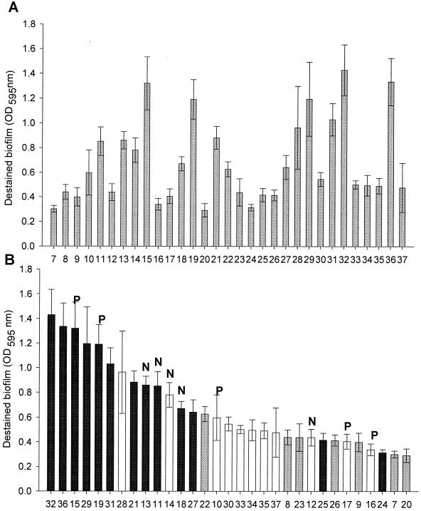
(A) Destained biofilm (measured at OD595) of coded L. monocytogenes strains incubated at 32°C for 40 h in MWB. (B) Three distinct lineages of decoded L. monocytogenes strains, sorted in descending manner. Lineage I strains are represented in black, lineage II is in white, and lineage III is in gray. Bars marked with letters were originally isolated from smoked fish processing plants. P represents persistently isolated strains, while N represents nonpersistent isolates.
The microtiter plate biofilm assay was performed three times for all L. monocytogenes strains, and the averages and standard deviations were calculated for all repetitions of the experiment. Upon completion of all microtiter plate and microscopic assays, all L. monocytogenes strains were decoded (Table 1).
Microscopic biofilm formation assay.
A total of eight coded L. monocytogenes strains were selected for microscopic biofilm formation analysis. The assay used was a modification of the methods described by Jeong and Frank (13) and Blackman and Frank (4). The surfaces used for attachment were stainless steel (type 304, no. 2B), and PVC slide dimensions were 5 by 1.2 cm. Stainless steel slides were cleaned as described previously (13), placed in 12- by 1.5-cm tubes, and autoclaved for 15 min at 15 lb/in2. PVC slides were soaked for 10 min in 70% ethanol, aseptically washed with sterile double-distilled water, and placed in sterile 12- by 1.5-cm tubes. Ten milliliters of MWB was placed into each tube (sufficient to completely immerse or cover the stainless steel or PVC slides) and inoculated with 0.1 ml of L. monocytogenes overnight culture grown in TSBYE. Tubes were incubated at 32°C for 20 and 40 h, allowing biofilm development. After incubation, slides were removed from the growth medium, washed with distilled water to remove any loosely attached cells, and fixed with 95% ethanol for 45 s.
Epifluorescence microscopy.
Slides with fixed cells were stained in Hoechst 33258 DNA-binding fluorescent stain (Aldrich Chemical Co.) for 5 min (0.5-mg/ml solution in double-distilled H2O) in the dark. Slides were then washed three times with distilled water to rinse off residual stain and air dried in the dark.
The area covered by biofilm was measured using a fluorescence microscope (Eclipse E400; Nikon) attached to a computer. Fifty fields per slide were observed under the microscope at 100× oil-immersion objective to obtain a representative area covered by cells. Each image was analyzed using NIH Image 1.62 Analysis software for Macintosh (http://rsb.info.nih.gov/nih-image/), and the average percentage of area covered by L. monocytogenes cells was calculated (by computing the percentage of area covered for each field observed) for both stainless steel and PVC slides. For each material and strain, the microscopic analysis was repeated three times, with duplicate slides used for each replicate. L. monocytogenes strains were decoded after completing all of the microscopic biofilm formation assays (Table 1).
Statistical analysis.
Statistical comparisons among different strains (all pairwise multiple comparison procedures, Tukey test, and Spearman rank order correlation, with statistically significant differences when P was <0.050) were performed by using Sigma Stat statistical software (SPSS, Inc.).
RESULTS
Growth of L. monocytogenes strains in MWB.
A total of 31 coded L. monocytogenes strains obtained from the Cornell University Listeria strain collection (coded LM7 to LM37) were tested using the PVC microtiter biofilm screening assay as described by O'Toole and Kolter (21) (Fig. 1). This assay was adapted to evaluate biofilm formation of L. monocytogenes after 20 and 40 h of incubation at 32°C.
FIG. 1.
Representative biofilm formation of L. monocytogenes coded strains in PVC microtiter plate wells arranged from low to high biofilm production. Wells from left to right: blank (A); LM12 (B); LM7 (C); LM20 (D); LM19 (E); LM15 (F); and LM36 (G) stained with 1% crystal violet solution after 40 h of incubation at 32°C in MWB.
Preliminary studies investigated growth and biofilm formation of six strains of L. monocytogenes in rich undefined medium (TSBYE) and minimal defined medium (MWB) (22) with either glucose or trehalose as a sole carbon source (14). Biofilm production in minimal defined media with either carbon source was much greater than that observed with TSAYE for all L. monocytogenes strains (data not shown). Carbon sources did not significantly affect biofilm formation; therefore, MWB with glucose was selected for this study. Biofilm production as a function of growth temperature was also evaluated. Biofilm formation at 32°C was observed as early as 20 h after inoculation and was much greater than that observed at 20°C. Even with extended incubation times at 20°C, biofilm formation never reached the levels observed at 32°C. Therefore, MWB with glucose and 32°C were selected as the medium and temperature to be used in this study.
Growth as measured by OD versus biofilm formation.
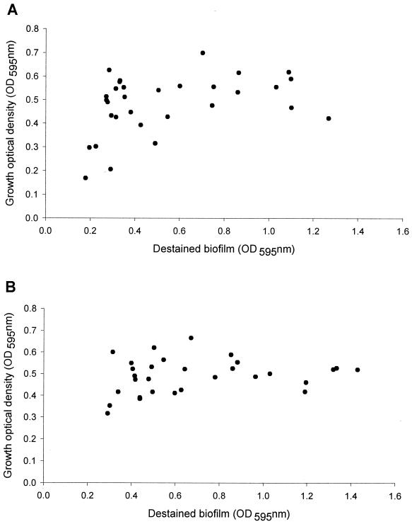
Each of the 31 coded L. monocytogenes strains were inoculated (1%) into MWB and grown in 100-μl volumes in eight replicate microtiter wells at 32°C. Growth was monitored over time by turbidity (OD595). At two time points, 20 and 40 h, unattached biomass was removed and the remaining biofilms were stained. All strains reached stationary phase between 18 and 24 h (data not shown). A weak correlation (P = 0.0286) between biofilm formation and cell density was found at 20 h (Fig. 2A). No significant relationship was found between biofilm formation and final cell density at 40 h (Fig. 2B) (P > 0.050). In addition, the exponential-phase growth rate (K) was calculated for each L. monocytogenes strain and there was no significant correlation between growth rates and the biofilm formation of different strains at 20 or 40 h (data not shown). Since biofilm formation was independent of growth at 40 h, this time point was selected for analysis. Vatanyoopaisarn et al. (30) showed that bacterial motility was an important factor in initial adhesion; therefore, we also examined the motility of all strains tested in MWB with 0.5% agar. No differences in motility were observed among strains, and all strains tested were motile after 40 h of incubation at 32°C in MWA (data not shown). Therefore, differences in biofilm formation do not appear to be due to differences in motility.
FIG. 2.
Scatter plot of final cell density (OD595) versus destained biofilm for 31 coded L. monocytogenes strains at 20 (A) or 40 (B) h. Each data point represents the average reading from three replicates.
Biofilm formation at 40 h in various lineages.
Biofilm production at 40 h of incubation varied greatly between different strains of L. monocytogenes, ranging from an OD595 of 0.3 to 1.4 (Fig. 3A). After the microtiter plate biofilm formation assay and the microscopic analysis were completed, L. monocytogenes strains were decoded into three different lineages (Table 1). Our results indicated that most of the lineage I strains were at the upper end of the continuum, with the exception of strains LM24 and LM25 (Fig. 3B). Moreover, lineage I strains were significantly different from lineage II and lineage III strains, P < 0.05 (Table 2). There was no significant difference found between biofilm production of lineage II and lineage III strains (P = 0.550) (Table 2).
TABLE 2.
Comparison of biofilm formation from three genetic lineages
| Comparison | Difference of meansa | P valueb,c |
|---|---|---|
| Lineage I vs lineage III | 0.518 | <0.001; S |
| Lineage I vs lineage II | 0.385 | 0.004; S |
| Lineage II vs lineage III | 0.133 | 0.550; NS |
Means for biofilm formation assessed by microtiter plate assay for all strains within the same lineage were calculated by using Sigma Stat software. The difference of means between various lineages was obtained by subtracting means of the two different lineages indicated.
P value was calculated for all pairwise multiple comparison procedures (Tukey test).
S, significant (P < 0.050); NS, not significant (P > 0.050).
L. monocytogenes strains used in this study included isolates from seafood-processing plants. Some strains were isolated repeatedly (persistent), while others were isolated only once (nonpersistent) (17). No relationship between environmental persistence of strains and biofilm formation was found under the conditions tested (Fig. 3B; Table 1).
Microscopic analysis on PVC and stainless steel.
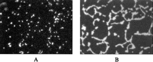
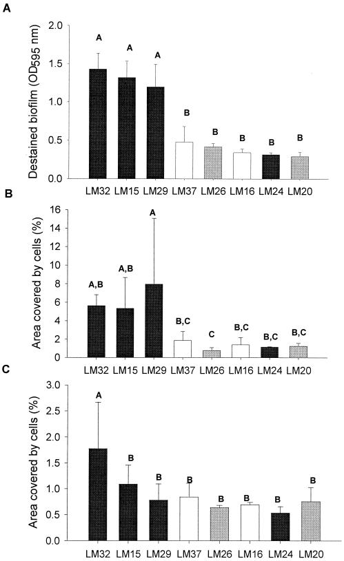
Quantitative microscopic analysis was performed on biofilms formed on PVC and stainless steel slides to compare results to those obtained with the microtiter assay. Representative images (strain LM15) are shown in Fig. 4. For this analysis, eight L. monocytogenes strains were selected based upon level of biofilm production as determined in the microtiter plate assay (Fig. 3A). Biofilm levels between the microtiter plate assay (Fig. 5A) and the PVC microscopic evaluation (Fig. 5B) were positively correlated, indicating a significant relationship (P > 0.050) in biofilm formation on PVC between these two assays (Table 3). For all strains tested, adhesion coverage on stainless steel was dramatically lower than that observed on PVC (Fig. 5C and B, respectively). A significant relationship (P > 0.050) was observed on both PVC and stainless steel on microscopic observation. No significant relationship (P < 0.050) was found between the levels from microtiter plate biofilm formation and microscopic assessment of coverage on stainless steel (Table 3); however, a weak correlation (correlation coefficient = 0.571) was found (Table 3). The consistency in ranking of biofilm producers assessed by these two different methods supports our proposal that biofilm production assessed by microtiter plate can be used as a rapid and simple screen for the prediction of biofilm formation as assessed by microscopy.
FIG. 4.
L. monocytogenes strain, coded LM15, grown in MWB at 32°C for 40 h on stainless steel (A) or PVC (B). The fields were observed under a 100× oil-immersion objective (Eclipse E400; Nikon).
FIG. 5.
(A) Destained biofilm production (OD595) determined in a microtiter plate biofilm assay at 32°C, 40 h in MWB. The area covered by biofilm cells was determined by using the microscopy biofilm formation assay at 32°C, 40 h (B), or PVC (C), on stainless steel. Lineage I strains are represented in black, lineage II is in white, and lineage III is in gray. Statistical analysis was done by using all pairwise multiple comparison procedures (Tukey test). For each graph, the same letters indicate no significant difference between strains (P > 0.050).
TABLE 3.
Relationship of biofilm formation assessed microscopically and biofilm formation assessed by microtiter plate assay
| Comparison | Correlation coefficienta | P valueb,c |
|---|---|---|
| Microscopic PVC vs microscopic stainless steel | 0.714 | 0.0347; S |
| PVC microtiter plate vs microscopic stainless steel | 0.571 | 0.120; NS |
| PCV microtiter plate vs microscopic PVC | 0.810 | 0.0096; S |
Correlation coefficient for biofilm formation of eight L. monocyotogenes strains (Fig. 5) was calculated by Sigma Stat software using Spearman rank order correlation.
P value was calculated by Spearman rank order correlation.
S, significant (P < 0.050); NS, not significant (P > 0.050).
DISCUSSION
The objective of this study was to standardize a PVC microtiter plate assay to compare the ability of 31 coded L. monocytogenes strains to form biofilms. Cellular growth rates and final cell density did not correlate with biofilm formation after 40 h at 32°C, indicating that differences in biofilm formation were due to factors other than the ability to grow in MWB (Fig. 1). The majority of lineage I strains fell within the upper end of biofilm formation, and overall this lineage had significantly greater biofilm formation than lineage II and lineage III strains. In addition, L. monocytogenes biofilm formation on PVC and stainless steel was analyzed by quantitative epifluorescence microscopy and results were compared to the PVC microtiter plate biofilm assay. Similar trends in biofilm formation were observed using the microscopic and the microtiter plate assays, indicating that the PVC microtiter plate assay may be used as a rapid, simple method to assess biofilm production.
L. monocytogenes strains were observed to differ in the amount of biofilm produced at 40 h (Fig. 3A), and the microtiter plate assay appeared to detect differences in biofilm-forming ability among L. monocytogenes strains. It has been suggested that motility and flagella formation can play a role in initial phases of biofilm formation (21, 30). In this study, initial cell attachment was not studied and biofilm formation was examined over a longer time period (40 h), so that possible flagella formation and its influence in the early stages of cell attachment (30) could be excluded. However, motility was observed for all strains after 40 h of incubation at 32°C in MWA.
It is important to note that the assay presented here measured biofilm production under a minimal nutrient environment. L. monocytogenes growing in a food-processing environment may be exposed to fluctuating levels of nutrients, depending upon location in the plant. For example, populations existing in floor drains will be exposed to high levels of nutrients during prerinsing of equipment prior to cleaning and sanitizing. On the other hand, if living on walls, ceilings, or within condensate (on the outside of cooling equipment or pipes), it is likely that these bacteria are surviving under reduced nutrient conditions. Although this research has focused upon biofilms of pure L. monocytogenes, in the food-processing environment they most likely exist as a member of a complex bacterial community consisting of many different types of bacteria. This is supported by the research of Sasahara and Zottola in which higher numbers of L. monocytogenes ScottA were observed in biofilms when cocultured with Pseudomonas fragi (27). If growing under low nutrient conditions in the processing plant, L. monocytogenes could possibly use other environmental bacteria as a source for the seven essential amino acids needed for growth.
After all testing, L. monocytogenes strains were decoded into three lineages (Table 1) (19, 31) and arranged from the highest to the lowest biofilm producers (Fig. 3B). It was observed that most of the lineage I strains (Table 1) were at the upper end of the continuum, with the exception of strains LM24 and LM25 (Fig. 3B). In general, lineage II and III strains had lower destained biofilm values (Fig. 3B). Using a one-way analysis of variance for statistical analysis of the mean, biofilm production by lineage I strains was found to be higher and statistically different from biofilm formation of lineage II and lineage III strains (Table 2). According to Wiedmann et al. (31), lineage I contains all food-borne epidemic strains and isolates from sporadic cases among humans and animals (Table 1). Lineage II contains both human and animal isolates, and lineage III contains strains isolated from animals only (19, 31). We thus hypothesize that the ability of lineage I strains to form biofilms may contribute to their prevalence among humans. Lineage I strains may represent an evolutionarily distinct group of L. monocytogenes, and the increased biofilm formation of isolates in this lineage could be due to the presence or absence of specific genes or by lineage-specific allelic variations. The presence or absence of specific genes within subtypes of the same species has been observed within group A streptococci (26).
Biofilm formation assessed by microtiter plate assay was compared to biofilm formation assessed by quantitative epifluorescence microscopy. The microtiter plate biofilm assay revealed greater differences in biofilm production than did the microscopy biofilm assay. In general, though, trends observed with the microtiter plate assay were also observed with the microscopic analysis on both PVC and stainless steel, although differences were not always statistically significant in the microscopic analysis.
Microscopic analysis indicated that biofilm coverage was higher on PVC slides for all strains tested than on stainless steel slides (Fig. 5B and C). This may be due to differences in surface hydrophobicity compared to cellular hydrophobicity when grown under low nutrient environments; however, others have found L. monocytogenes LO28 to be negatively charged and hydrophilic in nature when grown in a defined medium at various temperatures (8). Beresford et al. (3) observed that L. monocytogenes attachment was higher on PVC than on stainless steel after a short contact time and after 2 h of incubation, but listerial attachment was not significantly different between these two materials (3). Helke et al. (11) and Sommer et al. (28) proposed that the interactions between bacterial cells and an inorganic surface are different for adhesion onto hydrophobic or hydrophilic surfaces. Besides material properties, it has been observed that bacterial characteristics such as charge, hydrophobicity, and surface appendages can enhance the adhesion process as well (6). It has been reported that factors other than hydrophobic interactions, such as electrostatic and exopolymer interactions, account for the attachment of L. monocytogenes to various materials (4). Moreover, Sommer et al. (28) showed that cell wall hydrophobicity may be closely related to adhesion on inert substrata, either to hydrophobic or hydrophilic surfaces. For hydrophobic surfaces such as PVC, hydrophobic interactions are considered the main forces and, thus, cell wall hydrophobicity of strains plays a major role (28). For hydrophilic surfaces such as stainless steel, electrostatic interactions are considered dominant and the charge of the bacterial cell wall is more important than other factors (28). Since PVC and stainless steel differ in hydrophobicity, we believe that adhesion on PVC may be higher due to its hydrophobic nature compared to stainless steel.
When comparing the microtiter plate assay with the quantitative microscopic biofilm assay, it is important to consider the inherent advantages and disadvantages of each assay. The major advantage of microscopic analysis is the ability to observe the biofilm directly. The microscopic method, on the other hand, is relatively time-consuming compared to the microtiter plate assay. Blackman and Frank (4) also reported that epifluorescence microscopy may overestimate the area covered by cells, since some extracellular polymer is stained as well (4). A possibility for bias in selecting fields for analysis and usage of the program for microscopy data analysis (NIH Image software was used for calculation of the percentage of area covered by cells) could result in a human error by over- or underestimation of the percentage of area covered. In addition, the magnification for determining surface coverage should be considered. We used high magnification (×1,000) since fluorescence was brightest but, ideally, surface coverage should be observed at lower magnification to obtain a larger field. In addition, the surface areas examined by each method are different: in the microtiter plate assay the whole individual well area covered by cells (approximately 130 mm2) is evaluated, while the area of the 50 fields examined by microscopy represents only about 0.135 mm2.
The microtiter plate has the advantage of enabling researchers to rapidly analyze adhesion of multiple bacterial strains or growth conditions within each experiment. The major disadvantage is that the microtiter plate method is an indirect indication of the level of biofilm produced; the adsorption of the crystal violet in the destaining solution was used as an indication of the amount of biofilm. In order to assure consistency between experiments, the manufacturer's lot numbers of the PVC plates were monitored, since researchers have reported dramatic differences in adhesion between different lots of PVC (1). In addition, care was taken to thoroughly dry films before staining and to adhere to consistent timing of staining and destaining steps. Despite the shortfall as an indirect method, we found more consistent results (lower standard deviations between replicates within an experiment and between experiments) with the microtiter plate assay than with direct microscopy.
The results of the microtiter plate assay had a strong correlation to microscopic coverage on PVC. Although the microscopic assay showed greater coverage on PVC than on stainless steel, a significant correlation (P < 0.05) was observed between both materials. Direct observation will always be very important to studying adhesive cells and biofilms, but we believe that the microtiter plate assay can be used in conjugation with microscopy as a rapid and reproducible screening method. This assay will allow researchers to rapidly screen for differences between strains or growth conditions prior to performing labor-intensive microscopic quantification.
Acknowledgments
This research was supported in part by a Faculty Research Grant awarded by the University of Massachusetts Research Council and the Cooperative State Research Extension, Education Service, U.S. Department of Agriculture, Massachusetts Agricultural Experiment Station, under Project No. MAS00837.
REFERENCES
- 1.An, Y. H., and R. J. Friedman. 2000. Handbook of bacterial adhesion: principles, methods, and applications. Humana Press, Totowa, N.J.
- 2.Anwar, H., J. L. Strap, and J. W. Costerton. 1992. Eradication of biofilm cells of Staphylococcus aureus with tobramycin and cephalexin. Can. J. Microbiol. 38:618-625. [DOI] [PubMed] [Google Scholar]
- 3.Beresford, M. R., P. W. Andrew, and G. Shama. 2001. Listeria monocytogenes adheres to many materials found in food-processing environments. J. Appl. Microbiol. 90:1000-1005. [DOI] [PubMed] [Google Scholar]
- 4.Blackman, I. C., and J. F. Frank. 1996. Growth of Listeria monocytogenes as a biofilm on various food-processing surfaces. J. Food Prot. 59:827-831. [DOI] [PubMed] [Google Scholar]
- 5.Bolton, K. J., C. E. R. Dodd, G. C. Mead, and M. Waites. 1988. Chlorine resistance of strains of Staphylococcus aureus isolates from poultry processing plants. Lett. Appl. Microbiol. 6:31-34. [Google Scholar]
- 6.Bower, C. K., J. McGuire, and M. A. Daeschel. 1996. The adhesion and detachment of bacteria and spores on food-contact surfaces. Trends Food Sci. Technol. 7:152-157. [Google Scholar]
- 7.Brackett, R. E. 1992. Shelf stability and safety of fresh produce as influenced by sanitation and disinfection. J. Food Prot. 55:808-814. [DOI] [PubMed] [Google Scholar]
- 8.Chavant, P., B. Martinie, T. Meylheuc, M.-N. Bellon-Fontaine, and M. Hebraud. 2002. Listeria monocytogenes LO28: surface physiochemical properties and ability to form biofilms at different temperatures and growth phases. Appl. Environ. Microbiol. 68:728-737. [DOI] [PMC free article] [PubMed] [Google Scholar]
- 9.Doyle, M. P., L. R. Beuchat, and T. M. Montville. 1997. Food microbiology: fundamentals and frontiers. American Society for Microbiology, Washington, D.C.
- 10.Frank, J. F., and R. A. Koffi. 1990. Surface-adherent growth of Listeria monocytogenes is associated with increased resistance to surfactant sanitizers and heat. J. Food Prot. 53:550-554. [DOI] [PubMed] [Google Scholar]
- 11.Helke, D. M., E. B. Somers, and A. C. L. Wong. 1993. Attachment of Listeria monocytogenes and Salmonella typhimurium to stainless steel and buna-n rubber in the presence of milk and individual milk components. J. Food Prot. 56:479-484. [DOI] [PubMed] [Google Scholar]
- 12.Herald, P. J., and E. A. Zottola. 1988. Attachment of Listeria monocytogenes to stainless steel surfaces at various temperatures and pH values. J. Food Sci. 53:1549-1552. [Google Scholar]
- 13.Jeong, D. K., and J. F. Frank. 1994. Growth of Listeria monocytogenes at 10°C in biofilms with microorganisms isolated from meat and dairy processing environments. J. Food Prot. 57:576-586. [DOI] [PubMed] [Google Scholar]
- 14.Kim, K. Y., and J. F. Frank. 1995. Effect of nutrients on biofilm formation by Listeria monocytogenes on stainless steel. J. Food Prot. 58:24-28. [DOI] [PubMed] [Google Scholar]
- 15.Lad, T. L., and J. W. Costerton. 1990. Methods for studying biofilm bacteria. Methods Microbiol. 22:285-307. [Google Scholar]
- 16.Lindsay, D., and A. von Holy. 1997. Evaluation of dislodging methods for laboratory-grown bacterial biofilms. Food Microbiol. 14:383-390. [Google Scholar]
- 17.Lunden, J. M., M. K. Miettinen, T. J. Autio, and H. J. Korkeala. 2000. Persistent Listeria monocytogenes strains show enhanced adherence to food contact surface after short contact times. J. Food Prot. 63:1204-1207. [DOI] [PubMed] [Google Scholar]
- 18.Nickel, J. C., and J. W. Costerton. 1992. Bacterial biofilms and catheters: a key to understanding bacterial strategies in catheter-associated urinary tract infections. Can. J. Infect. Dis. 3:619-624. [DOI] [PMC free article] [PubMed] [Google Scholar]
- 19.Norton, M. D., J. M. Scarlett, K. Horton, D. Sue, J. Thimothe, K. J. Boor, and M. Wiedmann. 2001. Characterization and pathogenic potential of Listeria monocytogenes isolates from the smoked fish industry. Appl. Environ. Microbiol. 67:646-653. [DOI] [PMC free article] [PubMed] [Google Scholar]
- 20.Notermans, S., J. Dufrenne, P. Teunis, and T. Chackraborty. 1998. Studies on the risk assessment of Listeria monocytogenes. J. Food Prot. 61:244-248. [DOI] [PubMed] [Google Scholar]
- 21.O'Toole, G. A., and R. Kolter. 1998. Initiation of biofilm formation in Pseudomonas fluorescens WCS365 proceeds via multiple, convergent signaling pathways: a genetic analysis. Mol. Microbiol. 28:449-461. [DOI] [PubMed] [Google Scholar]
- 22.Poulsen, V. L. 1999. Microbial biofilm in food processing. Lebensm.-Wiss. U.-Technol. 32:321-326. [Google Scholar]
- 23.Premaratne, R. J., W. J. Lin, and E. A. Johnson. 1991. Development of an improved chemically defined minimal medium for Listeria monocytogenes. Appl. Environ. Microbiol. 57:3046-3048. [DOI] [PMC free article] [PubMed] [Google Scholar]
- 24.Rasmussen, O. F., P. Skouboe, L. Dons, L. Rossen, and J. E. Olsen. 1995. Listeria monocytogenes exists in at least three evolutionary lines: evidence from flagellin, invasive associated protein and listeriolysin O genes. Microbiology 141:2053-2061. [DOI] [PubMed] [Google Scholar]
- 25.Reid, G., C. Tieszer, R. Foerch, H. J. Busscher, A. H. Khoury, and A. W. Bruce. 1993. Adsorption of ciprofloxacin to urinary catheters and effect on subsequent bacterial adhesion and survival. Coll. Surf. Biointerfaces 1:9-16. [Google Scholar]
- 26.Reid, S. D., N. M Nicole, J. K. Buss, B. Lei, and J. M. Musser. 2001. Multilocus analysis of extracellular putative virulence proteins made by group A Streptococcus: population genetics, human serologic response and gene transcription. Proc. Natl. Acad. Sci. USA 98:7552-7557. [DOI] [PMC free article] [PubMed] [Google Scholar]
- 27.Sasahara, K. C., and E. A. Zottola. 1993. Biofilm formation by Listeria monocytogenes utilizes a primary colonizing microorganism in flowing systems. J. Food Prot. 56:1022-1028. [DOI] [PubMed] [Google Scholar]
- 28.Sommer, P., C. Martin-Rouas, and E. Mettler. 1999. Influence of the adherent population level on biofilm population, structure and resistance to chlorination. Food Microbiol. 16:503-515. [Google Scholar]
- 29.Tompkin, R. B., V. N. Scott, D. T. Bernard, W. H. Sveum, and K. S. Gombas. 1999. Guidelines to prevent post-processing contamination from Listeria monocytogenes. Dairy Food Environ. Sanit. 19:551-562. [Google Scholar]
- 30.Vatanyoopaisarn, S., A. Nazli, C. E. R. Dodd, C. E. D. Rees, and W. M. Waites. 2000. Effect of flagella on initial attachment of Listeria monocytogenes to stainless steel. Appl. Environ. Microbiol. 66:860-863. [DOI] [PMC free article] [PubMed] [Google Scholar]
- 31.Wiedmann, M., J. L. Bruce, C. Keating, A. E. Johnson, P. L. McDonough, and C. A. Batt. 1997. Ribotypes and virulence gene polymorphism suggest three distinct Listeria monocytogenes lineages with differences in their pathogenic potential. Infect. Immun. 65:2707-2716. [DOI] [PMC free article] [PubMed] [Google Scholar]


