Abstract
To develop prophylaxis against mother-to-child human immunodeficiency virus (HIV) transmission, we established a simian-human immunodeficiency virus (SHIV) infection model in neonatal macaques that mimics intrapartum mucosal virus exposure (T. W. Baba et al., AIDS Res. Hum. Retroviruses 10:351–357, 1994). Using this model, neonates were protected from mucosal SHIV-vpu+ challenge by pre- and postnatal treatment with a combination of three human neutralizing monoclonal antibodies (MAbs), F105, 2G12, and 2F5 (Baba et al., Nat. Med. 6:200–206, 2000). In the present study, we used this MAb combination only postnatally, thereby significantly reducing the quantity of antibodies necessary and rendering their potential use in humans more practical. We protected two neonates with this regimen against oral SHIV-vpu+ challenge, while four untreated control animals became persistently infected. Thus, synergistic MAbs protect when used as immunoprophylaxis without the prenatal dose. We then determined in vitro the optimal MAb combination against the more pathogenic SHIV89.6P, a chimeric virus encoding env of the primary HIV89.6. Remarkably, the most potent combination included IgG1b12, which alone does not neutralize SHIV89.6P. We administered the combination of MAbs IgG1b12, 2F5, and 2G12 postnatally to four neonates. One of the four infants remained uninfected after oral challenge with SHIV89.6P, and two infants had no or a delayed CD4+ T-cell decline. In contrast, all control animals had dramatic drops in their CD4+ T cells by 2 weeks postexposure. We conclude that our triple MAb combination partially protected against mucosal challenge with the highly pathogenic SHIV89.6P. Thus, combination immunoprophylaxis with passively administered synergistic human MAbs may play a role in the clinical prevention of mother-to-infant transmission of HIV type 1.
The role of neutralizing antibodies (NAbs) in protecting against the human immunodeficiency virus (HIV) has recently been investigated in macaques (3, 37, 38, 56) by using chimeric simian/human immunodeficiency viruses (SHIV) (51, 52, 57). These viruses contain a simian immunodeficiency virus (SIV) isolate mac239 backbone and encode envelope glycoproteins derived from HIV type 1 (HIV-1), which makes it possible to test antibodies directed against HIV-1 envelope in rhesus macaques.
Recently, passively infused antibodies were found to protect against an intravenous (i.v.) SHIV challenge in macaques (3, 37, 56). We found complete protection in four adult rhesus macaques challenged with SHIV-vpu+ after an infusion of F105, 2G12, and 2F5 (3). These human monoclonal antibodies (MAbs) are directed against conserved epitopes on HIV-1. F105 recognizes the CD4 binding site (CD4BS) on gp120 (49). 2G12 binds to a conformation-sensitive, glycosylation-dependent, discontinuous epitope centered around the C3/V4 domain on gp120 of HIV (62), and 2F5 is directed against a specific sequence, ELDKWA, within the external domain of the gp41 (17, 45). We and others have also shown that infusion of anti-HIV antibodies protected against mucosal transmission of SHIV (3, 38). Mascola et al. (38) infused MAbs 2F5 and 2G12 with or without high-titer anti-HIV immunoglobulin into adult rhesus monkeys 24 h prior to vaginal SHIV89.6PD challenge. The best protection was found in the cohort that received the triple combination. Four of five animals were protected against infection, and the fifth monkey did not develop CD4+ T-cell depletion. In contrast, all control monkeys given irrelevant control immunoglobulins developed high plasma viremia and rapid CD4+ T-cell decline.
Our goal is to develop immune prophylaxis against mother-to-child HIV-1 transmission. Previously, we established an SIV/macaque model that mimics mucosal HIV-1 exposure of neonates that can occur during delivery (2). Using this model, we achieved complete protection of four neonatal rhesus macaques with the human MAbs 2F5, F105, and 2G12 against oral challenge with SHIV-vpu+, a chimeric virus that encodes the env gene of the laboratory-adapted, T-cell-tropic HIV-1 IIIB (3). The neonates received transplacental MAbs before birth, by passive therapy of the pregnant dams, as well as by direct i.v. infusion after birth. Prenatal passive antibody therapy of pregnant women requires large amounts of MAbs compared to those needed to infuse only neonates. Thus, prenatal immunoprophylaxis might be too costly and could render large-scale use in humans impractical. In the present study, we assessed the efficacy of immunoprophylaxis using only postnatal MAb treatment of infants. First, we conducted a pilot study with two neonatal rhesus macaques that both received 2F5, F105, and 2G12 prior to oral challenge with SHIV-vpu+.
In a second experiment, we determined whether human MAbs administered postnatally would also inhibit mucosal infection by a chimeric virus carrying the env gene of a primary, and hence less neutralization-sensitive, HIV-1 isolate. Thus, neonates were treated with MAbs after birth and challenged orally with SHIV89.6P, an in vivo-passaged, pathogenic virus which expresses envelope glycoproteins of a primary HIV-1 isolate (52, 53). The optimal combination of human neutralizing MAbs against SHIV89.6P was determined in vitro. Although primary HIV-1 isolates are more resistant to neutralization than laboratory-adapted strains (19, 44), MAbs 2G12, 2F5, and IgG1b12, a MAb which binds to a confirmation-sensitive epitope that overlaps but is not precisely continuous with the CD4BS (10), have broad neutralizing activity against primary virus isolates (8, 10, 11, 17, 20, 61). In addition, we detected strong synergism and complete neutralization of the primary isolate, HIV89.6, in human peripheral blood mononuclear cells (PBMC) with the triple combination F105, 2F5, and 2G12 (3). Remarkably, although IgG1b12 alone did not neutralize SHIV89.6P or SHIV-KB9, a fourth-passage molecular clone of SHIV89.6P (18, 21, 27), the triple combination 2G12, IgG1b12, and 2F5 showed the strongest synergism. Thus, we used this combination for the in vivo immunoprophylaxis studies with oral SHIV89.6P challenge.
MATERIALS AND METHODS
Human IgG1 MAbs.
The human immunoglobulin G1 (IgG1) anti-HIV-1 MAbs used were 2F5 (17, 45), 2G12 (8, 62), IgG1b12 (10), and F105 (49). The MAb preparations were of clinical-grade purity and endotoxin-free. For the in vivo immunoprophylaxis studies, each of these MAbs was given at a dose of 10 mg/kg.
MAb IgG1b12-secreting CHO cells (kindly provided by Dennis Burton, Scripps Research Institute, La Jolla, Calif.) were expanded in endotoxin-free Glasgow minimum essential medium (Glasgow MEM; Sigma, St. Louis, Mo.). The medium was supplemented with 100 μM MEM nonessential amino acids (Gibco-BRL, Life Technologies, Grand Island, N.Y.), glutamic acid (60 μg/ml), asparagine (60 μg/ml), 1 mM sodium pyruvate (Sigma), heat-inactivated fetal calf serum (FCS; Gemini Bio-Products, Woodland, Calif.), penicillin (100 U/ml), streptomycin (100 μg/ml), 50 μM methionine sulfoximine (Sigma), and the nucleosides adenosine, guanosine, cytidine, uridine, and thymidine (Sigma), each at 7 μg/ml. The FCS concentration was stepwise reduced from initially 10% to 2%. Supernatants containing the MAb were collected on day 7 and clarified by centrifugation. The antibody was purified by protein G chromatography (Amersham Pharmacia Biotech Inc., Piscataway, N.J.), dialyzed against phosphate-buffered saline under endotoxin-free conditions, tested for endotoxin, and stored at 4°C until use. If necessary, endotoxin was removed by Detoxi-Gel (Pierce, Rockford, Il.) according to the manufacturer's instruction.
Virus stocks.
We used two different chimeric SHIVs as challenge viruses. SHIV-vpu+ contains env from the laboratory-adapted, T-cell-tropic HIV-1 IIIB on an SIVmac239 backbone (31, 36). SHIV89.6P encodes env of the primary, dualtropic HIV-1 strain 89.6 originally isolated from PBMC of a 47-year-old man with AIDS (16). The biological isolate was derived from SHIV89.6 after four serial passages in rhesus macaques (51, 52) and is acutely pathogenic, causing profound CD4+ T-cell depletion within 2 weeks of virus exposure (52). Both cell-free challenge virus stocks were propagated in mitogen-stimulated rhesus macaque PBMC in the presence of human interleukin-2 (IL-2). Supernatants were clarified by centrifugation, filtered, and stored in vapor-phase liquid nitrogen. Stocks were titrated by endpoint titration on CEMx174 cells as described elsewhere (3). The SHIV-vpu+ stock and the SHIV89.6P stock contained 2 × 10⁵ and 2.6 × 10⁴ 50% tissue culture infectious doses per ml, respectively. To determine the oral 50% animal infectious doses (AID50), seven neonatal rhesus monkeys were exposed orally to serial dilutions of SHIV-vpu+; eight neonates were used to titrate SHIV89.6P in a similar manner. The SHIV-vpu+ stock contained 3.79 oral AID50/ml (3); the SHIV89.6P stock yielded 30 oral AID50/ml.
In vitro SHIV89.6P neutralization assay.
We adapted an MT-2 cell viability assay (43) to measure neutralization of SHIV89.6P. MAbs were diluted serially in RPMI 1640 (Gibco-BRL) supplemented with 15% heat-inactivated FCS, penicillin (100 U/ml), streptomycin (100 μg/ml), and 2 mM l-glutamine (Sigma). Antibodies (50 μl) were preincubated in triplicate with 50 μl of SHIV89.6P (diluted 1:5 in ice-cold medium; 50 μl containing 4.2 pg of p27) in flat-bottom 96-well plates (Corning Costar Corporation, Boston, Mass.) at 37°C for 1 h. Log-phase MT-2 cells (50 μl of a 106-cell/ml suspension) were added to the virus-antibody mixture and incubated at 37°C with 5% CO2. Virus-only controls (containing SHIV89.6P and MT-2 cells only, without MAbs) and cells-only controls were cultured in parallel. After 2 h, each well was supplemented with 100 μl of medium and incubation was continued. Cell densities were reduced after 3 days of incubation by replacing 100 μl of each cell suspension with medium. On day 5, cells were fed by exchanging 100 μl of medium. Infection led to extensive (>80%) syncytium formation after approximately 7 days in the absence of MAb (virus-only wells). Cells were harvested, and viability was tested by neutral red uptake (43). Cell suspensions (100 μl) were transferred to 96-well plates precoated with poly-l-lysine (Becton Dickinson, Franklin Lakes, N.J.). A volume of 100 μl of neutral red (1:300 solution; ICN Pharmaceuticals, Costa Mesa, Calif.), diluted 1:10 in medium, was added to each well. The plates were incubated for 1 h at 37°C with 5% CO2, followed by aspiration of the medium and 2 gentle washes with phosphate-buffered saline. The neutral red absorbed by viable cells was extracted with 1% acetic acid in 50% ethanol for 1 h at room temperature under constant agitation. Optical density was read with a multiscan Microplate Reader (Du Pont Instruments, Wilmington, Del.) at 540 nm. The percentage of protection was defined as the difference in mean absorption between test wells (cells plus virus plus antibody) and virus-only wells, divided by the difference in mean absorption between cells-only wells and virus-only wells.
Determination of synergism (CI) and DRI.
The complex interactions of MAb combinations were assessed as outlined earlier (29, 30) by the Chou-Talalay method (14, 15). Basically, this method yields two indices describing the interactions among MAbs in a given combination: the combination index (CI) and the dose reduction index (DRI). A CI of <1 indicates synergism, a CI of 1 indicates additive effects, and a CI of >1 indicates antagonism. The DRI reflects the factor by which the dose of each MAb in a combination may be reduced at a given percent neutralization compared with the dose when each MAb is used alone (12, 13). A high DRI correlates with a strong synergistic effect, and the amount of MAb may be decreased accordingly in the combination.
Animals, animal care, and experimental design of the in vivo immunoprophylaxis study.
We used 14 neonatal rhesus monkeys (Macaca mulatta) from a retrovirus-free colony. The animals were kept according to National Institutes of Health guidelines on the care and use of laboratory animals at the University of Texas M. D. Anderson Cancer Center and at the Yerkes Regional Primate Center, Emory University, Atlanta, Ga. These facilities are fully accredited by the Association for Assessment and Accreditation of Laboratory Animal Care International. Animal experiments were approved by the animal care and use committees of these institutions and the Dana-Farber Cancer Institute. Monkeys were anaesthetized intramuscularly with ketamine (5 mg/kg) before all procedures that required removal from their cages. All animals described were found to be negative for simian T-lymphotropic virus type 1 (32) and simian retrovirus type D by PCR (34).
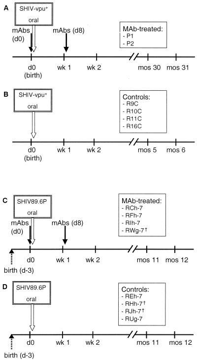
Two neonates, P1 and P2, were treated i.v. with the triple MAb combination of F105, 2G12, and 2F5 on the day of birth (Fig. 1A). One hour after completion of the infusion, P1 and P2 were challenged orally in a nontraumatic manner (2) with 10 oral AID50 of SHIV-vpu+. A second MAb dose was given on day 8. Four untreated control neonates (R9C, R10C, R11C, and R16C) were challenged identically (Fig. 1B).
FIG. 1.
Experimental design of the in vivo oral challenge studies with SHIV-vpu+ (A and B) and SHIV89.6P (C and D). (A) Two neonatal rhesus monkeys (P1 and P2) were infused twice i.v. with human MAbs F105, 2G12, and 2F5 (10 mg/kg; solid black arrows). d0 and d8, days 0 and 8. (B) Four neonates (R9C, R10C, R11C, and R16C) served as untreated control animals. All six animals were challenged orally with 10 oral AID50 of SHIV-vpu+ 1 h after the first MAb treatment on the day of birth (open arrows). Treated animals were observed for 31 months, and serial blood samples were collected. Control animals were sacrificed 6 to 12 months after virus challenge. (C) Four neonatal rhesus monkeys (RCh-7, RFh-7, RIh-7, and RWg-7) were infused i.v. twice with human MAbs IgG1b12, 2G12, and 2F5 (10 mg/kg; solid black arrows). (D) Four neonates (REh-7, RHh-7, RJh-7, and RUg-7) served as untreated control animals. All eight animals were challenged orally with SHIV89.6P (15 oral AID50) 1 h after the first MAb treatment or at the corresponding time point (3 days after birth) (open arrows). Animals were observed for 12 months, and serial blood samples were collected. †, animals RWg-7, RHh-7, RJh-7 were sacrificed 10, 7, and 9 weeks postexposure, respectively.
Four neonates (RCh-7, RFh-7, RIh-7, RWg-7) were treated i.v. with a combination of three MAbs (IgG1b12, 2G12, and 2F5) within the first 3 days of vaginal delivery (Fig. 1C). The treated animals were challenged orally with 15 oral AID50 of SHIV89.6P 1 hour after completion of the MAb infusion. This dose yielded a 99% probability of infection (58). A second MAb dose was given on day 8. Four control group neonates (REh-7, RHh-7, RJh-7, and RUg-7) were untreated prior to oral virus challenge (Fig. 1D).
Blood samples were collected from all animals on day 0 prior to MAb infusion and virus inoculation (baseline collection) and on day 8, prior to the second MAb infusion, to determine infection/protection of animals. Although desirable, no additional samples could be collected after the MAb infusions to determine the plasma titer of NAbs or to assess pharmacokinetics. The maximum blood volume that can be sampled from approximately 500-g neonates (6 ml/kg of body weight/month) did not allow it. MAb-treated animals were observed for 31 months after SHIV-vpu+ challenge and for 12 months after SHIV89.6P challenge, during which time samples were collected regularly. When necessary, animals with progressive disease were sacrificed via i.v. sodium pentobarbital injection.
Virus isolation; determination of proviral DNA and viral RNA loads.
Methods for PBMC coculture and DNA PCR have been described previously (2, 33). The lower limit of detection of the DNA PCR was one copy per 150,000 cells (33). Viral RNA loads were quantified by a one-tube fluorogenic probe-based real-time reverse transcription (RT)-PCR (24). RNA was extracted from 140 μl of cell-free plasma with a QIAamp Viral RNA Mini kit (Qiagen, Valencia, Calif.). The sensitivity of the RT-PCR is 50 copies per ml of undiluted plasma. Sodium citrate-anticoagulated samples were not available from animal R16C for RT-PCR or neutralization assays.
Serological assays.
Plasma samples were analyzed as described elsewhere (2, 33) for the presence of specific antibodies, using commercially available Western blot strips prepared from HIV-2 antigens which had been shown to cross-react with SIV antisera. HIV-1 Western blot analyses were conducted as instructed by the manufacturer (Epitope, Beaverton, Oreg.).
NAbs in saliva and plasma.
Saliva was collected from the oral cavities of the neonatal macaques using Weck-cel sponges (Xomed Surgical Products, Jacksonville, Fla.). Saliva samples were collected 1 h after the first MAb infusion and/or prior to virus inoculation. After collection, sponges were saturated with 400 μl of medium (see above) for 15 min. Gentle squeezing with a sterile pipette eluted the saliva, and the liquid (250 μl) was sterile filtered. To measure neutralization, serial dilutions of salivary filtrates were assayed in duplicate in an adapted MT-2 cell infection assay with SHIV89.6P as described above or with SHIV-vpu+ as described earlier (30). The salivary NAb titer is the reciprocal dilution which protected 50% of MT-2 cells from virus-induced cytotoxicity. This corresponds to 90% reduction of the viral Gag synthesis (9). The assay's lower limit of detection titer was 10 for undiluted samples. The same assays were applied to quantify NAbs in serially diluted, heat-inactivated (56°C, 1 h) plasma samples. Neutralizing activities against other SHIV variants, SHIV89.6 (51) and SHIV-KU2 (26), and the heterologous T-cell-line-adapted HIV-1 strain MN (23) were measured in an identical manner.
Quantification of CTL activity in the completely protected animal RCh-7.
An autologous B-lymphoblastic cell line (BLCL) was prepared by transformation of PBMC from RCh-7 with herpesvirus papio (50). PBMC collected 46 weeks after SHIV89.6P challenge were stimulated with paraformaldehyde-fixed, autologous BLCL infected with SHIV antigen-encoding vaccinia virus constructs at 2 to 3 PFU per cell (Therion, Cambridge, Mass.) Recombinant human IL-2 (20 U/ml) was added every 3 days. Cultures were tested on day 7 in a standard 5-h cytotoxic T-lymphocyte (CTL) assay using autologous, 51Cr-labeled BLCL target cells infected with vaccinia virus constructs encoding SIV gag-pol, SIV nef, or HIV-1 IIIB env. Background cytotoxicity was measured using wild-type vaccinia virus-infected control target cells. A PBMC culture from an animal with known CTL activity was included as a positive control for the autologous stimulation and cytotoxicity arms of the assay.
In vitro challenge of PBMC from the completely protected animal RCh-7.
PBMC were purified by Ficoll-Hypaque sedimentation from a sodium citrate-anticoagulated blood sample collected from uninfected animal RCh-7 36 weeks after SHIV89.6P challenge. Cells were washed and stimulated for 3 days with concanavalin A (5 μg/ml) in RPMI 1640 supplemented with 10% human type AB serum, l-glutamine, and penicillin-streptomycin. Cells were washed, and CD8+ T cells were removed by negative isolation using anti-CD8 magnetic beads as instructed by the manufacturer (Dynal, Lake Success, N.Y.). CD8-depleted cells were exposed to SHIV89.6P (100 ng of p27) for 12 h at 37°C. The cells were washed and then incubated for 17 days in the presence of recombinant human IL-2 (40 U/ml). Supernatants were collected every 2 to 4 days and analyzed for the presence of SIV p27 antigen, using a commercially available enzyme-linked immunoassay kit specific for SIV (Beckman/Coulter, Hialeah, Fla.). PBMC from an uninfected normal control animal were stimulated, CD8+ T-cell depleted, and infected in the same manner as a positive control.
Statistical analysis.
For analysis, the viral loads were expressed as a log10 transformation based on the distribution of HIV RNA in human studies. Viral loads below the limit of detection were set at half the limit of detection for calculation. No imputation for missing or truncated data was employed. It is noted that with four animals per group, there is low statistical power.
RESULTS
Synergistic neutralization of SHIV89.6P by a triple combination of human anti-HIV-1 MAbs in vitro.
We previously tested a large panel of human anti-HIV-1 MAbs for their ability to neutralize SHIV-vpu+ in vitro. MAbs IgG1b12, 2F5, 2G12, and F105 were synergistic when used in combinations (3, 29, 30). In addition, the triple combination of F105, 2G12, and 2F5 was used successfully to protect macaques from i.v. and oral SHIV-vpu+ challenge (3). In the present study, we explored the capacity of these four MAbs, used either alone or in combination, to neutralize pathogenic SHIV89.6P. We also compared the synergistic potency of F105 to that of IgG1b12 to determine whether different MAbs directed against the CD4BS were equally effective.
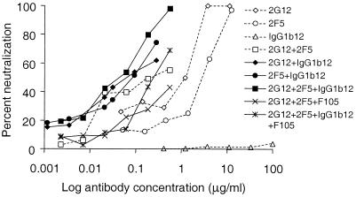
MAb IgG1b12 was inactive against SHIV89.6P; even high IgG1b12 concentrations up to 100 μg/ml did not significantly neutralize SHIV89.6P (Table 1; Fig. 2). Nevertheless, IgG1b12 (included at a constant concentration of 50 μg/ml) increased the neutralization potency of other MAbs (2G12 and 2F5) when used in combinations, as indicated by a leftward shift of the curves (Fig. 2; Table 1). This allowed us to use a reduced concentration of antibodies to achieve an equivalent level of neutralization. For example, when used alone, 1.243 μg of 2F5 per ml was required to obtain 50% SHIV89.6P neutralization (Table 1); by adding IgG1b12, the dose of 2F5 necessary to achieve the same result could be reduced 18.8 times (DRI) to 0.066 μg/ml. The addition of IgG1b12 to the combination of 2F5 and 2G12 led to very strong synergism and to even greater DRIs of 110 for 2F5 and 25.7 for 2G12 (Table 1). This effect was stronger than that observed using F105. The addition of F105 (also at a constant concentration of 50 μg/ml) to a serial dilution of 2F5 and 2G12 led to synergism at low concentrations, whereas antagonism was seen at the desirable, high degrees of neutralization. Furthermore, the quadruple MAb combination was less synergistic than the triple 2F5-2G12-IgG1b12 combination, presumably because of competition between F105 and IgG1b12 for the CD4BS. Therefore, we selected the triple combination of 2G12, IgG1b12, and 2F5 for the in vivo SHIV89.6P studies.
TABLE 1.
Synergistic neutralization of SHIV89.6P by human IgG1 MAbs in MT-2 cells
| Single antibody (specificity) or antibody combination (ratio) | Concn (μg/ml) for 50% neutralizationa | CIb at:
|
DRIc
|
Interaction | ||
|---|---|---|---|---|---|---|
| ED50 | ED90 | 2F5 | 2G12 | |||
| Single agents | ||||||
| IgG1b12 (anti-CD4BS) | >100 | |||||
| 2G12 (anti-gp120) | 0.291 | |||||
| 2F5 (anti-gp41) | 1.243 | |||||
| Combinations of 2 MAbs | ||||||
| 2G12 + IgG1b12e | 0.076 | 3.8 | Potentiation of 2G12 by IgG1b12d | |||
| 2F5 + IgG1b12e | 0.066 | 18.8 | Potentiation of 2F5 by IgG1b12d | |||
| 2F5 + 2G12 (1:1) | 0.191 | 0.41 | 1.25 | 13.1 | 3.06 | Synergism |
| Combinations of 3 MAbs | ||||||
| 2F5 + 2G12 (1:1) + IgG1b12e | 0.0226 | 0.048 | 0.067 | 110 | 25.7 | Very strong synergism |
| 2F5 + 2G12 (1:1) + F105e | 1.56 | 3.20 | 126 | 1.92 | 0.37 | Antagonism at high degrees of neutralization; synergism at low degrees of neutralization |
| Combination of 4 MAbs | ||||||
| 2F5 + 2G12 (1:1) + IgG1b12 + F105e | 0.265 | 0.54 | 0.80 | 11.3 | 2.2 | Synergism |
The neutralization dose for combinations of two and more MAbs was the sum of the dose of each MAb used in the combination regimen with the exception of IgG1b12 and F105. See footnote e.
Calculated by the Chou-Talalay method as described in Materials and Methods. CI < 1, CI = 1, and CI > 1, indicate synergism, additive effects, and antagonism, respectively. ED50 and ED90, 50 and 90% effective doses.
Measured by comparing the doses required to reach 50% virus neutralization when the antibody was used alone and in combination with other antibodies.
When one component in combination has no or negligible effect by itself (e.g., IgG1b12), no CI value can be calculated due to lack of dose-effect parameters. In this case, the term “potentiation” is used instead of “synergism.”
Always used at a concentration of 50 μg/ml.
FIG. 2.
Human MAb neutralization of SHIV89.6P in MT-2 cells. Neutralization was measured colorimetrically by the percentage of viable cells after incubation with the virus-antibody mixture (y axis). The antibody concentration (x axis) in combinations of two or more MAbs is the sum of the concentrations of each MAb with the exception of IgG1b12 and F105. When used in combinations, MAbs IgG1b12 and F105 were at 50 μg/ml; as potentiators, these amounts are not included in the total antibody concentration indicated on the x axis. In addition, when MAbs 2G12 and 2F5 were tested alone or in combination with other antibodies, the concentration of each was identical. Results are representative of two or three separate experiments for each antibody.
Outcome of oral SHIV-vpu+ challenge.
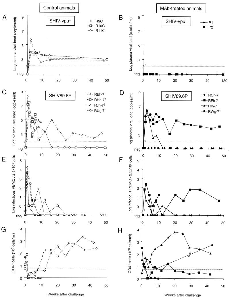
Two neonatal rhesus macaques were treated i.v. with a combination of 2G12, F105, and 2F5 on the day of birth (day 0 [Fig. 1]) prior to oral SHIV-vpu+ challenge. Both animals received a second infusion of this triple MAb combination 8 days later. Four untreated control animals were also challenged orally. These four monkeys served as controls also for another study arm, reported previously (3). All four untreated neonates, R9C, R10C, R11C, and R16C, became infected. They were positive for virus isolation and proviral DNA PCR, and all four animals seroconverted (3). Plasma samples from R9C, R10C, and R11C were tested by RT-PCR and yielded positive results throughout the observation period (Fig. 3A).
FIG. 3.
Plasma viral RNA load (A to D), virus isolation by coculture (E and F), and peripheral absolute CD4+ T-cell counts (G and H) of neonatal macaques challenged orally with SHIV-vpu+ (A and B) or SHIV89.6P (C to H). P1 and P2 (B) received MAbs F105, 2G12, and 2F5. No RT-PCR data were obtained for the fourth SHIV-vpu+ control animal, R16C (not shown in panel A), because sodium citrate-anticoagulated blood samples were not available. RCh-7, RFh-7, RIh-7, and RWg-7 (D, F, and H) received MAbs IgG1b12, 2G12, and 2F5. The sensitivity of the RT-PCR assay is 50 copies/ml (A to D, dotted line). CD4+ T-cell counts were not available for animal RCh-7 between weeks 9 and 43 (H). A dotted line is drawn in panels G and H to indicate 750 CD4+ T cells/μl, which defines severe T-cell depletion in human infants less than 12 months of age. †, animals RWg-7, RHh-7, and RJh-7 were sacrificed 10, 7, and 9 weeks postexposure, respectively, due to progressed disease.
In contrast, both MAb-treated animals were protected from oral SHIV-vpu+ challenge. No plasma viral RNA was detectable (Fig. 3B), virus isolation was persistently negative, and no proviral DNA was detectable in PBMC samples obtained from these animals between weeks 1 and 6 postexposure (data not shown). Furthermore, no virus-specific antibodies were detected by p27 enzyme-linked immunosorbent assay or by Western blotting using HIV-2 strips (data not shown). Taken together, our results confirm that the triple combination of neutralizing MAbs protects from oral SHIV-vpu+ challenge and demonstrate that protection is achieved with only postnatal treatment.
Virological and clinical outcome of SHIV89.6P challenge.
Four neonatal rhesus macaques were treated i.v. with a combination of 2G12, IgG1b12, and 2F5 within the first 3 days of vaginal delivery (day 0 [Fig. 1]) and were challenged orally with 15 oral AID50 of SHIV89.6P 1 h after completing the antibody infusion. They received a second infusion of this triple MAb combination 8 days later. Four untreated control animals were also challenged.
All four untreated control animals (REh-7, RHh-7, RJh-7, and RUg-7) became highly viremic (Fig. 3C and E). The peak viral RNA levels in the three MAb-treated infants that developed viremia were 10 to 100 times lower than those in the two control infants RHh-7 and RJh-7 (Fig. 3C and D), but because of the low number of study animals, statistical significance could not be reached. Without exception, the untreated animals developed rapid and profound CD4+ T-cell depletion, leading to nearly complete loss of peripheral CD4+ T cells by 2 weeks after exposure (Fig. 3G), similar to SHIV89.6P infection of adult macaques (52, 53). The two untreated control animals that had very high initial plasma viral RNA loads (RJh-7 and RHh-7) had complete and persistent CD4+ T-cell depletion (Fig. 3C, E, and G). They subsequently developed progressive disease (diarrhea, weakness, opportunistic infections, pneumonia, and lymphadenopathy) and succumbed to AIDS 7 and 9 weeks postchallenge, respectively. Remarkably, the other two untreated control animals (REh-7 and RUg-7) recovered gradually from declines in CD4+ T cells by week 8 postexposure, even though both had peak viral RNA loads exceeding 106 copies/ml.
One of four MAb-treated neonates (RCh-7) was completely protected from the oral virus challenge. No evidence of viral replication was observed, as viral RNA remained undetectable in the plasma by real-time RT-PCR (Fig. 3D) and PBMC coculture was negative (Fig. 3F). Proviral DNA could not be amplified from PBMC from this animal by DNA PCR (data not shown). Furthermore, no evidence of virus-induced disease was observed. The peripheral CD4+ T-cell count from RCh-7 did not decrease after challenge (Fig. 3H). The remaining three MAb-treated animals (RFh-7, RIh-7, and RWg-7) became infected, as determined by virus isolation and RT-PCR (Fig. 3D and F). Infant RIh-7 maintained its CD4+ T-cell counts above 955 cells/μl (Fig. 3H), in contrast to all four controls, which suffered severe, acute drops of their CD4+ T cells to 0 to 170 cells/μl at week 2. The MAb-treated infant RFh-7 showed a delay in an eventual decrease in CD4+ T cells (Fig. 3H). RFh-7 partially recovered and thereafter had slowly declining CD4+ T-cell counts. In contrast, RWg-7 had rapid CD4+ T-cell depletion and never recovered. This animal was sacrificed 10 weeks after the SHIV89.6P challenge because of disease that progressed to AIDS (diarrhea, weakness, opportunistic infections, severe thymic atrophy, and no detectable CD4+ T cells).
Seroconversion and clinical correlation after SHIV89.6P challenge.
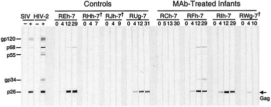
Assessment of Western blot-reactive antibodies and NAb titers revealed three different patterns of humoral immune responses among the animals in the SHIV89.6P study. The protected neonate, RCh-7, neither seroconverted (Fig. 4) nor developed a significant NAb titer (Table 2). This is consistent with undetectable virus replication in this animal (Fig. 3D and F). Two untreated control animals, RHh-7 and RJh-7, also had no detectable anti-SIV or anti-HIV-1 antibodies (Fig. 4 and data not shown) and no NAbs (Table 2). In these animals, however, rapid disease progression apparently precluded the ability to mount any antiviral antibody responses. A second pattern, observed in MAb-treated macaques RIh-7 and RFh-7 and untreated control animals REh-7 and RUg-7, was the development of antibodies to SIV Gag (Fig. 4) and HIV Env (data not shown), as well as significant titers (>100) of plasma NAb (Table 2). In three of the four animals (RIh-7, REh-7, and RUg-7), an increase in plasma NAbs preceded or coincided with a decrease in plasma viral load and an increase in CD4+ T-cell counts (data not shown). A third pattern was observed in the MAb-treated animal RWg-7, which transiently developed low level anti-Gag antibodies (Fig. 4). Animal RWg-7 progressed to disease and AIDS. In HIV-infected humans (7) as well as in SIV-infected macaques (2), selective loss of anti-Gag antibodies is a grave prognostic sign heralding the development of AIDS. The humoral response of RWg-7 followed just such a pattern.
FIG. 4.
Western blot analysis of plasma collected from SHIV89.6P-exposed animals. Serial samples were analyzed using HIV-2 strips. The number of weeks after challenge is indicated. Migration of HIV-2 proteins is shown on the left. The arrow on the right indicates migration of Gag antigen. Plasma from SIV-free macaques or normal human serum and plasma from SIV-infected macaques or human anti-HIV-2 serum were negative and positive controls, respectively (leftmost 4 lanes). †, sacrificed animal.
TABLE 2.
SHIV89.6P plasma neutralization titers after passive antibody infusion
| Group | Plasma neutralization titera
|
|
|---|---|---|
| 27–30b | 47–49 | |
| MAb-treated animals | ||
| RCh-7 | <10 | <10 |
| RFh-7 | 1,564 | 1,900 |
| RIh-7 | 279 | 155 |
| RWg-7 | <10 | |
| Untreated control animals | ||
| REh-7 | 1,182 | 933 |
| RHh-7 | <10 | |
| RJh-7 | 12 | |
| RUg-7 | 979 | 520 |
Reciprocal dilution which protected 50% of MT-2 cells from virus-induced cytotoxicity. This corresponds to 90% reduction of the viral Gag synthesis (9).
Weeks after virus exposure. Animals RWg-7, RHh-7, and RJh-7 were sacrificed in weeks 10, 7, and 9, respectively. Therefore, samples obtained 7 weeks postchallenge were analyzed.
Potential mechanisms of the protection observed in three MAb-treated animals.
Neonates P1, P2, and RCh-7 were not infected by all criteria tested and neither seroconverted nor developed NAb responses (Fig. 4, Table 2, and data not shown). P1, P2, and RCh-7 appeared to be protected from mucosal virus challenge by well-characterized pure human IgG MAbs. No mucosal NAbs of the IgA subtype were present in our MAb combination. How could systemically administered IgG MAbs protect against mucosal virus challenge? We tested saliva samples collected from P1, P2, and RCh-7 1 h after the first MAb infusion just prior to virus challenge in neutralization assays. They had no more neutralizing activity than the prechallenge samples collected from untreated control animals (all titers < 20). This observation was true for all MAb-treated animals as well (all titers < 20). Neutralizing titers, if present at all in saliva at the time of virus challenge, were below the lower detection limit of our assay. Thus, direct inactivation of SHIV-vpu+ and SHIV89.6P in saliva seems unlikely.
Given these results, it is possible that virus crossed the mucosal barrier and went through an initial round of infection in target cells of the local mucosal gastrointestinal tissue. The potent neutralizing MAbs would have stopped further waves of viral spread from these primary target cells. Nevertheless, limited local infection could have permitted the development of virus-specific, cellular immune responses, which in turn could have subsequently eliminated the small number of infected cells. To test this possibility, we evaluated CTL activity of PBMC obtained from RCh-7 46 weeks postchallenge; no significant specific lysis was detected in 51Cr release assays (data not shown). However, measuring systemic CTL activity in peripheral blood may not reflect specific antiviral cellular immunity at the level of the upper gastrointestinal mucosa itself.
To test for evidence of persistent mucosal immune protection, we rechallenged animal RCh-7 with 15 oral AID50 SHIV89.6P 54 weeks after the first challenge. We detected viral RNA in plasma obtained 1 week after the rechallenge (71,565 RNA copies/ml of plasma), and the CD4+ T-cell counts decreased from 1,516 to 684 cells/μl. Clearly, no protection was seen.
Potential mechanism that contributed to the recovery from the acute pathogenic effects of SHIV89.6P.
To gain more insight into what immune mechanisms might have contributed to the recovery of two of our four untreated controls (REh-7 and RUg-7) from the acute pathogenic effects of SHIV89.6P, we analyzed the humoral and cellular immune responses of these animals. As mentioned above, both animals developed high titers of NAbs against SHIV89.6P (Table 2). We evaluated the breadth of NAbs by assessing their ability to neutralize other SHIV variants and an HIV-1 strain as described earlier (18, 42, 43). We included plasma samples from REh-7 and RUg-7 collected 79 and 81 weeks postexposure, respectively. In addition, we assayed samples collected 35 and 135 weeks postexposure, respectively, from two other macaques, RDt-7 and RGt-6, that had been inoculated orally with SHIV89.6P as neonates and had recovered from the pathogenic manifestations of the infection. The plasma samples contained—in addition to anti-SHIV89.6P NAbs—variable and sometimes potent neutralizing activity against the heterologous T-cell-line-adapted HIV-1 strain MN (titers as high as 500) but no activity against SHIV89.6 and SHIV-KU2 (all titers < 20). We also tested specific cellular immune responses in five of our total six recovered macaques by using enzyme-linked immunospot assays (28) and found a significant number of SIVmac239 Gag- and HIV89.6 Env-specific T lymphocytes in all five animals (data not shown).
Safety of human MAb administration to neonatal macaques.
Infusions of MAb combinations were tolerated well in more than 15 neonatal macaques treated so far; no acute reactions were reported after any of the more than 30 i.v. infusions. One MAb-treated neonate (RCh-7) suffered a seizure approximately 2 days after the first MAb infusion. RCh-7 had a low birth weight (420 g) but subsequently gained body weight normally (1.68 kg at necropsy). No further seizures were reported. As in all of the other animals, the MAb infusion and the virus inoculation had been uneventful, suggesting that a connection between these manipulations and the observed seizure is unlikely. It is also improbable that it interfered with the challenge protocol, since 2 days had elapsed between virus challenge and the neurological events. Twenty-four days after virus challenge, physical examination of RCh-7 revealed partial paralysis of both lower legs (loss of reflexes); 7 days later, contracture and atrophy of the affected musculature were reported. Necropsy performed 55 weeks postchallenge revealed that RCh-7 was in reasonably good condition with the exception of the previously observed pelvic and leg muscle atrophy. In addition, focal cortical atrophy with sparing of the underlying white matter and cavitation of the underlying tissue were present in the left occipital-parietal junction, extending from the parietal lobe into the premotor cortex. Hemosiderin-laden macrophages were present at the borders of cavitated areas consistent with previous cortical hemorrhage. No evidence of vasculitis was detected. The unilateral lesion in the brain is not the likely cause of motor dysfunction, given the sparing of the motor areas and intactness of the cervical spinal cord. No specific diagnosis for the neurologic deficits could be obtained; however, the time course of events renders an etiologic association with the MAb treatment unlikely.
DISCUSSION
We infused triple combinations of human MAbs postnatally into six neonatal rhesus macaques to evaluate the protective potential against mucosal challenge with two different SHIV strains. We achieved complete protection in two of two MAb-treated neonates challenged with SHIV-vpu+ and one of four MAb-treated neonates exposed to SHIV89.6P, while all control animals exposed to either virus became viremic. An additional MAb-treated animal was protected from the rapid CD4+ T-cell depletion observed in all control animals after SHIV89.6P challenge. Thus, the MAb combination was partially successful in protecting animals from the pathogenic effects of the SHIV89.6P infection. Furthermore, protection occurred without administration of large prenatal maternal MAb doses.
The triple MAb combination of F105, 2G12, and 2F5 protected neonates against oral SHIV-vpu+ challenge after pre- and postnatal immunoprophylaxis (3); it was therefore used in the present study with SHIV-vpu+ as well. For the SHIV89.6P oral challenge study, F105 was replaced by IgG1b12 in the triple combination because this regimen produced the strongest in vitro neutralization against this virus. Interestingly, neutralization of SHIV89.6P by 2G12 and 2F5 was potentiated by the addition of IgG1b12. This was unexpected, since IgG1b12 alone did not inhibit SHIV89.6P replication in vitro. This result supports earlier studies that used either SHIV89.6P or SHIV-KB9, a molecular clone derived after the fourth passage of SHIV89.6P (18, 21, 27). Neutralization resistance of viruses might be due to a number of factors, including point mutations leading to loss of the relevant epitope (41) or to global conformational changes that make the epitope inaccessible to the MAb (47, 60, 64). The IgG1b12 epitope is present on the envelope protein of SHIV-KB9; IgG1b12 binds soluble KB9 gp120 monomers with undiminished capacity (21). However, the epitope seems less accessible for antibody binding in the context of the naturally folded oligomeric envelope glycoprotein complex on the SHIV-KB9 virion surface (21). IgG1b12 binds other viral strains with equal or better avidity to the oligomeric form of the envelope glycoprotein; this probably accounts for its exceptional potency (22, 54). Resistance to IgG1b12 has been mapped to the V1, V2, and V3 loops of the HIV-1 envelope (21, 41, 54). Antibodies 2G12 and 2F5 recognize epitopes unrelated to the V1, V2, or V3 loop or to the CD4BS (8, 17, 45, 62). We postulate that binding of either 2G12 or 2F5 induces a conformational change in the oligomeric envelope glycoprotein complex of SHIV89.6P that allows contact of IgG1b12 with its previously hidden cognate epitope.
The addition of a fourth antibody, F105, to the triple combination of IgG1b12, 2G12, and 2F5 led to a decrease in neutralization synergism against SHIV89.6P. F105 and IgG1b12 seem to inhibit each other, although they use different mechanisms to neutralize HIV. F105 inhibits the attachment of the neutralized virus to the target cell (49), while IgG1b12 inhibits the fusion entry process (39). Since they both recognize epitopes that at least partially overlap with the CD4BS, the diminished combined neutralization activity may be due to steric hindrance and competition for the binding site.
We achieved complete protection against oral SHIV-vpu+ challenge in two neonatal rhesus macaques that received postnatal treatment with a triple combination of human neutralizing MAbs. This chimeric virus encodes env of the laboratory-adapted HIV-1 IIIB. To mimic HIV infection of human newborns more closely, we then used a highly pathogenic chimeric virus that encodes env derived from the primary, dualtropic HIV89.6 and achieved partial protection. It is well known that primary strains of HIV are more difficult to neutralize than laboratory-adapted strains (19, 44). However, we had optimized the MAb combination previously for high in vitro neutralization of SHIV89.6P. Another factor that could have contributed to the different outcome of the two immunoprophylaxis studies could also be the higher challenge dose used of SHIV89.6P (15 AID50) than of SHIV-vpu+ (10 AID50).
Protection in the orally challenged neonates was achieved with only postnatal treatment. We chose this approach because the large amounts of each MAb required for treatment of the mother during pregnancy may render passive immunoprophylaxis approaches to prevent maternal HIV transmission in clinical trials too costly. Furthermore, we modeled our primate study on the successful prevention of maternal transmission of the hepatitis B virus, an enveloped virus that is transmitted through the same routes as HIV. Anti-hepatitis B immunoglobulin therapy is administered to newborn infants within 12 h of birth; no prenatal treatment is given to infected mothers. Passive immunization alone is 71% effective in preventing passage of hepatitis B virus to infants (5).
To our knowledge, the present study gives the first description of the course and outcome of SHIV89.6P infection of neonatal rhesus macaques. In adult rhesus monkeys, i.v. inoculation of SHIV89.6P led to high initial viral peak RNA loads and to profound, persistent depletion of CD4+ T cells within 2 weeks after virus challenge (4, 25, 53, 59, 63). According to most reports (42, 53, 63), SHIV89.6P infection leads to rapid disease progression and to AIDS in adult macaques, leaving the animals unable to seroconvert or mount any virus-specific CTL responses. This course of infection was seen in only two of our four naive neonates (RHh-7 and RJh-7). Interestingly, the other two untreated control animals (REh-7 and RUg-7) showed a rebound of CD4+ T-cell counts 8 weeks after virus exposure. These neonates seroconverted readily, developed high titers of NAbs, and remained clinically well throughout the 1-year observation period. A similar phenomenon was described recently by Barouch and coworkers (4). Two of eight rhesus monkeys that were challenged i.v. with 100 AID50 of SHIV89.6P did not show complete depletion of CD4+ T cells; one animal developed no significant disease, and only four of the eight macaques died by day 140 postexposure (4). The animals developed low virus-specific CD8+ CTL responses. However, in accordance with our observation in infant macaques, two of the adult animals developed high NAb titers by day 28 after virus exposure (4).
To further analyze the immune mechanisms that might have contributed to the recovery from the acute SHIV89.6P pathogenicity in our two untreated neonates, we assessed the breadth of their NAbs using in vitro neutralization assays. We included samples from REh-7, RUg-7, and two additional macaques that had been inoculated orally with SHIV89.6P as neonates and had recovered from the acute CD4+ T-cell depletion. Plasma samples from all four animals had NAb activity against SHIV89.6P and the HIV-1 strain MN but not against SHIV89.6 and SHIV-KU2. These results are similar to those reported for plasma samples from SHIV89.6PD-infected macaques (18, 42) and demonstrate the restricted nature of the induced NAbs also in SHIV89.6P-infected animals. In addition, all four untreated, recovered macaques had strong virus-specific cellular immune responses. In conclusion, these SHIV89.6P-infected macaques that recovered from the severe CD4+ T-cell depletion and survived for a prolonged period without significant disease demonstrated both strong NAb activity and cellular immune responses. It remains to be determined what additional factors are responsible for the variable outcomes observed among individual SHIV89.6P-infected adult and neonatal macaques.
Virus replication was undetectable in the peripheral blood of MAb-treated animals P1, P2, and RCh-7. The animals neither seroconverted nor developed NAb responses. Successful in vitro infection of CD8+-depleted PBMC collected 36 weeks after virus exposure from animal RCh-7 (data not shown) excluded the possibility of natural resistance to SHIV89.6P due to genetic predisposition, such as lack of coreceptors. Therefore, the triple combination of human neutralizing MAbs must have protected RCh-7 from systemic infection. It is not clear at what level the infused IgG MAbs stopped the virus. As in our previous study, in which neonates were completely protected against an oral SHIV-vpu+ challenge (3), we could not detect any significant neutralizing activity in the saliva of P1, P2, and RCh-7 at the time of challenge. While this does not rule out the possibility that low amounts of MAbs interacted with the virus in saliva, it is more likely that virus crossed the mucosal membrane and underwent a single round of replication in target cells of the local gastrointestinal tissues. Secondary waves of virus spread might then have been prevented either through direct virus neutralization by the parenterally infused IgG MAbs or via elimination of infected MAb-coated cells by antibody-dependent cell-mediated cytotoxicity (ADCC) or complement activation. Indeed, human anti-HIV MAb such as F105 were found to mediate ADCC (48), and 2G12 has both ADCC- and complement-mediated activity (62). Recently, ADCC or other cell-killing mechanisms were also suggested to cause transient reductions of viral load in SIVmac251-infected macaques infused with immunoglobulins (6). Alternatively, the initial infection of a set of susceptible target cells might have induced virus-specific cellular immune responses. However, no direct evidence for cellular immunity was seen—virus-specific CTL activity was not detected in the peripheral blood, and there was no protection from oral rechallenge 54 weeks after the first virus exposure.
The protection that we achieved in the four MAb-treated neonatal rhesus macaques after oral challenge with pathogenic SHIV89.6P may be compared to the level of protection reported for antibody-treated, adult macaques challenged vaginally with plasma-derived SHIV89.6PD (38). Similar doses (15 versus 10 to 50 AID50) and strains of virus (SHIV89.6P versus SHIV89.6PD [35, 52]) were used in the two studies. However, to achieve reproducible infection, the adult macaques were treated with progesterone prior to vaginal challenge, resulting in a thinning of the vaginal epithelium. This might have influenced the MAb efficacy by allowing more antibodies to seep across the mucosal barrier. In addition, we used a lower dose of antibody (10 mg/kg) than used in the adult study (15 mg/kg), and different combinations of antibodies were employed.
In conclusion, postnatal triple MAb combination overall prevented infection in three of six treated infants. Among SHIV89.6P-challenged animals, the MAb combination was partially successful in preventing infection; half of the treated infants were protected from the acute, severe T-cell depletion. The failure to protect all MAb-treated animals from SHIV89.6P infection did not seem to be related to neutralization resistance. Viruses from MAb-treated animal RFh-7 and untreated control animal REh-7, recovered 37 weeks postexposure, were still as sensitive to neutralization with the triple MAb combination as the original SHIV89.6P inoculum (data not shown). Even the partial protection that we report here is encouraging since the macaque-adapted SHIV89.6P (52) is more pathogenic in monkeys than HIV is in humans. To increase the degree of protection in future primate studies, we plan to include other synergistically acting MAbs in the combination regimen and/or increase the neonatal MAb doses.
Our approach is directly relevant for the development of a new strategy against maternal-fetal HIV-1 transmission in humans, since the MAbs used are human antibodies directed against HIV-1 glycoproteins. It may represent an important addition to the antiretroviral therapy protocols established to reduce mother-to-child HIV transmission. Being natural human proteins, human MAbs can be expected to have low toxicity. In symptomatic HIV-infected children, the prophylactic use of i.v. immunoglobulin for the prevention of bacterial infections was safe and was suggested as standard therapy (1, 55). In addition, MAbs 2G12 and 2F5 have been infused safely into HIV-infected adults in a phase I clinical trial (H. Katinger and G. Stiegler, unpublished observations). The stability and long half-lives of neutralizing human MAbs that we had noted in our earlier study in neonates (3) may yield another clinical benefit: the protection of infants against oral HIV transmission through infected breast milk in the neonatal period. Protection against this mode of maternal HIV transmission may be easier to achieve than against our SHIV89.6P oral challenge, since the infectivity of human breast milk is lower and does not yield a 99% probability of infection (40, 46) as our SHIV89.6P challenge dose did.
ACKNOWLEDGMENTS
We thank Matthew Frosch (Department of Pathology, Brigham and Women's Hospital, Boston, Mass.) for review of the neuropathological slides and helpful discussion. CHO cells producing MAb IgG1b12 were kindly provided by Dennis Burton (Department of Immunology and Molecular Biology, Scripps Research Institute, La Jolla, Calif.). We also thank Yulan Wang for technical assistance and C. Gallegos and S. Sharp for preparation of the manuscript.
This work was supported in part by National Institutes of Health grants RO1 AI34266, R21 AI46177, and PO1 AI48240 awarded to R.M.R., RR00165 to H.M.M., AI26926 to M.R.P. and AI45320 to L.A.C. It was also supported by the Pediatric AIDS Foundation grant 50864PG23 to R.M.R. and by Center for AIDS Research (CFAR) core grant IP30 28691 awarded to the Dana-Farber Cancer Institute. D.C.M. was supported by National Institutes of Health contract NO1 AI85343. R.H.-L. was supported by a grant from the Swiss National Science Foundation (fellowship 823A-50315) and is the recipient of a scholar award from the Friends of Switzerland, Inc. T.W.B. was a recipient of National Institutes of Health Clinical Investigator Development Award 30/35 KO8-AI01327.
REFERENCES
- 1.Anonymous. Intravenous immune globulin for the prevention of bacterial infections in children with symptomatic human immunodeficiency virus infection. The National Institute of Child Health and Human Developments Intravenous Immunoglobulin Study Group. N Engl J Med. 1991;325:73–80. doi: 10.1056/NEJM199107113250201. [DOI] [PubMed] [Google Scholar]
- 2.Baba T W, Koch J, Mittler E S, Greene M, Wyand M, Pennick D, Ruprecht R M. Mucosal infection of neonatal rhesus monkeys with cell-free SIV. AIDS Res Hum Retroviruses. 1994;10:351–357. doi: 10.1089/aid.1994.10.351. [DOI] [PubMed] [Google Scholar]
- 3.Baba T W, Liska V, Hofmann-Lehmann R, Vlasak J, Xu W, Ayehunie S, Cavacini L A, Posner M R, Katinger H, Stiegler G, Bernacky B J, Rizvi T A, Schmidt R, Hill L R, Keeling M E, Lu Y, Wright J E, Chou T C, Ruprecht R M. Human neutralizing monoclonal antibodies of the IgG1 subtype protect against mucosal simian-human immunodeficiency virus infection. Nat Med. 2000;6:200–206. doi: 10.1038/72309. [DOI] [PubMed] [Google Scholar]
- 4.Barouch D H, Santra S, Schmitz J E, Kuroda M J, Fu T M, Wagner W, Bilska M, Craiu A, Zheng X X, Krivulka G R, Beaudry K, Lifton M A, Nickerson C E, Trigona W L, Punt K, Freed D C, Guan L, Dubey S, Casimiro D, Simon A, Davies M E, Chastain M, Strom T B, Gelman R S, Montefiori D C, Lewis M G. Control of viremia and prevention of clinical AIDS in rhesus monkeys by cytokine-augmented DNA vaccination. Science. 2000;290:486–492. doi: 10.1126/science.290.5491.486. [DOI] [PubMed] [Google Scholar]
- 5.Beasley R P, Hwang L Y, Lee G C, Lan C C, Roan C H, Huang F Y, Chen C L. Prevention of perinatally transmitted hepatitis B virus infections with hepatitis B immune globulin and hepatitis B vaccine. Lancet. 1983;2:1099–1102. doi: 10.1016/s0140-6736(83)90624-4. [DOI] [PubMed] [Google Scholar]
- 6.Binley J M, Clas B, Gettie A, Vesanen M, Montefiori D C, Sawyer L, Booth J, Lewis M, Marx P A, Bonhoeffer S, Moore J P. Passive infusion of immune serum into simian immunodeficiency virus-infected rhesus macaques undergoing a rapid disease course has minimal effect on plasma viremia. Virology. 2000;270:237–249. doi: 10.1006/viro.2000.0254. [DOI] [PubMed] [Google Scholar]
- 7.Binley J M, Klasse P J, Cao Y, Jones I, Markowitz M, Ho D D, Moore J P. Differential regulation of the antibody responses to Gag and Env proteins of human immunodeficiency virus type 1. J Virol. 1997;71:2799–2809. doi: 10.1128/jvi.71.4.2799-2809.1997. [DOI] [PMC free article] [PubMed] [Google Scholar]
- 8.Buchacher A, Predl R, Strutzenberger K, Steinfellner W, Trkola A, Purtscher M, Gruber G, Tauer C, Steindl F, Jungbauer A, Katinger H. Generation of human monoclonal antibodies against HIV-1 proteins; electrofusion and Epstein-Barr virus transformation for peripheral blood lymphocyte immortalization. AIDS Res Hum Retroviruses. 1994;10:359–369. doi: 10.1089/aid.1994.10.359. [DOI] [PubMed] [Google Scholar]
- 9.Bures R, Gaitan A, Zhu T, Graziosi C, McGrath K, Tartaglia J, Caudrelier P, El Habib R, Klein M, Lazzarin A, Stablein D M, Corey L, Greenberg M L, Schwartz D H, Montefiori D C. Immunization with recombinant canarypox vectors expressing membrane-anchored gp120 followed by soluble gp160 boosting fails to generate antibodies that neutralize R5 primary isolates of human immunodeficiency virus type 1. AIDS Res Hum Retroviruses. 2000;16:2019–2035. doi: 10.1089/088922200750054756. [DOI] [PubMed] [Google Scholar]
- 10.Burton D R, Pyati J, Koduri R, Sharp S J, Thornton G B, Parren P W, Sawyer L S, Hendry R M, Dunlop N, Nata P L, Lamacchia M, Garratty E, Stiehm E R, Bryson Y J, Cao Y, Moore J P, Ho D D, Barbas C F. Efficient neutralization of primary isolates of HIV-1 by a recombinant human monoclonal antibody. Science. 1994;266:1024–1027. doi: 10.1126/science.7973652. [DOI] [PubMed] [Google Scholar]
- 11.Capon D J, Chamow S M, Mordenti J, Marsters S A, Gregory T, Mitsuya H, Byrn R A, Lucas C, Wurm F M, Groopman J E, et al. Designing CD4 immunoadhesins for AIDS therapy. Nature. 1989;337:525–531. doi: 10.1038/337525a0. [DOI] [PubMed] [Google Scholar]
- 12.Chou T C. The median-effect principle and the combination index for quantitation of synergism and antagonism. In: Chou T C, Rideout D C, editors. Synergism and antagonism in chemotherapy. San Diego, Calif: Academic Press; 1991. pp. 61–102. [Google Scholar]
- 13.Chou T C, Hayball M. CalcuSyn for Windows. Multi-drug dose-effect analyzer and manual. Cambridge, United Kingdom: Biosoft; 1996. [Google Scholar]
- 14.Chou T C, Talalay P. Generalized equations for the analysis of inhibitions of Michaelis-Menten and higher-order kinetic systems with two or more mutually exclusive and nonexclusive inhibitors. Eur J Biochem. 1981;115:207–216. doi: 10.1111/j.1432-1033.1981.tb06218.x. [DOI] [PubMed] [Google Scholar]
- 15.Chou T C, Talalay P. Quantitative analysis of dose-effect relationships: the combined effects of multiple drugs or enzyme inhibitors. Adv Enzyme Regul. 1984;22:27–55. doi: 10.1016/0065-2571(84)90007-4. [DOI] [PubMed] [Google Scholar]
- 16.Collman R, Balliet J W, Gregory S A, Friedman H, Kolson D L, Nathanson N, Srinivasan A. An infectious molecular clone of an unusual macrophage-tropic and highly cytopathic strain of human immunodeficiency virus type 1. J Virol. 1992;66:7517–7521. doi: 10.1128/jvi.66.12.7517-7521.1992. [DOI] [PMC free article] [PubMed] [Google Scholar]
- 17.Conley A J, Kessler J A, II, Boots L J, Tung J S, Arnold B A, Keller P M, Shaw A R, Emini E A. Neutralization of divergent human immunodeficiency virus type 1 variants and primary isolates by IAM-41-2F5, an anti-gp41 human monoclonal antibody. Proc Natl Acad Sci USA. 1994;91:3348–3352. doi: 10.1073/pnas.91.8.3348. [DOI] [PMC free article] [PubMed] [Google Scholar]
- 18.Crawford J M, Earl P L, Moss B, Reimann K A, Wyand M S, Manson K H, Bilska M, Zhou J T, Pauza C D, Parren P W, Burton D R, Sodroski J G, Letvin N L, Montefiori D C. Characterization of primary isolate-like variants of simian-human immunodeficiency virus. J Virol. 1999;73:10199–10207. doi: 10.1128/jvi.73.12.10199-10207.1999. [DOI] [PMC free article] [PubMed] [Google Scholar]
- 19.Daar E S, Li X L, Moudgil T, Ho D D. High concentrations of recombinant soluble CD4 are required to neutralize primary human immunodeficiency virus type 1 isolates. Proc Natl Acad Sci USA. 1990;87:6574–6578. doi: 10.1073/pnas.87.17.6574. [DOI] [PMC free article] [PubMed] [Google Scholar]
- 20.D'Souza M P, Livnat D, Bradac J A, Bridges S H. Evaluation of monoclonal antibodies to human immunodeficiency virus type 1 primary isolates by neutralization assays: performance criteria for selecting candidate antibodies for clinical trials. AIDS Clinical Trials Group Antibody Selection Working Group. J Infect Dis. 1997;175:1056–1062. doi: 10.1086/516443. [DOI] [PubMed] [Google Scholar]
- 21.Etemad-Moghadam B, Sun Y, Nicholson E K, Karlsson G B, Schenten D, Sodroski J. Determinants of neutralization resistance in the envelope glycoproteins of a simian-human immunodeficiency virus passaged in vivo. J Virol. 1999;73:8873–8879. doi: 10.1128/jvi.73.10.8873-8879.1999. [DOI] [PMC free article] [PubMed] [Google Scholar]
- 22.Fouts T R, Binley J M, Trkola A, Robinson J E, Moore J P. Neutralization of the human immunodeficiency virus type 1 primary isolate JR-FL by human monoclonal antibodies correlates with antibody binding to the oligomeric form of the envelope glycoprotein complex. J Virol. 1997;71:2779–2785. doi: 10.1128/jvi.71.4.2779-2785.1997. [DOI] [PMC free article] [PubMed] [Google Scholar]
- 23.Gallo R C, Salahuddin S Z, Popovic M, Shearer G M, Kaplan M, Haynes B F, Palker T J, Redfield R, Oleske J, Safai B, et al. Frequent detection and isolation of cytopathic retroviruses (HTLV-III) from patients with AIDS and at risk for AIDS. Science. 1984;224:500–503. doi: 10.1126/science.6200936. [DOI] [PubMed] [Google Scholar]
- 24.Hofmann-Lehmann R, Swenderten R K, Liska V, Leutenegger C M, Lutz H, McClure H M, Ruprecht R M. Sensitive and robust one-tube real-time reverse transcriptase-polymerase chain reaction to quantify SIV RNA load: Comparison of one- vs. two-enzyme systems. AIDS Res Hum Retroviruses. 2000;16:1247–1257. doi: 10.1089/08892220050117014. [DOI] [PubMed] [Google Scholar]
- 25.Iida T, Ichimura H, Shimada T, Ibuki K, Ui M, Tamaru K, Kuwata T, Yonehara S, Imanishi J, Hayami M. Role of apoptosis induction in both peripheral lymph nodes and thymus in progressive loss of CD4+ cells in SHIV-infected macaques. AIDS Res Hum Retroviruses. 2000;16:9–18. doi: 10.1089/088922200309557. [DOI] [PubMed] [Google Scholar]
- 26.Joag S V, Li Z, Foresman L, Pinson D M, Raghavan R, Zhuge W, Adany I, Wang C, Jia F, Sheffer D, Ranchalis J, Watson A, Narayan O. Characterization of the pathogenic KU-SHIV model of acquired immunodeficiency syndrome in macaques. AIDS Res Hum Retroviruses. 1997;13:635–645. doi: 10.1089/aid.1997.13.635. [DOI] [PubMed] [Google Scholar]
- 27.Karlsson G B, Halloran M, Li J, Park I W, Gomila R, Reimann K A, Axthelm M K, Iliff S A, Letvin N L, Sodroski J. Characterization of molecularly cloned simian-human immunodeficiency viruses causing rapid CD4+ lymphocyte depletion in rhesus monkeys. J Virol. 1997;71:4218–4225. doi: 10.1128/jvi.71.6.4218-4225.1997. [DOI] [PMC free article] [PubMed] [Google Scholar]
- 28.Larsson M, Jin X, Ramratnam B, Ogg G S, Engelmayer J, Demoitie M A, McMichael A J, Cox W I, Steinman R M, Nixon D, Bhardwaj N. A recombinant vaccinia virus based ELISPOT assay detects high frequencies of Pol-specific CD8 T cells in HIV-1-positive individuals. AIDS. 1999;13:767–777. doi: 10.1097/00002030-199905070-00005. [DOI] [PubMed] [Google Scholar]
- 29.Li A, Baba T W, Sodroski J, Zolla-Pazner S, Gorny M K, Robinson J, Posner M R, Katinger H, Barbas C F, III, Burton D R, Chou T C, Ruprecht R M. Synergistic neutralization of a chimeric SIV/HIV type 1 virus with combinations of human anti-HIV type 1 envelope monoclonal antibodies or hyperimmune globulins. AIDS Res Hum Retroviruses. 1997;13:647–656. doi: 10.1089/aid.1997.13.647. [DOI] [PubMed] [Google Scholar]
- 30.Li A, Katinger H, Posner M R, Cavacini L, Zolla-Pazner S, Gorny M K, Sodroski J, Chou T C, Baba T W, Ruprecht R M. Synergistic neutralization of simian-human immunodeficiency virus SHIV-vpu+ by triple and quadruple combinations of human monoclonal antibodies and high-titer anti-human immunodeficiency virus type 1 immunoglobulins. J Virol. 1998;72:3235–3240. doi: 10.1128/jvi.72.4.3235-3240.1998. [DOI] [PMC free article] [PubMed] [Google Scholar]
- 31.Li J T, Halloran M, Lord C I, Watson A, Ranchalis J, Fung M, Letvin N L, Sodroski J G. Persistent infection of macaques with simian-human immunodeficiency viruses. J Virol. 1995;69:7061–7067. doi: 10.1128/jvi.69.11.7061-7067.1995. [DOI] [PMC free article] [PubMed] [Google Scholar]
- 32.Liska V, Fultz P N, Su L, Ruprecht R M. Detection of simian T cell leukemia virus type I infection in seronegative macaques. AIDS Res Hum Retroviruses. 1997;13:1147–1153. doi: 10.1089/aid.1997.13.1147. [DOI] [PubMed] [Google Scholar]
- 33.Liska V, Khimani A H, Hofmann-Lehmann R, Fink A N, Vlasak J, Ruprecht R M. Viremia and AIDS in rhesus macaques after intramuscular inoculation of plasmid DNA encoding full-length SIVmac239. AIDS Res Hum Retroviruses. 1999;15:445–450. doi: 10.1089/088922299311196. [DOI] [PubMed] [Google Scholar]
- 34.Liska V, Lerche N W, Ruprecht R M. Simultaneous detection of simian retrovirus type D serotypes 1, 2, and 3 by polymerase chain reaction. AIDS Res Hum Retroviruses. 1997;13:433–437. doi: 10.1089/aid.1997.13.433. [DOI] [PubMed] [Google Scholar]
- 35.Lu Y, Pauza C D, Lu X, Montefiori D C, Miller C J. Rhesus macaques that become systemically infected with pathogenic SHIV 89.6-PD after intravenous, rectal, or vaginal inoculation and fail to make an antiviral antibody response rapidly develop AIDS, J. Acquir Immune Defic Syndr. 1998;19:6–18. doi: 10.1097/00042560-199809010-00002. [DOI] [PubMed] [Google Scholar]
- 36.Lu Y, Salvato M S, Pauza C D, Li J, Sodroski J, Manson K, Wyand M, Letvin N, Jenkins S, Touzjian N, Chutkowski C, Kushner N, LeFaile M, Payne L G, Roberts B. Utility of SHIV for testing HIV-1 vaccine candidates in macaques. J Acquir Immune Defic Syndr. 1996;12:99–106. doi: 10.1097/00042560-199606010-00001. [DOI] [PubMed] [Google Scholar]
- 37.Mascola J R, Lewis M G, Stiegler G, Harris D, VanCott T C, Hayes D, Louder M K, Brown C R, Sapan C V, Frankel S S, Lu Y, Robb M L, Katinger H, Birx D L. Protection of macaques against pathogenic simian/human immunodeficiency virus 89.6PD by passive transfer of neutralizing antibodies. J Virol. 1999;73:4009–4018. doi: 10.1128/jvi.73.5.4009-4018.1999. [DOI] [PMC free article] [PubMed] [Google Scholar]
- 38.Mascola J R, Stiegler G, VanCott T C, Katinger H, Carpenter C B, Hanson C E, Beary H, Hayes D, Frankel S S, Birx D L, Lewis M G. Protection of macaques against vaginal transmission of a pathogenic HIV-1/SIV chimeric virus by passive infusion of neutralizing antibodies. Nat Med. 2000;6:207–210. doi: 10.1038/72318. [DOI] [PubMed] [Google Scholar]
- 39.McInerney T L, McLain L, Armstrong S J, Dimmock N J. A human IgG1 (b12) specific for the CD4 binding site of HIV-1 neutralizes by inhibiting the virus fusion entry process, but b12 Fab neutralizes by inhibiting a postfusion event. Virology. 1997;233:313–326. doi: 10.1006/viro.1997.8547. [DOI] [PubMed] [Google Scholar]
- 40.Miotti P G, Taha T E, Kumwenda N I, Broadhead R, Mtimavalye L A, Van der Hoeven L, Chiphangwi J D, Liomba G, Biggar R J. HIV transmission through breastfeeding: a study in Malawi. JAMA. 1999;282:744–749. doi: 10.1001/jama.282.8.744. [DOI] [PubMed] [Google Scholar]
- 41.Mo H, Stamatatos L, Ip J E, Barbas C F, Parren P W, Burton D R, Moore J P, Ho D D. Human immunodeficiency virus type 1 mutants that escape neutralization by human monoclonal antibody IgG1b12. J Virol. 1997;71:6869–6874. doi: 10.1128/jvi.71.9.6869-6874.1997. [DOI] [PMC free article] [PubMed] [Google Scholar]
- 42.Montefiori D C, Reimann K A, Wyand M S, Manson K, Lewis M G, Collman R G, Sodroski J G, Bolognesi D P, Letvin N L. Neutralizing antibodies in sera from macaques infected with chimeric simian-human immunodeficiency virus containing the envelope glycoproteins of either a laboratory-adapted variant or a primary isolate of human immunodeficiency virus type 1. J Virol. 1998;72:3427–3431. doi: 10.1128/jvi.72.4.3427-3431.1998. [DOI] [PMC free article] [PubMed] [Google Scholar]
- 43.Montefiori D C, Robinson W B, Schuffman S S. Evaluation of antiviral drugs and neutralizing antibodies to human immunodeficiency virus by a rapid and sensitive microtiter infection assay. J Clin Microbiol. 1988;26:231–235. doi: 10.1128/jcm.26.2.231-235.1988. [DOI] [PMC free article] [PubMed] [Google Scholar]
- 44.Moore J P, Cao Y, Qing L, Sattentau Q J, Pyati J, Koduri R, Robinson J, Barbas III C F, Burton D R, Ho D D. Primary isolates of human immunodeficiency virus type 1 are relatively resistant to neutralization by monoclonal antibodies to gp120, and their neutralization is not predicted by studies with monomeric gp120. J Virol. 1995;69:101–109. doi: 10.1128/jvi.69.1.101-109.1995. [DOI] [PMC free article] [PubMed] [Google Scholar]
- 45.Muster T, Steindl F, Purtscher M, Trkola A, Klima A, Himmler G, Ruker F, Katinger H. A conserved neutralizing epitope on gp41 of human immunodeficiency virus type 1. J Virol. 1993;67:6642–6647. doi: 10.1128/jvi.67.11.6642-6647.1993. [DOI] [PMC free article] [PubMed] [Google Scholar]
- 46.Nduati R, John G, Mbori-Ngacha D, Richardson B, Overbaugh J, Mwatha A, Ndinya-Achola J, Bwayo J, Onyango F E, Hughes J, Kreiss J. Effect of breastfeeding and formula feeding on transmission of HIV-1: a randomized clinical trial. JAMA. 2000;283:1167–1174. doi: 10.1001/jama.283.9.1167. [DOI] [PubMed] [Google Scholar]
- 47.Parren P W, Wang M, Trkola A, Binley J M, Purtscher M, Katinger H, Moore J P, Burton D R. Antibody neutralization-resistant primary isolates of human immunodeficiency virus type 1. J Virol. 1998;72:10270–10274. doi: 10.1128/jvi.72.12.10270-10274.1998. [DOI] [PMC free article] [PubMed] [Google Scholar]
- 48.Posner M R, Elboim H S, Cannon T, Cavacini L, Hideshima T. Functional activity of an HIV-1 neutralizing IgG human monoclonal antibody: ADCC and complement-mediated lysis. AIDS Res Hum Retroviruses. 1992;8:553–558. doi: 10.1089/aid.1992.8.553. [DOI] [PubMed] [Google Scholar]
- 49.Posner M R, Hideshima T, Cannon T, Mukherjee M, Mayer K H, Byrn R A. An IgG human monoclonal antibody that reacts with HIV-1/GP120, inhibits virus binding to cells, and neutralizes infection. J Immunol. 1991;146:4325–4332. [PubMed] [Google Scholar]
- 50.Rabin H, Neubauer R H, Hopkins R F d, Dzhikidze E K, Shevtsova Z V, Lapin B A. Transforming activity and antigenicity of an Epstein-Barr-like virus from lymphoblastoid cell lines of baboons with lymphoid disease. Intervirology. 1977;8:240–249. doi: 10.1159/000148899. [DOI] [PubMed] [Google Scholar]
- 51.Reimann K A, Li J T, Lekutis C, Tenner-Racz K, Racz P, Lin W, Montefiori D C, Lee-Paritz D E, Lu Y, Collman R G, Sodroski J, Letvin N L. An env gene derived from a primary human immunodeficiency virus type 1 isolate confers high in vivo replicative capacity to a chimeric simian/human immunodeficiency virus in rhesus monkeys. J Virol. 1996;70:3198–3206. doi: 10.1128/jvi.70.5.3198-3206.1996. [DOI] [PMC free article] [PubMed] [Google Scholar]
- 52.Reimann K A, Li J T, Veazey R, Halloran M, Park I W, Karlsson G B, Sodroski J, Letvin N L. A chimeric simian/human immunodeficiency virus expressing a primary patient human immunodeficiency virus type 1 isolate env causes an AIDS-like disease after in vivo passage in rhesus monkeys. J Virol. 1996;70:6922–6928. doi: 10.1128/jvi.70.10.6922-6928.1996. [DOI] [PMC free article] [PubMed] [Google Scholar]
- 53.Reimann K A, Watson A, Dailey P J, Lin W, Lord C I, Steenbeke T D, Parker R A, Axthelm M K, Karlsson G B. Viral burden and disease progression in rhesus monkeys infected with chimeric simian-human immunodeficiency viruses. Virology. 1999;256:15–21. doi: 10.1006/viro.1999.9632. [DOI] [PubMed] [Google Scholar]
- 54.Roben P, Moore J P, Thali M, Sodroski J, Barbas III C F, Burton D R. Recognition properties of a panel of human recombinant Fab fragments to the CD4 binding site of gp120 that show differing abilities to neutralize human immunodeficiency virus type 1. J Virol. 1994;68:4821–4828. doi: 10.1128/jvi.68.8.4821-4828.1994. [DOI] [PMC free article] [PubMed] [Google Scholar]
- 55.Schaad U B, Gianella-Borradori A, Perret B, Imbach P, Morell A. Intravenous immune globulin in symptomatic paediatric human immunodeficiency virus infection. Eur J Pediatr. 1988;147:300–303. doi: 10.1007/BF00442700. [DOI] [PubMed] [Google Scholar]
- 56.Shibata R, Igarashi T, Haigwood N, Buckler-White A, Ogert R, Ross W, Willey R, Cho M W, Martin M A. Neutralizing antibody directed against the HIV-1 envelope glycoprotein can completely block HIV-1/SIV chimeric virus infections of macaque monkeys. Nat Med. 1999;5:204–210. doi: 10.1038/5568. [DOI] [PubMed] [Google Scholar]
- 57.Shibata R, Maldarelli F, Siemon C, Matano T, Parta M, Miller G, Fredrickson T, Martin M A. Infection and pathogenicity of chimeric simian-human immunodeficiency viruses in macaques: determinants of high virus loads and CD4 cell killing. J Infect Dis. 1997;176:362–373. doi: 10.1086/514053. [DOI] [PubMed] [Google Scholar]
- 58.Spouge J L. Statistical analysis of sparse infection data and its implications for retroviral treatment trials in primates. Proc Natl Acad Sci USA. 1992;89:7581–7585. doi: 10.1073/pnas.89.16.7581. [DOI] [PMC free article] [PubMed] [Google Scholar]
- 59.Ten Haaft P, Verstrepen B, Uberla K, Rosenwirth B, Heeney J. A pathogenic threshold of virus load defined in simian immunodeficiency virus- or simian-human immunodeficiency virus-infected macaques. J Virol. 1998;72:10281–10285. doi: 10.1128/jvi.72.12.10281-10285.1998. [DOI] [PMC free article] [PubMed] [Google Scholar]
- 60.Thali M, Charles M, Furman C, Cavacini L, Posner M, Robinson J, Sodroski J. Resistance to neutralization by broadly reactive antibodies to the human immunodeficiency virus type 1 gp120 glycoprotein conferred by a gp41 amino acid change. J Virol. 1994;68:674–680. doi: 10.1128/jvi.68.2.674-680.1994. [DOI] [PMC free article] [PubMed] [Google Scholar]
- 61.Trkola A, Pomales A B, Yuan H, Korber B, Maddon P J, Allaway G P, Katinger H, Barbas C F, Burton D R, Ho D D, Moore J P. Cross-clade neutralization of primary isolates of human immunodeficiency virus type 1 by human monoclonal antibodies and tetrameric CD4-IgG. J Virol. 1995;69:6609–6617. doi: 10.1128/jvi.69.11.6609-6617.1995. [DOI] [PMC free article] [PubMed] [Google Scholar]
- 62.Trkola A, Purtscher M, Muster T, Ballaun C, Buchacher A, Sullivan N, Srinivasan K, Sodroski J, Moore J P, Katinger H. Human monoclonal antibody 2G12 defines a distinctive neutralization epitope on the gp120 glycoprotein of human immunodeficiency virus type 1. J Virol. 1996;70:1100–1108. doi: 10.1128/jvi.70.2.1100-1108.1996. [DOI] [PMC free article] [PubMed] [Google Scholar]
- 63.Wyand M S, Manson K, Montefiori D C, Lifson J D, Johnson R P, Desrosiers R C. Protection by live, attenuated simian immunodeficiency virus against heterologous challenge. J Virol. 1999;73:8356–8363. doi: 10.1128/jvi.73.10.8356-8363.1999. [DOI] [PMC free article] [PubMed] [Google Scholar]
- 64.Wyatt R, Sodroski J. The HIV-1 envelope glycoproteins: fusogens, antigens, and immunogens. Science. 1998;280:1884–1888. doi: 10.1126/science.280.5371.1884. [DOI] [PubMed] [Google Scholar]


