Abstract
The ability of dendritic cells (DCs) to shape the adaptive immune response to viral infection is mediated largely by their maturation and activation state as determined by the surface expression of HLA molecules, costimulatory molecules, and cytokine production. Dengue is an emerging arboviral disease where the severity of illness is influenced by the adaptive immune response to the virus. In this report, we have demonstrated that dengue virus infects and replicates in immature human myeloid DCs. Exposure to live dengue virus led to maturation and activation of both the infected and surrounding, uninfected DCs and stimulated production of tumor necrosis factor alpha (TNF-α) and alpha interferon (IFN-α). Activation of the dengue virus-infected DCs was blunted compared to the surrounding, uninfected DCs, and dengue virus infection induced low-level release of interleukin-12 p70 (IL-12 p70), a key cytokine in the development of cell-mediated immunity (CMI). Upon the addition of IFN-γ, there was enhanced activation of dengue virus-infected DCs and enhanced dengue virus-induced IL-12 p70 release. The data suggest a model whereby DCs are the early, primary target of dengue virus in natural infection and the vigor of CMI is modulated by the relative presence or absence of IFN-γ in the microenvironment surrounding the virus-infected DCs. These findings are relevant to understanding the pathogenesis of dengue hemorrhagic fever and the design of new vaccination and therapeutic strategies.
Dendritic cells (DCs) are bone marrow-derived cells that form a system of professional antigen-presenting cells and are an important component of the innate immune response. They are comprised of at least three distinct subpopulations, one in the lymphoid/plasmacytoid lineage and two in the myeloid lineage (1, 20, 26). Myeloid DCs are found in most nonlymphoid organs including the epidermis (Langerhans cells), dermis, gastrointestinal and respiratory mucosa, and the interstitia of vascular organs (37). Following the uptake and processing of antigen in the periphery, immature myeloid DCs differentiate to an activated/mature state and migrate to the T-cell-rich areas of lymphoid organs. Activated DCs are the unique stimulators of primary T-cell responses and potent stimulators of memory responses, and they produce an array of cytokines and chemokines (26, 44, 50, 55). Thus, DCs are critical in the initiation of antimicrobial immunity, and they provide a crucial step in the development of adaptive immunity.
Dengue is an emerging arboviral disease where the adaptive immune response plays a significant role in determining the severity of clinical illness. The dengue viruses are a group of four antigenically related mosquito-borne flaviviruses that produce a spectrum of clinical illness and significant morbidity throughout the tropics (30, 35). Dengue hemorrhagic fever (DHF) and dengue shock syndrome (DSS) represent the most severe and potentially life-threatening manifestations of a dengue viral infection. DHF/DSS is characterized by the rapid onset of plasma leakage and coagulopathy near the time of defervescence and viremia resolution. The most significant risk factor for the development of DHF/DSS is acquisition of a second, heterotypic dengue virus infection (3, 11, 13). During this second dengue virus infection, it is postulated that the preexisting, cross-reactive, adaptive immune response leads to excessive cytokine production, complement activation, and the release of other phlogistic factors which produce DHF/DSS. Both the humoral and cellular components of adaptive immunity have been implicated in this process (12, 40). The principal target of dengue virus infection has been presumed to be blood monocytes and tissue macrophages (14, 46). However, myeloid DCs residing in the epidermis (Langerhans cells) and dermis are the predominant cells of the innate immune system that dengue virus encounters following the bite of an infected mosquito. A recent study demonstrated the permissiveness of immature myeloid DCs to dengue virus infection but did not address the effect of viral infection on the DCs (53). In this study, we further investigated the interaction between dengue virus and myeloid DCs. Immature myeloid DCs were generated from plastic-adherent peripheral blood mononuclear cells (PBMC) and were considered representative of myeloid interstitial DCs (1). Viral replication, DC maturation and activation, and cytokine production were examined in the hope of understanding the factors that guide formation of antiviral adaptive immunity, and, under certain conditions, increase the risk of developing severe disease.
MATERIALS AND METHODS
Generation of DCs.
Immature myeloid DCs were generated from PBMC using previously described techniques (38, 39, 44). Peripheral blood was collected in heparinized tubes from healthy adult volunteers. PBMC were isolated on Histopaque gradients (Sigma Chemical Co., St. Louis, Mo.), washed two times with RPMI 1640 medium (Gibco BRL, Gaithersburg, Md.), and incubated with neuraminidase-treated sheep red blood cells for 1 h on ice. Erythrocyte rosette-negative cells were collected and isolated using Histopaque gradient centrifugation. The T-cell-depleted, erythrocyte rosette-negative cells were cultured (3 × 10⁶ cells/well) for 1 h in 24-well plates at 37°C in a CO2 incubator with RPMI 1640 and 10% heat-inactivated fetal calf serum (FCS; Gibco BRL). Nonadherent cells were removed, and medium was replaced with the addition of human recombinant interleukin-4 (rIL-4; 500 U/ml; Endogen Inc., Woburn, Mass.) and human recombinant granulocyte-macrophage colony-stimulating factor (rGM-CSF; 800 U/ml; Endogen). Fresh medium and cytokines (rIL-4 plus rGM-CSF) were replaced every 2 to 3 days. After 7 days, the loosely adherent DCs were collected by vigorous pipetting. The cells obtained expressed typical myeloid DC markers, being CD3−, CD14−, CD16−, CD20−, CD56−, CD40+, HLA-DR+, and CD1a+. They had an immature DC phenotype, being CD83−, and comprised 80 to 85% of the cell population. The remaining cells were predominantly B lymphocytes and were not infected following dengue virus exposure (data not shown).
Dengue virus infection of DCs.
Dengue type 2 virus strain 16681 (DEN-2 16681) was grown and propagated in the mosquito cell line C6/36. The titers of virus stocks were determined by limiting dilution plaque assay on LLC-MK2 cells (41). In some experiments, virus was inactivated by UV irradiation from a germicidal lamp (60 min) or heat treatment (65°C for 30 min); the absence of viable virus was confirmed by plaque assay.
Immature myeloid DCs were plated in three wells (1.5 × 10⁶/well) and infected with DEN-2 16681 at a multiplicity of infection (MOI) of 5 (two wells) or mock infected (one well). After a 2-h incubation at 37°C in medium containing RPMI 1640 and 5% heat-inactivated FCS, supernatant was removed and the cells were washed twice. The DCs were then cultured in 24 well plates at 37°C in a CO2 incubator with RPMI 1640–10% FCS–rIL-4–rGM-CSF (final volume, 1 ml). In some experiments, human recombinant gamma interferon (rIFN-γ; Endogen) was added to one well with DEN-2-infected DCs at a final concentration of 100 U/ml. At 48 h, DCs were collected by vigorous pipetting and aliquoted into tubes for fluorescence-activated cell sorting (FACS) immunostaining; viable DCs were counted by trypan blue staining. Cell-free supernatants were collected, aliquoted, and stored at −70°C. The supernatant virus titers were determined by plaque assay on LLC-MK2 cells as previously noted. Cytokine levels were also determined in the supernatants by enzyme-linked immunosorbent assay (ELISA).
The presence of contaminating lipopolysaccharide (LPS) in the culture supernatants, media, and virus stock was assessed with the Limulus amoebocyte lysate assay (Whittaker M. A. Bioproducts, Walkersville, Md.). Mycoplasma contamination in the culture supernatants, virus stock, and C6/36 cell line was evaluated by the Mycoplasma Rapid Detection System (Gen-Probe, San Diego, Calif.).
Antibodies for flow cytometry.
The following monoclonal antibodies (MAbs) were purchased from Pharmingen (San Diego, Calif.): fluorescein isothiocyanate (FITC)-conjugated mouse anti-human CD3, anti-human CD14, anti-human CD20, and isotype control (immunoglobulin G1 [IgG1]) MAbs; phycoerythrin-conjugated mouse anti-human HLA-DR, anti-human HLA-ABC, anti-human CD83, anti-human CD86, anti-human CD40, anti-human CD16, anti-human CD56, and isotype control (IgG1) MAbs; and CyChrome-conjugated mouse anti-human CD1a MAb. Phycoerythin-conjugated mouse anti-human CD80 (IgG1) MAb was purchased from Immunotech (Marseille, France). Mouse polyclonal anti-DEN-2 antibody (Ab) was purified from hyperimmune mouse ascitic fluid (AFRIMS, Bangkok, Thailand) and directly conjugated to FITC (FITC isomer I; Sigma).
Immunostaining and flow cytometry analysis.
DCs were resuspended in staining buffer (phosphate-buffered saline, 1% heat-inactivated FCS, 0.09% sodium azide) and aliquoted at 1.5 × 10⁵ cells/tube. After washing twice, cells were incubated for 30 min at 4°C with fluorescent MAbs to cell surface antigens (10 μl/tube). Cells were then fixed and permeabilized with Cytofix/Cytoperm according to the recommendations of the manufacturer (Pharmingen). Intracellular staining for dengue virus was accomplished by incubation with the FITC-conjugated anti-DEN-2 Ab (1 μg/tube) at 4°C for 30 min. After being washed twice, cells were resuspended in 0.3 ml of staining buffer prior to FACS analysis. Samples were analyzed using a FACScan flow cytometer (Becton Dickinson) and the CellQuest software package. In all experiments, three-color staining was simultaneously performed for (i) intracellular dengue virus, (ii) cell surface expression of CD83, costimulatory molecules, or HLA antigens, and (iii) surface CD1a expression. All analyses were performed on the CD1a+ cell population gate; a minimum of 50,000 events was acquired.
Determination of antiflavivirus antibody.
The presence of antiflavivirus antibody was determined in the sera of all donors. Neutralizing Ab titers to dengue virus serotypes 1 to 4 and Japanese encephalitis virus were measured by a plaque reduction neutralization assay (41). The 50% plaque reduction neutralization titer was used for comparisons; a titer of >1:10 was considered evidence of prior exposure.
ELISAs for TNF-α and IFN-α.
ELISA plates (Immulon 1; Dynex Technologies, Chantilly, Va.) were coated overnight at room temperature with the appropriate Ab (for TNF-α, mouse anti-human TNF-α MAb 2TNF-H34A [2.0 μg/ml]; for IFN-α, sheep polyclonal anti-human IFN-α Ab [0.5 μg/ml]) (Endogen), each at 100 μl/well. Plates were incubated with 4% bovine serum albumin (200 μl/well; Sigma) in phosphate-buffered saline for 1 h at room temperature; 50-μl aliquots of each sample were then added to each well. Samples were incubated at room temperature for 1 h, and standard dilutions for the appropriate cytokines (human rTNF-α [Endogen] and human rIFN-αA [Biosource International, Camarillo, Calif.]) were also evaluated. All samples and standard dilutions were assayed in duplicate. Biotinylated detecting Ab (for TNF-α, mouse anti-human TNF-α MAb 2TNF-H33 [0.25 μg/ml]; for IFN-α, mouse anti-human IFN-α MAb 7N41 [0.75 μg/ml]) (Endogen) was added to the plate (50 μl/well) and incubated for 1 h at room temperature. This was followed by a 30-min incubation with streptavidin-peroxidase (poly-HRP Streptavidin [Endogen]; 1:20,000 dilution, 100 μl/well). Peroxidase substrate solution (Kirkegaard & Perry Laboratories Inc., Gaithersburg, Md.) was added, and the optical density was read in an ELISA reader (Spectra Max 340; Molecular Devices) at a wavelength of 450 nm.
ELISA for IL-12 p70 and IL-10.
IL-12 p70 and IL-10 levels in the cell-free culture supernatants were measured using a commercial ELISA kit (Quantikine; R&D Systems, Minneapolis, Minn.) according to the manufacturer's instructions. All samples and standard dilutions were assayed in duplicate.
Statistics.
All values are presented as mean ± standard error of the mean. Statistical significance of differences after infection or treatment of groups was calculated using the paired-samples Student t test for normally distributed variables or the Wilcoxon signed ranks test for nonparametric variables. Statistical significance of differences between two independent groups was calculated using the Student t test for normally distributed variables or the Mann-Whitney U test for nonparametric variables. Correlational analysis was performed using the Pearson statistical test. The statistical software package SPSS version 10.0 was used for all statistical analyses.
RESULTS
Dengue virus productively infects immature myeloid DCs, and infection is decreased the presence of IFN-γ.
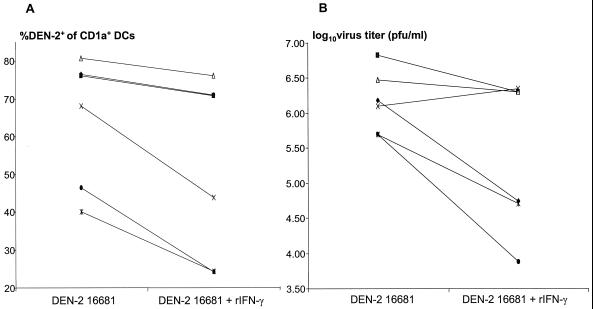
Immature myeloid DCs were infected with a laboratory-adapted strain of dengue virus, DEN-2 16681, in the absence or presence of rIFN-γ. At 48 h, cells were stained for surface CD1a expression and intracellular DEN-2 virus; the percentage of DEN-2-infected CD1a+ DCs was quantitated by flow cytometry. The intracellular location of virus in DCs was confirmed by immunofluorescence microscopy; adsorption with heat-killed DEN-2 16681 or UV-inactivated virus did not result in staining with the anti-DEN-2 Ab (data not shown). DCs were >95% viable by trypan blue staining. In the absence of IFN-γ, the percentage of DEN-2-infected CD1a+ DCs at 48 h was 65% ± 7% (range, 40 to 81%), and the log10 virus titer (PFU per milliliter) in the cell-free supernatants was 6.16 ± 0.18 (range, 5.70 to 6.83) (n = 6). The supernatant log10 virus titer (PFU per milliliter) correlated with the percentage of DEN-2-infected DCs (Pearson correlation coefficient = 0.86, P = 0.03). When rIFN-γ was added after virus adsorption, the percentage of DEN-2-infected CD1a+ DCs at 48 h decreased to 52% ± 10% (range, 24 to 76%) (P = 0.02). The supernatant log10 virus titer (PFU per milliliter) also decreased to 5.38 ± 0.44 (range, 3.88 to 6.35), although this decrease did not achieve statistical significance (P = 0.06) (Fig. 1).
FIG. 1.
Dengue virus productively infects immature myeloid DCs, and infection is decreased in the presence of IFN-γ. Immature myeloid DCs were infected with DEN-2 16681 (MOI = 5) in the absence or presence of rIFN-γ (100 U/ml). At 48 h, cells were stained for surface CD1a and intracellular DEN-2, and cell culture supernatants were collected. (A) Percent DEN-2-infected CD1a+ DCs determined by cytofluorometry; (B) virus titer in cell culture supernatants. The results of six independent experiments are shown. Each symbol represents one individual.
Antibody-dependent enhancement of infection (ADE) is a well-described in vitro phenomenon among the flaviviruses (15, 31). Although unlikely, ADE could not be entirely excluded as a mode of infection in some experiments. All six donors had detectable levels of neutralizing antiflavivirus Ab in their serum (four had anti-dengue virus Ab; two had only anti-Japanese encephalitis virus Ab). However, the level of anti-dengue virus Ab in the donors' sera was negatively correlated with the percentage of DEN-2-infected DCs (Pearson correlation coefficient = −0.95, P = 0.004). ADE of dengue virus infection in myeloid DCs was unable to be demonstrated in a previous study (53). The data show that dengue virus can infect and replicate in immature myeloid DCs. IFN-γ decreases the permissiveness of myeloid DCs to dengue virus infection.
IFN-γ enhances the activation of dengue virus-infected DCs.
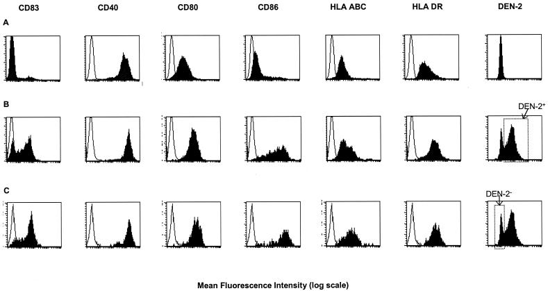
DC maturation and activation is characterized by upregulation of the costimulatory molecules CD40, CD80, CD86 (43, 44, 54), increased cell surface expression of HLA classes I and II (43, 54), and upregulation of the specific DC marker CD83 (54, 56). Immature myeloid DCs were infected with DEN-2 16681 or DEN-2 16681 plus rIFN-γ (100 U/ml) or were mock infected. At 48 h, the cell surface expression of CD83, CD40, CD80, CD86, HLA class I, and HLA class II was assessed by flow cytometry in three groups: mock-infected (control) CD1a+ DCs (Fig. 2A), DEN-2-infected CD1a+ DCs following virus adsorption (Fig. 2B), and uninfected (bystander) CD1a+ DCs following virus adsorption (Fig. 2C). Significant increases in the cell surface expression of CD83, CD40, CD80, CD86, and HLA classes I and II were seen in the DEN-2-infected and uninfected (bystander) DCs compared to the mock-infected controls (Fig. 2; Table 1). In control experiments, exposure of immature myeloid DCs to UV-inactivated dengue virus did not induce maturation or activation as determined by CD83, CD80, and CD86 expression (data not shown).
FIG. 2.
Dengue virus infection induces DC maturation and activation. Immature myeloid DCs were infected with DEN-2 16681 (MOI = 5) or mock infected. At 48 h following virus adsorption, three-color cytofluorometry of the DCs was performed. The following markers were evaluated: (i) presence of intracellular DEN-2 virus; (ii) surface expression of CD83, CD40, CD80, CD86, HLA class I, or HLA class II; and (iii) surface expression of CD1a. All analyses were performed on the CD1a+ cell population gate. (A) FACS profiles of mock-infected (control) CD1a+ DCs; (B) FACS profiles of dengue virus-infected CD1a+ DCs after virus adsorption (DEN-2+ gate, far right histogram); (C) FACS profiles of uninfected (bystander) DCs after virus adsorption (DEN-2− gate, far right histogram). Open peaks, isotype control; shaded peaks, specific antibody staining. One representative experiment of six is shown.
TABLE 1.
Dengue virus infection induces DC maturation and activationa
| DCs | Surface molecule expression (MFU)b
|
|||||
|---|---|---|---|---|---|---|
| CD83 (n = 6) | CD40 (n = 5) | CD80 (n = 6) | CD86 (n = 6) | HLA class I (n = 6) | HLA class II (n = 6) | |
| Mock infected (control) | 4 ± 1 | 1,053 ± 109 | 49 ± 9 | 65 ± 19 | 23 ± 3 | 84 ± 20 |
| Dengue virus infected | 27 ± 3c | 1,771 ± 186c | 159 ± 22c | 508 ± 105c | 64 ± 13c | 187 ± 33c |
| Uninfected (bystander) | 46 ± 6cd | 1,862 ± 159c | 210 ± 26cd | 1,063 ± 171cd | 114 ± 20cd | 202 ± 37c |
| Dengue virus infected in the presence of IFN-γ (100 U/ml) | 25 ± 4 | 1,954 ± 224e | 240 ± 23f | 936 ± 64f | 107 ± 13f | 233 ± 34f |
Cell surface activation marker expression in CD1a+ DCs at 48 h following dengue virus infection in the absence or presence of IFN-γ.
Expressed as specific mean fluorescence intensity units (MFU) calculated by subtracting the MFU obtained with the corresponding isotype control. Data are presented as the mean ± standard error of the mean.
P ≤ 0.01 compared to mock-infected (control) DCs.
P ≤ 0.01 compared to dengue virus-infected DCs.
P = 0.02 compared to dengue virus-infected DCs.
P < 0.005 compared to dengue virus-infected DCs.
Mean surface levels of the mature DC marker CD83, costimulatory molecules CD80/CD86, and HLA class I were significantly lower in the DEN-2-infected DCs than in the uninfected (bystander) DCs. Addition of rIFN-γ overcame this suppression and increased expression of costimulatory and HLA molecules on the cell surface of the DEN-2-infected DCs (Table 1). Mean surface levels of CD80, CD86, and HLA class I in the DEN-2-infected DCs with IFN-γ approached or exceeded the levels seen in the uninfected (bystander) DCs without IFN-γ.
LPS and Mycoplasma are potent inducers of DC maturation and activation (5, 6, 42, 52). LPS concentrations in the virus stock and culture supernatants of the dengue virus exposed DCs were <5 pg/ml. The virus stock, C6/36 cell line, and DC culture supernatants did not contain Mycoplasma, as determined by a nucleic acid hybridization assay. Thus, DC maturation and activation were not due to LPS or Mycoplasma contamination. The data demonstrate that both directly infected and uninfected myeloid DCs undergo maturation and activation after exposure to live dengue virus. The degree of maturation and activation appears to be less in the virus infected DCs than in the adjacent, uninfected DCs. Activation of dengue virus-infected DCs can be enhanced by the addition of IFN-γ.
Dengue virus induces TNF-α and IFN-α production from myeloid DCs.
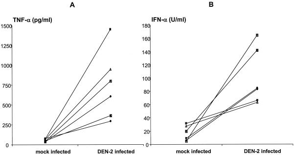
Elevated TNF-α and IFN-α levels have been detected in the sera of dengue virus-infected persons. We measured dengue virus-induced production of TNF-α and IFN-α from myeloid DCs. Immature myeloid DCs were infected with DEN-2 16681 or mock infected. Cell-free supernatants were collected at 48 h and assayed for TNF-α and IFN-α by ELISA. TNF-α levels increased from 54 ± 7 pg/ml in the mock-infected DCs to 743 ± 174 pg/ml in the dengue virus-exposed DCs (P = 0.01) (Fig. 3A). IFN-α levels increased from 16 ± 5 U/ml in the mock-infected DCs to 101 ± 17 U/ml in the dengue virus-exposed DCs (P = 0.009) (Fig. 3B). In control experiments, exposure of DCs to UV-inactivated dengue virus did not induce production of TNF-α or IFN-α (for TNF-α, mock infection, 92 ± 29 pg/ml; for UV-inactivated virus exposure, 56 ± 40, n = 2, P = 0.2; for IFN-α, mock infection and UV-inactivated virus exposure, <4 U/ml, n = 2). The data indicate that replicating dengue virus induces significant TNF-α and IFN-α release from myeloid DCs.
FIG. 3.
Dengue virus infection induces TNF-α and IFN-γ release from myeloid DCs. Immature myeloid DCs were infected with DEN-2 16681 (MOI = 5) or mock infected. Cell-free supernatants were collected at 48 h and assayed for TNF-α (A) and IFN-γ (B) release by ELISA. The results of six independent experiments are shown. Values are the means of duplicate determinations. Each symbol represents one individual.
Dengue virus induces IL-12 p70 secretion from myeloid DCs in the presence of IFN-γ and does not induce IL-10 secretion.
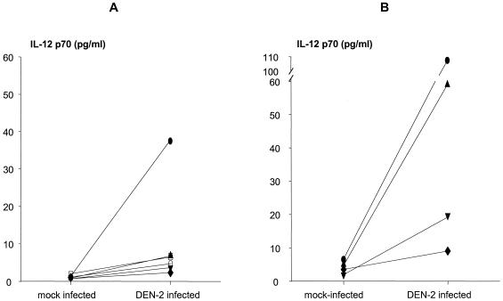
Myeloid DCs have been shown to be a major source of the IL-12 that is necessary for generation of a type 1 T-cell response and cell-mediated immunity (CMI) (5). IL-10 is the predominant type 2 cytokine that downregulates IL-12 production and CMI (7). We measured dengue virus-induced production of the biologically active heterodimer, IL-12 p70, and IL-10 from myeloid DCs. Immature myeloid DCs were infected with DEN-2 16681 or DEN-2 16681 plus rIFN-γ (100 U/ml) or were mock infected. Cell-free supernatants were collected at 48 h and assayed for IL-12 p70 and IL-10 by ELISA. Production of IL-12 p70 was low following dengue virus infection. IL-12 p70 concentrations were 1.1 ± 0.1 pg/ml in the mock-infected DCs and 10.2 ± 5.5 pg/ml in the dengue virus-exposed DCs (P = 0.2) (Fig. 4A). IL-12 p70 remained unchanged and at the level of detection (1.0 pg/ml) following exposure to UV-inactivated dengue virus.
FIG. 4.
IFN-γ enhances dengue virus-induced IL-12 p70 release from myeloid DCs. Immature myeloid DCs were infected with DEN-2 16681 (MOI = 5) or mock infected in the presence or absence of IFN-γ (100 U/ml). Cell-free supernatants were collected at 48 h and assayed for IL-12 p70 release by ELISA. (A) IL-12 p70 production in the absence of exogenous IFN-γ (n = 6); (B) IL-12 p70 production in the presence of exogenous IFN-γ (100 U/ml) (n = 4). Values are the means of duplicate determinations. Each symbol represents one individual.
In the presence of exogenous IFN-γ, IL-12 p70 levels increased from 4.2 ± 1.0 pg/ml in the mock-infected DCs to 48.7 ± 22.3 pg/ml in the dengue virus-exposed DCs, though this did not reach statistical significance (P = 0.07) (Fig. 4B). The level of IL-12 p70 induced by dengue virus in the presence of IFN-γ was greater than in the absence of IFN-γ (48.7 ± 22.3 pg/ml and 10.2 ± 5.5 pg/ml, respectively, P = 0.03).
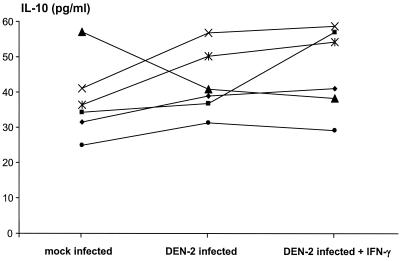
Dengue virus infection did not induce significant IL-10 release from immature myeloid DCs and was not affected by the addition of IFN-γ (mock-infected DCs, 37.5 ± 4.5 pg/ml; dengue virus-exposed DCs, 42.5 ± 3.8 pg/ml; dengue virus-exposed DCs plus IFN-γ [100 U/ml], 46.4 ± 4.9, P = 0.3) (Fig. 5).
FIG. 5.
Dengue virus infection does not induce IL-10 release from myeloid DCs. Immature myeloid DCs were infected with DEN-2 16681 (MOI = 5) or mock infected in the presence or absence of IFN-γ (100 U/ml). Cell-free supernatants were collected at 48 h and assayed for IL-10 release by ELISA. The results of six independent experiments are shown. Values are the means of duplicate determinations. Each symbol represents one individual.
These data demonstrate that exposure of immature myeloid DCs to live dengue virus does not induce significant IL-12 p70 production. This weak induction of IL-12 p70 secretion is not due to increased IL-10 release. IFN-γ can augment dengue virus-induced IL-12 p70 release.
DISCUSSION
Although dengue virus infection of myeloid DCs has been previously reported (53), this study is the first to describe the effects of dengue virus infection on the DCs. The results of this study provide evidence that exposure of immature myeloid DCs to dengue virus leads to productive infection with DC maturation and activation. Activation of the dengue virus-infected myeloid DCs appears blunted compared to the surrounding, uninfected DCs. While dengue virus infection of myeloid DCs results in production of TNF-α and IFN-α, secretion of IL-12 p70 is low. Addition of IFN-γ enhances the activation of dengue virus-infected myeloid DCs and enhances virus-induced IL-12 p70 release.
We have demonstrated that immature human myeloid DCs are permissive to dengue virus infection and replication, though wide variability to infection was seen among the blood-derived DCs from six individuals. The addition of IFN-γ (100 U/ml) immediately following exposure to dengue virus enhanced activation of the virus-infected DCs yet decreased the percentage of infected myeloid DCs and virus production. This is consistent with a recent report that found immature, but not mature or activated, myeloid DCs to be permissive to infection with dengue virus (53). Others have also demonstrated a decreased permissiveness to viral infection in activated DCs but have found the phenomenon to be stimulus dependent (4, 36). IFN-γ (100 U/ml) also decreased dengue virus replication in human peripheral blood monocytes (48). Virus replication and production in immature CD1a+ DCs, and their location in the epidermis and dermis, suggest that myeloid DCs are an important early target for dengue virus infection in vivo. Dengue virus infection of human skin CD1a+ DCs has been recently demonstrated ex vivo (53).
When exposed to live dengue virus, immature myeloid DCs underwent maturation, as characterized by the upregulation of CD83 and surface activation markers and the secretion of cytokines. Measles virus, influenza virus, and a variety of other microbial pathogens and products are also inducers of DC maturation (4, 37, 45). The effects of dengue virus infection on the maturation and activation of myeloid DCs appear to be pleiotropic. Dengue virus infection of immature myeloid DCs induced significant release of TNF-α and IFN-α, low levels of IL-12 p70 release, and no appreciable IL-10 release. Whether cytokines were produced solely by the virus-infected DCs or additionally by the adjacent uninfected DCs could not be determined; however, a productive viral infection was required. TNF-α and IFN-α production have also been demonstrated following dengue virus infection of human monocytes or monocytic cell lines (18, 21). TNF-α and IFN-α can drive maturation of both the virus-infected DCs and the adjacent uninfected DCs, resulting in an increase in the cell surface expression of CD83, HLA classes I and II, and costimulatory molecules (28, 34, 44, 54). The lower mean levels of HLA class I and CD80/CD86 on the surface of the dengue virus-infected DCs compared to the adjacent uninfected DCs suggest that dengue virus may exert a small inhibitory effect on the cytokine mediated upregulation of these cell surface molecules. Flavivirus-induced upregulation of class I major histocompatibility complex and CD80 expression has been previously described (19, 27, 33, 47). Depending on the cell culture system used, this upregulation has been found to be either cytokine dependent (47) or cytokine independent (33). In dengue virus infection of myeloid DCs, it appears that a cytokine-mediated effect dominates the expression of HLA class I and CD80/CD86 on the cell surface. Whether the upregulation of these T-cell stimulatory molecules is also counteracted by a direct viral effect will require further study.
Dengue virus-induced IL-12 p70 release from myeloid DCs was low but was enhanced by IFN-γ. Similar findings have been seen among other virus-exposed myeloid DCs. Adenovirus infection of peripheral blood-derived myeloid DCs produced incomplete maturation and failed to induce IL-12 p70 release (36). IFN-α has been shown to inhibit the production of IL-12 p70 from DCs and may contribute to a suppression of IL-12 release during viral infection (29). IL-12 production from myeloid DCs activated by measles virus requires coculture with lymphocytes or other CD40 ligand (CD40L)-bearing cells (8). The engagement of CD40 on DCs by CD40L-bearing T cells requires IFN-γ in order to produce IL-12 p70 (16, 49). Thus, for several viral pathogens, there appears to be a pattern whereby IFN-γ is an important second signal for the secretion of bioactive IL-12 from DCs. In dengue virus-infected DCs, IFN-γ also increases expression of the molecules involved in stimulating the antigen-specific T-cell response. This two-signal process may represent a regulatory mechanism for modulating potentially harmful type 1 T-cell activation early in the immune response.
These data have relevant implications for the pathogenesis of dengue virus-induced disease. Early in a primary infection, dengue virus-infected DCs would be weakly activated and would not produce significant amounts of IL-12 p70, as there is a paucity of IFN-γ and dengue virus-reactive T cells (bearing CD40L) in the local microenvironment. With continued viral replication and stimulation of the innate immune response, there is eventual production of IFN-γ with T- and B-cell activation. Viremia is cleared; CMI and humoral immunity develop without an early, anamnestic burst of type 1 cytokines. In this setting, the risk of proceeding to DHF/DSS is low. During a heterotypic, second dengue virus infection, cross-reactive memory T cells would be activated in response to dengue virus-infected DCs. These T cells are a source of early IFN-γ and CD40L in the DC microenvironment, leading to greater DC activation, greater T-cell stimulatory capacity, a burst of IL-12 p70 release, increases in TNF-α (2) and IFN-γ (51) secretion, and potential dysregulation of the type 1 cytokine response. Viremia is cleared, yet the cascade of events initiated by an early, poorly controlled type 1 cytokine response likely contributes to the pathogenesis of DHF/DSS. Plasma levels of IFN-γ, TNF-α, and soluble TNF-α receptors have been found to be elevated in DHF/DSS compared to dengue fever (10, 17).
We have postulated that the relative presence or absence of IFN-γ in the local microenvironment surrounding dengue virus-infected DCs is a key determinant of disease manifestation and severity. In our experiments, DCs were exposed to 100 U of rIFN-γ per ml. Although in vivo concentrations of IFN-γ in lymph nodes containing dengue virus-infected DCs are unknown, there is evidence to suggest that 100 U/ml is within an achievable range. Dengue virus-immune CD4+ T cells have produced 10 to 280 U of IFN-γ per ml in response to dengue virus antigens, and serum levels of IFN-γ in dengue virus-infected patients and dengue virus vaccine recipients have reached 50 U/ml. A critical question that remains is what other factors modulate the appearance of early IFN-γ, DC activation, and IL-12 p70 production. Dengue virus-stimulated natural killer (NK) cells are a potential source of early IFN-γ in both primary and secondary infections. The percent of circulating activated NK cells was found to be greater in children who developed DHF compared to dengue fever (9). Activated platelets may provide an alternative source of CD40L. The number of DCs infected (viral load), differences in virus strains, and polymorphisms in the transcriptional promoters of IL-12 (p40 and p35) and IFN-γ are potential modulators of the early type 1 cytokine response to dengue virus infection in vivo.
The data presented here underscore the multifaceted role of DCs in viral infection. Immature myeloid DCs are an early target of dengue virus infection and a source of virus replication and production. At the same time, they play a critical role in the development of both protective and pathogenic aspects of antiviral immunity. Further studies to delineate the interaction between dengue virus and DCs should provide new opportunities for vaccination strategies and therapeutic interventions in this emerging arboviral disease.
ACKNOWLEDGMENTS
We thank Khun Naowayubol Nutkumhang and Khun Somsak Imlap for performing the virus titration plaque assays, Khun Napaporn Lutthiwongsakorn for performing the plaque reduction neutralization assays, Khun Kosol Yongvanitchit for FACS analysis, and Khun Utaiwon Kum-arb for assistance with DC preparation. We thank Ananda Nisalak for overall supervision of the diagnostic virology laboratory at AFRIMS and Alan Rothman and Sharone Green for assistance with manuscript preparation.
This work was supported by the National Institutes of Health (grant NIH-P01AI34533) and the U.S. Army Medical Research and Materiel Command.
REFERENCES
- 1.Banchereau J, Steinman R. Dendritic cells and the control of immunity. Nature. 1998;392:245–252. doi: 10.1038/32588. [DOI] [PubMed] [Google Scholar]
- 2.Boehm U, Klamp T, Groot M, Howard J. Cellular responses to interferon-γ. Annu Rev Immunol. 1997;15:749–795. doi: 10.1146/annurev.immunol.15.1.749. [DOI] [PubMed] [Google Scholar]
- 3.Burke D, Nisalak A, Johnson D, Scott R. A prospective study of dengue infections in Bangkok. Am J Trop Med Hyg. 1988;38:172–180. doi: 10.4269/ajtmh.1988.38.172. [DOI] [PubMed] [Google Scholar]
- 4.Cella M, Salio M, Sakakibara Y, Langen H, Julkunen I, Lanzavecchia A. Maturation, activation, and protection of dendritic cells induced by double-stranded RNA. J Exp Med. 1999;189:821–829. doi: 10.1084/jem.189.5.821. [DOI] [PMC free article] [PubMed] [Google Scholar]
- 5.Cella M, Scheidegger D, Palmer-Lehmann K, Lane P, Lanzavecchia A, Alber G. Ligation of CD40 on dendritic cells triggers production of high levels of interleukin-12 and enhances T cell stimulatory capacity: T-T help via APC activation. J Exp Med. 1996;184:747–752. doi: 10.1084/jem.184.2.747. [DOI] [PMC free article] [PubMed] [Google Scholar]
- 6.Corinti S, Fanales-Belasio E, Albanesi C, Cavani A, Angelisova P, Girolomoni G. Cross-linking of membrane CD43 mediates dendritic cell maturation. J Immunol. 1999;162:6331–6336. [PubMed] [Google Scholar]
- 7.D'Andrea A, Aste-Amezaga M, Valiante N, Ma X, Kubin M, Trinchieri G. Interleukin-10 inhibits human lymphocyte IFN-γ production by suppressing natural killer cell stimulatory factor/interleukin 12 synthesis in accessory cells. J Exp Med. 1993;178:1041–1048. doi: 10.1084/jem.178.3.1041. [DOI] [PMC free article] [PubMed] [Google Scholar]
- 8.Fugier-Vivier I, Servet-Delprat C, Rivailler P, Rissoan M-C, Liu Y-J, Rabourdin-Combe C. Measles virus suppresses cell mediated immunity by interfering with the survival and functions of dendritic and T cells. J Exp Med. 1997;186:813–823. doi: 10.1084/jem.186.6.813. [DOI] [PMC free article] [PubMed] [Google Scholar]
- 9.Green S, Pichyangkul S, Vaughn D, Kalaynarooj S, Nimmannitya S, Nisalak A, Kurane I, Rothman A, Ennis F. Early CD69 expression on peripheral blood lymphocytes from children with dengue hemorrhagic fever. J Infect Dis. 1999;180:1429–1435. doi: 10.1086/315072. [DOI] [PubMed] [Google Scholar]
- 10.Green S, Vaughn D, Kalayanarooj S, Nimmannitya S, Suntayakorn S, Nisalak A, Lew R, Innis B, Kurane I, Rothman A, Ennis F. Early immune activation in acute dengue illness is related to development of plasma leakage and disease severity. J Infect Dis. 1999;179:755–762. doi: 10.1086/314680. [DOI] [PubMed] [Google Scholar]
- 11.Guzman M, Kouri G, Bravo J, Soler M, Vazquez S, Morier L. Dengue hemorrhagic fever in Cuba. Am J Trop Med Hyg. 1990;42:179–184. doi: 10.4269/ajtmh.1990.42.179. [DOI] [PubMed] [Google Scholar]
- 12.Halstead S. Antibody, macrophages, dengue virus infection, shock, and hemorrhage: a pathogenetic cascade. Rev Infect Dis. 1989;11:S830–S839. doi: 10.1093/clinids/11.supplement_4.s830. [DOI] [PubMed] [Google Scholar]
- 13.Halstead S, Nimmannitya S, Cohen S. Observations related to pathogenesis of dengue hemorrhagic fever. IV. Relation of disease severity to antibody response and virus recovered. Yale J Biol Med. 1970;42:311–328. [PMC free article] [PubMed] [Google Scholar]
- 14.Halstead S, O'Rourke E, Allison A. Dengue viruses and mononuclear phagocytes. II. Identity of blood and tissue leukocytes supporting in vitro infection. J Exp Med. 1977;146:218–229. doi: 10.1084/jem.146.1.218. [DOI] [PMC free article] [PubMed] [Google Scholar]
- 15.Halstead S, Porterfield J, O'Rourke E. Enhancement of dengue virus infection in monocytes by flavivirus antisera. Am J Trop Med Hyg. 1980;29:638–642. doi: 10.4269/ajtmh.1980.29.638. [DOI] [PubMed] [Google Scholar]
- 16.Hilkens C, Kalinski P, de Boer M, Kapsenberg M. Human dendritic cells require exogenous interleukin-12 inducing factors to direct the development of naive T-helper cells toward the Th1 phenotype. Blood. 1997;90:1920–1926. [PubMed] [Google Scholar]
- 17.Hober D, Delannoy A-S, Benyoucef S, De Groote D, Wattre P. High levels of sTNFR p75 and TNFα in dengue-infected patients. Microbiol Immunol. 1996;40:569–573. doi: 10.1111/j.1348-0421.1996.tb01110.x. [DOI] [PubMed] [Google Scholar]
- 18.Hober D, Shen L, Benyoucef S, De Groote D, Deubel V, Wattre P. Enhanced TNFα production by monocytic-like cells exposed to dengue virus antigens. Immunol Lett. 1996;53:115–120. doi: 10.1016/s0165-2478(96)02620-x. [DOI] [PubMed] [Google Scholar]
- 19.Johnston L, Halliday G, King N. Phenotypic changes in Langerhans' cells after infection with arboviruses: a role in the immune response to epidermally acquired viral infection? J Virol. 1996;70:4761–4766. doi: 10.1128/jvi.70.7.4761-4766.1996. [DOI] [PMC free article] [PubMed] [Google Scholar]
- 20.Klagge I, Schneider-Schaulies S. Virus interactions with dendritic cells. J Gen Virol. 1999;80:823–833. doi: 10.1099/0022-1317-80-4-823. [DOI] [PubMed] [Google Scholar]
- 21.Kurane I, Ennis F. Production of interferon alpha by dengue virus-infected human monocytes. J Gen Virol. 1988;69:445–449. doi: 10.1099/0022-1317-69-2-445. [DOI] [PubMed] [Google Scholar]
- 22.Kurane I, Innis B, Hoke C J, Eckels K, Meager A, Janus J, Ennis F. T cell activation in vivo by dengue virus infection. J Clin Lab Immunol. 1995;46:35–40. [PubMed] [Google Scholar]
- 23.Kurane I, Innis B, Nimmannitya S, Nisalak A, Meager A, Ennis F. High levels of interferon alpha in the sera of children with dengue virus infection. Am J Trop Med Hyg. 1993;48:222–229. doi: 10.4269/ajtmh.1993.48.222. [DOI] [PubMed] [Google Scholar]
- 24.Kurane I, Innis B, Nimmannitya S, Nisalak A, Meager A, Janus J, Ennis F. Activation of T lymphocytes in dengue virus infections. J Clin Investig. 1991;88:1473–1480. doi: 10.1172/JCI115457. [DOI] [PMC free article] [PubMed] [Google Scholar]
- 25.Kurane I, Innis B, Nisalak A, Hoke C J, Nimmannitya S, Meager A, Ennis F. Human T cell responses to dengue virus antigens. J Clin Investig. 1989;83:506–513. doi: 10.1172/JCI113911. [DOI] [PMC free article] [PubMed] [Google Scholar]
- 26.Lane P, Brocker T. Developmental regulation of dendritic cell function. Curr Opin Immunol. 1999;11:308–313. doi: 10.1016/s0952-7915(99)80049-1. [DOI] [PubMed] [Google Scholar]
- 27.Lobigs M, Blanden R, Mullbacher A. Flavivirus-induced up regulation of MHC class I antigens; implications for the induction of CD8+ T-cell-mediated autoimmunity. Immunol Rev. 1996;152:5–19. doi: 10.1111/j.1600-065X.1996.tb00908.x. [DOI] [PMC free article] [PubMed] [Google Scholar]
- 28.Luft T, Pang K, Thomas E, Hertzog P, Hart D, Trapani J, Cebon J. Type I IFNs enhance the terminal differentiation of dendritic cells. J Immunol. 1998;161:1947–1953. [PubMed] [Google Scholar]
- 29.McRae B, Semmani R, Hayes M, van Seventer G. Type I IFNs inhibit human dendritic cell IL-12 production and Th1 cell development. J Immunol. 1998;160:4298–4304. [PubMed] [Google Scholar]
- 30.Monath T. Dengue: the risk to developed and developing countries. Proc Natl Acad Sci USA. 1994;91:2395–2400. doi: 10.1073/pnas.91.7.2395. [DOI] [PMC free article] [PubMed] [Google Scholar]
- 31.Morens D. Antibody-dependent enhancement of infection and the pathogenesis of viral disease. Clin Infect Dis. 1994;19:500–512. doi: 10.1093/clinids/19.3.500. [DOI] [PubMed] [Google Scholar]
- 32.Mori M, Kurane I, Janus J, Ennis F. Cytokine production by dengue virus antigen-responsive human T lymphocytes in vitro examined using a double immunocytochemical technique. J Leukoc Biol. 1997;61:338–345. doi: 10.1002/jlb.61.3.338. [DOI] [PubMed] [Google Scholar]
- 33.Mullbacher A, Lobigs M. Up-regulation of MHC class I by flavivirus-induced peptide translocation into the endoplasmic reticulum. Immunity. 1995;3:207–214. doi: 10.1016/1074-7613(95)90090-x. [DOI] [PubMed] [Google Scholar]
- 34.Paquette R, Hsu N, Kiertscher S, Park A, Tran L, Roth M, Glaspy J. Interferon-alpha and granulocyte-macrophage colony stimulating factor differentiate peripheral blood monocytes into potent antigen-presenting cells. J Leukoc Biol. 1998;64:358–367. doi: 10.1002/jlb.64.3.358. [DOI] [PubMed] [Google Scholar]
- 35.Pinheiro F, Corber S. Global situation of dengue and dengue haemorrhagic fever, and its emergence in the Americas. World Health Stat Q. 1997;50:161–169. [PubMed] [Google Scholar]
- 36.Rea D, Schagen F, Hoeben R, Mehtali M, Havenga M, Toes R, Melief C, Offringa R. Adenoviruses activate human dendritic cells without polarization toward a T-helper type 1-inducing subset. J Virol. 1999;73:10245–10253. doi: 10.1128/jvi.73.12.10245-10253.1999. [DOI] [PMC free article] [PubMed] [Google Scholar]
- 37.Reis e Sousa C, Sher A, Kaye P. The role of dendritic cells in the induction and regulation of immunity to microbial infection. Curr Opin Immunol. 1999;11:392–399. doi: 10.1016/S0952-7915(99)80066-1. [DOI] [PubMed] [Google Scholar]
- 38.Rissoan M-C, Soumelis V, Kadowaki N, Grouard G, Briere F, Malefyt R, Liu Y-J. Reciprocal control of T helper cell and dendritic cell differentiation. Science. 1999;283:1183–1186. doi: 10.1126/science.283.5405.1183. [DOI] [PubMed] [Google Scholar]
- 39.Romani N, Gruner S, Brang D, Kampgen E, Lenz A, Trockenbacher B, Konwalinka G, Fritsch P, Steinman R, Schuler G. Proliferating dendritic cell progenitors in human blood. J Exp Med. 1994;180:83–93. doi: 10.1084/jem.180.1.83. [DOI] [PMC free article] [PubMed] [Google Scholar]
- 40.Rothman A L, Ennis F A. Immunopathogenesis of dengue hemorrhagic fever. Virology. 1999;257:1–6. doi: 10.1006/viro.1999.9656. [DOI] [PubMed] [Google Scholar]
- 41.Russell P, Nisalak A, Sukhavachana P, Vivona S. A plaque reduction test for dengue virus neutralization antibodies. J Immunol. 1967;99:285–290. [PubMed] [Google Scholar]
- 42.Salio M, Cerundolo V, Lanzavecchia A. Dendritic cell maturation is induced by mycoplasma infection but not by necrotic cells. Eur J Immunol. 2000;30:705–708. doi: 10.1002/1521-4141(200002)30:2<705::AID-IMMU705>3.0.CO;2-P. [DOI] [PubMed] [Google Scholar]
- 43.Sallusto F, Cella M, Danieli C, Lanzavecchia A. Dendritic cells use macropinocytosis and the mannose receptor to concentrate macromolecules in the major histocompatibility complex class II compartment: downregulation by cytokines and bacterial products. J Exp Med. 1995;182:389–400. doi: 10.1084/jem.182.2.389. [DOI] [PMC free article] [PubMed] [Google Scholar]
- 44.Sallusto F, Lanzavecchia A. Efficient presentation of soluble antigen by cultured human dendritic cells is maintained by granulocyte/macrophage colony-stimulating factor plus interleukin 4 and downregulated by tumor necrosis factor α. J Exp Med. 1994;179:1109–1118. doi: 10.1084/jem.179.4.1109. [DOI] [PMC free article] [PubMed] [Google Scholar]
- 45.Schnorr J-J, Xanthakos S, Keikavoussi P, Kampgen E, Meulen V, Schneider-Schaulies S. Induction of maturation of human blood dendritic cell precursors by measles virus is associated with immunosuppression. Proc Natl Acad Sci USA. 1997;94:5326–5331. doi: 10.1073/pnas.94.10.5326. [DOI] [PMC free article] [PubMed] [Google Scholar]
- 46.Scott R, Nisalak A, Cheamudon U, Seridoranakhul S, Nimmannitya S. Isolation of dengue viruses from peripheral blood leukocytes of patients with hemorrhagic fever. J Infect Dis. 1980;141:1–6. doi: 10.1093/infdis/141.1.1. [DOI] [PubMed] [Google Scholar]
- 47.Shen J, To S T-, Schrieber L, King N. Early E-selectin, VCAM-1, ICAM-1, and late major histocompatibility complex antigen induction on human endothelial cells by flavivirus and comodulation of adhesion molecule expression by immune cytokines. J Virol. 1997;71:9323–9332. doi: 10.1128/jvi.71.12.9323-9332.1997. [DOI] [PMC free article] [PubMed] [Google Scholar]
- 48.Sittisombut N, Maneekarn N, Kanjanahaluethai A, Kasinrerk W, Viputtikul K, Supawadee J. Lack of augmenting effect of interferon-g on dengue virus multiplication in human peripheral blood monocytes. J Med Virol. 1995;45:43–49. doi: 10.1002/jmv.1890450109. [DOI] [PubMed] [Google Scholar]
- 49.Snijders A, Kalinski P, Hilkens C, Kapsenberg M. High level IL-12 production by human dendritic cells requires two signals. Int Immunol. 1998;10:1593–1598. doi: 10.1093/intimm/10.11.1593. [DOI] [PubMed] [Google Scholar]
- 50.Steinman R. The dendritic cell system and its role in immunogenicity. Annu Rev Immunol. 1991;9:271–296. doi: 10.1146/annurev.iy.09.040191.001415. [DOI] [PubMed] [Google Scholar]
- 51.Trinchieri G, Scott P. Interleukin-12: a proinflammatory cytokine with immunoregulatory functions. Res Immunol. 1995;146:423–431. doi: 10.1016/0923-2494(96)83011-2. [DOI] [PubMed] [Google Scholar]
- 52.Verhasselt V, Buelens C, Willems F, Groote D, Haeffner Cavaillon N, Goldman M. Bacterial lipopolysaccharide stimulates the production of cytokines and the expression of costimulatory molecules by human peripheral blood dendritic cells. J Immunol. 1997;158:2919–2925. [PubMed] [Google Scholar]
- 53.Wu S, Grouard-Vogel G, Sun W, Mascola J, Brachtel E, Putvatana R, Louder M, Filgueira L, Marovich M, Wong H, Blauvelt A, Murphy G, Robb M, Innis B, Birx D, Hayes C, Frankel S. Human skin Langerhans cells are targets of dengue virus infection. Nat Med. 2000;6:816–820. doi: 10.1038/77553. [DOI] [PubMed] [Google Scholar]
- 54.Zhou L-J, Tedder T. CD14+ blood monocytes can differentiate into functionally mature CD83+ dendritic cells. Proc Natl Acad Sci USA. 1996;93:2588–2592. doi: 10.1073/pnas.93.6.2588. [DOI] [PMC free article] [PubMed] [Google Scholar]
- 55.Zhou L-J, Tedder T. A distinct pattern of cytokine gene expression by human CD83+ blood dendritic cells. Blood. 1995;86:3295–3301. [PubMed] [Google Scholar]
- 56.Zhou L-J, Tedder T. Human blood dendritic cells selectively express CD83, a member of the immunoglobulin superfamily. J Immunol. 1995;8:3821–3835. [PubMed] [Google Scholar]


