Abstract
Carbohydrates comprise about 50% of the mass of gp120, the external envelope glycoprotein of simian immunodeficiency virus (SIV) and human immunodeficiency virus. We identified 11 replication-competent derivatives of SIVmac239 lacking two, three, four, or five potential sites for N-linked glycosylation. These sites were located within and around variable regions 1 and 2 of the surface envelope protein of the virus. Asn (AAT) of the canonical N-linked glycosylation recognition sequence (Asn X Ser/Thr) was changed in each case to the structurally similar Gln (CAG or CAA) such that two nucleotide changes in the codon would be required for reversion. Replication of one triple mutant (g456), however, was severely impaired. A revertant of the g456 mutant was recovered from CEMx174 cells with a Met-to-Val compensatory substitution at position 144, 2 amino acids upstream of attachment site 5. Thus, a debilitating loss of sites for N-linked glycosylation can be compensated for by amino acid changes not involving the Asn-X-Ser/Thr consensus motif. These results provide a framework to begin testing the hypothesis that carbohydrates form a barrier that can limit the humoral immune responses to the virus.
Carbohydrates comprise about 50% of the mass of gp120, the external envelope glycoprotein of the simian and human immunodeficiency viruses (SIV and HIV). Both N-linked and O-linked forms of glycosylation have been detected (5, 8, 18). While the consensus sequence for the addition of N-linked oligosaccharides is known (Asn-X-Ser/Thr), the determinants of O-linked glycosylation are less well understood (28). The number of N-linked sites on gp120 varies with the strain of SIV and HIV but is usually around 24, and the locations are generally conserved within each group (24). The detailed structural analysis by Leonard et al. demonstrated that all 24 N-linked sites are utilized in the gp120 of the HIV-1 IIIB strain produced in Chinese hamster ovary cells (18).
When the envelope precursor of gp120 is produced in mammalian cells in the presence of agents that inhibit early steps in N-linked oligosaccharide biosynthesis, there is a marked decrease in both viral infectivity and syncytium formation (13, 14, 27, 30). Inhibition of mannose trimming and later steps in the processing of N-glycans do not significantly interfere with infectivity and cell fusion (14, 27). Deficits imparted by complete lack of glycosylation, e.g., when synthesis occurs in the presence of tunicamycin, include lack of proper folding, retention in the Golgi complex, lack of proteolytic processing, and inability to bind to CD4 (10, 20). When fully glycosylated gp120 is deglycosylated enzymatically in the absence of detergents, gp120 apparently retains its native structure and can bind CD4 (11, 20). Thus, carbohydrates appear to be required to generate a properly folded, properly processed protein, but once formed the carbohydrates do not appear to be required to maintain the native structure. Despite this general requirement for carbohydrates, many individual N-linked sites can be eliminated without impairing the native structure or the ability of the virus to replicate (6, 17). However, other N-linked sites are essential for the virus (22, 32).
Since the extensive glycosylation of HIV and SIV envelope proteins was initially recognized, it has been speculated that the carbohydrates may form a barrier that can limit the humoral immune response and protect the virus from immune recognition. However, little evidence has been presented in actual support of this hypothesis. In one study, the reactivity of some monoclonal antibodies was increased and the reactivity of others was decreased when gp120 was synthesized in the presence of an oligosaccharide-processing inhibitor (12). The authors concluded that peripheral structures of N-glycans are involved in modulating the overall conformation of gp120 (12). In another study, the neutralizing potential of some monoclonal antibodies was enhanced slightly when strains of virus lacking an N-linked glycosylation site in V3 were used (4). We are interested in using the SIV monkey model to provide a more rigorous investigation of the hypothesis.
In this report, we describe the extent to which N-linked glycosylation sites within and around the V1 and V2 regions of gp120 of SIVmac239 are dispensable for viral replication.
MATERIALS AND METHODS
Site-specific mutagenesis and subcloning.
To reduce the size of the plasmids to be mutated, a SphI-ClaI fragment of the proviral SIVmac239 DNA containing 1469 nucleotides of env coding sequence (proviral nucleotides 6450 to 8073 in the numbering of Regier and Desrosiers [29]) was subcloned into pSP72 (Promega), resulting in pSP72SC. The SacI-EcoRI fragment containing the 3′ 1,050 bases of the proviral genome was subcloned into pSP72 to create pSP72SE. Mutations of env were created by recombinant PCR mutagenesis (9). Carbohydrates were numbered according to their order of appearance within the envelope sequence of SIVmac239. The following mutagenic primers were used: for g4, (6931 to 6970) 5′-ACTATGAGATGCCAGAAAAGTGAGACAGATAGATGGGGAT-3′ and (6957 to 6919) 5′-TGTCTCACTTTTCTGGCATCTCATAGTAATGCATAATGG-3′; for g5, (7027 to 7068) 5′-GTAGACATGGTCCAGGAGACTAGTTCTTGTATAGCCCAGGAT-3′ and (7053 to 7014) 5′-AGAACTAGTCTCCTGGACCATGTCTACTTTTGCTGATGCT-3′; for g6, (7057 to 7097) 5′-ATAGCCCAGGATCAATGCACAGGCTTGGAACAAGAGCAAAT-3′ and (7084 to 7045) 5′-CCAAGCCTGTGCATTGATCCTGGGCTATACAAGAACTAGT-3′; for M144V, (7026 to 7062) 5′-AGTAGACGTGGTCAATGAGACTAGTTCTTGTATAGCC-3′ and (7045 to 7010) 5′-GTCTCATTGACCACGTCTACTTTTGCTGATGCTGTCG-3′; for g456 (M144V), (7022 to 7055) 5′-CAAAAGTAGACGTGGTCCAGGAGACTAGTTCTTG-3′ and (7042 to 7009) 5′-CCTGGACCACGTCTACTTTTGCTGATGCTGTCGT-3′; for g7, (7103 to 7137) 5′-GCTGTAAATTCCAGATGACAGGGTTAAAAAGAGAC-3′ and (7126 to 7092) 5′-ACCCTGTCATCTGGAATTTACAGCTTATCATTTGC-3′; for g8, (7145 to 7180) 5′-AAGAGTACCAGGAAACTTGGTACTCTGCAGATTTGG-3′ and (7164 to 7130) 5′-CCAAGTTTCCTGGTACTCTTTTTTCTTGTCTCTTTT-3′; for g9, (7186 to 7220) 5′-GAACAAGGGCAGAACACTGGTAATGAAAGTAGATG-3′ and (7206 to 7171) 5′-ACCAGTGTTCTGCCCTTGTTCACATACCAAATCTGC-3′; for g10, (7198 to 7231) 5′-AACACTGGTCAGGAAAGTAGATGTTACATGAACC-3′ and (7218 to 7184) 5′-TCTACTTTCCTGACCAGTGTTATTCCCTTGTTCAC-3′; for g11, (7228 to 7264) 5′-ACCACTGTCAGACTTCTGTTATCCAAGAGTCTTGTG-3′ and (7249 to 7213) 5′-TAACAGAAGTCTGACAGTGGTTCATGTAACATCTACT-3′; and for g12 and g13, (7228 to 7364) 5′-GATGTCAGGACACACAATATTCAGGCTTTATGCCTAA-3′ and (7349 to 7313) 5′-GAATATTGTGTGTCCTGACATCTAAGCAAAGCATAAC-3′. The primers were synthesized on a Cyclone DNA synthesizer (Biosearch, Inc.) or purchased from Genosys Biotechnologies, Inc. (Woodlands, Texas). The SphI-ClaI fragment containing the mutated env sequence was excised and subcloned into the 3′ parental clone, pSP72-239-3′ (15). For transient gene expression, the wild-type envelope sequence was subcloned into the unique XhoI and BamHI sites of the expression vector pSVL (Pharmacia) after creation of a BamHI site 3′ of the env coding sequence by using the mutagenic primers 27 (9268 to 9302) 5′-GTATATGAAGGATCCATGGAGAAACCCAGCTGAAG-3′ and 28 (9286 to 9253) 5′-CCATGGATCCTTCATATACTGTCCCTGATTGTAT-3′. The mutant envelope sequences were subcloned into the resultant pSVLenv via the unique XhoI and ClaI sites.
DNA transfection of cultured cells.
For viral stocks, the 5′ and 3′ clones of SIVmac239 were digested with SphI and heated to 65°C for 15 min. Each right-half clone was ligated with the left-half clone p239SpSp5′ by using T4 DNA ligase. A 3-μg portion of the ligated DNA was used to transfect CEMx174 cells treated with DEAE-dextran (25). The vector pSVL, which expresses the simian virus 40 late promoter, was used for transient transfection of the wild-type and mutant env genes. A 1-μg portion of DNA was combined with DEAE-dextran, and transfection of COS-1 cells at 80% confluence in 35-mm diameter plates (Falcon Primaria) was performed by the procedure of Levesque et al. (19).
Virus stocks and cell culture.
Rhesus monkey peripheral blood mononuclear cells (PBMC), CEMx174, 221, and COS-1 cells were maintained as described previously (21, 23). For virus stocks, CEMx174 cells were transfected as described above. The medium was changed every 2 days, and the supernatants were harvested at or near the peak of virus production. Cells and debris were removed by centrifugation, and virus contained in the supernatant was aliquoted and stored at −70°C. The concentration of p27 antigen was measured by an antigen capture assay (Coulter Corp., Hialeah, Fla.). For virus infections, 5 ng of p27 was used to infect 2.5 million pelleted cells.
DNA sequencing and PCR amplification.
Cloned fragments containing mutated envelope DNA were sequenced in their entirety on an ABI377 automated DNA sequencer by using dye terminator cycle-sequencing chemistry as specified by the manufacturer (Perkin-Elmer Inc., Foster City, Calif.). Total genomic DNA was isolated with the AmpPrep kit (HRI Research, Inc., Concord, Calif.) and used as a template for nested PCR amplification with primers located outside of the viral env sequence. The outer primers were 39 (6320 to 6348) 5′-GAGGGAGCAGGAGAACTCATTAGAATCCTCC-3′ and 40 (9373 to 9405) 5′-GTTCTTAGGGGAACTTTTGGCCTCACTGATACC-3′. The inner mutagenic primers created XhoI and BamHI sites used for cloning the PCR products into pSP72 and were 38 (6423 to 6465) 5′-CTCAGCTATACCGCCCTCGAGAAGCATGCTATAAC-3′ and 32 (9256 to 9288) 5′-CTCCATGGATCCTTCATATACTGTCCCTGATTG-3′. Each 100-μl reaction mix contained 1 μg of total DNA, 2 mM Mg2+, 200 μM each deoxynucleoside triphosphate, 0.2 μM each primer, and 2 U of Vent polymerase (New England Biolabs, Beverly, Mass.), and the mixtures were amplified for 30 cycles. Each cycle consisted of denaturation at 93°C for 1 min, annealing at 50°C for 1 min, and elongation at 72°C for 3 min 15 s, ending with a 10-min final extension at 72°C.
Immunoblotting and CD4 binding.
For Western analysis, transfected COS-1 cells were rinsed three times with phosphate-buffered saline (PBS) and lysed in 0.5 ml of lysis buffer (1% Triton X-100, 0.5% sodium deoxycholate, 10 mM NaCl, 1.5 mM MgCl2, 10 mM Tris-HCl [pH 7.4], 5 mM EDTA, 1 mM phenylmethylsulfonyl fluoride, 1 mM Pefabloc, 1 mg of iodoacetamide). A 20-μl volume of extract was mixed with an equal amount of sample buffer and boiled for 4 min. Following electrophoresis, the proteins were transferred to a polyvinylidene difluoride membrane (Millipore) and incubated sequentially with a rhesus polyclonal antibody generated against SIVmac239 followed by horseradish peroxidase-conjugated anti-rhesus immunoglobulin G (Southern Biotechnology Associates). The membranes were reacted with a chemiluminescent substrate (ECL reagents; Amersham) and placed against film (Kodak BioMax) for 5 to 200 s. For metabolic labeling, COS-1 cells were transfected and then cultured for 3 days. The cells were starved for 30 min by replacing the culture medium with labeling medium (minimum essential medium without methionine or cysteine but with 10% dialyzed fetal calf serum). The cold medium was replaced with 1 ml of the same medium containing 100 μCi of [35S]methionine and [35S]cysteine (Dupont NEN, Boston, Mass.), and the cells were returned to culture at 37°C. The length of the labeling period varied depending on the assay and is indicated in the figure legends. At the end of the labeling period, the cells were washed twice in PBS and lysed in 0.5 ml of lysis buffer. All the lysates were frozen at −20°C, thawed, and vortexed vigorously, and the cell debris was pelleted by centrifugation for 5 min. For CD4 binding assays, 100 μl of each lysate was incubated with either PBS or 250 ng of soluble CD4 as described previously (23). For the shedding assay (see Fig. 9), the transfected cells were cultured for 3 days. The culture medium was replaced with labeling medium, and the cells were metabolically labeled for 44 h. The assay was repeated twice with shorter labeling times of 18 and 30 h and gave similar results. For immunoprecipitations, after the labeling period, supernatants were clarified by centrifugation for 5 min and the labeled proteins in the supernatants or 100 μl of lysates were immunoprecipitated with serum obtained from a rhesus macaque infected with SIVmac239. For the pulse-chase experiment (see Fig. 10), the entire extract was immunoprecipitated. The immune complexes were precipitated with protein A-agarose (Santa Cruz), washed, and resolved on a 10% polyacrylamide–sodium dodecyl sulfate (SDS) gel.
FIG. 9.
gp120 shedding. Transfected COS-1 cells expressing wild-type (WT) or mutant envelope proteins were labeled for 44 h at 3 days posttransfection. Proteins in the supernatant were immunoprecipitated with polyclonal antiserum to SIVmac239. Similar results were obtained after repetitions of the assay with labeling periods of 18 and 30 h.
FIG. 10.
Pulse-chase analysis. COS-1 cells were transfected in quadruplicate and cultured for 3 days. All the cultures were pulse-labeled for 4 h. The labeling medium was replaced with cold medium, and samples were harvested at the end of the pulse and at chase times of 4, 8, and 20 h as indicated above the lanes. At the end of the pulse or the chase periods, the supernatants were collected and labeled envelope proteins within the cell extract or supernatants were immunoprecipitated as described previously. Cell extracts, lanes 1 to 4, 9 to 12, and 17 to 20; supernatants, lanes 5 to 8, 13 to 16, and 21 to 24. P, pulse label, lanes 1, 5, 9, 13, 17, and 21.
N-Glycosidase F digestions.
Immunoprecipitated proteins were denatured by boiling for 2 min in 20 μl of 0.1% SDS–1% 2-mercaptoethanol–20 mM sodium phosphate buffer (pH 8). The sample volume was brought up to 80 μl in 1% Triton–20 mM sodium phosphate buffer–10 mM EDTA. The samples were split into aliquots, and either 2 U of N-glycosidase F (Boehringer) in a volume of 10 μl was added or, for mock digestions, 10 μl of sodium phosphate buffer was added. The samples were incubated at 37°C for 20 h, dried, and resuspended in 30 μl of sample buffer containing 5% 2-mercaptoethanol.
RESULTS
Replication of mutant viruses involving fourth, fifth, and sixth sites.
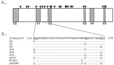
The fourth, fifth, and sixth sites containing Asn-X-Ser/Thr in the gp120 sequence of SIVmac239 were initially selected for mutagenesis (Fig. 1). These sites are located in the vicinity of the highly variable region 1 but nonetheless are strongly conserved among SIV sequences in the database (24). The Asn codon at all three sites of SIVmac239 is AAT. The AAT codon at sites 4 and 5 was changed to CAG (Gln), and at site 6 it was changed to CAA (Gln). Glutamine is structurally similar to asparagine, differing only by a single CH2 group. Since only AAT and AAC can code for Asn, two nucleotide changes would be required for the codon to revert to an Asn codon. All seven possible mutant forms of these sites were created. These are referred to as g4, g5, g6, g45, g46, g56, and g456.
FIG. 1.
(A) Linear depiction of the SIVmac239 envelope gp120 subunit showing the approximate location of potential sites for N-linked oligosaccharides (Ψ) in relation to the five regions exhibiting variability in amino acid sequence (shaded boxes labeled V1 to V5) and signal sequence (V0). (B) Wild-type and mutant amino acid sequences. Amino acids 114 to 158 of the V1 and adjacent regions are shown. The fourth, fifth, and sixth potential glycosylation sites are underlined. The amino acid substitutions made in the viral genome are shown. The N-to-Q substitution removes the potential for the addition of N-linked carbohydrate. The mutation nomenclature is shown on the left. The M144V compensatory mutation is also shown.
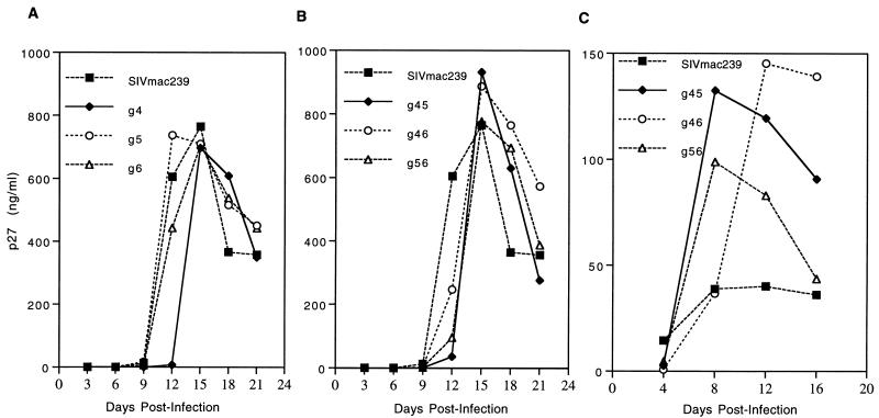
All six single and double mutants (g4, g5, g6, g45, g46, and g56) replicated similarly to the parental virus upon transfection of cloned DNA into CEMx174 cells (data not shown). Normalized amounts of mutant and parental virus stocks produced from CEMx174 transfection were used to analyze viral replication in CEMx174 cells, the rhesus monkey 221 cell line, and primary rhesus monkey PBMC cultures. Again, all single- and double-mutant forms of the virus replicated similarly to the parental virus in CEMx174 cells (Fig. 2A and B), 221 cells (data not shown), and stimulated rhesus monkey PBMC cultures (Fig. 2C). Slight delays or differences in peak heights were observed with the mutants in some experiments, but it is uncertain whether these represent a significant difference.
FIG. 2.
Replication of cloned SIVmac239 and env variants generated by site-directed mutagenesis in CEMx174 cells (A and B) and rhesus PBMC (C). Virus containing 5 ng of p27 was used for all infections. Virus production was monitored by an assay of p27 antigen at the indicated number of days postinfection.
Utilization of sites.
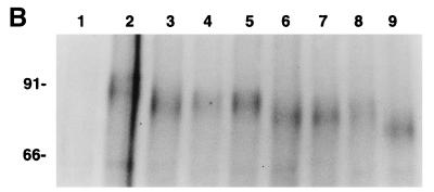
Utilization of the fourth, fifth, and sixth potential sites was investigated by examining the mobility of the proteins in SDS-polyacrylamide gels. Vectors for transient expression of the wild-type and all mutant env sequences were constructed and transfected into COS-1 cells. Only slight size differences were apparent when the gp160 molecules of the panel of labeled envelope proteins were immunoprecipitated and resolved by gel electrophoresis (Fig. 3A, lanes 10 to 18). When N-glycans were removed by treatment with N-glycosidase F, all precursor molecules comigrated, as expected, at approximately 110 kDa, indicating that any size differences between the envelope proteins were due entirely to differences in N-linked glycosylation (lanes 1 to 9). To enhance the detection of size differences among the mutant envelope proteins, a smaller fragment of the envelope protein was generated by acid hydrolysis. Labeled proteins were immunoprecipitated from transfected COS-1 cells and treated with dilute acetic acid. This treatment hydrolyzes proteins between the aspartic acid and proline residues at positions 385 and 740 of the SIV gp160, resulting in fragments of 385, 355, and 140 residues. The first 385 amino acids of gp160 contain 20 signals for N-linked carbohydrates, and each glycan adds approximately 2,500 Da to the relative mass of the fragment (16). Thus, this 385-aa fragment of the wild-type protein is predicted to migrate at approximately 91 kDa. Protein fragments representing these amino-terminal 385 residues of the wild-type and mutant envelope proteins are shown in Fig. 3B. The three single mutants showed an increase in mobility of approximately 3 kDa with respect to the wild-type protein which migrated, as expected, between 90 and 92 kDa (Fig. 3B, lanes 2 to 5). The double mutants showed an additional increase in mobility (lanes 6 to 8), while the triple mutant, g456, had a further increase in mobility (lane 9). These results are consistent with utilization of each of the fourth, fifth, and sixth N-linked glycosylation sites.
FIG. 3.
Utilization of sites. COS-1 cells were transfected with pSVL plasmid containing wild-type (wt) or mutant env DNA. (A) On day 3 posttransfection, the cells were radioactively labeled for 5.5 h. Proteins present in the cell extracts were immunoprecipitated with rhesus anti-SIV polyclonal sera and digested (lanes 1 to 9) or mock digested (lanes 10 to 18) with N-glycosidase F. Lanes: 1 and 10, vector DNA; 2 and 11, wild type; 3 and 12 g4; 4 and 13, g5; 5 and 14, g6; 6 and 15, g45; 7 and 16, g46; 8 and 17, g56; 9 and 18, g456. (B) On day 3 posttransfection, the cells were radioactively labeled for 19 h. Proteins present in the cell extracts were immunoprecipitated, and the pelleted material was hydrolyzed in 10% glacial acetic acid for 66 h at 40°C. The hydrolyzed samples were dried before being dissolved in sample buffer and boiled for 3 min. Lanes: 1, parental vector DNA; 2, wild type; 3, g4; 4, g5; 5, g6; 6, g45; 7, g46; 8, g56; 9, g456. Numbers on the left indicate the relative positions of molecular mass markers and are shown in kilodaltons. Lanes 1 to 9 in panel B are in the same order as lanes 1 to 9 in panel A.
Identification of a revertant of the triple mutant.
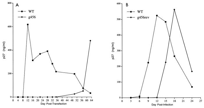
In contrast to the results with the single and double mutants, replication of the triple mutant (g456) was undetectable after transfection of CEMx174 and 221 cells (data not shown). In one CEMx174 culture transfected with g456 viral DNA, detectable virus began to appear beyond 40 days after transfection (Fig. 4A). When virus from day 63 of this transfection was used to infect CEMx174 cells, the virus replicated with only a slight delay compared to the parental virus (Fig. 4B). These findings suggested that reversions or compensatory changes had appeared in the culture to allow wild-type or near-wild-type levels of viral replication.
FIG. 4.
(A) Growth kinetics of wild-type (WT) virus and mutant virus g456 following transfection of CEMx174 cells. (B) Growth kinetics in CEMx174 cells of 5 ng of p27 of wild-type and uncloned revertant virus stock obtained from culture shown in panel A on day 63 posttransfection.
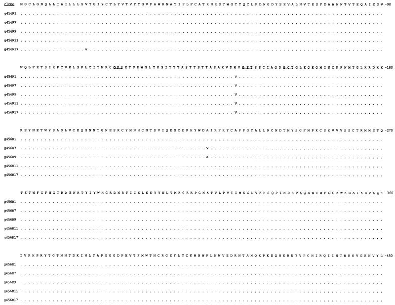
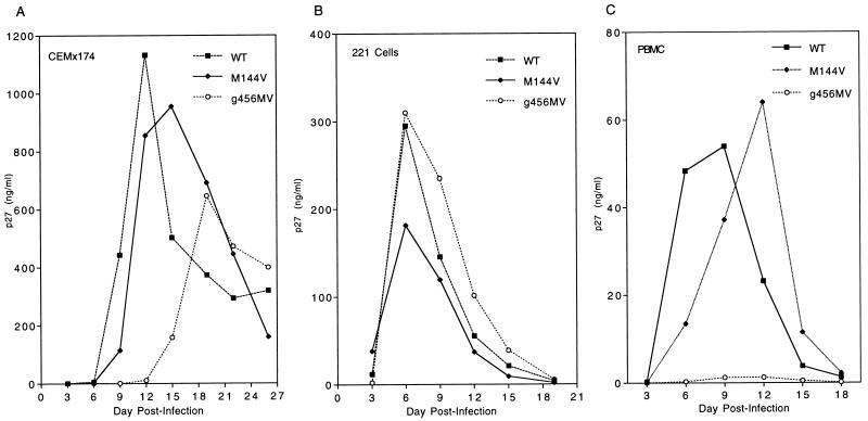
Sequence analysis of viral DNA derived from CEMx174 cells infected with the g456 revertant revealed a single predominant change of Met to Val at position 144 (Fig. 5). This position is located 2 amino acids upstream of the mutated fifth N-linked site. No changes were observed in the fourth, fifth, and sixth QXS/T sites themselves (Fig. 5). We introduced the Val-to-Met change into the parental SIV239 DNA and into the g456 mutant in the absence of any other changes. Virus containing the M144V change in the 239 background replicated similarly to the parental SIVmac239 upon transfection (data not shown) and infection of both the CEMx174 and 221 cell lines (Fig. 6A and B). Virus containing the M144V change in the g456 background replicated with only a slight delay compared to SIVmac239 upon both transfection (data not shown) and infection of both CEMx174 and 221 cells (Fig. 6A and B). The virus with the M144V mutation in the g456 background replicated with kinetics similar to that of the revertant recovered from the original transfection (Fig. 4B). Thus, the change of Met to Val at position 144 is able to compensate for the loss of the fourth, fifth, and sixth NXS/T sites. However, replication of the g456MV variant was severely impaired after infection of stimulated rhesus monkey PBMC (Fig. 6C).
FIG. 5.
Amino acid sequences deduced from env clones obtained from CEMx174 cells infected with the g456rev virus stock for 14 days. Genomic DNA was recovered, and five clones were obtained by PCR. Both strands of the amino-terminal 1,350 bases of each clone were sequenced. The first 450 residues of the parental g456 envelope sequence are shown. The three asparagine-to-glutamine substitutions are shown in boldface type. Sites g4, g5, and g6 are underlined. Dots represent identity. Lowercase letters indicate silent mutations. Capital letters indicate amino acid substitutions.
FIG. 6.
Replication of wild-type SIVmac239 and M144V mutants following infection of CEMx174 cells (A), 221 cells (B), and rhesus PBMC (C). Virus containing 5 ng of p27 was used for all infections. Virus production was monitored by an assay of p27 antigen at the indicated number of days postinfection.
Nature of the block to replication.
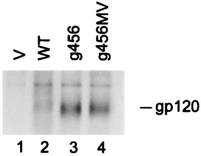
We performed experiments to evaluate both the nature of the block to replication with the g456 mutant and the way in which the M-to-V substitution may relieve this block. Vectors for transient expression of wild-type, g456, and g456MV envelope proteins were transfected into COS-1 cells. Envelope protein expression, processing, and size were evaluated by Western blot analysis of cell extracts on days 2, 3, 4, and 5 after transfection (Fig. 7). The parental clone yielded both the gp160 precursor and the proteolytically processed gp120 external surface subunit as expected during the 2- to 5-day period that was examined (Fig. 7). Both the g456 and g456MV mutants yielded a precursor that migrated faster than the gp160 precursor of the parental virus (Fig. 7). Less processed “gp120” was observed in cells expressing the g456 mutant than in cells expressing the parental envelope, while g456MV expressed intermediate levels (Fig. 7).
FIG. 7.
Proteolytic processing of wild-type and mutant envelope protein expressed in COS-1 cells. COS-1 cells were transfected with pSVL vector expressing wild-type or mutant envelope sequence. Cell lysates were obtained on the days indicated and electrophoresed in an 8% polyacrylamide–SDS gel, transferred to a membrane, and reacted with a polyclonal serum obtained from a rhesus monkey infected with SIVmac239. V, parental plasmid; WT, SIVmac239 env; g456 and g456MV env substitutions are depicted in Fig. 1.
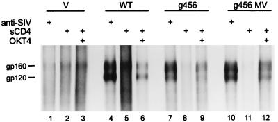
We next assessed the ability of the g456 envelope protein to bind CD4 receptor by measuring binding to sCD4. Transfected COS-1 cells were cultured for 3 days and then labeled with [35S] methionine and [35S]cysteine for 24 h. Labeled envelope protein in the cell lysate was tested for its ability to bind to soluble CD4. CD4-env protein complexes were immunoprecipitated with the OKT4 anti-CD4 monoclonal antibody, whose reactivity is not blocked by the CD4-env interaction. Under the conditions used for this assay, both mutant envelope proteins were able to bind to the soluble CD4 (Fig. 8). We have shown previously that the gp160 precursor of SIVmac239 is capable of binding to soluble CD4 (23).
FIG. 8.
CD4 binding ability of mutant envelope proteins. Transfected COS-1 cells expressing wild-type or mutant envelope proteins were labeled for 24 h with [35S]cysteine and [35S]methionine as described in Materials and Methods. Proteins in the labeled cell extracts were immunoprecipitated with polyclonal antiserum to SIVmac239 (lanes 1, 4, 7, and 10). Labeled cell extracts were incubated with soluble CD4, and the env-CD4 complexes were immunoprecipitated with anti-CD4 MAb OKT4 (lanes 3, 6, 9, and 12). Antigen-antibody complexes were precipitated with protein A/protein G-agarose and analyzed by electrophoresis in a 10% polyacrylamide–SDS gel. Lanes: 1 to 3, vector (v); 4 to 6, wild-type (WT) envelope; 7 to 9, g456 envelope; 10 to 12, g456MV envelope.
Since only a small amount of g456 gp120 equivalent was detected in cells both by Western blot analysis and immunoprecipitation, loss of “gp120” by release into the medium was evaluated by labeling transfected COS-1 cells with [35S]methionine and [35S]cysteine and immunoprecipitating “gp120” from the cell-free supernatants. “gp120” was detected in the supernatant in all cases (Fig. 9). Upon two repetitions of the assay, the g456 virus consistently released higher levels of gp120 than did the wild-type. Processing of “gp160” precursor and loss of “gp120” by release into the medium was also evaluated by a pulse-chase experiment (Fig. 10). The results of these assays were consistent with those of the other methods, suggesting that there were decreased amounts of processed “gp120” of g456 in cells and increased amounts of “gp120” released into the media.
Replication of additional mutants.
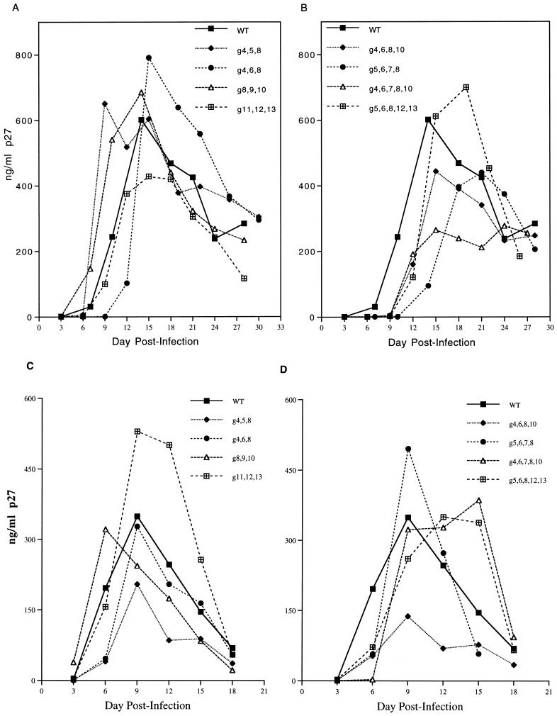
To examine the importance for replication of other sites for N-linked carbohydrates around the V1 and V2 regions, additional asparagine codons were replaced with the codon for glutamine. Mutants were created that lacked various combinations of the 4th through 13th glycosylation sites. Alteration of the 8th or 10th site or both the 12th and 13th sites for N-linked glycosylation had no effect on viral replication in CEMx174 cells (data not shown). Viral replication was not significantly affected when three consecutive or nonconsecutive potential glycosylation sites were altered (Fig. 11A and C). Furthermore, multiple combinations of env mutants lacking four or five glycosylation sites were also replication competent in CEMx174 cells (Fig. 11B) as well as 221 cells (not shown) and stimulated rhesus PBMCs (Fig. 11D). Thus, elimination of multiple combinations of carbohydrate attachment sites between g4 and g13 yielded virus that was still replication competent.
FIG. 11.
Replication of SIV variants lacking additional N-linked carbohydrate attachment sites. Virus containing 5 ng of p27 was used to infect PBMCs or CEMx174 cells as described in the legend to Fig. 2. The nomenclature indicates the position of the asparagine residues in the potential glycosylation sites that were substituted with a glutamine. (A and B) Replication in CEMx174 cells. (C and D) Replication in stimulated rhesus PBMCs.
DISCUSSION
V1 has been described as being the most variable of the hypervariable regions of the external subunit of SIVmac (3, 7). In spite of this variation, potential sites for carbohydrate addition are present near the boundaries of the V1 region in most isolates of SIV and HIV-2 listed in the Los Alamos database (24). This conservation suggests that selective pressure is working to maintain the presence of these V1 carbohydrate attachment sites. In addition, the positions of the fourth and fifth carbohydrate sites are absolutely conserved for all SIVs (24). The positioning of the sixth N-linked glycosylation site is slightly less conserved and sometimes shifts in position by 2 amino acids (24). One might expect from this conservation an important role for the g4 through g6 canonical N-linked sites for some aspect of the viral life cycle. Our study of SIV gp120 mutants has shown that the asparagine residues of the fourth, fifth, and sixth canonical Asn-Xaa-Ser/Thr N-linked glycosylation sites can be replaced individually and in all pairwise combinations with a glutamine residue without detectably influencing the kinetics of viral replication in cell culture.
SDS-polyacrylamide gel electrophoresis following metabolic labeling and acid hydrolysis demonstrated increased mobility of the g4, g5, and g6 individual mutant envelope proteins. The double mutants exhibited even further increased mobility, while the triple mutant had an additional increase in mobility. Removal of the N-linked glycans resulted in the comigration of all envelope species. These results are consistent with the attachment of N-linked carbohydrates to the fourth, fifth, and sixth canonical sites in the parental sequence.
The g456 mutant exhibited decreased levels of its gp120 equivalent associated with cells and increased shedding of its “gp120” into the cell-free supernatant. These results suggest that the g456 mutant is deficient in the association of its “gp120” with the gp41 transmembrane subunit. The growth defect of the g456 virus may thus be due in part to the decrease in association of the g456 surface protein with the gp41 transmembrane protein. Consistent with this, the g456MV variant exhibited increased association of its “gp120” with the cell and decreased shedding into the cell-free supernatant. However, the severe growth defect of the g456 mutant cannot be easily explained by the somewhat subtle differences in processing and gp41 association that were observed. Underglycosylated viral glycoproteins have previously been shown to have impaired assembly (26), and it is possible that the g456 env molecule is deficient in its oligomerization, since this would not necessarily have been detected by the methods used. Differences in the affinity of association with primary or secondary receptors are also possible.
The g456MV variant replicated well in the rhesus monkey cell line 221 but, surprisingly, replicated poorly in stimulated rhesus monkey PBMC cultures. The 221 cell line is immortalized by herpesvirus saimiri (2) and could possibly express the virus-encoded G-protein-coupled receptor which is a homolog of the interleukin-8 (IL-8) receptor (vIL-8R) (1). Thus, one possible explanation for the differential growth of g456MV in 221 and rhesus monkey PBMC is the use of alternate or unusual second receptors that allow entry into CEMx174 and 221 cells but not into stimulated rhesus PBMC.
The finding that only the M144V substitution arose to compensate for the absence of all three potential carbohydrates was surprising due to the large number of serines and threonines within the V1 region that could be used to regenerate new glycosylation sites. Curiously, this methionine-to-valine substitution is found naturally in some MNE and SMM isolates which are not derived from the SIVmac239 or SIVmac251 strains (24). We did not see any difference in viral replication when only the M144V substitution was introduced into the parental SIV239 genome. Our results with SIV are similar in some respects to those described by Wang et al. for HIV-1 (31). In that study, the authors found that simultaneously altering three of six N-linked glycosylation sites in the V1-V2 sequence of HIV-1 HXB2 resulted in gross impairment of virus infectivity. Study of revertants of this mutant revealed that an upstream isoleucine-to-valine substitution conferred the revertant phenotype (31). Similarly, Willey et al. found that the absence of viral replication following the loss of N-linked glycosylation at residue 276 of HIVNL4-3 could be restored with substitutions that do not create a new glycosylation site (32).
We have identified strains simultaneously lacking as many as five of the potential N-linked sites in gp120. This represents a loss of half of all the N-linked glycan addition sites in the region covered by residues 114 to 247. No one, to our knowledge, has identified replication-competent strains of SIV or HIV that are this deficient in glycosylation. In addition, viral replication was tolerant to removal of a series of adjacent carbohydrate-addition sites. This was indicated by growth of the g5678 variant. Replication of the g11, g12, g13 virus is especially noteworthy. The g11 site is absolutely conserved among the 54 HIV-2 and SIV isolates listed in the 1996 database, while the g12 and g13 sites are just slightly less conserved (24). Our results indicate that none of the 10 sites for the addition of N-linked carbohydrate in the vicinity of the V1 and V2 regions is individually required for viral replication. The mutants described in this report will allow us to begin investigating the effects of loss of N-linked carbohydrate attachment sites in the V1 and V2 regions on the nature and quality of the humoral immune response.
ACKNOWLEDGMENTS
We thank Dean Regier for assistance with DNA sequencing and Joanne Newton for preparation of the manuscript.
This work was supported by Public Health Service grants AI35365 and RR00168.
REFERENCES
- 1.Ahuja S K, Murphy P M. Molecular piracy of mammalian interleukin-8 receptor type B by herpesvirus saimiri. J Biol Chem. 1996;268:20691–20694. [PubMed] [Google Scholar]
- 2.Alexander L, Du Z, Rosenzweig M, Jung J U, Desrosiers R C. A role for natural simian immunodeficiency virus and human immunodeficiency virus type 1 nef alleles in lymphocyte activation. J Virol. 1997;71:6094–6099. doi: 10.1128/jvi.71.8.6094-6099.1997. [DOI] [PMC free article] [PubMed] [Google Scholar]
- 3.Almond N, Jenkins A, Heath A B, Kitchin P. Sequence variation in the env gene of simian immunodeficiency virus recovered from immunized macaques is predominantly in the V1 region. J Gen Virol. 1993;74:865–871. doi: 10.1099/0022-1317-74-5-865. [DOI] [PubMed] [Google Scholar]
- 4.Back N K T, Smit L, De Jong J-J, Keulen W, Schutten M, Goudsmit J, Tersmette M. An N-glycan within the human immunodeficiency virus type 1 gp120 V3 loop affects virus neutralization. Virology. 1994;199:431–438. doi: 10.1006/viro.1994.1141. [DOI] [PubMed] [Google Scholar]
- 5.Bernstein H B, Tucker S P, Hunter E, Schutzbach J S, Compans R W. Human immunodeficiency virus type 1 envelope glycoprotein is modified by O-linked oligosaccharides. J Virol. 1994;68:463–468. doi: 10.1128/jvi.68.1.463-468.1994. [DOI] [PMC free article] [PubMed] [Google Scholar]
- 6.Bolmstedt A, Hemming A, Flodby P, Berntsson P, Travis B, Lin J P C, Ledbetter J, Tsu T, Wigzell H, Hu S-L, Olofsson S. Effects of mutations in glycosylation sites and disulphide bonds on processing, CD4-binding and fusion activity of human immunodeficiency virus envelope glycoproteins. J Gen Virol. 1991;72:1269–1277. doi: 10.1099/0022-1317-72-6-1269. [DOI] [PubMed] [Google Scholar]
- 7.Burns D P W, Desrosiers R C. Selection of genetic variants of simian immunodeficiency virus in persistently infected rhesus monkeys. J Virol. 1991;65:1843–1854. doi: 10.1128/jvi.65.4.1843-1854.1991. [DOI] [PMC free article] [PubMed] [Google Scholar]
- 8.Chackerian B, Rudensey L M, Overbaugh J. Specific N-linked and O-linked glycosylation modifications in the envelope V1 domain of simian immunodeficiency virus that evolve in the host alter recognition by neutralizing antibodies. J Virol. 1997;71:7719–7727. doi: 10.1128/jvi.71.10.7719-7727.1997. [DOI] [PMC free article] [PubMed] [Google Scholar]
- 9.Du Z, Regier D A, Desrosiers R C. An improved recombinant PCR mutagenesis procedure that uses alkaline denatured plasmid template. BioTechniques. 1995;18:376–378. [PubMed] [Google Scholar]
- 10.Fennie C, Lasky L A. Model for intracellular folding of the human immunodeficiency virus type 1 gp120. J Virol. 1989;63:639–646. doi: 10.1128/jvi.63.2.639-646.1989. [DOI] [PMC free article] [PubMed] [Google Scholar]
- 11.Fenouillet E, Gluckman J C, Bahraoui E. Role of N-linked glycans of envelope glycoproteins in infectivity of human immunodeficiency virus type 1. J Virol. 1990;64:2841–2848. doi: 10.1128/jvi.64.6.2841-2848.1990. [DOI] [PMC free article] [PubMed] [Google Scholar]
- 12.Fischer P B, Karlsson G B, Butters T D, Dwek R A, Platt F M. N-Butyldeoxynojirimycin-mediated inhibition of human immunodeficiency virus entry correlates with changes in antibody recognition of the V1/V2 region of gp120. J Virol. 1996;70:7143–7152. doi: 10.1128/jvi.70.10.7143-7152.1996. [DOI] [PMC free article] [PubMed] [Google Scholar]
- 13.Fischer P B, Karlsson G B, Dwek R A, Platt F M. N-Butyldeoxynojirimycin-mediated inhibition of human immunodeficiency virus entry correlates with impaired gp120 shedding and gp41 exposure. J Virol. 1996;70:7153–7160. doi: 10.1128/jvi.70.10.7153-7160.1996. [DOI] [PMC free article] [PubMed] [Google Scholar]
- 14.Gruters R A, Neefjes J J, Tersmette M, de Goede R E Y, Tulp A, Huisman H G, Miedema F, Ploegh H L. Interference with HIV-induced syncytium formation and viral infectivity by inhibitors of trimming glucosidase. Nature. 1987;330:74–77. doi: 10.1038/330074a0. [DOI] [PubMed] [Google Scholar]
- 15.Ilyinskii P O, Desrosiers R C. Efficient transcription and replication of simian immunodeficiency virus in the absence of NF-κB and Sp1 binding elements. J Virol. 1996;70:3118–3126. doi: 10.1128/jvi.70.5.3118-3126.1996. [DOI] [PMC free article] [PubMed] [Google Scholar]
- 16.Kornfeld R, Kornfeld S. Assembly of asparagine-linked oligosaccharides. Annu Rev Biochem. 1985;54:631–664. doi: 10.1146/annurev.bi.54.070185.003215. [DOI] [PubMed] [Google Scholar]
- 17.Lee W-R, Syu W-J, Du B, Matsuda M, Tan S, Wolf A, Essex M, Lee T-H. Nonrandom distribution of gp120 N-linked glycosylation sites important for infectivity of human immunodeficiency virus type 1. Proc Natl Acad Sci USA. 1992;89:2213–2217. doi: 10.1073/pnas.89.6.2213. [DOI] [PMC free article] [PubMed] [Google Scholar]
- 18.Leonard C K, Spellman M W, Riddle L, Harris R J, Thomas J N, Gregory T J. Assignment of intrachain disulfide bonds and characterization of potential glycosylation sites of the type 1 recombinant human immunodeficiency virus envelope glycoprotein (gp120) expressed in Chinese hamster ovary cells. J Biol Chem. 1990;265:10373–10382. [PubMed] [Google Scholar]
- 19.Levesque J-P, Sanilvestri P, Hatzfeld A, Hatzfeld J. DNA transfection in Cos cells. BioTechniques. 1991;11:313–318. [PubMed] [Google Scholar]
- 20.Li Y, Luo L, Rasool N, Kang C Y. Glycosylation is necessary for the correct folding of human immunodeficiency virus gp120 in CD4 binding. J Virol. 1993;67:584–588. doi: 10.1128/jvi.67.1.584-588.1993. [DOI] [PMC free article] [PubMed] [Google Scholar]
- 21.Mori K, Ringler D J, Kodama T, Desrosiers R C. Complex determinants of macrophage tropism in env of simian immunodeficiency virus. J Virol. 1992;66:2067–2075. doi: 10.1128/jvi.66.4.2067-2075.1992. [DOI] [PMC free article] [PubMed] [Google Scholar]
- 22.Morikawa Y, Moore J P, Wilkinson A J, Jones I M. Reduction in CD4 binding affinity associated with removal of a single glycosylation site in the external glycoprotein of HIV-2. Virology. 1991;180:853–856. doi: 10.1016/0042-6822(91)90106-l. [DOI] [PubMed] [Google Scholar]
- 23.Morrison H G, Kirchhoff F, Desrosiers R C. Evidence for the cooperation of gp120 amino acids 322 and 448 in SIVmac entry. Virology. 1993;195:167–174. doi: 10.1006/viro.1993.1357. [DOI] [PubMed] [Google Scholar]
- 24.Myers G, Foley B, Mellors J W, Korber B, Jeang K-T, Wain-Hobson S. A compilation and analysis of nucleic acid and amino acid sequences. Los Alamos, N.M: Los Alamos National Laboratory; 1996. [Google Scholar]
- 25.Naidu Y M, Kestler H W, III, Li Y, Butler C V, Silva D P, Schmidt D K, Troup C D, Sehgal P K, Sonigo P, Daniel M D, Desrosiers R C. Characterization of infectious molecular clones of simian immunodeficiency virus (SIVmac) and human immunodeficiency virus type 2: persistent infection of rhesus monkeys with molecularly cloned SIVmac. J Virol. 1988;62:4691–4696. doi: 10.1128/jvi.62.12.4691-4696.1988. [DOI] [PMC free article] [PubMed] [Google Scholar]
- 26.Ng D T W, Hiebert S W, Lamb R A. Different roles of individual N-linked oligosaccharide chains in folding, assembly, and transport of the simian virus 5 hemagglutinin-neuraminidase. Mol Cell Biol. 1990;10:1989–2001. doi: 10.1128/mcb.10.5.1989. [DOI] [PMC free article] [PubMed] [Google Scholar]
- 27.Pal R, Hoke G M, Sarngadharan M G. Role of oligosaccharides in the processing and maturation of envelope glycoproteins of human immunodeficiency virus type 1. Proc Natl Acad Sci USA. 1989;86:3384–3388. doi: 10.1073/pnas.86.9.3384. [DOI] [PMC free article] [PubMed] [Google Scholar]
- 28.Pinter A, Honnen W J. O-linked glycosylation of retroviral envelope gene products. J Virol. 1988;62:1016–1021. doi: 10.1128/jvi.62.3.1016-1021.1988. [DOI] [PMC free article] [PubMed] [Google Scholar]
- 29.Regier D A, Desrosiers R C. The complete nucleotide sequence of a pathogenic molecular clone of simian immunodeficiency virus. AIDS Res Hum Retroviruses. 1990;6:1221–1231. doi: 10.1089/aid.1990.6.1221. [DOI] [PubMed] [Google Scholar]
- 30.Walker B D, Kowalski M, Goh W C, Kozarsky K, Krieger M, Rosen C, Rohrschneider L, Haseltine W A, Sodroski J. Inhibition of human immunodeficiency virus syncytium formation and virus replication by castanospermine. Proc Natl Acad Sci USA. 1987;84:8120–8124. doi: 10.1073/pnas.84.22.8120. [DOI] [PMC free article] [PubMed] [Google Scholar]
- 31.Wang W-K, Essex M, Lee T-H. Single amino acid substitution in constant region 1 or 4 of gp120 causes the phenotype of a human immunodeficiency virus type 1 variant with mutations in hypervariable regions 1 and 2 to revert. J Virol. 1996;70:607–611. doi: 10.1128/jvi.70.1.607-611.1996. [DOI] [PMC free article] [PubMed] [Google Scholar]
- 32.Willey R L, Smith D H, Lasky L A, Theodore T S, Earl P L, Moss B, Capon D J, Martin M A. In vitro mutagenesis identifies a region within the envelope gene of the human immunodeficiency virus that is critical for infectivity. J Virol. 1988;62:139–147. doi: 10.1128/jvi.62.1.139-147.1988. [DOI] [PMC free article] [PubMed] [Google Scholar]


