Abstract
The development of efficient non-viral methodologies for genome-wide insertional mutagenesis and gene tagging in mammalian cells is highly desirable for functional genomic analysis. Here we describe transposon mediated mutagenesis (TRAMM), using naked DNA vectors based on the Drosophila hydei transposable element Minos. By simple transfections of plasmid Minos vectors in HeLa cells, we have achieved high frequency generation of cell lines, each containing one or more stable chromosomal integrations. The Minos-derived vectors insert in different locations in the mammalian genome. Genome-wide mutagenesis in HeLa cells was demonstrated by using a Minos transposon containing a lacZ–neo gene-trap fusion to generate a HeLa cell library of at least 105 transposon insertions in active genes. Multiple gene traps for six out of 12 active genes were detected in this library. Possible applications of Minos-based TRAMM in functional genomics are discussed.
INTRODUCTION
The complete sequence of the human genome is expected to be available soon and a versatile method for large scale insertional mutagenesis will be a sine qua non for its functional analysis. Insertional mutagenesis and gene trapping with plasmid or retroviral vectors has been successfully applied in mammalian cultured cells (Gossler et al., 1989; Friedrich and Soriano, 1991; Wurst et al., 1995; Hicks et al., 1997; Bonaldo et al., 1998; Whitney et al., 1998; Zambrowicz et al., 1998; Wiles et al., 2000). The efficiency of plasmid vectors is limited by low insertion rates, while retroviral vectors are relatively difficult to manipulate. Transposable elements present an attractive alternative. Transposable elements have been used previously as vectors for gene and enhancer trapping in various complex organisms, but not in mammals (Bier et al., 1989; Bellen et al., 1989; Wilson et al., 1989; Parinov and Sundaresan, 2000; Walbot, 2000). Most elements are mobilized through a cut-and-paste process catalysed by an element-encoded transposase that excises the element from its original plasmid or chromosomal site and re-integrates it elsewhere in the genome. Minos (Franz and Savakis, 1991) belongs to the Tc1/mariner superfamily of transposable elements (Franz et al., 1994), members of which are active in a wide range of species from protozoa to mammals (Gueiros-Filho and Beverley, 1997; Ivics et al., 1997; Schouten et al., 1998; Luo et al., 1998; Zhang et al., 1998; Yant et al., 2000). Transposition of Minos in cultured cells or embryos of several insect species suggests that this element may serve as a universal gene transfer and mutagenesis vector in insects (Loukeris et al., 1995a,b; Catteruccia et al., 2000a,b; Klinakis et al., 2000; Shimizu et al., 2000). Here we show that Minos is functional in human HeLa cells and that Minos-based vectors can be used for genome-wide mutagenesis and functional genomic analysis. We propose that Minos-based TRAMM (transposon mediated mutagenesis) may be a powerful tool for functional analysis of the human genome.
RESULTS AND DISCUSSION
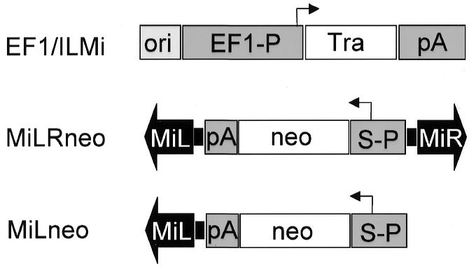
To test for transposase-catalysed chromosomal integration of Minos in human cells, two circular Minos-based plasmids were co-transfected into HeLa cells: a helper plasmid (pEF1/ILMi) carrying the Minos transposase gene driven by the human EF1α (elongation factor 1α) promoter, and a donor plasmid (pMiLRneo) bearing a neomycin resistance gene driven by the SV40 promoter and flanked by Minos inverted repeats (Figure 1). Approximately 4% of the cells from each co-transfection, corresponding to 105 clones, were stably transformed to G418 resistance. A reduction ranging between 9- and 50-fold was observed in the absence of helper plasmid or in transfections with helper and pMiLneo, a ‘wings clipped’ donor lacking one of the two inverted repeats (Table I). These differences were reproducible and independent of transfection efficiencies, as indicated by transient expression of a co-transfected CMV/GFP construct.

Fig. 1. Minos-derived vectors. Minos inverted terminal repeats are shown as block arrows. Arrowheads indicate the direction of transcription of the transposase (Tra) and neo genes. pA is polyadenylation signal; ori and S-P are the SV40 origin of replication and promoter, respectively.
Table I. G418-resistant HeLa colonies generated after transfection with Minos plasmids.
| Plasmids used in transfection | Experiment 1 | Experiment 2 |
|---|---|---|
| pMiLRneo + EF1 | 3350 | 6240 |
| pMiLRneo + EF1/ILMi | 32 600 | 102 960 |
| pMiLneo + EF1/ILMi | 3400 | 2040 |
Numbers are averages of two duplicate transfections.
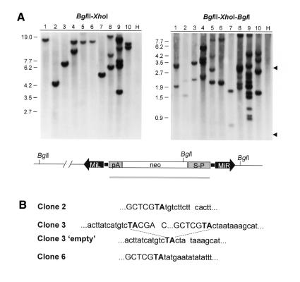
Genomic analysis of randomly selected clones derived from pMiLRneo and pEF1/ILMi co-transfection, indicated that single copies of the transposon had integrated at different genomic positions (Figure 2A). Restriction analysis failed to detect diagnostic fragments derived from integration of the entire donor plasmid (Figure 2A, triple digest). Absence of plasmid vector sequences was confirmed by re-probing the blots with an appropriate probe (data not shown). Analysis of 30 clones showed between one and seven insertions per clone, with a mean of 2.1.
Fig. 2. MiLRneo insertions in individual HeLa clones. (A) Southern blot analysis of individual colonies. Genomic DNA (10 µg) was digested with the enzymes indicated before electrophoresis. The probe used is indicated by a grey bar. Arrowheads indicate the expected positions of diagnostic fragments derived from plasmid vector sequences. Lane H is from non-transfected cells. (B) Sequences flanking MiLRneo insertions. Transposon sequences and the target TA dinucleotide are in upper case.
As with all Tc1/mariner elements, Minos insertions invariably disrupt a TA dinucleotide, which is duplicated upon insertion (Loukeris et al., 1995a). We determined the DNA sequences flanking the MiLRneo transposon of three different clones bearing single insertions. All insertions had occurred at a TA dinucleotide and the flanking sequences were different from the original Minos flanks. In clone 3, the pre-insertion site was cloned and sequenced; the TA duplication was the only alteration introduced (Figure 2B).
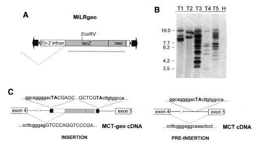
The observed high integration frequencies suggested that genome-wide gene trapping is possible. A Minos-based gene-trap construct (pMiLRgeo) was used, utilizing the first splice acceptor and part of exon 2 of the mouse Engrailed-2 gene fused in-frame to an AUG-less lacZ–neo fusion (geo, Figure 3A). Upon insertion of geo vectors into introns of active genes in the appropriate orientation, in-frame splicing of the geo fusion to the upstream exon confers resistance to G418 and occasionally gives detectable galactosidase activity. Co-transfection of HeLa cells with pMiLRgeo plus pEF1/ILMi resulted in 10- to 100-fold lower frequency of G418-resistant clones compared with pMiLRneo. Transposase-induced insertions were 10-fold more frequent than in the absence of transposase. Southern blot analysis of five randomly chosen clones indicates that insertions occured in different genomic positions, varying in number from 1 to 10 per clone (Figure 3B). From 16 individual G418-resistant clones, eight were positive for β-galactosidase activity (data not shown). As in all random mutagenesis schemes, occurrence of multiple hits can complicate downstream analysis. This problem is of lesser concern in gene trap experiments, since gene traps are identified at the level of cDNA rather than at the genomic level.
Fig. 3. MiLRgeo gene-trap insertions. (A) The gene-trap construct MiLRgeo. The probe used for Southern blotting is indicated by a grey bar. (B) Southern blot analysis of individual HeLa lines stably transfected with MiLRgeo. Ten micrograms of DNA were digested with EcoRV. Lane H is from non-transfected cells. (C) Fusion cDNA and flanking intron sequences of a gene-trap insertion from clone T4. Transposon sequences and the target TA dinucleotide are in upper case.
A fusion transcript from clone T4 was recovered by 5′ RACE and the sequence of the splice junction was determined. Integration had occurred into the fourth intron of the monocarboxylate transporter 3 (MCT3) gene. Analysis of the insertion sequence showed that the transposon had inserted at a TA ∼1.7 kb upstream from exon 5, generating the characteristic target TA duplication (Figure 3C).
The restriction patterns shown in Figures 2 and 3 were unchanged after >50 passages, indicating that Minos insertions in HeLa cells are stable (data not shown).
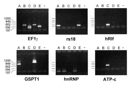
Using standard protocols, transfection of 106 cells can yield ∼4 × 10⁴ clones carrying Minos insertions. Since the average number of insertions per clone is ∼2 (Figure 2A), this corresponds to a total number of 8 × 10⁴ individual insertions per 106 transfected cells. Therefore, a library containing 3 × 10⁶ insertions can be generated from 4 × 10⁷ cells. Such a library should contain, on average, one insertion per kb. To further challenge Minos as a tool for genome-wide gene trapping, 4 × 10⁷ HeLa cells distributed in twenty 100 mm tissue culture dishes were co-transfected with gene trap vector pMiLRgeo and helper pEF1/ILMi. G418 selection yielded ∼105 G418-resistant colonies. Given that only a fraction of genes should be active in HeLa cells, Minos insertions should have occurred in the majority of active genes. To test this, five sets of four plates each were pooled to create sub-libraries of ∼2 × 10⁴ clones each. Poly(A)+ RNA was isolated from each pool, and was used to detect gene-trap fusions of 12 genes whose intron–exon structure is known, and which have been reported or are expected to be active in HeLa cells (Ryo et al, 1998; see legend to Figure 4). Fusion RT–PCR products were detectable for six of these genes without the need to optimize PCR conditions or use alternate primers. Four of the genes were hit in more than one pool and two were hit in more than one intron (Figure 4 and Table II). It is notable that only two of the nine exon traps detected were in-frame. This suggests that the remaining fusions represent ‘by-standers’, i.e. that the respective cells may carry at least one other gene trap that confers G418 resistance. Considering the limitations of RT–PCR, that HeLa cells are largely polyploid, and taking into account that our sample (12 genes) is rather small, our experiments indicate that at least 50% of the genes that are active in HeLa cells have been tagged. Further experiments are required to determine whether the element inserts preferentially into transcriptionally active regions of the genome.
Fig. 4. Trapping of six genes in HeLa cells with MiLRgeo. Each panel shows agarose gel electrophoresis separated, ethidium bromide stained RT–PCR products that were recovered from five independent pools (A–E) of G418-resistant clones. The control lane corresponds to a reaction without template. The panels correspond to genes (accession numbers in brackets): elongation factor 1γ (EF1γ) (Z11531); ribosomal protein 18 (rs18) (X69150); DNA replication licensing factor homolog (hRlf) (D39073); G1 to S phase transition protein 1 homolog (GSPT1) (X17644); heterogeneous nuclear ribonucleoprotein A2/B1 (hnRNP) (D28877); ATP synthase c-subunit (ATP-c) (X69908). Genes that were not detected are: thymidine kinase (M15205), ATP synthase γ-subunit (D16561), F-actin capping protein (U03271), a putative protein kinase C inhibitor (U51004), hypoxantine phosphoribosyltransferase 1 (NM_000194) and cytochrome c core I protein (L16842).
Table II. Sequences of gene-trap chimeric cDNAs.
| Gene | Exon trapped | No. of hits | Approx. intron size (kb) | Sequencea |
|---|---|---|---|---|
| EF1γ | 4 | 1 | 3.4 | …gct gac atc aca gtt gtc tgc acc ctg ttg tgg ctc tat aag cag GT CCC AGG… |
| EF1γ | 5 | 2 | 0.4 | …tc ttg ggg gaa gtg aaa ctg tgt gag aag atg gcc cag ttt gat gGT CCC AGG… |
| EF1γ | 6 | 2 | 5.7 | …gct gag ccc aag gcc aag gac ccc ttc gct cac ctg ccc aag ag GT CCC AGG… |
| rs18 | 1 | 2 | 3.0 | …att gat ggg cgg cgg aaa ata gcc ttt gcc atc act gcc att aag GT CCC AGG… |
| hRlf | 9 | 1 | 2.3 | …gcc cgc tgc agt gtt ttg gca gct gcc aac cct gtc tac ggc agg GT CCC AGG… |
| GSPT1 | 9 | 1 | 5.4 | …gga tct att tgt aaa ggc cag cag ctt gtg atg atg cca aac aag GT CCC AGG… |
| GSPT1 | 13 | 1 | 2.5 | …tt aaa gac ttc cct cag atg ggt cgt ttc acc tta aga gatgag gGT CCC AGG… |
| hnRNP | 1 | 3 | 2.7 | …ga aat cgg gct gaa gcg act gag tcc gcg atg gag GT CCC AGG… |
| ATP-c | 4 | 1 | 3.7 | …tgg gat tgg aac tgt gtt tgg gag cct cat cat tgg tta tgc cag GT CCC AGG… |
aTrapped exon sequences are in lower case; the first eight En-2 exon 2 bases are in upper case. Codons are separated by spaces.
We show for the first time that a transposable element can potentially tag all genes of the human genome. Furthermore, we demonstrate that Minos-based TRAMM can be used for high efficiency gene trapping in a human cell line. Biological phenomena such as differentiation, response to external stimuli, and disease, frequently involve alterations of transcriptional levels of specific genes. This work opens the way to use Minos-based TRAMM for efficient genome-wide functional identification of transcriptionally modulated genes in cell lines. Minos-based TRAMM could also be useful for high-throughput screening of ES cells for potential knockouts.
METHODS
Plasmids. Helper plasmid pEF1/ILMi was constructed by inserting the spliced transposase coding region (Klinakis et al., 2000) into the ClaI–XbaI sites of plasmid pEF-BOS (Goldman et al., 1996). Donor plasmid pMiLRneo was constructed by replacing the tetracycline resistance gene in pMiLRTetR (Klinakis et al., 2000) by a SV40-neo cassette (pRcCMV, Invitrogen). ‘Wings-clipped’ pMiLneo is pMiLRneo lacking a SacII–SacI fragment containing the Minos right-hand inverted repeat. The gene-trap plasmid vector pMiLRgeo was constructed by replacing the SV40-neo cassette from pMiLRneo with an EcoRI–HindIII geo cassette from plasmid pGT1.8geo (Skarnes et al., 1995).
Cell culture and transfection. HeLa cells were cultured in Dulbecco’s Modified Eagle’s Medium (DMEM) supplemented with 10% fetal calf serum and 50 µg/ml gentamycin at 37°C, 5% CO2. Exponentially growing HeLa cells (6 × 10⁵) were transfected in 3 ml DMEM with 8 µg pMiLRneo or pMiLneo and 2 µg pEF-BOS or pEF1/ILMi, using calcium phosphate precipitation. One microgram of plasmid pQBI25 (Quantum) containing a GFP gene under the control of the cytomegalovirus promoter was included in all mixes for reference. Two days post-transfection, the cells were transferred to medium containing 600 µg/ml G418 (Gibco).
Amplification and cloning of the insertion sites. Genomic DNA from three clones with single insertions was digested with EcoRV and probed with a neo probe. The gel regions containing the Minos flanks were cut out and DNA was isolated and digested with either Sau3AI or AluI. The Sau3AI fragments were ligated to vectorettes (Vectorette II kit, Genosys) and PCR was performed with vectorette-specific primers and the Minos primers: IMio1 (5′-AAGAGAATAAAATTCTCTTTGAGACG-3′) for the first PCR and IMio2 (5′-GATAATATAGTGTGTTAAACATTGCGC-3′) for the ‘nested’ PCR. The AluI fragments were circularized after dilution and inverse PCR was performed with Minos primers, IMio1 and IMii1 (5′-CAAAAATATGAGTAATTTATTCAAACGG-3′), followed by a second round of ‘nested’ PCR with primers IMio2 and IMii2 (5′-GCTTAAGAGATAAGAAAAAAGTGACC-3′). At least one flank of each insertion was recovered. The ‘right-hand’ flank from one of the clones (clone 3) was recovered by screening a human genomic library with the left flank as probe and using the sequence adjacent to the known flank for primer design. The pre-insertion site sequence of clone 3 was recovered from non-transfected HeLa DNA by PCR.
Amplification and cloning of fusion cDNAs. A modification of the 5′ RACE protocol (cRACE) was used to identify fusion cDNAs (Maruyama et al., 1995). Total RNA was reverse transcribed with AMV reverse transcriptase (Gibco) using the geo specific primer ZRT (5′-GACCGTAATGGGATAGGTTAC-3′). The geo specific primers used for inverse PCR were: ZF1 (5′-CAGTTGCGCAGCCTGAATGGG-3′) and ZR1 (5′-CTTCGCTATTACGCCAGCTGG-3′) for the first PCR; and ZF2 (5′-GCTGGCTGGAGTGCGATCTTC-3′) and ZR2 (5′-CAGGGTTTTCCCAGTCACGAC-3′) for the ‘nested’ PCR. The right-hand flank of the insertion was isolated by PCR using Minos primer IMio1 and MCT3 exon 5 primer GGCTTCTTCCTAATGCAG. The left flank and the pre-insertion site in the MCT3 intron were recovered as described above for the MiLRneo insertion.
Identification of trapped genes by RT–PCR. Reverse transcription was performed with the Superscript II system (Gibco) using 0.5 µg poly(A)+ RNA from each pool as template and primer ZR1. Gene-trap insertions were identified using nested PCR with two geo-specific primers (ZR2 for the first and TGCTCTGTCAGGTACCTGTTGG for the second PCR) and two gene-specific primers (sequences of gene-specific primers available upon request). Amplification products were analysed by agarose gel electrophoresis. Individual bands were eluted from the gel and cloned in a TA vector (Promega T-easy).
Acknowledgments
ACKNOWLEDGEMENTS
We thank G. Vrentzos for technical help and S. Oehler for critically reading the manuscript. This work was supported by a grant from the European Union to C.S. (BIO4-CT98-0203) and a Career Development Award grant from the Greek Secretariat for Research and Technology to D.K.V.
REFERENCES
- Bellen H.J., O’Kane, C.J., Wilson, C., Grossniklaus, U., Pearson, R.K. and Gehring, W.J. (1989) P-element-mediated enhancer detection: a versatile method to study development in Drosophila. Genes Dev., 3, 1288–1300. [DOI] [PubMed] [Google Scholar]
- Bier E. et al. (1989) Searching for pattern and mutation in the Drosophila genome with a P-lacZ vector. Genes Dev., 3, 1273–1287. [DOI] [PubMed] [Google Scholar]
- Bonaldo P., Chowdhury, K., Stoykova, A., Torres, M. and Gruss, P. (1998) Efficient gene trap screening for novel developmental genes using IRES β geo vector and in vitro preselection. Exp. Cell Res., 244, 125–136. [DOI] [PubMed] [Google Scholar]
- Catteruccia F., Nolan, T., Blass, C., Muller, H.M., Crisanti, A., Kafatos, F.C. and Loukeris, T.G. (2000a) Toward Anopheles transformation: Minos element activity in anopheline cells and embryos. Proc. Natl Acad. Sci. USA, 97, 2157–2162. [DOI] [PMC free article] [PubMed] [Google Scholar]
- Catteruccia F., Nolan, T., Loukeris, T.G., Blass, Savakis, C., Kafatos, F.C. and Crisanti, A. (2000b) Stable germline transformation of the malaria mosquito Anopheles stephensi. Nature, 405, 959–962 [DOI] [PubMed] [Google Scholar]
- Franz G. and Savakis, C. (1991) Minos, a new transposable element from Drosophila hydei, is a member of the Tc1-like family of transposons. Nucleic Acids Res., 19, 6646. [DOI] [PMC free article] [PubMed] [Google Scholar]
- Franz G., Loukeris, T.G., Dialektaki, G., Thompson, C.R. and Savakis, C. (1994) Mobile Minos elements from Drosophila hydei encode a two-exon transposase with similarity to the paired DNA-binding domain. Proc. Natl Acad. Sci. USA, 91, 4746–4750. [DOI] [PMC free article] [PubMed] [Google Scholar]
- Friedrich G. and Soriano, P. (1991) Promoter traps in embryonic stem cells: a genetic screen to identify and mutate developmental genes in mice. Genes Dev., 5, 1513–1523. [DOI] [PubMed] [Google Scholar]
- Goldman L.A., Cutrone, E.C., Kotenko, S.V., Krause, C.D. and Langer, J.A. (1996) Modifications of vectors pEF-BOS, pcDNA1 and pcDNA3 result in improved convenience and expression. Biotechniques, 21, 1013–1015. [DOI] [PubMed] [Google Scholar]
- Gossler A., Joyner, A.L., Rossant, J. and Skarnes, W.C. (1989) Mouse embryonic stem cells and reporter constructs to detect developmentally regulated genes. Science, 244, 463–465. [DOI] [PubMed] [Google Scholar]
- Gueiros-Filho F.J. and Beverley, S.M. (1997) Trans-kingdom transposition of the Drosophila element mariner within the protozoan Leishmania. Science, 276, 1716–1719. [DOI] [PubMed] [Google Scholar]
- Hicks G.G., Shi, E.G., Li, X.M, Li, C.H., Pawlak, M. and Ruley, H.E. (1997) Functional genomics in mice by tagged sequence mutagenesis. Nature Genet., 16, 338–244. [DOI] [PubMed] [Google Scholar]
- Ivics Z., Hackett, P.B., Plasterk, R.H. and Izsvak, Z. (1997) Molecular reconstruction of Sleeping Beauty, a Tc1-like transposon from fish, and its transposition in human cells. Cell, 91, 501–510. [DOI] [PubMed] [Google Scholar]
- Klinakis A.G., Loukeris, T.G., Pavlopoulos, A. and Savakis, C. (2000) Mobility assays confirm the broad host range activity of the Minos transposable element and validate new transformation tools. Insect Mol. Biol., 9, 269–275. [DOI] [PubMed] [Google Scholar]
- Loukeris T.G., Arca, B., Livadaras, I., Dialektaki, G. and Savakis, C. (1995a) Introduction of the transposable element Minos into the germ line of Drosophila melanogaster. Proc. Natl Acad. Sci. USA, 92, 9485–9489. [DOI] [PMC free article] [PubMed] [Google Scholar]
- Loukeris T.G., Livadaras, I., Arca, B., Zabalou, S. and Savakis, C. (1995b) Gene transfer into the medfly, Ceratitis capitata, with a Drosophila hydei transposable element. Science, 270, 2002–2005. [DOI] [PubMed] [Google Scholar]
- Luo G., Ivics, Z., Izsvak, Z. and Bradley, A. (1998) Chromosomal transposition of a Tc1/mariner-like element in mouse embryonic stem cells. Proc. Natl Acad. Sci. USA, 95, 10769–10773. [DOI] [PMC free article] [PubMed] [Google Scholar]
- Maruyama I.N., Rakow, T.L. and Maruyama. H.I. (1995) cRACE: a simple method for identification of the 5′ end of mRNAs. Nucleic Acids Res., 23, 3796–3797. [DOI] [PMC free article] [PubMed] [Google Scholar]
- Parinov S and Sundaresan, V. (2000) Functional genomics in arabidopsis: large-scale insertional mutagenesis complements the genome sequencing project. Curr. Opin. Biotechnol., 11, 157–161. [DOI] [PubMed] [Google Scholar]
- Ryo A., Kondoh, N., Wakatsuki, T., Hada, A., Yamamoto, N. and Yamamoto, M. (1998) A method for analysing the qualitative and quantitative aspects of gene expression: a transcriptional profile revealed for HeLa cells. Nucleic Acids Res., 26, 2586–2592. [DOI] [PMC free article] [PubMed] [Google Scholar]
- Schouten G.J., van Luenen, H.G., Verra, N.C., Valerio, D. and Plasterk, R.H. (1998) Transposon Tc1 of the nematode Caenorhabditis elegans jumps in human cells. Nucleic Acids Res., 26, 3013–3017. [DOI] [PMC free article] [PubMed] [Google Scholar]
- Shimizu K., Kamba, M., Sonobe, H., Kanda, T., Klinakis, A.G., Savakis, C. and Tamura, T. (2000) Extrachromosomal transposition of the transposable element Minos occurs in embryos of the silkworm Bombyx mori.Insect Mol. Biol., 9, 277–281. [DOI] [PubMed] [Google Scholar]
- Skarnes W.C., Moss, J.E., Hurtley, S.M. and Beddington, R.S. (1995) Capturing genes encoding membrane and secreted proteins important for mouse development. Proc. Natl Acad. Sci. USA, 92, 6592–6596. [DOI] [PMC free article] [PubMed] [Google Scholar]
- Walbot V. (2000) Saturation mutagenesis using maize transposons. Curr. Opin. Plant Biol., 3, 103–107. [DOI] [PubMed] [Google Scholar]
- Whitney M., Rockenstein, E., Cantin, G., Knapp, T., Zlokarnik, G., Sanders, P., Durick, K., Craig, F.F. and Negulescu, P.A. (1998) A genome-wide functional assay of signal transduction in living mammalian cells. Nature Biotechnol., 16, 1329–1333. [DOI] [PubMed] [Google Scholar]
- Wiles M.V. et al. (2000) Establishment of a gene-trap sequence tag library to generate mutant mice from embryonic stem cells. Nature Genet., 24, 13–14. [DOI] [PubMed] [Google Scholar]
- Wilson C., Pearson, R.K., Bellen, H.J., O’Kane, C.J., Grossniklaus, U. and Gehring, W.J. (1989) P-element-mediated enhancer detection: an efficient method for isolating and characterizing developmentally regulated genes in Drosophila. Genes Dev., 3, 1301–1313. [DOI] [PubMed] [Google Scholar]
- Wurst W. et al. (1995) A large-scale gene-trap screen for insertional mutations in developmentally regulated genes in mice. Genetics, 139, 889–899. [DOI] [PMC free article] [PubMed] [Google Scholar]
- Yant S.R., Meuse, L., Chiu, W., Ivics, Z., Izsvak, Z. and Kay, M.A. (2000) Somatic integration and long-term transgene expression in normal and haemophilic mice using a DNA transposon system. Nature Genet., 25, 35–41. [DOI] [PubMed] [Google Scholar]
- Zambrowicz B.P., Friedrich, G.A., Buxton, E.C., Lilleberg, S.L., Person, C., and Sands, A.T. (1998) Disruption and sequence identification of 2000 genes in mouse embryonic stem cells. Nature, 392, 608–611. [DOI] [PubMed] [Google Scholar]
- Zhang L., Sankar, U., Lampe, D.J., Robertson, H.M., and Graham, F.L. (1998) The Himar1 mariner transposase cloned in a recombinant adenovirus vector is functional in mammalian cells. Nucleic Acids Res., 26, 3687–93. [DOI] [PMC free article] [PubMed] [Google Scholar]


